2-Butyl-5-pentylbenzene-1,3-diol
Juangjun Jumpathong,aPascal Retailleau,b* Muna Ali Abdalla,cJamal Ouazzaniband Saisamorn Lumyonga*
a
Department of Biology, Faculty of Science, Chiang Mai University, Chiang Mai 50200, Thailand,bInstitut de Chimie des Substances Naturelles - CNRS, 1 avenue de la Terrasse, 91198 Gif sur Yvette, France, andcDepartment of Organic and
Biomolecular Chemistry, Georg-August-Universita¨t Go¨ttingen, Tammannstrasse 2, D-37077 Go¨ttingen, Germany
Correspondence e-mail: [email protected], [email protected]
Received 7 May 2009; accepted 18 May 2009
Key indicators: single-crystal X-ray study;T= 293 K; mean(C–C) = 0.003 A˚; Rfactor = 0.048;wRfactor = 0.131; data-to-parameter ratio = 16.7.
In the title compound, C15H24O2, a natural dialkylresorcinol
commonly named stemphol, the molecules are linked into
C(6) andC2 2
(4) chains andR4 4
(16) rings by intermolecular O— H O hydrogen bonds, creating molecular sheets parallel to the (010) plane. The alkyl chains are directed orthogonally away from these planes in almost complete extension.
Related literature
For general background, synthesis, biological activity and related structures, see: Achenbach & Kohl (1979); Andersen & Frisvad (2004); Marumoet al.(1985); Solfrizzoet al.(1994); Stodola et al. (1973). For structural discussion, see: Etter (1990); Bernsteinet al.(1995).
Experimental
Crystal data
C15H24O2 Mr= 236.34 Monoclinic,P21=c a= 4.654 (2) A˚
b= 25.450 (5) A˚
c= 12.790 (4) A˚
= 108.12 (1)
V= 1439.8 (8) A˚3 Z= 4
MoKradiation
= 0.07 mm1 T= 293 K
0.500.100.08 mm
Data collection
Nonius KappaCCD diffractometer Absorption correction: multi-scan
(SCALEPACK; Otwinowski &
Minor, 1997)
Tmin= 0.881,Tmax= 0.994
15852 measured reflections
1703 reflections withI> 2(I)
Refinement
R[F2> 2(F2)] = 0.048 wR(F2) = 0.131 S= 1.04 2631 reflections
158 parameters
H-atom parameters constrained
max= 0.18 e A˚
3
min=0.16 e A˚
3
Table 1
Hydrogen-bond geometry (A˚ ,).
D—H A D—H H A D A D—H A
O1—H1 O2i
0.82 1.96 2.767 (2) 167 O2—H2 O1ii
0.82 1.95 2.750 (2) 165
Symmetry codes: (i)xþ1;yþ1 2;zþ
1
2; (ii)x;yþ 1 2;z
1 2.
Data collection: DENZO (Otwinowski & Minor, 1997) and
COLLECT(Nonius, 1999); cell refinement:DENZO; data reduction:
SCALEPACK (Otwinowski & Minor, 1997); program(s) used to solve structure:SIR97 (Altomareet al., 1999); program(s) used to refine structure: SHELXL97 (Sheldrick, 2008) and CrystalBuilder
(Welter, 2006); molecular graphics: PLATON (Spek, 2009) and
Mercury(Macraeet al., 2006); software used to prepare material for publication:SHELXL97andpublCIF(Westrip, 2009).
JJ thanks the Thailand Research Fund (Royal Golden Jubilee PhD Program 4.V.CM/47/D.1) and the French Embassy for financial support. Professor H. Laatsch (Department of Organic and Biomolecular Chemistry, Georg-August-Universita¨t Go¨ttingen, Germany) is thanked for the structure elucidation.
Supplementary data and figures for this paper are available from the IUCr electronic archives (Reference: DN2455).
References
Achenbach, H. & Kohl, W. (1979).Chem. Ber.112, 209–217.
Altomare, A., Burla, M. C., Camalli, M., Cascarano, G. L., Giacovazzo, C., Guagliardi, A., Moliterni, A. G. G., Polidori, G. & Spagna, R. (1999).J. Appl. Cryst.32, 115–119.
Andersen, B. & Frisvad, J. C. (2004).J. Agric. Food Chem.52, 7507–7513. Bernstein, J., Davis, R. E., Shimoni, L. & Chang, N.-L. (1995).Angew. Chem.
Int. Ed. Engl.34, 1555–1573.
Etter, M. C. (1990).Acc. Chem. Res.23, 120–126.
Macrae, C. F., Edgington, P. R., McCabe, P., Pidcock, E., Shields, G. P., Taylor, R., Towler, M. & van de Streek, J. (2006).J. Appl. Cryst.39, 453–457. Marumo, S., Hattori, H. & Katayama, M. (1985).Agric. Biol. Chem.49, 1521–
1522.
Nonius (1999).COLLECT. Nonius BV, Delft, The Netherlands.
Otwinowski, Z. & Minor, W. (1997). Methods in Enzymology, Vol. 276,
Macromolecular Crystallography, Part A, edited by C. W. Carter Jr & R. M. Sweet, pp. 307–326. New York: Academic Press.
Sheldrick, G. M. (2008).Acta Cryst.A64, 112–122.
Solfrizzo, M., Strange, R. N., Sabia, C. & Visconti, A. (1994).Nat. Toxins,2, 14–18.
Spek, A. L. (2009).Acta Cryst.D65, 148–155.
Stodola, F. H., Weisleder, D. & Vesonder, R. F. (1973).Phytochemistry,12, 1797–1798.
Welter, R. (2006).Acta Cryst.A62, s252. Westrip, S. P. (2009).publCIF. In preparation.
Structure Reports
Online
supporting information
Acta Cryst. (2009). E65, o1366 [doi:10.1107/S1600536809018820]
2-Butyl-5-pentylbenzene-1,3-diol
Juangjun Jumpathong, Pascal Retailleau, Muna Ali Abdalla, Jamal Ouazzani and Saisamorn
Lumyong
S1. Comment
Stemphol was first isolated from Stemphylium majusculum (Stodola et al., 1973) and its related compounds were reported
with antimicrobial activities against fungus (Mucor hiemalis), yeast (Schizosaccharomyces pombe) and Gram positive
bacteria (Bacillus subtilis and Staphylococcus aureus) (Achenbach & Kohl, 1979). Later on, stemphol was isolated from
Pleospora herbarum and described as self-inhibitor (Marumo et al., 1985). Five of eleven isolates of Stemphylium
botryosum Wallr. from oilseed rape produced the phytotoxin, stemphol when cultured on rice (Solfrizzo et al., 1994).
Stemphylium cf. lycopersici was postharvest spoiler of fresh tomatoes and was first detected of stemphols in naturally
contaminated tomatoes (Andersen & Frisvad, 2004). Screening investigation for polyketide and novel substance from
new endophytic fungus led to the finding of stemphol from Gaeumannomyces amomi BCC4066 isolated from healthy
pseudostem of Alpinia malaccensis, which was collected in the Suthep- Pui National Park, Chiang Mai, in the northern
part of Thailand. This molecule has been reported for the first time in endophytic fungus and its X-ray structure (Fig. 1) is
presented herein.
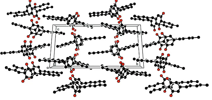
The two hydroxyl groups of the stemphol molecules lead to the formation of chains and rings through O—H···O
hydrogen bonds (Table 1) with graph set motifs C(6), C22(4) and R44(16) (Etter, 1990; Bernstein et al., 1995) all contained
within the glide planes (Fig. 2). Both butyl and pentyl chains, directed in opposite directions and running up and down
with respect to the resorcinol mean plane, adopt essentially fully extended conformations, but with 12.7 (2)° of deviation
between their mean planes defined by C1–C7/C10 and C4–C12/C15 respectively (Fig. 3). The dihedral angles they form
with respect to the (010) plane are 84.3 (2)° and 73.5 (2)° respectively.
S2. Experimental
Gaeumannomyces amomi BCC4066 was cultivated in 20 l of liquid culture and incubated for 21 d at room temperature
(20 °C). Liquid culture was filtrated and extracted separately. The residue (5.0 g) obtained after evaporation of solvent
was subjected to column chromatography over sephadex to afford 30 mg of Stemphol. Colourless needle-shaped crystals
were obtained by re-crystallization from 20% EtOAc in Heptane (m.p. 91 °C).
S3. Refinement
All H atoms were located in difference maps but were treated as riding on their parent atoms, with O—H = 0.82 Å, and C
Figure 1
Molecular view of the compound with the atom labelling scheme. Displacement ellipsoids are drawn at the 50%
probability level. H atoms are represented as small spheres of arbitrary radii.
Figure 2
The crystal packing of the title molecule, viewed down the b axis, showing the molecules connected by O—H···O
[image:3.610.125.482.308.551.2]Figure 3
The crystal packing of the title molecule, viewed down the a axis, showing the segregration between alkyl chains and
resorcinol moieties; H atoms are omitted for clarity.
2-Butyl-5-pentylbenzene-1,3-diol
Crystal data
C15H24O2 Mr = 236.34 Monoclinic, P21/c Hall symbol: -P 2ybc
a = 4.654 (2) Å
b = 25.450 (5) Å
c = 12.790 (4) Å
β = 108.12 (1)°
V = 1439.8 (8) Å3 Z = 4
F(000) = 520
Dx = 1.090 Mg m−3 Melting point: 364 K
Mo Kα radiation, λ = 0.71069 Å Cell parameters from 2700 reflections
θ = 0.4–25.4°
µ = 0.07 mm−1 T = 293 K Needle, colourless 0.50 × 0.10 × 0.08 mm
Data collection
Nonius KappaCCD diffractometer
Radiation source: fine-focus sealed tube Graphite monochromator
φ and ω scans
Absorption correction: multi-scan
(SCALEPACK; Otwinowski & Minor, 1997)
Tmin = 0.881, Tmax = 0.994
15852 measured reflections 2631 independent reflections 1703 reflections with I > 2σ(I)
Rint = 0.029
θmax = 25.3°, θmin = 2.3° h = −5→5
k = −30→30
l = −15→15
Refinement
Refinement on F2 Least-squares matrix: full
R[F2 > 2σ(F2)] = 0.048 wR(F2) = 0.131 S = 1.04 2631 reflections 158 parameters 0 restraints
Secondary atom site location: difference Fourier map
Hydrogen site location: difference Fourier map H-atom parameters constrained
w = 1/[σ2(F
o2) + (0.0625P)2 + 0.1805P] where P = (Fo2 + 2Fc2)/3
Geometry. All e.s.d.'s (except the e.s.d. in the dihedral angle between two l.s. planes) are estimated using the full covariance matrix. The cell e.s.d.'s are taken into account individually in the estimation of e.s.d.'s in distances, angles and torsion angles; correlations between e.s.d.'s in cell parameters are only used when they are defined by crystal symmetry. An approximate (isotropic) treatment of cell e.s.d.'s is used for estimating e.s.d.'s involving l.s. planes.
Refinement. Refinement of F2 against ALL reflections. The weighted R-factor wR and goodness of fit S are based on F2, conventional R-factors R are based on F, with F set to zero for negative F2. The threshold expression of F2 > σ(F2) is used only for calculating R-factors(gt) etc. and is not relevant to the choice of reflections for refinement. R-factors based on F2 are statistically about twice as large as those based on F, and R-factors based on ALL data will be even larger.
Fractional atomic coordinates and isotropic or equivalent isotropic displacement parameters (Å2)
x y z Uiso*/Ueq
O1 0.0951 (3) 0.27129 (5) 0.28490 (10) 0.0555 (4)
H1 0.2648 0.2719 0.3299 0.083*
O2 −0.2986 (2) 0.22721 (5) −0.09324 (9) 0.0517 (4)
H2 −0.2074 0.2253 −0.1386 0.077*
C1 −0.1145 (3) 0.24879 (6) 0.09661 (13) 0.0358 (4)
C2 −0.1012 (3) 0.21631 (7) 0.01107 (13) 0.0383 (4)
C3 0.0994 (4) 0.17495 (7) 0.02531 (14) 0.0428 (4)
H3 0.1000 0.1542 −0.0345 0.051*
C4 0.3006 (3) 0.16426 (7) 0.12888 (14) 0.0414 (4)
C5 0.2925 (3) 0.19620 (7) 0.21530 (14) 0.0432 (5)
H5 0.4233 0.1896 0.2855 0.052*
C6 0.0935 (3) 0.23774 (7) 0.19898 (13) 0.0394 (4)
C7 −0.3211 (4) 0.29564 (7) 0.07705 (15) 0.0454 (5)
H7A −0.3674 0.3038 0.1441 0.054*
H7B −0.5092 0.2871 0.0207 0.054*
C8 −0.1801 (4) 0.34346 (7) 0.04135 (17) 0.0557 (5)
H8A 0.0020 0.3528 0.0999 0.067*
H8B −0.1210 0.3341 −0.0226 0.067*
C9 −0.3832 (5) 0.39095 (8) 0.0136 (2) 0.0756 (7)
H9A −0.5625 0.3822 −0.0468 0.091*
H9B −0.4472 0.3999 0.0766 0.091*
C10 −0.2332 (6) 0.43810 (9) −0.0182 (3) 0.1069 (10)
H10A −0.0721 0.4500 0.0445 0.160*
H10B −0.3790 0.4657 −0.0430 0.160*
H10C −0.1529 0.4286 −0.0762 0.160*
C11 0.5085 (4) 0.11754 (7) 0.14655 (16) 0.0523 (5)
H11A 0.6758 0.1228 0.2134 0.063*
H11B 0.5910 0.1151 0.0858 0.063*
C12 0.3515 (4) 0.06650 (7) 0.15543 (18) 0.0604 (6)
H12A 0.1767 0.0628 0.0905 0.073*
H12B 0.2783 0.0687 0.2185 0.073*
C13 0.5435 (4) 0.01760 (7) 0.16697 (17) 0.0594 (5)
H13A 0.6171 0.0153 0.1040 0.071*
H13B 0.7178 0.0211 0.2322 0.071*
H14A 0.3137 −0.0304 0.2391 0.102*
H14B 0.2060 −0.0354 0.1109 0.102*
C15 0.5672 (6) −0.08146 (9) 0.1840 (2) 0.0970 (9)
H15A 0.6319 −0.0848 0.1200 0.145*
H15B 0.4468 −0.1114 0.1892 0.145*
H15C 0.7409 −0.0796 0.2485 0.145*
Atomic displacement parameters (Å2)
U11 U22 U33 U12 U13 U23
O1 0.0453 (7) 0.0768 (9) 0.0399 (8) 0.0016 (7) 0.0069 (6) −0.0146 (7)
O2 0.0446 (7) 0.0744 (9) 0.0314 (7) 0.0080 (6) 0.0052 (5) 0.0047 (7)
C1 0.0292 (8) 0.0443 (10) 0.0339 (9) −0.0028 (7) 0.0099 (7) 0.0024 (8)
C2 0.0316 (8) 0.0488 (10) 0.0311 (9) −0.0041 (8) 0.0046 (7) 0.0039 (8)
C3 0.0432 (9) 0.0484 (11) 0.0367 (10) 0.0004 (9) 0.0120 (8) −0.0031 (8)
C4 0.0345 (8) 0.0442 (10) 0.0442 (11) −0.0015 (8) 0.0104 (8) 0.0053 (8)
C5 0.0333 (9) 0.0552 (11) 0.0350 (10) −0.0007 (8) 0.0018 (7) 0.0063 (9)
C6 0.0342 (9) 0.0511 (11) 0.0325 (10) −0.0050 (8) 0.0101 (7) −0.0029 (8)
C7 0.0369 (9) 0.0538 (11) 0.0450 (11) 0.0028 (8) 0.0119 (8) −0.0009 (9)
C8 0.0491 (10) 0.0527 (12) 0.0640 (13) 0.0043 (9) 0.0156 (9) 0.0016 (10)
C9 0.0815 (15) 0.0596 (14) 0.0889 (17) 0.0159 (12) 0.0310 (13) 0.0062 (13)
C10 0.131 (2) 0.0567 (16) 0.138 (3) 0.0108 (15) 0.050 (2) 0.0224 (16)
C11 0.0440 (10) 0.0511 (11) 0.0592 (12) 0.0069 (9) 0.0124 (8) 0.0052 (10)
C12 0.0517 (11) 0.0524 (12) 0.0753 (14) 0.0037 (9) 0.0168 (10) 0.0068 (11)
C13 0.0604 (12) 0.0521 (12) 0.0638 (14) 0.0075 (10) 0.0167 (10) 0.0006 (10)
C14 0.0828 (16) 0.0543 (14) 0.113 (2) 0.0030 (12) 0.0242 (15) 0.0058 (13)
C15 0.118 (2) 0.0560 (15) 0.113 (2) 0.0078 (14) 0.0293 (17) −0.0002 (14)
Geometric parameters (Å, º)
O1—C6 1.3899 (19) C9—H9A 0.9700
O1—H1 0.8200 C9—H9B 0.9700
O2—C2 1.3924 (18) C10—H10A 0.9600
O2—H2 0.8200 C10—H10B 0.9600
C1—C2 1.388 (2) C10—H10C 0.9600
C1—C6 1.394 (2) C11—C12 1.512 (3)
C1—C7 1.503 (2) C11—H11A 0.9700
C2—C3 1.381 (2) C11—H11B 0.9700
C3—C4 1.391 (2) C12—C13 1.512 (3)
C3—H3 0.9300 C12—H12A 0.9700
C4—C5 1.382 (2) C12—H12B 0.9700
C4—C11 1.505 (2) C13—C14 1.497 (3)
C5—C6 1.378 (2) C13—H13A 0.9700
C5—H5 0.9300 C13—H13B 0.9700
C7—C8 1.519 (3) C14—C15 1.496 (3)
C7—H7A 0.9700 C14—H14A 0.9700
C8—H8B 0.9700 C15—H15C 0.9600
C9—C10 1.506 (3)
C6—O1—H1 109.5 H9A—C9—H9B 107.8
C2—O2—H2 109.5 C9—C10—H10A 109.5
C2—C1—C6 115.59 (15) C9—C10—H10B 109.5
C2—C1—C7 121.63 (14) H10A—C10—H10B 109.5
C6—C1—C7 122.55 (15) C9—C10—H10C 109.5
C3—C2—C1 122.97 (15) H10A—C10—H10C 109.5
C3—C2—O2 119.73 (15) H10B—C10—H10C 109.5
C1—C2—O2 117.29 (14) C4—C11—C12 112.77 (14)
C2—C3—C4 120.13 (16) C4—C11—H11A 109.0
C2—C3—H3 119.9 C12—C11—H11A 109.0
C4—C3—H3 119.9 C4—C11—H11B 109.0
C5—C4—C3 117.98 (15) C12—C11—H11B 109.0
C5—C4—C11 121.38 (16) H11A—C11—H11B 107.8
C3—C4—C11 120.57 (17) C11—C12—C13 115.46 (16)
C6—C5—C4 120.95 (15) C11—C12—H12A 108.4
C6—C5—H5 119.5 C13—C12—H12A 108.4
C4—C5—H5 119.5 C11—C12—H12B 108.4
C5—C6—O1 120.90 (14) C13—C12—H12B 108.4
C5—C6—C1 122.35 (16) H12A—C12—H12B 107.5
O1—C6—C1 116.74 (14) C14—C13—C12 114.58 (17)
C1—C7—C8 111.85 (14) C14—C13—H13A 108.6
C1—C7—H7A 109.2 C12—C13—H13A 108.6
C8—C7—H7A 109.2 C14—C13—H13B 108.6
C1—C7—H7B 109.2 C12—C13—H13B 108.6
C8—C7—H7B 109.2 H13A—C13—H13B 107.6
H7A—C7—H7B 107.9 C15—C14—C13 115.4 (2)
C9—C8—C7 114.57 (16) C15—C14—H14A 108.4
C9—C8—H8A 108.6 C13—C14—H14A 108.4
C7—C8—H8A 108.6 C15—C14—H14B 108.4
C9—C8—H8B 108.6 C13—C14—H14B 108.4
C7—C8—H8B 108.6 H14A—C14—H14B 107.5
H8A—C8—H8B 107.6 C14—C15—H15A 109.5
C10—C9—C8 113.16 (18) C14—C15—H15B 109.5
C10—C9—H9A 108.9 H15A—C15—H15B 109.5
C8—C9—H9A 108.9 C14—C15—H15C 109.5
C10—C9—H9B 108.9 H15A—C15—H15C 109.5
C8—C9—H9B 108.9 H15B—C15—H15C 109.5
C6—C1—C2—C3 1.2 (2) C7—C1—C6—C5 −176.77 (15)
C7—C1—C2—C3 175.85 (15) C2—C1—C6—O1 176.89 (14)
C6—C1—C2—O2 −177.88 (13) C7—C1—C6—O1 2.3 (2)
C7—C1—C2—O2 −3.2 (2) C2—C1—C7—C8 −83.9 (2)
C1—C2—C3—C4 −0.1 (2) C6—C1—C7—C8 90.4 (2)
C2—C3—C4—C5 −0.2 (2) C7—C8—C9—C10 178.26 (19)
C2—C3—C4—C11 176.65 (15) C5—C4—C11—C12 98.0 (2)
C3—C4—C5—C6 −0.8 (2) C3—C4—C11—C12 −78.7 (2)
C11—C4—C5—C6 −177.58 (15) C4—C11—C12—C13 176.76 (17)
C4—C5—C6—O1 −177.00 (15) C11—C12—C13—C14 −179.8 (2)
C4—C5—C6—C1 2.0 (2) C12—C13—C14—C15 178.6 (2)
C2—C1—C6—C5 −2.2 (2)
Hydrogen-bond geometry (Å, º)
D—H···A D—H H···A D···A D—H···A
O1—H1···O2i 0.82 1.96 2.767 (2) 167
O2—H2···O1ii 0.82 1.95 2.750 (2) 165