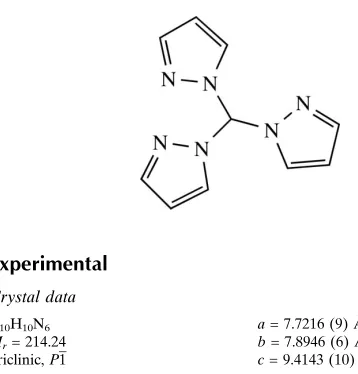
Trispyrazol-1-ylmethane
Tobias Kerscher, Philipp Pust, Richard Betz, Peter Klu¨fers* and Peter Mayer
Ludwig-Maximilians Universita¨t, Department Chemie und Biochemie, Butenandt-strasse 5–13 (Haus D), 81377 Mu¨nchen, Germany
Correspondence e-mail: [email protected]
Received 5 December 2008; accepted 9 December 2008
Key indicators: single-crystal X-ray study;T= 200 K; mean(C–C) = 0.003 A˚;
Rfactor = 0.037;wRfactor = 0.082; data-to-parameter ratio = 14.6.
In the title compound, C10H10N6, the three N atoms in the
2-positions of the pyrazole rings (the ones not bridging to the central C atom are acceptors for weak C—H N contacts with H N distances ranging from 2.49 to 2.59 A˚ ). These furnish the formation of layers perpendicular to [100]. An orthorhombic polymorph of the title compound has already been described [McLauchlan et al. (2004). Acta Cryst. E60, o1419–o1420].
Related literature
The compound was prepared according to a published procedure (Regeret al., 2000). For a structure analysis of the orthorhombic polymorph, see: McLauchlanet al.(2004). For classification of hydrogen bonds, see: Bernstein et al.(1995); Etteret al.(1990).
= 100.023 (9)
= 107.045 (9)
V= 526.36 (10) A˚3
Z= 2
= 0.09 mm T= 200 (2) K 0.340.200.14 mm
Data collection
Nonius KappaCCD diffractometer Absorption correction: analytical
(de Meulenaer & Tompa, 1965) Tmin= 0.975,Tmax= 0.989
4340 measured reflections 2117 independent reflections 1054 reflections withI> 2(I) Rint= 0.030
Refinement
R[F2> 2(F2)] = 0.037
wR(F2) = 0.082
S= 0.83 2117 reflections
145 parameters
H-atom parameters constrained max= 0.13 e A˚3
[image:1.610.51.230.497.684.2]min=0.18 e A˚3
Table 1
Hydrogen-bond geometry (A˚ ,).
D—H A D—H H A D A D—H A
C10—H10 N6i
1.00 2.49 3.451 (2) 161 C6—H6 N2ii
0.95 2.51 3.432 (2) 163 C1—H1 N4iii
0.95 2.59 3.353 (2) 138
Symmetry codes: (i)xþ1;yþ1;zþ1; (ii)x;y1;z; (iii)xþ1;y;z.
Data collection: CrysAlis CCD (Oxford Diffraction, 2005); cell refinement: CrysAlis RED (Oxford Diffraction, 2005); data reduc-tion:CrysAlis RED; program(s) used to solve structure:SHELXS97 (Sheldrick, 2008); program(s) used to refine structure:SHELXL97 (Sheldrick, 2008); molecular graphics:ORTEP-3(Farrugia, 1997) and Mercury(Macraeet al., 2006); software used to prepare material for publication:SHELXL97.
TK thanks the Hanns-Seidel-Stiftung for a PhD scholarship financed by the Bundesministerium fu¨r Bildung und Forschung.
Supplementary data and figures for this paper are available from the IUCr electronic archives (Reference: BT2828).
References
Bernstein, J., Davis, R. E., Shimoni, L. & Chang, N.-L. (1995).Angew. Chem. Int. Ed. Engl.34, 1555–1573.
Etter, M. C., MacDonald, J. C. & Bernstein, J. (1990).Acta Cryst.B46, 256–262. Farrugia, L. J. (1997).J. Appl. Cryst.30, 565.
Macrae, C. F., Edgington, P. R., McCabe, P., Pidcock, E., Shields, G. P., Taylor, R., Towler, M. & van de Streek, J. (2006).J. Appl. Cryst.39, 453–457. McLauchlan, C. C., Varda, A. N. & Giles, J. R. (2004).Acta Cryst.E60, o1419–
Structure Reports Online
supporting information
supporting information
Acta Cryst. (2009). E65, o108 [doi:10.1107/S1600536808041767]
Trispyrazol-1-ylmethane
Tobias Kerscher, Philipp Pust, Richard Betz, Peter Kl
ü
fers and Peter Mayer
S1. Comment
The title compound was synthesized as a neutral tridentate ligand for coordination studies with transition metals.
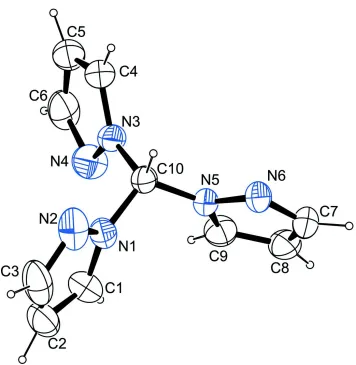
In the molecule, three pyrazole moieties are N-bound to a central C atom (Fig. 1). The molecule is found in a
non-symmetric conformation in the solid state. The highest possible symmetry C3v is broken by the ring containing N4 which
is flipped by about 180°.
If only such contacts whose range falls by about 0.2 Å below the sum of van der Waals radii are considered, the crystal
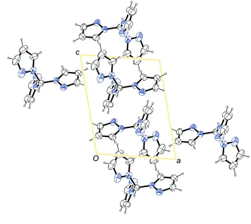
structure shows two C–H···N contacts. Infinite strands along [0 1 0] are formed by C6—H6···N2 contacts (Fig. 2). This
pattern can be described according to graph-set analysis (Etter et al., 1990; Bernstein et al., 1995) with a C(7) descriptor
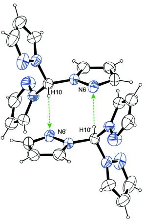
on the unitary level. In addition, dimeric units are formed by interaction of the H atom of C10 and N6 (Fig. 3). These
dimers can be described with a R2
2(8) descriptor on the unitary level. Both these described interactions give rise to tubes
along [0 1 0].
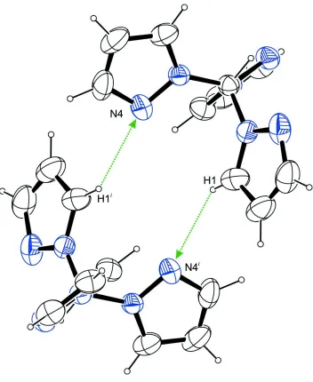
Considering also contacts whose range falls below the sum of van der Waals radii by only about 0.1 Å, a second dimeric
ring system is obvious with a R2
2(12) descriptor formed by the H atom of C1 and N4 (Fig. 4). In combination with the
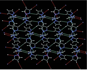
interactions described above, this second ring system features the formation of layers perpendicular to [1 0 0] (Fig. 5).
The molecular packing is shown in Figure 6.
S2. Experimental
The compound was prepared according to a published procedure (Reger et al., 2000) upon reaction of pyrazole and
chloroform in alkaline aqueous media in the presence of a phase-transfer catalyst (tetrabutylammonium chloride).
S3. Refinement
Carbon-bound H atoms were placed in calculated positions (C—H 1.00 Å for the tertiary C atom and C—H 0.95 Å for
Figure 1
The molecular structure of the title compound with atom labels and anisotropic displacement ellipsoids (drawn at 50%
[image:3.610.127.485.487.600.2]supporting information
Figure 3
Dimeric units in the crystal structure of the title compound, formed by intermolecular C–H···N contacts whose ranges fall
by about 0.2 Å below the sum of van der Waals radii of the corresponding atoms, viewed along [0 1 0]. Symmetry code: i
Figure 4
Dimeric units in the crystal structure of the title compound, formed by intermolecular C–H···N contacts whose ranges fall
by about 0.1 Å below the sum of van der Waals radii of the corresponding atoms, viewed along [0 1 0]. Symmetry code: i
supporting information
Figure 5
Figure 6
The packing of the title compound, viewed along [0 1 0].
Tripyrazol-1-ylmethane
Crystal data
C10H10N6
Mr = 214.24
Triclinic, P1 Hall symbol: -P 1 a = 7.7216 (9) Å b = 7.8946 (6) Å c = 9.4143 (10) Å α = 99.292 (8)° β = 100.023 (9)° γ = 107.045 (9)°
Z = 2 F(000) = 224 Dx = 1.352 Mg m−3
Mo Kα radiation, λ = 0.71073 Å Cell parameters from 1494 reflections θ = 3.9–26.3°
µ = 0.09 mm−1
supporting information
Refinement
Refinement on F2
Least-squares matrix: full R[F2 > 2σ(F2)] = 0.037
wR(F2) = 0.082
S = 0.83 2117 reflections 145 parameters 0 restraints
Primary atom site location: structure-invariant direct methods
Secondary atom site location: difference Fourier map
Hydrogen site location: inferred from neighbouring sites
H-atom parameters constrained w = 1/[σ2(F
o2) + (0.04P)2]
where P = (Fo2 + 2Fc2)/3
(Δ/σ)max < 0.001
Δρmax = 0.13 e Å−3
Δρmin = −0.18 e Å−3
Fractional atomic coordinates and isotropic or equivalent isotropic displacement parameters (Å2)
x y z Uiso*/Ueq
N5 0.36455 (16) 0.27051 (16) 0.27731 (13) 0.0334 (3)
N1 0.64066 (18) 0.38873 (17) 0.19270 (15) 0.0379 (3)
N6 0.28341 (18) 0.38479 (16) 0.33973 (14) 0.0409 (4)
N3 0.63243 (17) 0.18619 (16) 0.35571 (14) 0.0367 (3)
C10 0.5661 (2) 0.32745 (19) 0.31338 (16) 0.0334 (4)
H10 0.6125 0.4332 0.4005 0.040*
N2 0.78449 (19) 0.54772 (17) 0.22761 (17) 0.0539 (4)
N4 0.6002 (2) 0.02927 (18) 0.25599 (16) 0.0517 (4)
C7 0.1030 (2) 0.2989 (2) 0.28350 (19) 0.0491 (5)
H7 0.0086 0.3453 0.3072 0.059*
C4 0.7280 (2) 0.1888 (2) 0.49067 (19) 0.0489 (5)
H4 0.7652 0.2845 0.5766 0.059*
C9 0.2372 (2) 0.1197 (2) 0.18445 (18) 0.0444 (4)
H9 0.2619 0.0219 0.1283 0.053*
C8 0.0671 (2) 0.1351 (2) 0.18683 (18) 0.0497 (5)
H8 −0.0510 0.0511 0.1335 0.060*
C6 0.6797 (3) −0.0647 (2) 0.3351 (2) 0.0582 (5)
H6 0.6807 −0.1830 0.2961 0.070*
C1 0.5986 (3) 0.3080 (2) 0.0478 (2) 0.0535 (5)
H1 0.5039 0.1956 −0.0004 0.064*
C2 0.7163 (3) 0.4167 (3) −0.0163 (2) 0.0641 (6)
H2 0.7215 0.3975 −0.1175 0.077*
C3 0.8267 (3) 0.5611 (3) 0.0972 (3) 0.0656 (6)
H3 0.9231 0.6602 0.0842 0.079*
C5 0.7606 (3) 0.0283 (3) 0.4800 (2) 0.0599 (5)
H5 0.8254 −0.0117 0.5560 0.072*
Atomic displacement parameters (Å2)
N2 0.0499 (9) 0.0383 (9) 0.0822 (12) 0.0167 (7) 0.0294 (9) 0.0183 (8) N4 0.0703 (10) 0.0433 (9) 0.0505 (10) 0.0312 (8) 0.0175 (8) 0.0085 (8) C7 0.0349 (11) 0.0708 (13) 0.0519 (12) 0.0241 (9) 0.0156 (9) 0.0242 (10) C4 0.0463 (11) 0.0698 (13) 0.0410 (11) 0.0276 (9) 0.0133 (9) 0.0221 (9) C9 0.0423 (11) 0.0436 (11) 0.0377 (11) 0.0079 (9) 0.0037 (9) 0.0004 (8) C8 0.0372 (11) 0.0635 (13) 0.0390 (11) 0.0066 (9) 0.0030 (9) 0.0105 (9) C6 0.0707 (13) 0.0529 (12) 0.0802 (16) 0.0404 (11) 0.0395 (13) 0.0348 (12) C1 0.0595 (12) 0.0647 (12) 0.0408 (11) 0.0214 (10) 0.0187 (10) 0.0154 (10) C2 0.0760 (15) 0.0869 (15) 0.0625 (14) 0.0479 (12) 0.0409 (13) 0.0427 (13) C3 0.0698 (14) 0.0579 (13) 0.0996 (18) 0.0327 (11) 0.0552 (15) 0.0422 (13) C5 0.0639 (13) 0.0855 (15) 0.0598 (14) 0.0454 (11) 0.0279 (12) 0.0449 (12)
Geometric parameters (Å, º)
N5—C9 1.3516 (18) C7—H7 0.9500
N5—N6 1.3565 (15) C4—C5 1.354 (2)
N5—C10 1.4475 (18) C4—H4 0.9500
N1—C1 1.348 (2) C9—C8 1.357 (2)
N1—N2 1.3562 (16) C9—H9 0.9500
N1—C10 1.4486 (17) C8—H8 0.9500
N6—C7 1.3243 (19) C6—C5 1.378 (2)
N3—C4 1.3473 (19) C6—H6 0.9500
N3—N4 1.3566 (16) C1—C2 1.351 (2)
N3—C10 1.4397 (18) C1—H1 0.9500
C10—H10 1.0000 C2—C3 1.373 (3)
N2—C3 1.336 (2) C2—H2 0.9500
N4—C6 1.330 (2) C3—H3 0.9500
C7—C8 1.378 (2) C5—H5 0.9500
C9—N5—N6 111.92 (13) C5—C4—H4 126.7
C9—N5—C10 130.33 (14) N5—C9—C8 106.78 (15)
N6—N5—C10 117.71 (12) N5—C9—H9 126.6
C1—N1—N2 112.14 (13) C8—C9—H9 126.6
C1—N1—C10 130.61 (13) C9—C8—C7 105.04 (15)
N2—N1—C10 117.11 (13) C9—C8—H8 127.5
C7—N6—N5 103.53 (12) C7—C8—H8 127.5
C4—N3—N4 112.51 (13) N4—C6—C5 112.70 (17)
supporting information
N6—C7—C8 112.73 (15) C2—C3—H3 123.5
N6—C7—H7 123.6 C4—C5—C6 105.23 (17)
C8—C7—H7 123.6 C4—C5—H5 127.4
N3—C4—C5 106.61 (16) C6—C5—H5 127.4
N3—C4—H4 126.7
C9—N5—N6—C7 0.54 (15) C10—N3—N4—C6 −179.60 (13)
C10—N5—N6—C7 178.36 (12) N5—N6—C7—C8 −0.47 (17)
C4—N3—C10—N5 −113.15 (16) N4—N3—C4—C5 0.17 (17)
N4—N3—C10—N5 66.40 (16) C10—N3—C4—C5 179.76 (13)
C4—N3—C10—N1 121.92 (15) N6—N5—C9—C8 −0.41 (17)
N4—N3—C10—N1 −58.53 (17) C10—N5—C9—C8 −177.88 (13)
C9—N5—C10—N3 −51.4 (2) N5—C9—C8—C7 0.10 (17)
N6—N5—C10—N3 131.27 (13) N6—C7—C8—C9 0.24 (19)
C9—N5—C10—N1 73.53 (18) N3—N4—C6—C5 −0.21 (18)
N6—N5—C10—N1 −103.82 (13) N2—N1—C1—C2 −0.57 (19)
C1—N1—C10—N3 73.6 (2) C10—N1—C1—C2 −176.15 (15)
N2—N1—C10—N3 −101.77 (14) N1—C1—C2—C3 0.2 (2)
C1—N1—C10—N5 −51.5 (2) N1—N2—C3—C2 −0.5 (2)
N2—N1—C10—N5 133.13 (13) C1—C2—C3—N2 0.2 (2)
C1—N1—N2—C3 0.65 (17) N3—C4—C5—C6 −0.28 (18)
C10—N1—N2—C3 176.88 (13) N4—C6—C5—C4 0.3 (2)
C4—N3—N4—C6 0.02 (16)
Hydrogen-bond geometry (Å, º)
D—H···A D—H H···A D···A D—H···A
C10—H10···N6i 1.00 2.49 3.451 (2) 161
C6—H6···N2ii 0.95 2.51 3.432 (2) 163
C1—H1···N4iii 0.95 2.59 3.353 (2) 138