1
H
-Indole-3-carbaldehyde azine
Mohd. Razali Rizal, Hapipah M. Ali and Seik Weng Ng*
Department of Chemistry, University of Malaya, 50603 Kuala Lumpur, Malaysia Correspondence e-mail: [email protected]
Received 22 January 2008; accepted 28 January 2008
Key indicators: single-crystal X-ray study;T= 295 K; mean(C–C) = 0.002 A˚;
Rfactor = 0.043;wRfactor = 0.122; data-to-parameter ratio = 15.8.
The molecule of the title compound, C18H14N4, lies on a center of inversion such that there is one half-molecule in the asymmetric unit. The N—N single bond adopts a trans
configuration and the indole fused-ring system is nearly coplanar with the –CH N—N CH– fragment [dihedral angle = 9.8 (2)]. Adjacent molecules are linked by indole– azine N—H N hydrogen bonds into a layer motif.
Related literature
For the synthesis, see: Alemanyet al.(1970); Swaminathan & Narasimhan (1964). For the crystal structures of some aromatic azines, for example, benzalazine, see: Burke-Laing & Laing (1976); Mom & de With (1978); Sinha, 1970). For other heterocyclic aldehyde azines, see: Linet al.(2001a,b); Wuet al.
(2006).
Experimental
Crystal data
C18H14N4
Mr= 286.33
Monoclinic,P21=c
a= 5.0849 (2) A˚
b= 10.6708 (4) A˚
c= 13.4435 (5) A˚
= 94.366 (3)
V= 727.33 (5) A˚3
Z= 2
MoKradiation
= 0.08 mm1
T= 295 (2) K 0.330.270.17 mm
Data collection
Bruker APEX2 diffractometer Absorption correction: none 5388 measured reflections
1659 independent reflections 1085 reflections withI> 2(I)
Rint= 0.038
Refinement
R[F2> 2(F2)] = 0.042
wR(F2) = 0.121
S= 1.01 1659 reflections 105 parameters
H atoms treated by a mixture of independent and constrained refinement
max= 0.17 e A˚3 min=0.16 e A˚3
Table 1
Hydrogen-bond geometry (A˚ ,).
D—H A D—H H A D A D—H A
N1—H1 N2i
0.87 (2) 2.21 (2) 3.065 (2) 167 (2)
Symmetry code: (i)xþ1;yþ1 2;zþ
1 2.
Data collection:APEX2(Bruker, 2007); cell refinement:SAINT (Bruker, 2007); data reduction:SAINT; program(s) used to solve structure:SHELXS97(Sheldrick, 2008); program(s) used to refine structure: SHELXL97 (Sheldrick, 2008); molecular graphics: X-SEED (Barbour, 2001); software used to prepare material for publication:publCIF(Westrip, 2008).
We thank the Science Fund (12–02-03–2031) for supporting this study, and the University of Malaya for the purchase of the diffractometer.
Supplementary data and figures for this paper are available from the IUCr electronic archives (Reference: FL2186).
References
Alemany, A., Bernabe, M., Fernandez Alvarez, E., Lora-Tamayo, M. & Nieto Lopez, O. (1970).Anal. Quim.66, 681–688.
Barbour, L. J. (2001).J. Supramol. Chem.1, 189–191.
Bruker (2007).APEX2andSAINT. Bruker AXS Inc., Madison, Wisconsin, USA.
Burke-Laing, M. & Laing, M. (1976).Acta Cryst.B32, 3216–3224.
Lin, C.-J., Hwang, W.-S. & Chiang, M. J. (2001a).J. Organomet. Chem.640, 85– 92.
Lin, C.-J., Hwang, W.-S. & Chiang, M. J. (2001b).Polyhedron,20, 3257–3264. Mom, V. & de With, G. (1978).Acta Cryst.B34, 2785–2789.
Sheldrick, G. M. (2008).Acta Cryst.A64, 112–122. Sinha, U. C. (1970).Acta Cryst.B26, 889–895.
Swaminathan, S. & Narasimhan, K. (1964).Indian J. Chem.2, 423–424. Westrip, S. P. (2008).publCIF. In preparation.
Wu, C.-Y., Chen, Y., Jing, S.-Y., Lee, C.-S., Dinda, J. & Hwang, W.-S. (2006).
Polyhedron,25, 3053–3065.
Acta Crystallographica Section E
Structure Reports
Online
supporting information
Acta Cryst. (2008). E64, o555 [doi:10.1107/S1600536808003164]
1
H
-Indole-3-carbaldehyde azine
Mohd. Razali Rizal, Hapipah M. Ali and Seik Weng Ng
S1. Comment
Azines are readily synthesized by condensing hydrazine with an aldehyde; the crystal structures of a large number of
substituted benzaldehdye azines have been reported. The structure of the parent aromatic compound, benzalazine, has
been known for a long time (Burke-Laing & Laing, 1976; Mom & de With, 1978; Sinha, 1970). There are few examples
of heterocyclic azines, and their rarity can be attributed to the difficulty of synthesizing the starting aldehyde reactant.
Among the few are, for example, unsubstituted and methyl-subsituted thiophene-2-aldehyde azine (Lin et al., 2001a,
2001b) and a pyrrole derivative has recently been reported (Wu et al., 2006).
3-Indole azine has been known for some time; it was first synthesized from indole-3-carboxaldehyde and hydrazine in
order to examine its psychopharmacological activity (Alemany et al., 1970; Swaminathan Narasimhan, 1964). The title
compound was the unexpected decomposition product of the Schiff base derived from the condensation of
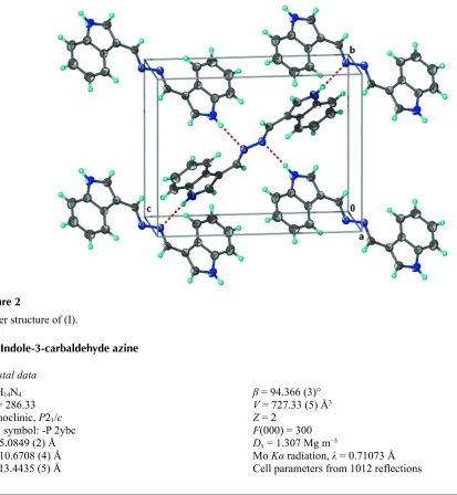
carbohydrazide and indole-3-carboxaldehyde. The molecule (Scheme I, Fig. 1) lies about a center-of-inversion such that
there is half a molecule in the asymmetric unit. The N–N single-bond adopts a trans configuration and the indolyl
fused-ring is nearly coplanar with the –CH=N–N=CH– fragment. Adjacent molecules are linked by an N–Hindole···Nazine hydrogen
bonds into layer motif (Fig. 2).
S2. Experimental
The reaction of carbohydrazide (0.3 g, 3.3 mmol) and indole -3-carboxaldehyde (1 g, 6.6 mmol) in ethanol under reflux
for 2 h gave the corresponding Schiff base. This compound (0.2 g, 0.6 mmol), zinc acetate (0.06 g,0.3 mmol) and several
drops of triethylamine were dissolved in 10 ml e thanol. The contents were heated in a 25-ml, stainless-steel Paar bomb
for for 2 d at 373 K. The bomb was cooled to room temperature over several hours. Well formed crystals were isolated
from the cooled bomb.
S3. Refinement
Carbon-bound H-atoms were placed in calculated positions (C—H 0.93 Å) and were included in the refinement in the
riding model approximation, with U(H) set to 1.2U(C). The amino H-atom was located in a difference Fourier map, and
Figure 1
Displacement ellipsoid plot of (I) at the 50% probability level. H atoms are drawn as spheres of arbitrary radiius.
Figure 2
Layer structure of (I).
1H-Indole-3-carbaldehyde azine
Crystal data
C18H14N4
Mr = 286.33
Monoclinic, P21/c
β = 94.366 (3)°
V = 727.33 (5) Å3
[image:3.610.77.490.290.738.2]θ = 2.3–23.6°
µ = 0.08 mm−1
T = 295 K
Irregular block, green–yellow 0.33 × 0.27 × 0.17 mm
Data collection
Bruker APEX2 diffractometer
Radiation source: fine-focus sealed tube Graphite monochromator
φ and ω scans
5388 measured reflections 1659 independent reflections
1085 reflections with I > 2σ(I)
Rint = 0.038
θmax = 27.5°, θmin = 2.4°
h = −6→6
k = −9→13
l = −17→17
Refinement
Refinement on F2
Least-squares matrix: full
R[F2 > 2σ(F2)] = 0.042
wR(F2) = 0.121
S = 1.01 1659 reflections 105 parameters 0 restraints
Primary atom site location: structure-invariant direct methods
Secondary atom site location: difference Fourier map
Hydrogen site location: inferred from neighbouring sites
H atoms treated by a mixture of independent and constrained refinement
w = 1/[σ2(F
o2) + (0.0645P)2 + ]
where P = (Fo2 + 2Fc2)/3
(Δ/σ)max = 0.001
Δρmax = 0.17 e Å−3
Δρmin = −0.16 e Å−3
Extinction correction: SHELXL97 (Sheldrick, 2008), Fc*=kFc[1+0.001xFc2λ3/sin(2θ)]-1/4
Extinction coefficient: 0.016 (6)
Fractional atomic coordinates and isotropic or equivalent isotropic displacement parameters (Å2)
x y z Uiso*/Ueq
N1 0.4730 (3) 0.8059 (1) 0.1978 (1) 0.0484 (4) N2 0.4464 (2) 0.5165 (1) 0.4519 (1) 0.0422 (4) C1 0.2718 (3) 0.7222 (1) 0.1726 (1) 0.0425 (4) C2 0.1064 (3) 0.7130 (2) 0.0860 (1) 0.0538 (5) C3 −0.0787 (3) 0.6201 (2) 0.0817 (1) 0.0579 (5) C4 −0.1034 (4) 0.5384 (2) 0.1612 (1) 0.0546 (4) C5 0.0606 (3) 0.5473 (1) 0.2473 (1) 0.0454 (4) C6 0.2548 (3) 0.6396 (1) 0.2540 (1) 0.0388 (4) C7 0.4578 (3) 0.6773 (1) 0.3285 (1) 0.0400 (4) C8 0.5819 (3) 0.7783 (1) 0.2901 (1) 0.0466 (4) C9 0.5376 (3) 0.6213 (1) 0.4228 (1) 0.0411 (4)
H1 0.524 (3) 0.865 (2) 0.159 (1) 0.062 (5)*
H2 0.1210 0.7680 0.0330 0.065*
H3 −0.1911 0.6111 0.0243 0.070*
H4 −0.2329 0.4767 0.1561 0.065*
H5 0.0419 0.4926 0.3002 0.054*
H8 0.7215 0.8217 0.3228 0.056*
Atomic displacement parameters (Å2)
U11 U22 U33 U12 U13 U23
N1 0.061 (1) 0.041 (1) 0.043 (1) −0.002 (1) 0.005 (1) 0.012 (1) N2 0.063 (1) 0.036 (1) 0.027 (1) −0.001 (1) −0.003 (1) 0.001 (1) C1 0.049 (1) 0.039 (1) 0.039 (1) 0.006 (1) 0.006 (1) 0.005 (1) C2 0.062 (1) 0.058 (1) 0.040 (1) 0.007 (1) −0.001 (1) 0.014 (1) C3 0.060 (1) 0.065 (1) 0.047 (1) 0.005 (1) −0.008 (1) 0.003 (1) C4 0.054 (1) 0.050 (1) 0.059 (1) −0.003 (1) −0.001 (1) 0.000 (1) C5 0.052 (1) 0.040 (1) 0.045 (1) 0.003 (1) 0.006 (1) 0.004 (1) C6 0.046 (1) 0.035 (1) 0.036 (1) 0.007 (1) 0.007 (1) 0.003 (1) C7 0.052 (1) 0.034 (1) 0.034 (1) 0.004 (1) 0.004 (1) 0.001 (1) C8 0.059 (1) 0.040 (1) 0.041 (1) −0.002 (1) 0.001 (1) 0.003 (1) C9 0.056 (1) 0.036 (1) 0.032 (1) −0.003 (1) −0.001 (1) −0.003 (1)
Geometric parameters (Å, º)
N1—C8 1.351 (2) C6—C7 1.440 (2)
N1—C1 1.381 (2) C7—C8 1.370 (2)
N2—C9 1.283 (2) C7—C9 1.432 (2)
N2—N2i 1.409 (2) N1—H1 0.87 (2)
C1—C2 1.387 (2) C2—H2 0.9300
C1—C6 1.412 (2) C3—H3 0.9300
C2—C3 1.365 (2) C4—H4 0.9300
C3—C4 1.393 (2) C5—H5 0.9300
C4—C5 1.377 (2) C8—H8 0.9300
C5—C6 1.393 (2) C9—H9 0.9300
C8—N1—C1 109.2 (1) N2—C9—C7 123.4 (1)
C9—N2—N2i 112.0 (1) C8—N1—H1 126 (1)
N1—C1—C2 129.9 (1) C1—N1—H1 125 (1)
N1—C1—C6 107.6 (1) C3—C2—H2 121.4
C2—C1—C6 122.5 (2) C1—C2—H2 121.4
C3—C2—C1 117.3 (2) C2—C3—H3 119.2
C2—C3—C4 121.7 (2) C4—C3—H3 119.2
C5—C4—C3 121.2 (2) C5—C4—H4 119.4
C4—C5—C6 118.9 (1) C3—C4—H4 119.4
C5—C6—C1 118.5 (1) C4—C5—H5 120.5
C5—C6—C7 135.2 (1) C6—C5—H5 120.5
C1—C6—C7 106.3 (1) N1—C8—H8 124.8
C8—C7—C9 123.7 (1) C7—C8—H8 124.8
C8—C7—C6 106.5 (1) N2—C9—H9 118.3
C9—C7—C6 129.7 (1) C7—C9—H9 118.3
N1—C8—C7 110.5 (1)
C6—C1—C2—C3 0.4 (2) C5—C6—C7—C9 5.1 (3)
C1—C2—C3—C4 0.7 (3) C1—C6—C7—C9 −175.9 (2)
C2—C3—C4—C5 −0.7 (3) C1—N1—C8—C7 −0.2 (2)
C3—C4—C5—C6 −0.3 (2) C9—C7—C8—N1 176.5 (1)
C4—C5—C6—C1 1.3 (2) C6—C7—C8—N1 −0.2 (2)
C4—C5—C6—C7 −179.7 (2) N2i—N2—C9—C7 −178.9 (1)
N1—C1—C6—C5 178.6 (1) C8—C7—C9—N2 −169.5 (1)
C2—C1—C6—C5 −1.4 (2) C6—C7—C9—N2 6.3 (3)
N1—C1—C6—C7 −0.6 (2)
Symmetry code: (i) −x+1, −y+1, −z+1.
Hydrogen-bond geometry (Å, º)
D—H···A D—H H···A D···A D—H···A
N1—H1···N2ii 0.87 (2) 2.21 (2) 3.065 (2) 167 (2)