ISSN Online: 2169-2483 ISSN Print: 2169-2475
DOI: 10.4236/act.2017.64004 Nov. 8, 2017 21 Advances in Computed Tomography
Acute Epiploic Appendagitis of the Vermiform
Appendix: Typical Computed Tomographic
Image with Pathologic Correlation
Jung Wook Seo
Department of Radiology, Ilsan Paik Hospital, Inje University College of Medicine, Goyang-Si, Korea
Abstract
Epiploic appendages can become ischemic and infarcted, which can present acute abdominal pain that mimics other diseases, such as diverticulitis or ap-pendicitis. Particularly, inflammation of an epiploic appendage attached to the vermiform appendix is a very rare cause of acute abdominal pain, and it is dif-ficult to diagnose this condition preoperatively. We present a case of epiploic appendagitis of the vermiform appendix that was identified with a typical ra-diologic image and was pathologically confirmed. In addition, we review the literature of similar cases and analyze the clinical and radiologic features of EA of the vermiform appendix.
Keywords
Acute Abdomen, Appendix Epiploica, Epiploica Appendagitis, Vermiform Appendix, Computed Tomography
1. Introduction
Epiploic appendagitis (EA) is an uncommon self-limited condition that is caused by inflammation and ischemia related to torsion, or venous thrombosis, of the epiploic appendages [1]. In particular, inflammation or torsion of the epiploic appendages in the vermiform appendix is an extremely rare cause of acute ab-dominal pain and it is difficult to diagnose this condition preoperatively [2]. To our knowledge, fewer than 10 cases have been reported in the English literature since Hambury first reported the disease in 1952 [3]. Although EA is rarely di-agnosed preoperatively; knowledge of this condition is important as it may mimic other forms of acute abdominal pain that require surgery, when it can be
How to cite this paper: Seo, J.W. (2017) Acute Epiploic Appendagitis of the Vermi-form Appendix: Typical Computed Tomo-graphic Image with Pathologic Correlation. Advances in Computed Tomography, 6, 21-27.
https://doi.org/10.4236/act.2017.64004
Received: September 22, 2017 Accepted: November 5, 2017 Published: November 8, 2017
Copyright © 2017 by author and Scientific Research Publishing Inc. This work is licensed under the Creative Commons Attribution International License (CC BY 4.0).
http://creativecommons.org/licenses/by/4.0/
DOI: 10.4236/act.2017.64004 22 Advances in Computed Tomography treated conservatively with pain management. Therefore, clinicians and radiolo-gists should be familiar with the characteristic radiologic features of EA to pre-vent unnecessary surgery.
We report the first case of EA of the vermiform appendix identified with a typical computed tomography image and pathologic correlation, and review 5 additional cases reported in the literature.
2. Case Presentation
A 32-year-old man visited the emergency department complaining of acute right lower abdominal pain that had started 6 hours previously. He had no underlying disease and no other associated symptoms, such as nausea, vomiting, or diarr-hea. His vital signs were immediately assessed: body temperature was 36.6˚C, pulse rate was 71 beats per minute, blood pressure was 145/75 mmHg, and res-piratory rate was 20 breaths per minute. Physical examination revealed marked tenderness in the right lower quadrant. Clinical laboratory results indicated leu-kocytosis (white blood cell count 10,950/mm3, normal: 4000 - 10,000 mm3) and elevated erythrocyte sedimentation rate of 14 mm/h (normal < 12 mm/h). Al-though an intravenous painkiller and fluid therapy were administered, his right lower quadrant pain worsened.
Abdominal radiography showed nonspecific distribution of bowel gas without any sign of bowel obstruction. Abdominopelvic computed tomography (APCT) was performed using a 64-channel multi-detector CT system (Aquilion; Toshiba, Tokyo, Japan), and the contrast medium used was Iohexol (IO-Brix; Taejoon Pharm, Seoul, Korea). APCT showed a 1.5-cm oval, fatty lesion with localized fat stranding and an engorged central vessel closely abutted to the tip of the vermi-form appendix. The proximal portion of the vermivermi-form appendix showed no de-finite evidence of inflammatory change and had a relatively normal diameter (Figure 1(a), Figure 1(b)). However, the surgeon considered the patient to have acute appendicitis and decided to perform an immediate surgical intervention before the CT results were provided.
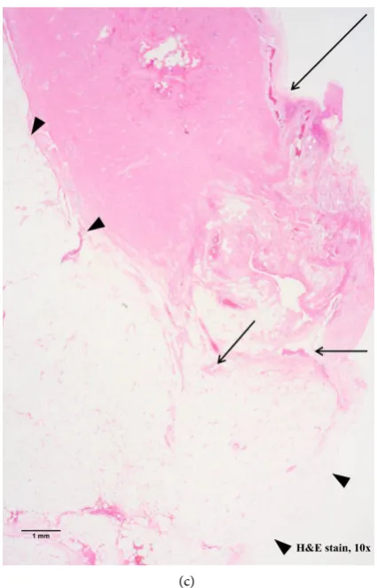
During surgery, the vermiform appendix was found to be normal in appear-ance, with mild inflammatory changes in the tip portion and inflamed epiploic appendages attached to the tip of the vermiform appendix. Therefore, a laparos-copic appendectomy was performed. Pathology showed lobulated fibrofatty tis-sue attached to tip of appendix, with fibrin components, acute inflammation, and a tubular structure suspected to be an engorged vessel, and showed reactive hyperemia in tip of appendix (Figure 1(c)).
3. Discussion
DOI: 10.4236/act.2017.64004 23 Advances in Computed Tomography are larger and more numerous in the descending and sigmoid colon, EA typical-ly manifests as left lower quadrant pain, mimicking acute diverticulitis. Less frequently, it can also involve the cecum and ascending colon, and this may clinically mimics appendicitis. Epiploic appendages can also exist in the vermi-form appendix and are usually smaller than those on the colon. However,
(a)
DOI: 10.4236/act.2017.64004 24 Advances in Computed Tomography (c)
Figure 1. A 32-year-old man with EAA. (a) Axial scan of contrast-enhanced abdominopelvic
computed tomography (CT) shows a fatty oval lesion (arrow) with hyperattenuating rim and fat stranding in the right lower quadrant, around the tip of the vermiform appendix; (b) Multiplanar reconstructed CT scan shows inflamed epiploic appendage (arrowhead) of the tip portion of a normal vermiform appendix (arrow), with a central linear high at-tenuation (short arrow). C = normal cecum; (c) Histopathologic examination shows large fibrofatty tissue (arrowhead) containing tubular structures (short arrows) suspected as engorged vessel and fibrin components, around the tip of the appendix (arrow) (hema-toxylin and eosin stain, × 10).
epiploic appendagitis of the vermiform appendix (EAA) is rare [1]. Since Ham-bury [3] reported the first case of primary EAA, similar cases have been reported by Purysko [1], Aslam [2], Sand [4] and Magnuson [5]; the authors diagnosed the patients as having acute appendicitis and immediate appendectomy was performed.
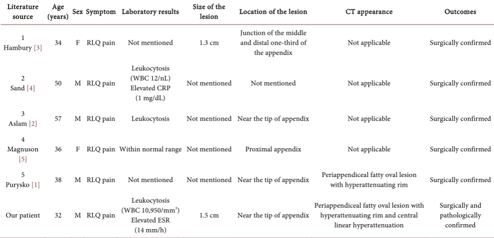
To our knowledge, 5 cases have been previously reported in the English lite-rature, and only one case included radiologic findings [1]. In contrast, our report correlates radiologic and histopathologic findings. The characteristics of the 5 previously reported cases, as well as our own, are summarized in Table 1.
DOI: 10.4236/act.2017.64004 25 Advances in Computed Tomography
Table 1. Clinical and radiologic features of 6 patients with epiploic appendagitis of the vermiform appendix.
Literature
source (years) Age Sex Symptom Laboratory results Size of the lesion Location of the lesion CT appearance Outcomes
1
Hambury [3] 34 F RLQ pain Not mentioned 1.3 cm
Junction of the middle and distal one-third of
the appendix Not applicable Surgically confirmed
2
Sand [4] 50 M RLQ pain
Leukocytosis (WBC 12/nL) Elevated CRP
(1 mg/dL)
Not mentioned Not mentioned Not applicable Surgically confirmed
3
Aslam [2] 57 M RLQ pain Leukocytosis Not mentioned Near the tip of appendix Not applicable Surgically confirmed
4 Magnuson
[5]
36 F RLQ pain Within normal range Not mentioned Proximal appendix Not applicable Surgically confirmed
5
Purysko [1] 38 M RLQ pain Not mentioned Not mentioned Near the tip of appendix Periappendiceal fatty oval lesion with hyperattenuating rim Surgically confirmed
Our patient 32 M RLQ pain
Leukocytosis (WBC 10,950/mm3)
Elevated ESR (14 mm/h)
1.5 cm Near the tip of appendix Periappendiceal fatty oval lesion with hyperattenuating rim and central linear hyperattenuation
Surgically and pathologically confirmed
F = female; M = male; RLQ = right lower quadrant; WBC = white blood cells; CRP = C-reactive protein; ESR = erythrocyte sedimentation rate.
one patient as 1.3 cm, which is similar to that of our patient, which was meas-ured as 1.5 cm. The location of the lesion was provided in 5 cases, including our case. The most common location of the inflamed appendage was near the tip of the vermiform appendix, in 3 cases. In two cases, the locations of the lesions were the proximal appendix the junction of the middle and distal one-third of the appendix.
The imaging features of acute EA have been well described in CT examina-tions, that is the most common modality applied for initial diagnosis of acute abdominal pain. The common CT appearance of EA is a fat attenuating pericolic oval lesion with a hyperattenuating rim. A high attenuated central dot, an irre-gular or a linear focus, may also be present and correspond to thrombosed or engorged central vessels, or central areas of hemorrhage or fibrosis. Wall ening of the adjacent colon may be present but it is most often normal in thick-ness [1]. Other findings include localized edema, which is often manifested as streaky fluid attenuation around the appendage that is the source of the image description of “fat stranding”, and visibility of the appendage serosa either due to fluid or vascular engorgement [6]. There is little difference in radiologic find-ings between epiploic appendagitis of the vermiform appendix and that of the colon. The only difference is that lesions of the vermiform appendix are rela-tively smaller (1.3 to 1.5 cm) than those on the serosal surface of the colon (2 to 4 cm) [1]. In our case, a 1.5-cm fatty, oval lesion was seen adjacent to the tip portion of the vermiform appendix with a hyperattenuating rim and central li-near high attenuation (Figure 1(a), Figure 1(b)).
DOI: 10.4236/act.2017.64004 26 Advances in Computed Tomography caliber, wall enhancement, and thickness, but there can be infiltration of the pe-riappendiceal fat observed on CT due to secondary inflammation [7]. In appen-diceal diverticulitis, the typical CT findings include the presence of a round, outpouching lesion beyond the margin of the appendix with prominent en-hancement of the diverticulum wall and surrounding fat stranding [1].
EA is generally a self-limited disease, with patients spontaneously recovering within 7 to 30 days. Conservative management with oral anti-inflammatory me-dication is currently considered the standard management, once an accurate ra-diological diagnosis has been established [8]. Antibiotics or surgical treatments are rarely warranted and surgical intervention is used only for complications such as inflammation-induced adhesions, secondary abscess, or intestinal occlu-sions [9] [10]. But in our case, laparoscopic appendectomy was performed by the surgeon, prior to confirmation by CT.
EAA is often misdiagnosed because clinical symptoms are nonspecific and similar to those of other diseases that induce right lower quadrant pain, such as acute appendicitis or diverticulitis, including abdominal pain, nausea, and vo-miting. However, if radiologists are familiar with the characteristic radiologic findings of EAA, it is possible to avoid unnecessary surgery, such as negative appendectomy.
We present the first case of EAA in which a typical radiologic image was con-firmed by the pathologic findings.
Financial Disclosure
No funding support or grant was received for this study.
References
[1] Purysko, A.S., Remer, E.M., Filho, H.M., Bittencourt, L.K., Lima, R.V. and Racy, D.J. (2011) Beyond Appendicitis: Common and Uncommon Gastrointestinal Caus-es of Right Lower Quadrant Abdominal Pain at Multidetector CT. Radiographics, 31, 927-947.https://doi.org/10.1148/rg.314105065
[2] Aslam, M.B. and Hasan, N. (2009) Torsion of an Appendix Epiploica Present at the Vermiform Appendix: A Rare Cause of Acute Abdomen. Turkish Journal of Trau-ma and Emergency Surgery (Ulusal TravTrau-ma Dergisi), 15, 509-510.
[3] Hambury, H.J. (1952) Torsion of an Appendix Epiploica of the Vermiform Appen-dix. British Journal of Surgery, 40, 176-177.
https://doi.org/10.1002/bjs.18004016019
[4] Sand, M., Bonhag, G., Bechara, F.G., Sand, D. and Mann, B. (2009) An Inflamed Necrotic Appendix Epiploicum with Immediate Contact to a Non-Inflamed Ap-pendix Vermiformis: A Case Report.Journal of Medical Case Reports, 3, 57.
https://doi.org/10.1186/1752-1947-3-57
[5] Magnuson, A., Reber, H., Reyna, P., Ries, D. and Aanning, H.L. (2006) Infarcted Epiploic Appendage of the Vermiform Appendix Masquerading as Acute Appendi-citis. The Journal of the South Dakota State Medical Association, 59, 511-513. [6] Eberhardt, S.C., Strickland, C.D. and Epstein, K.N. (2016) Radiology of Epiploic
DOI: 10.4236/act.2017.64004 27 Advances in Computed Tomography
History. Abdominal Radiology (New York), 41, 1653-1665.
https://doi.org/10.1007/s00261-016-0757-0
[7] Nadida, D., Amal, A., Ines, M., Makram, M., Amira, M., Leila, B.F. and Lotfi, H. (2016) Acute Epiploic Appendagitis: Radiologic and Clinical Features of 12 Patients.
International Journal of Surgery Case Reports, 28, 219-222.
https://doi.org/10.1016/j.ijscr.2016.09.015
[8] Almuhanna, A.F., Alghamdi, Z.M. and Alshammari, E. (2016) Acute Epiploic Ap-pendagitis: A Rare Cause of Acute Abdomen and a Diagnostic Dilemma. Journal of Family and Community Medicine, 23, 48-50.
https://doi.org/10.4103/2230-8229.172234
[9] Sand, M., Gelos, M., Bechara, F.G., Sand, D., Wiese, T.H., Steinstraesser, L. and Mann, B. (2007) Epiploic Appendagitis—Clinical Characteristics of an Uncommon Surgical Diagnosis. BMC Surgery, 7, 11.https://doi.org/10.1186/1471-2482-7-11
[10] Singh, A.K., Gervais, D.A., Hahn, P.F., Sagar, P., Mueller, P.R. and Novelline, R.A. (2005) Acute Epiploic Appendagitis and Its Mimics. Radiographics, 25, 1521-1534.
https://doi.org/10.1148/rg.256055030
Abbreviations
APCT abdominopelvic computed tomography CT computed tomography
EA epiploic appendagitis
EAA epiploic appendagitis of the vermiform appendix WBC white blood cells
CRP C-reactive protein