*Present address: Howard Florey Institute of Experimental Physiology and Medicine, University of Melbourne, Parkville, Victoria, Australia.
Key words: fish, Oreochromis mossambicus, Cyprinus carpio, Oncorhynchus mykiss, gills, Ca2+ transport, phosphatase, stanniocalcin, teleocalcin.
INDICATIONS FOR TWO BIOACTIVE PRINCIPLES IN
THE CORPUSCLES OF STANNIUS
P. M. VERBOST, A. BUTKUS*, P. WILLEMS and S. E. WENDELAAR BONGA
Department of Animal Physiology, Faculty of Science, University of Nijmegen, Toernooiveld, 6525 ED Nijmegen, The Netherlands
Accepted 14 December 1992
Summary
For a long time it was thought that the corpuscles of Stannius (CS) of holostean and teleostean fishes produce a single hormone reducing Ca2+influx from the water via the
gills. We here present data showing that two separate bioactive principles are present in the CS: stanniocalcin (STC), a 56kDa glycoprotein, and teleocalcin (TC), a 3kDa glycopeptide. STC indeed inhibits Ca2+influx (as reported many times before) but does
not affect the Ca2+- and Mg2+-dependent phosphatase activity located in the gill plasma
membrane. TC does not affect Ca2+influx but inhibits the Ca2+- and Mg2+-dependent
phosphatase activity. Thus, the Ca2+- and Mg2+-dependent phosphatase activity appears
not to be involved in transbranchial Ca2+transport. We conclude that STC is the pivotal
calcium-regulating hormone in fish and that TC has an as yet unidentified role in gill physiology through its phosphatase-reducing activity.
Introduction
Salmon TC (sTC) inhibits a Ca2+ (or Mg2+)-dependent phosphatase (CaP
iase). This activity was erroneously proposed to be the driving force for Ca2+uptake (Ma et al. 1974; Ma and Copp, 1978). Later it was argued and demonstrated by Flik et al. (1984) that a high-affinity Ca2+-ATPase is responsible for transcellular Ca2+ uptake. Ma and Copp (1982) claimed that sTC reduces Ca2+influx in eels, but this claim is not justified since in their influx experiments CS extracts containing both STC and TC were used. We have examined the effects of STC and TC from two different species, trout and carp, on branchial Ca2+influx in tilapia and carp. We used CS material from these two species to reduce the risk of measuring some species-specific effect. Furthermore, we tested the effects of TC and STC in the assay for CaPiase activity developed by Ma et al. (1974). Trout TC (tTC), trout STC (tSTC) and synthetic hormone fragments of Australian eel STC (eSTC), peptides U, V and W (Butkus et al. 1989), were compared. The CaPiase activity was determined on purified basolateral membranes that are essentially free of apical and intracellular membranes (Flik et al. 1985b). The role of the two bioactive principles in branchial Ca2+handling will be discussed.
Materials and methods
Fish
Tilapia, Oreochromis mossambicus, were obtained from a laboratory stock. Tilapia used for Ca2+flux studies weighed 60–100g and those used for gill membrane isolation around 200g. Fish were held in Nijmegen city tapwater (containing in mmol l21: 0.8 Ca2+; 0.20 Mg2+, 0.61 Na+, 0.05 K+, 0.66 Cl2, 0.32 SO
422, 3.15 HCO32, pH7.2) at 27˚C. Common carp, Cyprinus carpio, weighing 70–120g were obtained from laboratory stock (Agricultural University, Wageningen, The Netherlands) and held in Nijmegen city tapwater at 23˚C. Rainbow trout, Oncorhynchus mykiss, were obtained from a commercial dealer in Beek near Nijmegen.
Analytical techniques
The protein contents of cell membrane preparations and cell suspensions were determined with a commercial reagent kit (Biorad) according to the method of Bradford (1976). Concentrations of hormones were quantitated according to the method of Lowry
et al. (1951); this method is more suited for the determination of small peptides (ø3 kDa). Radiotracer activities were determined with a Wallac 1410 liquid scintillation counter (Pharmacia/LKB).
Hormones
desalting by ultrafiltration. A Western blot of purified tSTC has been published before (Flik et al. 1990).
Trout and carp teleocalcin (tTC and cTC) were purified from a CS homogenate using a Sephadex G-25 column (fine, Pharmacia; 10mm3450mm, 5–8mg protein per run, 0.5mlmin21elution rate) and ammonium acetate as eluent according to the procedure described by Ma and Copp (1978) for the purification of salmon TC. The major peak, measured spectrophotometrically at 280nm, was collected (Fig. 1) and freeze dried. The apparent size of the product was around 3kDa and it contained a carbohydrate moiety, as determined with a glycan detection kit (Boehringer). This teleocalcin has a negative charge (in polyacrylamide gel it runs faster than the front marker) and contains carbohydrates with terminal mannoses as determined with a glycan differentiation kit (Boehringer).
Synthetic fragments of Australian eel, Anguilla australis, STC (eSTC) were prepared at the Howard Florey Institute peptide laboratory (Butkus et al. 1989) based on the cDNA amino acid sequence of the mature hormone (Butkus et al. 1987). The following three peptides were used: peptide U (N-terminal 1–20 sequence), peptide V (mid 103–136 sequence) and peptide W (C-terminal 202–231 sequence). Numbering of the amino acids starts at the N terminus of the mature hormone.
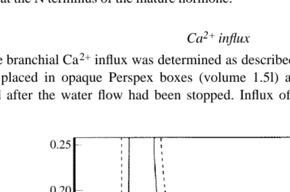
Ca2+ influx
[image:3.612.86.373.393.583.2]The branchial Ca2+influx was determined as described by Verbost et al. (1989). Fish were placed in opaque Perspex boxes (volume 1.5l) and 45CaCl2 (1.0MBq l21) was added after the water flow had been stopped. Influx of Ca2+ was calculated from the
Fig. 1. Elution pattern of aqueous corpuscle of Stannius (CS) extracts (___) and kidney extracts (dashed line) using gel permeation chromatography (G-25, fine). The STC (void volume) and TC fractions of trout CS extract and the corresponding fractions of trout kidney extract are indicated as vertical lines.
0.25
0.20
0.15
0.10
0.05
0
radioactivity accumulated in the fish after 3h of exposure to 45Ca (the radioactivity in the entire fish was determined) and the mean 45Ca specific activity of the water. Ca2+influx (Fin) data were normalized to fish mass according to Flik et al. (1985a) and expressed in mmol h21100 g21fish.
Hormones were administered by intraperitoneal injection 1h before the flux determination. Saline served as vehicle and was used in the controls.
Isolation of plasma membranes
The purification of the branchial plasma membranes was carried out as described by Flik et al. (1985b). After quick anaesthesia in Na2CO3-buffered MS-222 (1 gl21, pH7.4) the gill arches were excised. Branchial epithelium was scraped off with a glass microscope slide and collected in isotonic buffer (containing in mmol l21: 250 sucrose, 12.5 NaCl, 5 Hepes/Tris pH7.4, 0.1 EDTA and 25TIU l21aprotinin, where TIU is one trypsin inhibitor unit). After homogenization in a douncer device with a loosely fitting pestle, cellular debris and erythrocytes were separated from the membranes by centrifugation (550 g, 10min). The supernatant (H0) was centrifuged (250000 g, 30min), yielding a two-layered pellet of mitochondria and membranes. The fluffy layer of the pellet containing the plasma membranes was collected by mild swirling and subsequently resuspended in isotonic sucrose buffer by 100 strokes in a douncer. The suspension was further purified by differential centrifugation: 1000 g, 10min and 10000 g, 10min. Finally, the membranes were pelleted (50000 g, 20min) and resuspended in 0.3mol l21 sucrose for storage (at 220˚C for up to 14 days without significant loss of cyclase and phosphatase activity). The Na+/K+-ATPase activity, a marker enzyme for basolateral membranes, was purified 3.9 times in the final pellet compared to the initial homogenate H0 (determined before freezing the membranes). This is in good agreement with the purification (3.8 times) reported by Flik et al. (1985b) using the same isolation procedure.
Phosphatase assay
The phosphatase activity of the isolated membranes was determined as described by Ma et al. (1974) with minor modifications. In a differential assay, with or without 5 mmol l21Ca2+or Mg2+, the Ca2+/Mg2+-dependent release of inorganic phosphate, Pi, from Ca2+-ATP or Mg2+-ATP was determined. Apart from Ca2+and Mg2+, the reaction mixture contained 30mg ml21 membrane protein, 70mmol l21 NaCl, 20mmol l21 Hepes/Tris (pH8.0), 1mgml21 ouabain (to exclude Na+/K+-ATPase activity) and 3 mmol l21ATP in a total volume of 350ml. Assays were run at 37˚C for 30min.
The phosphatase inhibitory activity of CS compounds has been expressed in CS units per milligram (Ma and Copp, 1978), where 1 unit is the amount of CS material that causes 50% inhibition of Ca2+-dependent ATP hydrolysis.
Statistics
Results
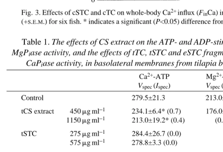
Effects of STC and TC on Ca2+influx
Ca2+influx in tilapia is reduced to 52% of the control level by injection of 56pmolg21 tSTC; tTC (up to 300pmol g21) had no effect (Fig. 2). Carp STC (24pmol g21) reduced Ca2+influx in tilapia to 53% of the control level, whereas cTC (up to 200pmol g21) had no effect. In carp, cSTC (7pmol g21) reduced Ca2+ influx to 59%; injection of 40pmol g21cTC was without effect on Ca2+influx (Fig. 3).
Effects of tSTC and tTC on phosphatase
Table 1 shows the effects of CS extracts, tSTC and tTC on the Ca2+-dependent phosphatase (CaPiase ) activity in tilapia gill plasma membranes. Extracts of CS and tTC inhibit CaPiase activity, whereas tSTC at concentrations up to 10mmol l21 does not. Incubation with tTC decreased CaPiase activity with an average specific inhibitory activity of 11.3. It follows from Table 1 that tTC was purified 20.1 times, based on its specific inhibition. Table 1 also shows that phosphate release was decreased by CS extracts when Mg2+-ATP or Ca2+-ADP was used as substrate. The corresponding controls show that when Ca2+was replaced by Mg2+, ATP hydrolysis decreased by 25%. With Ca2+-ADP as substrate, CaPiase activity reached 62% of the Ca2+-ATP value. The synthetic eSTC fragments U, V and W did not affect CaPiase activity at concentrations up to 50mmol l21(corresponding to 99mg ml21U, 186mg ml21V and 161mg ml21W).
Discussion
Two major conclusions can be drawn from this study. First, stanniocalcin (STC) inhibits Ca2+influx without affecting the Ca2+-dependent phosphatase (CaPiase). Second,
4
3
2
1
0
[image:5.612.81.362.70.249.2]Control tSTC 56pmolg−1 cSTC 24pmolg−1 tTC 300pmolg−1 cTC 24pmolg−1 cTC 200pmolg−1 Tilapia
Fig. 2. Effects of STC and TC from trout (t) and carp (c) on whole-body Ca2+influx (FinCa) in tilapia. Values are means (+S.E.M.) for six fish. * indicates a significant (P<0.05) difference from the control value.
5
4
3
2
1
0
Carp
[image:6.612.134.337.65.251.2]Control cSTC 7pmolg−1 cTC 40pmolg−1
*
Fig. 3. Effects of cSTC and cTC on whole-body Ca2+ influx (FinCa) in carp. Values are means (+S.E.M.) for six fish. * indicates a significant (P<0.05) difference from the control value.
Table 1. The effects of CS extract on the ATP- and ADP-stimulated CaPiase and
MgPiase activity, and the effects of tTC, tSTC and eSTC fragments on ATP-stimulated
CaPiase activity, in basolateral membranes from tilapia branchial epithelium
Ca2+-ATP Mg2+-ATP Ca2+-ADP
Vspec (Ispec) Vspec (Ispec) Vspec (Ispec)
Control 279.5±21.3 213.0±10.8 172.3±19.2
tCS extract 450mg ml−1 234.1±6.4* (0.7) 176.0±11.8* 142.7±4.6* 1150mg ml−1 213.0±19.2* (0.4) (0.8) (0.8) tSTC 275mg ml−1 284.4±26.7 (0.0)
575mg ml−1 278.8±3.3 (0.0) tTC 30mg ml−1 238.6±11.1* (9.8)
75mg ml−1 150.3±6.7* (12.3) Fragment U 1mmol l−1 277.2±11.5 (0.0)
25mmol l−1 281.6±35.6 (0.0) 50mmol l−1 285.7±11.4 (0.0) Fragment V 1mmol l−1 280.4±10.8 (0.0) 25mmol l−1 284.2±22.0 (0.0) 50mmol l−1 277.0±19.7 (0.0) Fragment W 1mmol l−1 177.6±14.3 (0.0) 25mmol l−1 268.6±16.9 (0.0) 50mmol l−1 277.9±20.2 (0.0) Values are means of five experiments (±S.D.).
Vspecin mmol Pih−1mg−1protein.
Ispec, between parentheses, is the specific inhibitory activity in unitsmg−1, where 1 unit of (CS) material causes 50% inhibition of CaPiase activity (Ma and Copp, 1978).
[image:6.612.50.416.332.581.2]teleocalcin (TC), a glycopeptide of around 3kDa from trout CS extracts, inhibits CaPiase but does not affect branchial Ca2+influx. The biochemistry and bioactivity of this CS fraction indicate similarities with the 3kDa glycopeptide isolated from salmon CS by Ma and Copp (1978). STC is a fast calcium-regulating hormone of the CS, inhibiting branchial Ca2+influx. Thus, the CaP
iase appears not to be involved in the regulation of transcellular Ca2+influx. An important conclusion from this work is that the CS contain at least two bioactive principles and, therefore, that studies showing the effects of CS extracts on branchial ion handling (Flik, 1990; Mayer-Gostan, 1992) should be re-evaluated.
Ca2+influx
Branchial influx of Ca2+was reduced by tSTC and cSTC, whereas tTC and cTC had no effect. Higher doses of TC than of STC were examined to show that TC did not have an effect on Ca2+influx, thus taking into account the possibility that TC is a fragment of the active principle and may need a higher dose to produce the same effect. It has been determined, for instance, that an N-terminal fragment of STC inhibits Ca2+ influx in tilapia to a similar extent as the native hormone when it is used at a ten times higher dose than the whole hormone (P. M. Verbost, A. Butkus, P. Willems and S. E. Wendelaar Bonga, in preparation).
To obtain a significant inhibition of influx in tilapia we needed a fairly high dose of tSTC (56pmol g21) compared with the dose that is required to accomplish the same inhibition of Ca2+influx in trout (10pmol g21). When testing cSTC in carp, 3.5 times lower doses than those needed in tilapia were sufficient to obtain significant inhibition of influx. It is tempting to conclude that there is a species-specificity whereby tilapia and carp STC are more closely related than tilapia and trout STC. However, we cannot exclude the possibility that the different hormone preparations had dissimilar contents of bioactive STC.
Phosphatase
The phosphatase activity in the plasma membranes from the gills of tilapia is activated by Ca2+as well as Mg2+. ATP is the preferred substrate for the CaPiase. The activation characteristics match those described previously for CaPiase activity in gill plasma membranes of rainbow trout (Ma et al. 1974) and eel (Flik et al. 1983). The only discrepancy seems to be the almost 30-fold higher specific enzyme activity in tilapia than in trout, which in all probability is explained by the difference in assay temperature (37˚C
versus 12˚C). Compared with the phosphatase activity in eel gill plasma membranes
measured at 37˚C by Flik et al. (1983) our tilapia value is five times higher.
It is not known whether TC is released by the CS and no data on plasma levels are available. The possibility that this factor is not released by the glands and is irrelevant for branchial ion regulation should be considered. We did exclude, however, the possibility that the factor originated from kidney tissue, a source of contamination in the collection of CS tissue. Kidney contaminants may be co-purified with CS material and exert effects that would erroneously be ascribed to the CS. However, the kidney extracts did not contain a 3kDa product corresponding to the CS-TC and none of the kidney compounds reduced the phosphatase activity: trout kidney extract (0.25–1.00mgml21) or its gel permeation fractions corresponding to STC (0.025–0.575mgml21) or TC (0.015– 0.100mgml21) from CS extracts had no effect on the CaPiase (tested five times, data not shown). This finding also argues against the (remote) possibility that the inhibitory effect is caused by salts that co-elute with TC in the isolation procedure (also see the amino acid composition below).
The CaPiase activity is changed neither by tSTC nor by the eSTC fragments U, V or W. Branchial Ca2+influx is reduced by tSTC and fragment U in tilapia and trout (Lafeber et
al. 1988a; Milliken et al. 1990; P. M. Verbost, J. Van Rooij, G. Flik, R. A. C. Lock and
S. E. Wendelaar Bonga, in preparation; this study). From these results, we conclude that STC does not exert its hypocalcaemic effects through changes in CaPiase activity in the gill. It has previously been shown that CaPiases cannot be responsible for the translocation of Ca2+across the basolateral membrane because the affinity for Ca2+is too low to be stimulated by intracellular Ca2+ concentrations and because the substrate specificity and pH optima are not characteristic for a transport Ca2+-ATPase (Flik et al. 1984). We conclude now that the CaPiase does not play a role in active branchial Ca2+ transport.
Teleocalcin
One could argue that TC is a fragment of the native STC. However, the carbohydrate moiety of TC carries terminal mannoses but no terminal sialic acids as STC does (P. M. Verbost, A. Butkus, P. Willems and S. E. Wendelaar Bonga, unpublished observations, obtained with a Boehringer glycan differentiation kit on dot blots). A polyclonal antibody against tSTC did not recognize TC (tested on dot blot; 1mg material per dot) but this does not exclude the possibility that TC is a fragment of STC because it may not contain the moiety that is reactive to the antibody.
as the sTC used for the analysis. However, the ratio of these seven amino acids relative to valine in tTC (Ala, 3.2; Asn, 1.2; Gln, 4.0; Gly, 4.8; Ser, 2.8; Thr, 1.7; Val, 1.0) was similar to that in sTC, indicating that tTC contains the TC sequence. A major difference between tTC and sTC seems to be the effect on Ca2+metabolism. Ca2+influx in tilapia was not influenced by tTC or cTC. Ma and Copp (1978) showed hypocalcaemic effects of purified sTC. They reported that the Sephadex G-50 fraction of salmon CS decreased the total plasma Ca2+concentration in American eels by 14% after four daily injections with 340pmol g21bodymass. However, in their experiments CS, not TC, extracts were used to show the inhibition of gill Ca2+uptake, and CS extracts contain both STC and TC. A possible explanation for the hypocalcaemic effect of TC is that it stimulated Ca2+efflux, thus reducing plasma calcium concentration. The hypothesis that should now be tested is whether TC controls a mechanism for the regulation of Ca2+efflux.
Ma and Copp (1978) called the 3kDa glycopeptide from the Stannius corpuscles TC. Therefore, in retrospect, it was confusing that Wagner et al. (1986) also called the 39kDa glycoprotein from salmon CS TC; this protein turned out to be homologous with STC (also named ‘hypocalcin’ till 1990), the genuine Ca2+-influx-reducing hormone. We suggest that the name TC should be reserved for the 3kDa moiety, the potentially interesting glycopeptide from the CS.
We thank Dr G. Flik and Dr R. G. J. M. Hanssen for valuable discussions during the performance of this study and for critical reading of the manuscript. The research of P.M.V. was made possible by a fellowship of the Royal Netherlands Academy of Arts and Sciences. The work of A. B. was supported by an Institute grant from the National Health and Medical Research Council of Australia.
References
BRADFORD, M. M. (1976). A rapid and sensitive method for the quantitation of microgram quantities of protein utilizing the principle of protein–dye binding. Analyt. Biochem. 72, 248–254.
BUTKUS, A., ROCHE, P. J., FERNLEY, R. T., HARALAMBIDIS, J., PENSCHOW, J. D., RYAN, G. B., TRAHAIR, J. F., TREGEAR, G. W. ANDCOGHLAN, J. P.(1987). Purification and cloning of a corpuscles of Stannius protein from Anguilla australis. Molec. cell. Endocr. 54, 123–133.
BUTKUS, A., YATES, N. A., COPP, D. H., MILLIKEN, C. E., MCDOUGALL, J. G., ROCHE, P. J., TREGEAR, G. W. ANDCOGHLAN, J. P. (1989). The corpuscles of Stannius protein: Processing and bioactivity.
Fish Physiol. 7, 359–365.
COPP, D. H. ANDMA, S. W. Y. (1981). Teleocalcin and calcium regulation in bony fish. In Hormonal
Control of Calcium Metabolism (ed. D. V. Cohn, V. Talmage and J. L. Matthews), pp. 298–306.
Amsterdam: Exerpta Medica.
FENWICK, J. C. (1982). Some evidence concerning the nature of the hypocalcemic factor in the corpuscles of Stannius. In Comparative Endocrinology of Calcium Regulation (ed. C. Oguro and P. K. T. Pang), 167pp. Tokyo: Jap. Sci. Soc. Press.
FLIK, G. (1990). Hypocalcin physiology. In Progress in Comparative Endocrinology (ed. A. Epple, C. G. Scanes and M. H. Stetson), vol. 342, pp. 578–585. New York: Wiley-Liss, Inc.
FLIK, G., FENWICK, J. C., KOLAR, Z., MAYER-GOSTAN, N. ANDWENDELAARBONGA, S. E. (1985a). Whole-body calcium flux rates in cichlid teleost fish Oreochromis mossambicus adapted to freshwater. Am. J. Physiol. 249, R432–R437.
FLIK, G., LABEDZ, T., NEELISSEN, J. A. M., HANSSEN, R. G. J. M., WENDELAARBONGA, S. E. ANDPANG, P. K. T. (1990). Rainbow trout corpuscles of Stannius: stanniocalcin synthesis in vitro. Am. J. Physiol. 258, R1157–R1164.
FLIK, G., VAN RIJS, J. H. ANDWENDELAARBONGA, S. E. (1985b). Evidence for high-affinity Ca2+ -ATPase activity and ATP-driven Ca2+-transport in membrane preparations of the gill epithelium of the cichlid fish Oreochromis mossambicus. J. exp. Biol. 119, 335–347.
FLIK, G., WENDELAARBONGA, S. E. ANDFENWICK, J. C. (1983). Ca2+-dependent phosphatase and Ca2+ -dependent ATPase activity in plasma membranes of eel gill epithelium. I. Identification of Ca2+ -activated ATPase activities with non-specific phosphatase activities. Comp. Biochem. Physiol. 76B, 745–754.
FLIK, G., WENDELAARBONGA, S. E. ANDFENWICK, J. C. (1984). Ca2+-dependent phosphatase and Ca2+ -dependent ATPase activity in plasma membranes of eel gill epithelium. II. Evidence for transport high affinity Ca2+-ATPase. Comp. Biochem. Physiol. 79B, 9–16.
LAFEBER, F. P. J. G., FLIK, G., WENDELAARBONGA, S. E. ANDPERRY, S. F. (1988a). Hypocalcin from Stannius corpuscles inhibits gill calcium uptake in trout. Am. J. Physiol. 254, R891–R896.
LAFEBER, F. P. J. G., HANSSEN, R. G. J. M., CHOY, Y. M., FLIK, G., HERMANN-ERLEE, M. P. M., PANG, P. K. T. AND WENDELAARBONGA, S. E. M. (1988b). Indentification of hypocalcin (teleocalcin) isolated from trout Stannius corpuscles. Gen. comp. Endocr. 69, 19–30.
LOWRY, O. H., ROSEBROUGH, N. J., FARR, A. L. ANDRANDALL, R. J. (1951). Protein measurement with the folin phenol reagent. J. biol. Chem. 193, 265–275.
MA, S. W. Y. ANDCOPP, D. H. (1978). Purification, properties and action of a glycopeptide from the corpuscles of Stannius which affects calcium metabolism in the teleost. In Comparative
Endocrinology (ed. P. J. Gaillard and H. H. Boer), pp. 283–286. Amsterdam: Elsevier.
MA, S. W. Y. ANDCOPP, D. H. (1982). Role of the corpuscles of Stannius and teleocalcin in calcium regulation in the teleost. In Comparative Endocrinology of Calcium Regulation (ed. C. Oguro and P. K. T. Pang), pp. 173–179. Tokyo: Jap. Sci. Soc. Press.
MA, S. W. Y., SHAMI, Y., MESSER, H. H. ANDCOPP, D. H. (1974). Properties of Ca2+-ATPase from the gill of rainbow trout (Salmo gairdneri). Biochim. biophys. Acta 345, 243–251.
MAYER-GOSTAN, N. (1992). Effects of stannius corpuscles removal on branchial sodium exchanges in the eel (Anguilla anguilla). Second International Symposium on Fish Endocrinology. p. 102. MILLIKEN, C. E., FARGHER, R. C., BUTKUS, A., MCDONALD, M. ANDCOPP, D. H. (1990). Effects of
synthetic peptide fragments of teleocalcin (hypocalcin) on calcium uptake in juvenile rainbow trout (Salmo gairdneri). Gen. comp. Endocr. 77, 416–422.
PANG, P. K. T., PANG, R. K., LIU, V. K. Y. ANDSOKABE, H. (1981). Effects of fish angiotensins and angiotensin-like substances on killifish calcium regulation. Gen. comp. Endocr. 43, 292–298. THIEDE, M. A., YOON, K., GLOLUB, E. E., NODA, M. ANDRODAN, G. (1988). Structure and expression of
rat osteosarcoma (ROS 17/2.8) alkaline phosphatase: product of a single copy gene. Proc. natn. Acad.
Sci. U.S.A. 85, 319–323.
VERBOST, P. M., VANROOIJ, J., FLIK, G., LOCK, R. A. C. ANDWENDELAARBONGA, S. E. (1989). The movement of cadmium through freshwater trout branchial epithelium and its interference with calcium transport. J. exp. Biol. 145, 185–197.
WAGNER, G. F., HAMPONG, M., PARK, C. M. ANDCOPP, D. H. (1986). Purification, characterization and bioassay of teleocalcin, a glycoprotein from salmon corpuscles of Stannius. Gen. comp. Endocr. 63, 481–491.
WENDELAAR BONGA, S. E. AND PANG, P. K. T. (1986). Stannius corpuscles. In Vertebrate
Endocrinology, Fundamentals and Biomedical Implications (ed. P. K. T. Pang and M. P.
Schreibman), pp. 439–464. New York: Academic Press.
WENDELAARBONGA, S. E., VANEYS, G. J. J. M., FLIK, G., LOWICK, C. W. G. M. ANDUCHIYAMA, M. (1985). Structure, function and biosynthetic activity of the teleost corpuscles of Stannius. In Current
Trends in Comparative Endocrinology (ed. B. Lofts and W. N. Holmes), 819pp. Hong Kong