The physiological changes that occur during the post-ovulatory (early to mid-luteal, L) phase of the menstrual cycle include rises in progesterone (P) and estrogen (E2) levels. During this phase of the ovarian cycle, compared to the pre-ovulatory (follicular, F) phase, normally menstruating women at sea level typically show an increase in resting ventilation (V.E) (Schoene et al., 1981; Takano, 1984; Takano et al., 1981),
although this is not evident in all subjects or all studies (Edwards et al., 1996; Regensteiner et al., 1990; Schoene et al., 1981; White et al., 1983). The increase in resting V.Eis probably
due to the action of P, potentiated by E2, on the carotid body and central nervous system (Hannhart et al., 1990). Concomitant with increased V.E is a probable increase in
ventilatory chemosensitivity, which manifests as an increase in the hypoxic ventilatory responsiveness (HVR) and the hypocapnic ventilatory reponsiveness (HCVR) (Dutton et al.,
1989; Jurkowski et al., 1981; Schoene et al., 1981; Takano, 1984). The menstrual cycle also affects systemic and renal hemodynamic systems. Cardiac output and plasma volume (Vp) are increased in the L phase (Bayliss and Millhorn, 1992;
Chapman et al., 1997), and a plasma volume expansion in the mid to late L phase may decrease the concentration of blood hemoglobin (Chapman et al., 1997; Cullinane et al., 1995; Pahwa et al., 1998; Vellar, 1974). Pulmonary diffusion capacity for carbon monoxide may be higher in the mid to late L phase due to an increase in pulmonary capillary blood volume (Sansores et al., 1995).
These physiological changes could have important secondary effects on maximal exercise performance, especially at high altitude (Moore, 1997) where exercise is characterized by arterial partial pressure (PaO∑) values that fall on the steep
part of the hemoglobin oxygen dissociation curve, by arterial JEB3745
At sea level normally menstruating women show increased ventilation (V.E) and hemodynamic changes due to increased progesterone (P) and estrogen (E2) levels during the mid-luteal (L) compared to the mid-follicular (F) phase of the ovarian cycle. Such changes may affect maximal exercise performance. This repeated-measures, randomized study, conducted at 3600 m, tests the hypothesis that a P-mediated increase in V.E increases maximal oxygen consumption (V.O∑max) during the L phase relative to the F phase in Bolivian women, either born and raised at high altitude (HA), or resident at HA since early childhood. Subjects (N=30) enrolled in the study were aged 27.7±0.7 years (mean ± S.E.M.) and non-pregnant, non-lactating, relatively sedentary residents of La Paz, Bolivia, who were not using hormonal contraceptives. Mean salivary P levels at the time of the exercise tests were 63.3 pg ml–1and 22.9 pg ml–1for the L and F phases, respectively. Subset analyses of submaximal (N=23) and
maximal (N=13) exercise responses were conducted only with women showing increased P levels from F to L and, in the latter case, with those also achieving true V.O∑max. Submaximal exercise V.Eand ventilatory equivalents were higher in the L phase (P<0.001). P levels were significantly correlated to the submaximal exercise V.E (r=0.487,
P=0.006). Maximal work output (W) was higher
(approximately 5 %) during the L phase (P=0.044), but
V.O∑max (l min–1) was unchanged (P=0.063). Post-hoc analyses revealed no significant relationship between changes in P levels and changes in V.O∑max from F to L (P=0.072). In sum, the menstrual cycle phase has relatively modest effects on ventilation, but no effect on V.O∑maxof HA native women.
Key words: progesterone, V.O∑max, luteal, follicular, ovarian cycle,
woman. Summary
Introduction
Effect of menstrual cycle phase on exercise performance of high-altitude native
women at 3600 m
Tom D. Brutsaert
1, Hilde Spielvogel
2, Esperanza Caceres
2, Mauricio Araoz
2, Robert T. Chatterton
3and Virginia J. Vitzthum
41Department of Anthropology, The University at Albany, State University of New York, 12222, USA, 2Instituto
Boliviano de Biología de Altura, La Paz, Bolivia, 3Department of Obstetrics/Gynecology, Northwestern University Medical School, Chicago, Illinois, USA and 4Department of Anthropology, Binghamton University, State University
of New York, 13901, USA
*Author for correspondence (e-mail: [email protected])
oxygen saturation levels (SaO∑) that typically drop throughout
exercise (Bender et al., 1989), and by a significant diffusion limitation that contributes in part to a large alveolar PO∑ –
arterial PO∑difference in the lung (Torre-Bueno et al., 1985).
Therefore, increased V.Eduring exercise at high altitude might counteract these reductions and increase arterial oxygen content (CaO∑). This would be expected to increase maximal
oxygen consumption (V.O∑max) during the L phase, compared to
the F phase, at high altitude.
One previous study did not confirm this hypothesis, but may have been limited by small sample size and a testing protocol restricted to acute hypoxic exposure (Beidleman et al., 1999). In a non-acclimatized state, differences in F-and L-phase exercise may be overwhelmed by sympatho-adrenal responses associated with ventilatory and renal acclimatization (Mazzeo et al., 1998; Muza et al., 1997). Thus, we take the alternative approach of testing HA native subjects. Using a repeated-measures design on a large study sample, our purpose was to determine whether ovarian cycle-phase affects maximal exercise performance (V.O∑max) in
women who had lived at an altitude of 3600 m since birth or early childhood.
Materials and methods
Subjects
Potential study participants were recruited during May and June 1995 through announcements and by word-of-mouth from among the students and personnel of the national medical school in La Paz (3600 m). A screening interview to evaluate inclusion criteria was administered in a woman’s native language. Informed consent was obtained from all participants according to the protocol approved by the Human Subjects Review Committee, University of California, Riverside. The resulting sample (N=30) comprises healthy Bolivian middle-class women living at high altitude (>3000 m) since birth or early childhood, 23–35 years of age, non-pregnant and non-lactating for a minimum of 6 months, not using hormonal contraceptives or other medicines (other than occasional stomach and/or cold remedies) for the previous 6 months, and reporting regular menstrual cycles of 25–35 days duration. Ethnically, these subjects may be broadly characterized as the admixed descendants of European (Spanish) and Amerindian (Aymara) peoples. Subject interview and unpublished 24 hour heart rate recordings indicated a sedentary lifestyle, i.e. none of these women engaged in strenuous daily physical labor or structured exercise programs.
Measurement of progesterone and documentation of menstrual cycle phase
Beginning with the first day of menstrual bleeding and throughout the course of the two sequential menstrual cycles, participants visited the Bioenergetics laboratory every other day at the Instituto Boliviano de Biología de Altura (IBBA) for collection of a saliva sample and recording of menstrual
bleeding. Exercise tests were scheduled during the mid-follicular (days 7–9) and mid-luteal (days 21–23) phases. To avoid potential bias from a ‘learning effect’, half of the participants were randomly assigned to a first test during the F phase followed by the second test during the L phase, and half to the alternative sequence.
Coincident with each exercise test, finger-prick blood samples were analyzed for hemoglobin concentration (Hb) (g dl–1) by photometry (Hemocue, Angelholm, Sweden), a
saliva sample was collected for assay of P (pg ml–1) (see
below), and anthropometrics were collected according to standard procedures by a single observer (Spielvogel). Women were weighed in underclothing on an electronic scale precise to 0.10 kg. Height was measured with a stadiometer precise to 1 mm.
Saliva samples were maintained at ambient temperature in La Paz until shipment to the Reproductive Laboratory at Northwestern University, under the direction of Chatterton, where they were stored at 4 °C, and radioimmunoassayed for P according to previously published protocols (Lu et al., 1999). Following more than two decades of refinement, assays of salivary progesterone are now well established to correlate highly with serum levels (O’Rorke et al., 1994). In the present study, using identical procedures in the same laboratory, correlations of P concentrations in matched saliva and serum samples collected over the course of the menstrual cycle in three Chicago subjects ranged from 0.80 to 0.97 (pg ml–1) (Lu
et al., 1999).
Measurement of V˙O2max
All V.O∑max tests were conducted at IBBA according to
standard protocols on an electrically braked cycle ergometer, affording the greatest level of comfort and ease of use to participants. During the study mean barometric pressure was 499 mmHg (1 mmHg=133.3 Pa) and mean temperature was 19 °C. This ergometer gave continuous 30 s work output values (W) based on the resistance setting applied and the subject’s pedal cadence. Validity of work output readings was subsequently confirmed by repeated measurements of oxygen consumption (V.O∑) at a given workload on a second mechanically braked cycle
ergometer that was routinely calibrated in the laboratory; measurements on the two ergometers did not significantly differ.
Subjects were connected to a low resistance breathing valve open to room air on the inspiratory side and connected to Douglas bags on the expiratory side. Resting measurements were made over a 5 min period prior to exercise with the subject seated on the cycle ergometer. To elicit V.O∑max, subjects
performed a brief warm-up, and then worked against an incremented series of increasing work loads starting at 30 W and increasing by approximately 20 W every 3 min until volitional fatigue. A typical V.O∑max test lasted 10–15 min.
During the last 1 min of each work load expired gases were collected in Douglas bags. Expired gas volume was measured with a Tissot Spirometer and corrected to STPD. The fractions of O2and CO2in expired gas samples were determined with
Mark III carbon dioxide analyzer, respectively. Both analyzers were calibrated using a two-point calibration procedure against standard gases before and after each exercise test. Heart rate (HR) was measured continuously during exercise testing by a Polar Heart Rate Monitor, model Vantage XL (Electric Oy, Sweden). Blood pressure was recorded at the end of every work load. V.O∑ and carbon dioxide production (V
.
CO∑) were
calculated according to conventional formulae and served to calculate the respiratory exchange ratio (RER=V.CO∑/V
.
O∑),
ventilatory equivalent for oxygen (V.E/V.O∑), and ventilatory
equivalent for carbon dioxide (V.E/V.CO∑). Although subjects
were verbally encouraged to achieve maximum work output, the study criterion for a true V.O∑maxwas observance of at least
two of the following: (1) a maximal HR within 5 % of the age predicted maximum (220 beats min–1– age in years); and/or (2)
an RER greater than 1.1; and/or (3) an increase in V.O∑max of
less than 5 % over the penultimate work load. Of 30 women, 14 met these stringent criteria for V.O∑max on both F- and
L-phase exercise tests.
Statistical analyses
Analyses of sub-maximal exercise response data are based on N=23 women (only those showing an increase in P levels from F to L), while analyses of maximal exercise data are limited to the subset of 13 women who achieved a true V.O∑max
by the above criteria and who showed an increase in P levels. Other than the exclusion criteria, excluded subjects did not differ from the overall study sample in age, anthropometrics or submaximal exercise response. F- and L-phase differences in maximal exercise response variables were analyzed by paired
t-test. F- and L-phase differences in the submaximal exercise
response variables (as in Fig. 2) were tested by ANOVA for repeated measures using the GLM procedure of the Systat Statistical Software (Versions 5.2 or 9.0). Likewise, the GLM procedure was used to calculate correlation coefficients for the relationship between V.E (ml min–1) during submaximal
exercise and P level (pg ml–1).
Multivariate analyses addressed the hypothesis that changes in V.O∑max from F to L are mediated either by the effect of P
on V.E, or P on Vp (using Hb as a proxy variable for Vp).
Because change depends upon the initial value of a given variable (Kaiser, 1989), initial (F) values of V.O∑max, Hb and P
were included as covariates. For all statistical tests, P<0.05 was the criterion for significance. Data are presented as means ±S.E.M.
Results
Within sample variation
The women in this study fell between the fifth and tenth percentiles of height for age compared to normative values from the US National Center for Health Statistics (NCHS) (Hamill et al., 1979) (Table 1). However, they were normative with respect to weight for height, falling at the fiftieth percentile of NCHS standards.
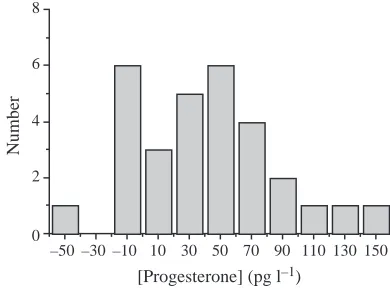
Fig. 1 is a histogram of the change in P levels from F to L
phases. While most women showed an increase in P level from F to L, seven women showed no increase and were subsequently eliminated from statistical testing of the hypothesis of a P-mediated increase in exercise capacity.
Follicular versus luteal phase differences in submaximal exercise
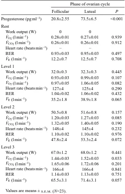
For the 23 women showing an increase in P level from F to L, exercise data at rest and during submaximal exercise are presented for three different levels of external work, corresponding to approximately 58 % (level 1), 72 % (level 2) and 86 % (level 3) of V.O∑max(Table 2). With the exception of V.O∑ at work level 3, there were no significant differences in
oxygen consumption and work output (W) at any given level of work. Repeated measures analysis shows higher V.E/V.O∑
(P<0.001, Fig. 2), V.E/V.CO∑ (P<0.001, not shown), and V
.
E
(P<0.001, not shown) over all workloads to maximum. During the L phase, exercise V.Eat the third work level was positively correlated to P levels (Fig. 3, r=0.487, P=0.006). No such correlation was evident during the F phase. Similarly, the ventilatory equivalents for V.O∑(r=0.476, P=0.008) and V
.
CO∑
(r=0.463, P=0.010) were positively correlated with P level in the L phase (data not shown).
–50 –30 –10 10 30 50 70 90 110 130 150 0
2 4 6 8
Number
[image:3.612.317.571.84.213.2][Progesterone] (pg l–1)
Table 1. Study group characteristics
Age (years) 27.7±0.7
Weight (kg) 54.4±1.3
Height (cm) 154.4±0.9
Body mass index (kg m2) 22.8±0.5
Hemoglobin
Follicular (g dl–1) 15.9±0.2
Luteal (g dl–1) 16.1±0.2
Progesterone
Follicular (pg ml–1) 22.9±2.3
Luteal (pg ml–1) 63.3±7.4
[image:3.612.344.539.547.692.2]Values are means ±S.E.M. (N=30).
Follicular versus luteal phase differences in maximal exercise
Analyses of maximal exercise data are presented for the subset of 13 women who showed an increase in P levels from F to L and who achieved a true V.O∑maxon both tests (Table 3).
These 13 women did not differ from the sample of 23 women with respect to their submaximal exercise response, or their P level increase from F to L. Calculations for N=13 indicate a power of approximately 75 % to detect a 5 % change in V.O∑max
from F to L at α=0.05 (Cohen, 1977).
Work output was slightly higher during the L phase (86.4 W
versus 82.2 W, P=0.044), but no other exercise parameters
measured at V.O∑maxdiffered between F and L phases (Table 3).
Unlike the findings at submaximal levels, no significant correlation was found between P level and V.O∑max, maximal V.E, or the maximal ventilatory equivalents.
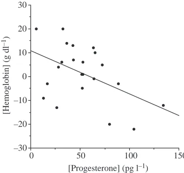
Post-hoc analysis in all women showing an increase in P
level revealed a negative relationship between the change in Hb and the change in P from F to L (r2=0.426, P=0.017), using
a general linear model and holding constant the initial (F) levels of Hb and P (Fig. 4). Thus, higher P levels were associated with a decrease in Hb, possibly reflecting an increase in Vpand a hemodilution effect during the L phase
(Chapman et al., 1997; Cullinane et al., 1995).
Given that P>0.05 but <0.08 for many maximal response variables (i.e. V.O∑max, V
.
E), a worthwhile alternative analytical approach is to model change variables from F to L phases using multivariate methods. This approach allows one to test hypotheses of phase differences in V.O∑max as a function of
[image:4.612.339.538.74.264.2]different interacting physiological pathways. The change in
Table 2. Submaximal exercise data during follicular and
luteal phases for La Paz women Phase of ovarian cycle
Follicular Luteal P
Progesterone (pg ml–1) 20.8±2.55 73.5±6.5 <0.001
Rest
Work output (W) 0 0
V˙O2(l min–1) 0.28±0.01 0.27±0.01 0.939
V˙CO2(l min–1) 0.26±0.01 0.26±0.01 0.912
Heart rate (beats min–1) – –
RER 0.93±0.03 0.95±0.03 0.497
V˙E(l min–1) 12.2±0.7 12.5±0.7 0.708
Level 1
Work output (W) 32.0±0.3 32.3±0.3 0.445 V˙O2(l min–1) 0.93±0.03 0.99±0.03 0.107
V˙O2(l min–1) 0.97±0.05 1.06±0.05 0.082
Heart rate (beats min–1) 127±4 125±4 0.290
RER 1.04±0.02 1.06±0.02 0.432
V˙E(l min–1) 35.2±1.8 38.9±1.8 0.065
Level 2
Work output (W) 50.5±0.8 51.6±0.8 0.157 V˙O2(l min–1) 1.20±0.03 1.27±0.03 0.085
V˙CO2(l min–1) 1.32±0.05 1.40±0.05 0.190
Heart rate (beats min–1) 148±4 145±4 0.232
RER 1.10±0.02 1.10±0.02 0.976
V˙E(l min–1) 47.8±2.4 53.3±2.4 0.072
Level 3
Work output (W) 67.0±1.2 68.0±1.2 0.441 V˙O2(l min–1) 1.44±0.03 1.52±0.03 0.033
V˙CO2(l min–1) 1.65±0.06 1.72±0.06 0.201
Heart rate (beats min–1) 166±4 166±4 0.841
RER 1.14±0.03 1.13±0.03 0.751
V˙E(l min–1) 65.5±3.1 71.4±3.1 0.057
Values are means ±S.E.M. (N=23).
RER, respiratory exchange ratio (V˙CO2/V˙O2).
100 80
60 40 20 0
30 40 50 60
Follicular Luteal
Work output (W)
V
E
/
V
. O2
200 150
100 50
0 20 40 60 80 100 120
[Progesterone] (pg ml–1)
V
ent
il
at
ion r
ate (ml m
in
–
[image:4.612.41.294.97.489.2]1)
Fig. 2. The ventilatory equivalent for oxygen (V.E/V
.
O∑) at 3600 m is
higher during the luteal phase at rest and during submaximal and maximal exercise in La Paz women by analysis for repeated measures (P<0.001; N=23). Similar results were evident for both the
V.E/V
.
CO∑and V
.
E.
Fig. 3. Submaximal exercise ventilation (V.E, ml min–1) during work
level 3 at 3600 m versus progesterone (P) level (pg ml–1) during the
luteal phase of the menstrual cycle in La Paz women (r=0.487,
[image:4.612.334.534.349.540.2]V.O∑maxfrom F to L was not explained by a change in Hb status
from F to L (P=0.980), holding constant the initial (F) values of V.O∑max and Hb. Similarly, the change in V
.
O∑max was not
explained by a change in P level, although this relationship reached P=0.072. When the change in Hb and change in P were entered together into a general linear model, neither main effects nor interaction effects explained the change in V.O∑max
from F to L.
Discussion
Unlike some previous studies of menstrual cycle phase and exercise performance, we measured levels of the ovarian hormone P to confirm the occurrence of a true L phase of the menstrual cycle. This is important because menses need not be accompanied by ovulation nor by a substantial rise in P and E2 (Prior and Vigna, 1991). Indeed, the women in this study showed considerable variability in P levels and variability in the P increase from F to L (Table 1) (Fig. 1). Seven of 30 women did not show a typical L-phase increase in P level and may have been anovulatory. These women were excluded from analyses in order to best test the main study hypothesis of a P-mediated increase in physical work capacity.
At a mean P level of 73.5 pg ml–1on the day of the L-phase
exercise test (for those whose P level did increase; N=23), the women in this study showed lower L-phase P values than a US reference group, but normative values compared to other samples from developing countries. For example, in samples of women from Boston, rural Poland in the post-harvest season, rural Nepal in the winter, and rural Zaire, the mean mid-L-phase P levels are 117 pg ml–1, 94 pg ml–1, 80 pg ml–1
and 63 pg ml–1, respectively (Ellison et al., 1993; Vitzthum,
2000).
These points established, this study demonstrated that an increase in P level during the L phase is directly related to a significant, although modest, increase in submaximal exercise
V.Ein HA native women (Table 2, Fig. 3). This increase in L phase V.Eis accompanied by a small increase in maximal work output (5 %), but no increase in V.O∑max(P=0.063). Additional
multivariate analyses revealed no relationship between the change in V.O∑max from F to L and the change in P level or
change in Hb status (the latter used as a proxy variable for Vp).
Thus, while HA native women do show modest physiological responses to increased P levels during the L phase of the menstrual cycle, our study does not support the hypothesis of secondary effects increasing work capacity.
The hypothesis of increased L phase V.O∑maxat high altitude
was based in part on studies conducted at sea level showing that resting V.Eis higher during the L phase (Dombovy et al., 1987; Edwards et al., 1996; England and Farhi, 1976; Jurkowski et al., 1981; Machida, 1981; Regensteiner et al., 1990; Schoene et al., 1981; Takano, 1984; Takano et al., 1981; White et al., 1983). Whether L-phase V.Eis also higher during exercise has not been as clearly demonstrated. Some studies report higher L-phase exercise V.E (Dombovy et al., 1987; Jurkowski et al., 1981; Schoene et al., 1981; Williams and Krahenbuhl, 1997), while other studies report no cycle-phase exercise V.Edifferences (Bemben et al., 1995; De Souza et al., 1990; Hackney, 1991; Lebrun et al., 1995). Increased exercise
V.E(should it occur), could serve to maintain alveolar and thus arterial PaO∑, and could be of substantial benefit during
exercise at high altitude. Unlike sea-level exercise, exercise in men at high altitude is characterized by PaO∑ values that fall
on the steep part of the hemoglobin oxygen dissociation curve, by SaO∑levels that typically drop throughout exercise (Bender
et al., 1989), and by significant diffusion limitation that contributes in part to a large alveolar PO∑ – arterial PO∑
difference (Torre-Bueno et al., 1985). Thus, there is reason to expect L-phase benefit at HA despite the fact that the literature almost uniformly reports no L-phase V.O∑max increase at sea
[image:5.612.348.536.74.251.2]level (De Souza et al., 1990; Dombovy et al., 1987; McCracken
Table 3. Maximal exercise data during follicular (F) and
luteal (L) phases for 13 subjects showing a progesterone increase from F to L and achieving a true V˙O2max
Phase of ovarian cycle
Follicular Luteal P
Progesterone (pg ml–1) 18.8±2.4 66.1±7.0 <0.001
Work output (W) 82.2±5.2 86.4±5.4 0.044 V˙O2(ml min–1kg–1) 32.5±0.9 34.1±0.8 0.068
V˙O2(l min–1) 1.68±0.06 1.77±0.06 0.063
V˙CO2(l min–1) 2.11±0.09 2.18±0.09 0.427
Heart rate (beats min–1) 176±2 177.0±2 0.406
RER 1.26±0.04 1.23±0.02 0.585
V˙E(l min–1) 90.8±3.3 100.6±4.4 0.078
V˙E/V˙O2 54.3±1.6 57.1±1.8 0.243
V˙E/V˙CO2 43.6±1.7 46.3±0.09 0.133
Values are means ±S.E.M.
RER, respiratory exchange ratio (V˙CO2/V˙O2).
150 100
50 0
–30 –20 –10 0 10 20 30
[Progesterone] (pg l–1)
[H
em
oglo
bi
n] (g
d
l
–
[image:5.612.49.300.108.277.2]1)
Fig. 4. Change in hemoglobin (Hb) status from mid-follicular to mid-luteal phase is negatively correlated with the change in progesterone (P) level (r2=0.426, P=0.017, in a linear model holding
et al., 1994; Schoene et al., 1981), or only a very small increase (Lebrun et al., 1995; Schoene et al., 1981).
Two previous studies have addressed the general possibility of L-phase benefit at HA. A study by Muza et al. (1997) tested the hypothesis that women ascending to HA in their L phase would show an accelerated ventilatory acclimatization compared to women in their F phase. The results of that study did not support the hypothesis of L-phase benefit as no significant differences in resting V.E, HVR or HCVR were reported between cycle phases for women during 12 days of altitude exposure at 4300 m. That study may have been limited by a small sample size (nine F-phase women and five different L-phase women) as well as by the fact that investigators did not use a randomized repeated-measures design. In addition, the authors noted a low mean P level during the L phase in their subjects, as well as the possibility that menstrual cycle effects were overwhelmed by the hypoxic stress of acute exposure. With respect to the latter, the ventilatory increase that is expected on sojourn to altitude is some threefold greater than that expected from F to L phases (Muza et al., 1997).
Beidleman et al. (1999) also tested the hypothesis that an increase in exercise V.E during the L phase would increase maximal exercise performance. Using a repeated-measures design, women were tested on exposure to hypoxia in a hypobaric chamber simulating 4300 m. SaO∑ was 3 % higher during the
L phase, but paradoxically, cycle-phase differences in V.E and
V.O∑max were not evident. Subjects showed a large L-phase
increase in P levels, but again the study may have been limited by a small sample size (eight women) and by the possibility that acclimatization to hypoxia overwhelms the ovarian hormone modulation of resting and exercise V.E. In the present study we were unable to measure SaO∑due to logistical/technical problems.
This measurement would have provided a link with which to test the hypothesis that there is a functional benefit due to increased
V.E, but the absence of a SaO∑ measure does not preclude our
ability to test the general hypothesis that P mediates aerobic capacity across the menstrual cycle.
We studied only women who were lifelong residents of high altitude, eliminating the potential problems of ventilatory, renal or hematological acclimatization. Unlike the study by Beidleman et al. (1999), we demonstrate a small increase in submaximal exercise V.Eduring the L phase, which is directly related to the P level (Fig. 3). The increase in V.E was not evident at maximal exercise in the subset of 13 women who achieved a true V.O∑max (P=0.057). Statistical significance, in
this regard, may depend on sample size, and it was unfortunate that only approx. 50 % of our study subjects (N=13) actually achieved a true V.O∑maxon both tests by objective criteria. It was
difficult to motivate all subjects as many were sedentary and unfamiliar with cycle exercise. Despite practice sessions, many subjects were visibly inefficient during the work protocol. Cycling inefficiency, and the additional cost of breathing at HA compared to sea-level, may explain why sub-maximal V.O∑
is higher than expected at any level of external work in these subjects (by approx. 20–30 %). The absence of any significant difference in PaO∑between the electronically braked cycle and
a mechanically braked ergometer suggests the absence of any systematic bias resulting from miscalibration of the electronic ergometer. Whatever the explanation, measures of V.O∑ are
internally consistent and the discrepancy does not affect the comparison of F- versus L-phase exercise response. Finally, despite the exclusion of subjects for the analyses of maximal exercise data, power analysis (see below) demonstrates that the sample size is sufficient to test the major hypotheses of this study.
We demonstrate a significant negative correlation between the increase in P and the decrease in Hb from F to L phases (Fig. 4). Estrogen and P are known to affect systemic and renal hemodynamic systems via activation of the renin–angiotensin– aldosterone axis, and this may account for a Vpexpansion in
the late L phase that has been associated with decreases in the concentration of serum lipids and/or blood Hb due to hemodilution (Cullinane et al., 1995; Pahwa et al., 1998; Vellar, 1974). Our finding of a decrease in Hb concentration in the L phase is consistent with some (Cullinane et al., 1995; Dombovy et al., 1987; Inoue and Sugiyama, 1998), but not all studies (Inoue and Sugiyama, 1998; Jurkowski et al., 1981; Kim et al., 1993; Lebrun et al., 1995), possibly because P has inhibitory effects on erythropoietin production (Moore et al., 1978; Peschle et al., 1973). Alterations in Hb, mediated through changes in arterial oxygen content, and alterations in blood volume, mediated through changes in cardiac output, can both have significant effects on V.O∑max(Gledhill et al., 1999).
Thus, P may have counteracting effects on V.O∑maxvia alternate
ventilatory and hemodynamic pathways. Additionally, it should also be considered that an increase in the work of breathing due to increased V.Ecould detract from any potential L-phase exercise benefit.
To further test these hypotheses, we initiated multivariate analyses of exercise variables as they changed from F to L phases. No significant relationships were revealed between the change in V.O∑maxand either the change in Hb status or P level
from F to L phases, although the latter relationship reaches
P=0.072. The consistently low P values for maximal exercise
variables that approach, but do not reach, statistical significance, raise the possibility of type II error. Power analysis revealed β=75 % to detect a 5 % difference in V.O∑max.
Thus, this study has reasonably good power to detect even small V.O∑max differences, but we cannot eliminate the
possibility of smaller effects, especially when considering the low P values attained (Table 3).
important in athletes and becomes more important at higher altitudes (Torre-Bueno et al., 1985). Therefore, small changes in V.O∑maxand work output may be of significance to a highly
trained woman or to a woman exercising at extreme altitude. These studies remain to be conducted.
Funding was provided to V.J.V. by the University Research Expeditions Program, the University of California, and Binghamton University, State University of New York. Our appreciation to Dr Enrigue Vargas, Director of Instituto Boliviano de Biología de Altura, for his gracious support, to the assistants who facilitated this research, and especially to the participants for their patience.
References
Bayliss, D. A. and Millhorn, D. E. (1992). Central neural mechanisms of
progesterone action: application to the respiratory system. J. Appl. Physiol.
73, 393–404.
Beidleman, B. A., Rock, P. B., Muza, S. R., Fulco, C. S., Forte, V. A., Jr. and Cymerman, A. (1999). Exercise V.E and physical performance at altitude are not affected by menstrual cycle phase. J. Appl. Physiol. 86, 1519–1526.
Bemben, D. A., Salm, P. C. and Salm, A. J. (1995). Ventilatory and blood
lactate responses to maximal treadmill exercise during the menstrual cycle.
J. Sports Med. Phys. Fitness 35, 257–262.
Bender, P. R., McCullough, R. E., McCullough, R. G., Huang, S. Y., Wagner, P. D., Cymerman, A., Hamilton, A. J. and Reeves, J. T. (1989).
Increased exercise SaO∑ independent of ventilatory acclimatization at
4,300 m. J. Appl. Physiol. 66, 2733–2738.
Chapman, A. B., Zamudio, S., Woodmansee, W., Merouani, A., Osorio, F., Johnson, A., Moore, L. G., Dahms, T., Coffin, C., Abraham, W. T. and Schrier, R. W. (1997). Systemic and renal hemodynamic changes in
the luteal phase of the menstrual cycle mimic early pregnancy. Am. J.
Physiol. 273, F777–782.
Cohen, J. (1977). Statistical Power Analysis for the Behavioral Sciences. New
York: Academic Press.
Cullinane, E. M., Yurgalevitch, S. M., Saritelli, A. L., Herbert, P. N. and Thompson, P. D. (1995). Variations in plasma volume affect total and
low-density lipoprotein cholesterol concentrations during the menstrual cycle.
Metabolism 44, 965–971.
De Souza, M. J., Maguire, M. S., Rubin, K. R. and Maresh, C. M. (1990).
Effects of menstrual phase and amenorrhea on exercise performance in runners. Med. Sci. Sports Exerc. 22, 575–580.
Dombovy, M. L., Bonekat, H. W., Williams, T. J. and Staats, B. A. (1987).
Exercise performance and ventilatory response in the menstrual cycle. Med.
Sci. Sports Exerc. 19, 111–1117.
Dutton, K., Blanksby, B. A. and Morton, A. R. (1989). CO2sensitivity
changes during the menstrual cycle. J. Appl. Physiol. 67, 517–522.
Edwards, N., Wilcox, I., Polo, O. J. and Sullivan, C. E. (1996). Hypercapnic
blood pressure response is greater during the luteal phase of the menstrual cycle. J. Appl. Physiol, 81, 2142–2146.
Ellison, P. T., Lipson, S. F., O’Rourke, M. T., Bentley, G. R., Harrigan, A. M., Panter-Brick, C. and Vizthum, V. J. (1993). Population variation
in ovarian function [letter]. Lancet 342, 433–434.
England, S. J. and Farhi, L. E. (1976). Fluctuations in alveolar CO2and in
base excess during the menstrual cycle. Resp. Physiol. 26, 157–161.
Gledhill, N., Warburton, D. and Jamnik, V. (1999). Haemoglobin, blood
volume, cardiac function, and aerobic power. Can. J. Appl. Physiol. 24, 54–65.
Hackney, A. C., Curley, C.S. and Nicklas, B. J. (1991). Physiological
responses to submaximal exercise at the follicular, ovulatory, and mid-luteal phases of the menstrual cycle. Scand. J. Med. Sci. Sport 1, 94–98.
Hamill, P. V., Drizd, T. A., Johnson, C. L., Reed, R. B., Roche, A. F. and Moore, W. M. (1979). Physical growth: National Center for Health
Statistics percentiles. Am. J. Clin. Nutr. 32, 607–629.
Hannhart, B., Pickett, C. K. and Moore, L. G. (1990). Effects of estrogen
and progesterone on carotid body neural output responsiveness to hypoxia.
J. Appl. Physiol. 68, 1909–1916.
Inoue, K. and Sugiyama, M. (1998). Changes in body iron status, heart rate,
and unspecific symptoms across menstrual cycle in marginally iron-deficient young women. J. Clin. Biochem. Nutr. 24, 99–104.
Jurkowski, J. E., Jones, N. L., Toews, C. J. and Sutton, J. R. (1981). Effects
of menstrual cycle on blood lactate, O2delivery, and performance during
exercise. J. Appl. Physiol. 51, 1493–1499.
Kaiser, L. (1989). Adjusting for baseline: change or percentage change? [see
comments]. Stat. Med. 8, 1183–1190.
Kim, I., Yetley, E. A. and Calvo, M. S. (1993). Variations in iron status
measures during the menstrual cycle. Am. J. Clin. Nutr. 58, 705–709.
Lebrun, C. M., McKenzie, D. C., Prior, J. C. and Taunton, J. E. (1995).
Effects of menstrual cycle phase on athletic performance. Med. Sci. Sports
Exerc. 27, 437–444.
Lu, Y.-C., Bentley, G. R., Gann, P. H., Hodges, K. R. and Chatterton, R. T. (1999). Salivary estradiol and progesterone levels in conception and
non-conception cycles in women. Evaluation of a new assay for salivary estradiol. Fertil. Steril. 71, 863–868.
Machida, H. (1981). Influence of progesterone on arterial blood and CSF
acid–base balance in women. J. Appl. Physiol. 51, 1433–1436.
Mazzeo, R. S., Child, A., Butterfield, G. E., Mawson, J. T., Zamudio, S. and Moore, L. G. (1998). Catecholamine response during 12 days of
high-altitude exposure (4300 m) in women. J. Appl. Physiol. 84, 1151–1157.
McCracken, M., Ainsworth, B. and Hackney, A. C. (1994). Effects of the
menstrual cycle phase on the blood lactate responses to exercise. Eur. J.
Appl. Physiol. 69, 174–175.
Moore, L. G. (1997). Women at Altitude: Overview. In Hypoxia: Women at Altitude (ed. C. S. Houston, and G. Coates), pp. 1–7. Burlington: Queen City
Printer.
Moore, L. G., McMurtry, I. F. and Reeves, J. T. (1978). Effects of sex
hormones on cardiovascular and hematologic responses to chronic hypoxia in rats. Proc. Soc. Exp. Biol. Med. 158, 658–662.
Muza, S. R., Rock, P. B., Fulco, C. S., Zamudio, S., Braun, B., Reeves, J. T., Butterfield, G. E. and Moore, L. G. (1997). Women at altitude:
Influence of menstrual cycle phase on ventilatory acclimatization. In
Hypoxia: Women at Altitude (ed. C. S. Houston, and G. Coates), pp. S1–S7.
Burlington: Queen City Printer.
O’Rorke, A., Kane, M. M., Gosling, J. P., Tallon, D. F. and Fottrell, P. F.
(1994). Development and validation of a monoclonal antibody enzyme immunoassay for measuring progesterone in saliva. Clin. Chem. 40, 454–458.
Pahwa, M. B., Seth, S. and Seth, R. K. (1998). Lipid profile in various phases
of menstrual cycle and its relationship with percentage plasma volume changes. Clin. Chim. Acta 273, 201–207.
Peschle, C., Rappaport, I. A., Sasso, G. F., Condorelli, M. and Gordon, A. S. (1973). The role of estrogen in the regulation of erythropoietin
production. Endocrinology 92, 358–362.
Prior, J. C. and Vigna, Y. M. (1991). Ovulation disturbances and exercise
training. Clin. Obst. Gynecol. 34, 180–190.
Regensteiner, J. G., McCullough, R. G., McCullough, R. E., Pickett, C. K. and Moore, L. G. (1990). Combined effects of female hormones and
exercise on hypoxic ventilatory response. Resp. Physiol. 82, 107–114.
Sansores, R. H., Abboud, R. T., Kennell, C. and Haynes, N. (1995). The
effect of menstruation on the pulmonary carbon monoxide diffusing capacity. Am. J. Resp. Crit. Care Med. 152, 381–384.
Schoene, R. B., Robertson, H. T., Pierson, D. J. and Peterson, A. P. (1981).
Respiratory drives and exercise in menstrual cycles of athletic and nonathletic women. J. Appl. Physiol. 50, 1300–1305.
Takano, N. (1984). Changes of ventilation and ventilatory response to hypoxia
during the menstrual cycle. Pflugers Arch. 402, 312–316.
Takano, N., Sakai, A. and Iida, Y. (1981). Analysis of alveolar PCO∑control
during the menstrual cycle. Pflugers Arch. 390, 56–62.
Torre-Bueno, J. R., Wagner, P. D., Saltzman, H. A., Gale, G. E. and Moon, R. E. (1985). Diffusion limitation in normal humans during exercise at sea
level and simulated altitude. J. Appl. Physiol. 58, 989–995.
Vellar, O. D. (1974). Changes in hemoglobin concentration and hematocrit
during the menstrual cycle. I. A cross-sectional study. Acta Obst. Gynecol.
Scand. 53, 243–246.
Vitzthum, V. J., Ellison, P.T., Sukalich, S., Caceres, E., Spielvogel, H.
(2000). Does Hypoxia Impair Ovarian Function in Bolivian Women Indigenous to High Altitude? High Altitude Medicine Biol. 1, 39–49.
White, D. P., Douglas, N. J., Pickett, C. K., Weil, J. V. and Zwillich, C. W. (1983). Sexual influence on the control of breathing. J. Appl. Physiol. 54, 874–89.
Williams, T. J. and Krahenbuhl, G. S. (1997). Menstrual cycle phase and