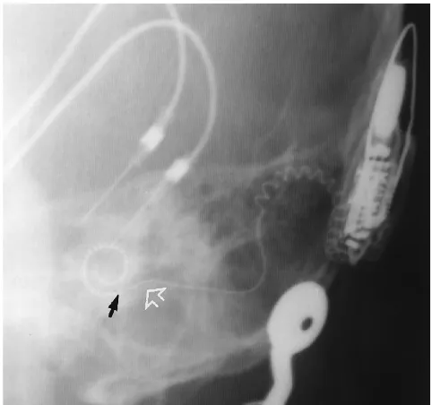
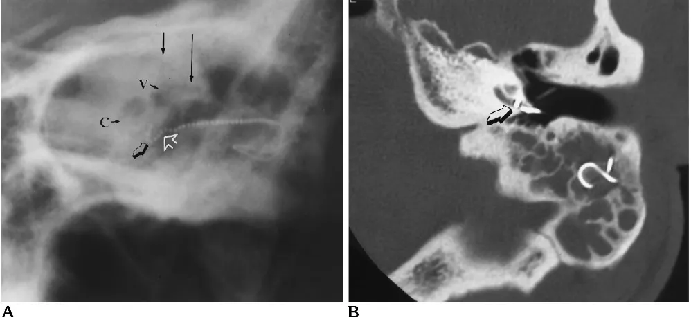
Postoperative imaging of the multichannel cochlear implant
8
0
0
Full text
Figure


+3


Related documents
