Original Article
High-sensitivity epidermal growth factor receptor
immunostaining for colorectal carcinomas, compared
with EGFR PharmDx
TM: A study of diagnostic accuracy
Kazuya Shiogama1, Trai Wongsiri2, Yasuyoshi Mizutani1, Ken-ichi Inada1, Yutaka Tsutsumi1
1Department of Pathology, Fujita Health University School of Medicine, Toyoake, Japan; 2Department of Pathology, Faculty of Medicine, Khon Kaen University, Khon Kaen, Thailand
Received October 18, 2012; Accepted November 6, 2012; Epub November 20, 2012; Published January 1, 2013
Abstract: Immunostaining for epidermal growth factor receptor (EGFR) is important in the contemporary therapeu-tic strategy of colorectal carcinomas. We tried to increase detection sensitivity, and compared the high-sensitivity EGFR immunostaining with a worldwide standard, EGFR PharmDxTM (Dako). In order to pursue high-sensitivity EGFR
detection, deparaffinized sections were pressure-cooked in 1 mM EDTA solution, pH 8.0. Two mouse monoclonal antibodies against EGFR, clone EGFR2.5 and DAK-H1-WT, and six kinds of secondary detection reagents, includ-ing biotin-free catalyzed signal amplification (CSA II), Simple Stain MAX-PO, PolyVue, Novolink, EnVisionTM FLEX+,
and MACH3, were evaluated to compare the results with those with EGFR PharmDxTM, employing a combination of
2-18-C9 as the primary monoclonal antibody and EnVisionTM as the secondary reagent. Furthermore, we replaced
EnVisionTM in the EGFR PharmDxTM kit with CSAII. EGFR detection sensitivity was higher with DAK-H1-WT than with
EGFR2.5, and among the secondary reagents, the strongest signals were observed with Novolink. All 30 colorectal carcinomas showed distinct expression of EGFR with our high-sensitivity EGFR immunostaining, while only 16 (53%) gave focal positivity with EGFR PharmDxTM. When EnVisionTM in EGFR PharmDxTM was replaced by CSA II, strong
sig-nals were seen in all cases, and the expression pattern was comparable with our sequence. Non-neoplastic crypt epithelial cells often showed weakly signal with the standard EGFR PharmDxTM, but consistently revealed strong
membrane staining in the two high-sensitivity sequences. EGFR PharmDxTM frequently gave false negativity.
Impor-tantly, EGFR was consistently and sensitively detected when the secondary polymer in the EGFR PharmDxTM kit was
simply replaced by CSA II.
Keywords: Colorectal cancer, epidermal growth factor receptor, immunohistochemistry, sensitivity and specificity, monoclonal antibody
Introduction
Epidermal growth factor receptor (EGFR), a 170 kD transmembrane protein, categorized in the tyrosine kinase family, regulates cell functions, including cell division and apoptosis [1, 2]. Reportedly, EGFR is expressed in approximate-ly 60% to 80% of colorectal carcinomas [3, 4], and molecular targeted therapy is given to EGFR-positive cases [5].
EGFR PharmDxTM, a Food and Drug Administration (FDA)-approved diagnostic kit for localizing EGFR in formalin-fixed, paraffin-embedded sections available from Dako Co., is widely utilized for determining the eligibility of anti-EGFR molecular target therapy Cetuximab
In the present study, we evaluated two anti-EGFR monoclonal antibodies and various sec-ondary detection reagents, and established high sensitivity EGFR immunostaining for colorectal cancer. Subsequently, we compared the results with those with EGFR PharmDxTM under both the standard and modified condi-tions. In the modified PharmDxTM method, the secondary polymer reagent (EnVisionTM) was replaced by the biotin-free catalyzed signal amplification system (CSAII) available also from Dako.
Materials and methods
High-sensitivity EGFR immunostaining
Samples: We analyzed a total of five advanced colorectal adenocarcinomas surgically removed in Fujita Health University Hospital, Toyoake, Japan. The tissues were routinely fixed in 10% formalin and embedded in paraffin wax. One block sampled from the normal/tumor junction was used for analysis in each case.
Immunohistochemistry: Sections were depar-affinized with xylene, and rehydrated in graded ethanol. Endogenous peroxidase activity was quenched with 0.3% hydrogen peroxide in methanol for 30 minutes at room temperature. Hydrated heat-assisted epitope retrieval was applied using a pressure pan cooker (Delicio 6L, T-FAL, Clithy, France) for 10 minutes. Preliminary study chose 1 mM ethylenediamine tetraacetic acid (EDTA) solution, pH 8.0, for the optimal soaking solution for heating. After pres-sure pan cooking, the sections were left for 30 minutes at room temperature for cooling. Anti-EGFR monoclonal antibodies, clone Anti-EGFR 2.5 (diluted at 1:100, NovoCastra, Newcastle, UK) and clone DAK-H1-WT (diluted at 1:100, Dako, Glostrup, Denmark), were incubated for 30 min-utes at room temperature. After rinsing in 50 mM Tris-HCl-buffered saline (TBS), pH 7.6, the sections were reacted with six different kinds of secondary detection reagents, principally according to manufacturer’s instructions. These included 1) tyramide amplification-assisted biotin-free catalyzed signal amplifica-tion (CSA II, Dako), and five different immuno-peroxidase polymer reagents, such as Histofine Simple Stain MAX-PO (SSMAX, Nichirei Bioscience, Tokyo, Japan), PolyVue (Japan Tanner, Osaka, Japan), Novolink (Novocastra), EnVision FLEX+ (Dako) and MACH3 (Biocare
Medical, Concord, CA, USA). Reaction products were visualized in 50 mM Tris-HCl buffer, pH 7.6, containing 20 mg/dl diaminobenzidine tet-rahydrochloride and 0.006% hydrogen peroxi-dase. The nuclei were lightly counterstained with Mayer’s hematoxylin.
Comparison with EGFR PharmDxTM
Samples: A total of 30 advanced colorectal adenocarcinomas surgically removed in Fujita Health University Hospital in the period from 2001 to 2009 were routinely fixed in 10% for-malin and embedded in paraffin wax. Again, samples at the tumor/normal junction were evaluated. The patients’ age ranged from 36 years to 78, with the average of 59.9. The male to female ratio was 14:16. No preoperative treatment was given in any case.
EGFR PharmDxTM immunohistochemistry: The immunohistochemical procedure for EGFR PharmDxTM followed the manufacturer’s instruc-tion. Briefly, sections were deparaffinized, rehy-drated, and treated with proteinase K for 5 min-utes at room temperature. After peroxidase inactivation, the primary antibody, clone 2-18-C9, was incubated for 30 minutes at room tem-perature. After rinsing in TBS, the sections were incubated with secondary polymer reagent (EnVisionTM) for 30 minutes at room tempera-ture. Diaminobenzidine coloration and nuclear counterstaining followed. In order to improve the detection sensitivity of EGFR PharmDxTM, the secondary reagent was simply replaced by CSA II available from the same company, Dako. We compared the findings obtained with the standard EGFR PharmDxTM with those with the modified EGFR PharmDxTM and the above-men-tioned high sensitivity EGFR immunostaining.
EGFR immunoreactivity interpretation
EGFR expression on the plasma membranes was evaluated according to the intensity of staining: 0, no reactivity, 1+, weak reactivity, 2+, moderate reactivity, 3+, strong reactivity. The judgment was independently confirmed by three authors (KS, WT and YT).
Ethical issue
University, Toyoake. The approved number is 11-091. Written informed consent was obtained from each patient.
Results
High-sensitivity EGFR immunostaining
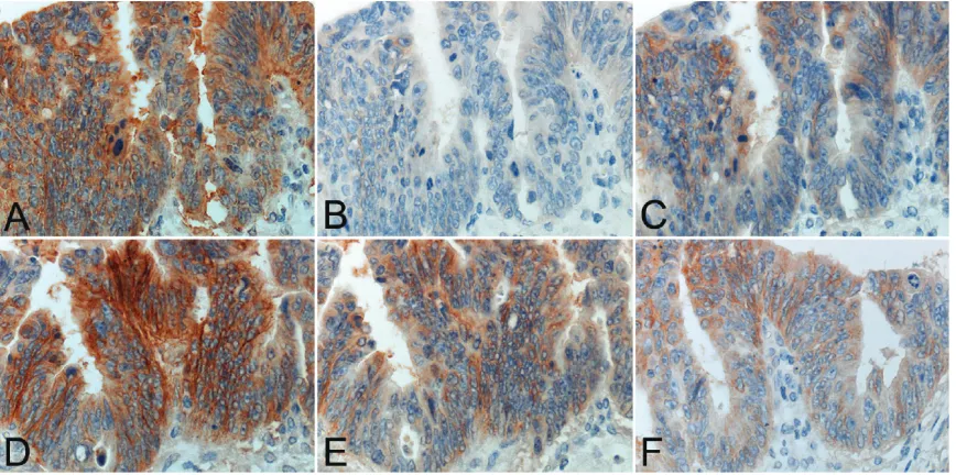
The immunostained results for EGFR using two monoclonal antibodies and six secondary detection reagents are summarized in Table 1. Representative staining patterns are illustrated in Figure 1. EGFR immunoreactivity was consis-tently localized on the plasma membranes. The clone DAK-H1-WT consistently gave stron-ger immunoreactivity than the clone EGFR2.5 in any experiment employing six different kinds
of the secondary reagents. The clone EGFR2.5 gave negativity in 4 of 5 (80%) lesions with SSMAX and 1 of 5 (20%) with PolyVue or MACH3. The clone DAK-H1-WT antibody showed negativity only in 2 of 5 (40%) with SSMAX. Meanwhile, the clone EGFR2.5 demonstrated 3+ immunoreactivity in 2 of 5 (40%) lesions with Novolink and 1 of 5 (20%) with EnVisionTM FLEX+ or MACH3. The clone DAK-H1-WT result-ed in 3+ immunoreactivity in 5 of 5 (100%) with Novolink, 2 of 5 (40%) with MACH3 and 1 of 5 (20%) with CSAII or EnVisionTM FLEX+. The area of positively stained cancer tissue paralleled with the staining intensity. The strongest and consistent signals were obtained in the combi-nation of DAK-H1-WT and Novolink. CSAII tend-ed to show cytoplasmic diffusion of the reac-Table 1. EGFR immunostaining in various combinations
Monoclonal antibody to EGFR Intensity Score CSA II SSMAX PolyVue Novolink EnVisionTM FLEX+ MACH3
EGFR 2.5
0 0 (0%) 4 (80%) 1 (20%) 0 (0%) 0 (0%) 1 (20%)
1+ 2 (40%) 1 (20%) 2 (40%) 0 (0%) 2 (40%) 0 (0%)
2+ 3 (60%) 0 (0%) 2 (40%) 3 (60%) 2 (40%) 3 (60%)
3+ 0 (0%) 0 (0%) 0 (0%) 2 (40%) 1 (20%) 1 (20%)
DAK-H1-WT
0 0 (0%) 2 (40%) 0 (0%) 0 (0%) 0 (0%) 0 (0%)
1+ 1 (20%) 2 (40%) 0 (0%) 0 (0%) 1 (20%) 1 (20%)
2+ 3 (60%) 1 (20%) 5 (100%) 0 (0%) 3 (60%) 2 (40%)
3+ 1 (20%) 0 (0%) 0 (0%) 5 (100%) 1 (20%) 2 (40%)
[image:3.612.91.523.83.198.2]0: negative, 1+: weak intensity, 2+: moderate intensity, 3+: strong intensity.
Figure 1. Comparative study employing six different secondary reagents for detecting EGFR immunoreactivity with a mouse monoclonal antibody, clone: DAK-H1-WT in a representative case of advanced colon cancer. A. CSA II, B. SSMAX, C. PolyVue, D. Novolink, E. EnVisionTM FLEX+ and F. MACH3. CSAII and Novolink give the strongest reactivity,
[image:3.612.91.525.233.449.2]tion product. With our high-sensitivity method,
[image:4.612.88.530.99.188.2]non-neoplastic crypt epithelial cells strongly expressed EGFR on the basolateral plasma membranes. Table 2. Comparison among the standard and modified EGFR PharmDxTM and higy-sensitivity immunos-taining for EGFR using DAK-H1-WT and Novolink
Intensity score
Standard EGFR PharmDxTM Modified EGFR PharmDxTM High-sensitivity immunostaining
Non-neoplastic
crypt epithelial cells cancer cellsColorectal crypt epithelial cellsNon-neoplastic cancer cellsColorectal crypt epithelial cellsNon-neoplastic cancer cellsColorectal 0 3 (10%) 14 (47%) 0 (0) 0 (0) 0 (0) 0 (0) 1+ 23 (77%) 6 (20%) 0 (0) 0 (0) 0 (0) 0 (0) 2+ 3 (10%) 6 (20%) 2 (7%) 2 (7%) 2 (7%) 2 (7%) 3+ 1 (3%) 4 (13%) 28 (93%) 28 (93%) 28 (93%) 28 (93%)
0: negative, 1+: weak intensity, 2+: moderate intensity, 3+: strong intensity.
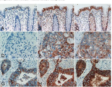
Figure 2. Comparative study of two colon cancer cases using the standard and modified EGFR PharmDxTM and
high-sensitivity EGFR immunostaining with DAK-H1-WT and Novolink. (A, D, G) standard EGFR PharmDxTM, (B, E,
H) modified EGFR PharmDxTM using CSAII as the secondary reagent, (C, F, I) high-sensitivity EGFR immunostaining
with DAK-H1-WT and Novolink. (A, B, C) non-neoplastic crypt epithelial cells, (D, E, F, G, H, I) advanced colorectal carcinomas. The top panels (A-C) and middle panels (D-F) were sampled from the same case. In the normal region shown in the top panels (A-C), 1+ signals are obtained with the standard EGFR pharmDxTM, and 3+ reactivities are
appreciated many of non-neoplastic crypt epithelial cells in the modified EGFR PharmDxTM and high-sensitivity EGFR
immunostaining. In the lesion indicated in the middle panels (D-F), no positivity is seen with the standard EGFR PharmDxTM, while the modified EGFR PharmDxTM and high-sensitivity EGFR immunostaining result in strong
reactiv-ity in a large number of cancer cells. In the lesion demonstrated in the bottom panels (G-I), 3+ signals are obtained with the standard EGFR PharmDxTM, and diffuse and very strong membrane plus cytoplasmic reactivities are seen
with the modified EGFR PharmDxTM and high-sensitivity sequence. Stromal deposition of the reaction product is
[image:4.612.90.526.197.538.2]Comparing the standard EGFR PharmDxTM with
the modified EGFR PharmDxTM and the
high-sensitivity EGFR immunostaining
Results are summarized in Table 2 and Figure 2. Positivity with the standard EGFR PharmDxTM was observed in 16 of 30 (53%) lesions evalu-ated. Only in four (13%), 3+ positivity was observed. Non-neoplastic crypt epithelial cells were often weakly signaled in 23 of 30 (77%) with the standard EGFR PharmDxTM. In contrast, the modified EGFR PharmDxTM employing CSA II, as well as our high-sensitivity EGFR immu-nostaining with DAK-H1-WT and Novolink, dem-onstrated positive signals in all of the 30 lesions, with 3+ reactivity seen in 28. The expression patterns were comparable with both of the latter methods in non-neoplastic crypt epithelial cells and colon cancer cells. In four cancerous lesions with 3+ reactivity with the standard EGFR PharmDxTM , both of the lat-ter demonstrated very strong reactivity with cytoplasmic diffusion of the reaction product, and the modified EGFR PharmDxTM gave rela-tively high background staining.
Discussion
Improvement of EGFR immunostaining has been reported using a variety of EGFR antibod-ies and secondary detection reagents [3, 12-16]. In the present study, by comparing six different secondary detection reagents, we showed that the one-step polymer reagent, SSMAX, was inferior to the two-step polymer reagents, and that Novolink yielded the stron-gest reactivity. CSA II tended to show signal dif-fusion into the cytoplasm. Rocha et al. also rec-ommended Novolink in estrogen receptor immunostaining for breast cancer [12]. However, Skaland et al. demonstrated EnVisionTM FLEX+ being the most suitable for immunolocalization among five polymer reagents, including Novolink [13].
With EGFR PharmDxTM, EGFR immunoreactivity was reportedly detected in 75% of stage IV colorectal adenocarcinomas [3]. In the present study, EGFR expression demonstrated by the standard EGFR PharmDxTM was observed in a bit more than half (16/30 = 53%) of the advanced colorectal cancer, and non-neoplas-tic crypt epithelial cells often showed weakly expression. In contrast, the modified EGFR PharmDxTM, with the secondary reagent being
simply replaced by CSAII, showed positive sig-nals in all 30 cancer lesions, and the results were comparable with those obtained with the high-sensitivity EGFR immunostaining with DAK-H1-WT and Novolink. Most of them (28/30 = 93%) yielded 3+ reactivity. It should be of note that non-neoplastic crypt epithelial cells consistently showed strong membrane reactiv-ity with both our high-sensitivreactiv-ity EGFR immu-nostaining and the modified EGFR PharmDxTM. Consistent expression of EGFR on the plasma membrane of normal crypt epithelial cells has been described [14].
Burkley et al. reported that a monoclonal anti-body, clone 31G7, slightly improved detection sensitivity when compared with EGFR PharmDxTM [15], while Bhargava et al. described that EGFR PharmDxTM and immunostaining with the same clone 31G7 gave the comparable results [16]. In non-small lung cancer, more than 80% of the lesions exhibited EGFR expres-sion with both EGFR PharmDxTM and the immu-nostaining with 31G7 [17]. Derecskei et al. rec-ommended the modification of EGFR PharmDxTM using microwave pretreatment [18]. It has been reported that fixatives affected the EGFR PharmDxTM reactivity of colorectal cancer [10]. Formalin overfixation may thus lead to false negativity.
We should discuss the fact that immunohisto-chemical evaluation of EGFR expression in colon cancer was proven not to be biologically meaningful. Frequent EGFR immunoreactivity in colon cancer was pointed out by Shia et al. [19], who also documented that EGFR gene amplification was observed only in 12% of pri-mary colon cancer tissues. In early clinical tri-als, the response rates to anti-EGFR monoclo-nal antibody Cetuximab were 10-20% [20, 21], and thereafter, it has been reported that activa-tion of downstream effectors, especially KRAS [22] and BRAF [23] oncogene mutations, con-fers the resistance to anti-EGFR therapy. We thus reconfirmed herein that the immunohisto-chemical detection of EGFR in colon cancer was not clinically validated.
and focal immunoreactivity is observed. Importantly, simple replacement of the second-ary reagent in the kit with CSAII gave the very sensitive detection for EGFR immunoreactivity, and the results were comparable with those obtained with the high-sensitivity sequence with DAK-H1-WT, the different primary mono-clonal, and Novolink as the secondary polymer reagent. The fact that with the improved meth-ods, all 30 lesions of colorectal cancer showed clear positivity of EGFR should be re-evaluated in view of the therapeutic response in respec-tive cancer patients. Our improved immunohis-tochemical sequences are readily applicable to daily diagnostic pathology services, in order to solve this point, we believe.
Acknowledgements
We thank Dako Japan for supplying the EGFR PharmDxTM. Prof. Kotaro Maeda, M.D., Depart-ment of Surgery, Fujita Health University School of Medicine, Toyoake, Japan, kindly supplied us with surgical specimens of colorectal carcino-ma. The skillful technical assistance by Ms. Mai Ito and Ms. Mika Maeshima, Department of Pathology, Fujita Health University School of Medicine, is cordially acknowledged.
The present work was supported by the Rese-arch Grant from Fujita Health University, 2011, where no specific grant number was given. Competing interest statement
The authors no conflict of interest.
Address correspondence to: Dr. Yutaka Tsutsumi, Department of Pathology, Fujita Health University School of Medicine, Toyoake, Aichi 470-1192, Japan. Tel: +81-562-93-2439; Fax: +81-562-93-3063; E-mail: tsutsumi@fujita-hu.ac.jp
References
[1] Kari C, Chan TO, Rocha de Quadros M and Ro-deck U. Targeting the epidermal growth factor receptor in cancer: apoptosis takes center stage. Cancer Res 2003; 63: 1-5.
[2] Jorissen RN, Walker F, Pouliot N, Garrett TP, Ward CW and Burgess AW. Epidermal growth factor receptor: mechanisms of activation and signaling. Exp Cell Res 2003; 284: 31-53. [3] Goldstein NS and Armin M. Epidermal growth
factor receptor immunohistochemical reactivi-ty in patients with American Joint Committee on Cancer Stage IV colon adenocarcinoma:
im-plications for a standardized scoring system. Cancer 2001; 92: 1331-1346.
[4] Nicholson RI, Gee JM and Harper ME. EGFR and cancer prognosis.Eur J Cancer 2001; 37 Suppl 4: S9-S15.
[5] Ciardiello F and Tortora G. Epidermal growth factor receptor (EGFR) as a target in cancer therapy: understanding the role of receptor ex-pression and other molecular determinants that could influence the response to anti-EGFR drugs. Eur J Cancer 2003; 39: 1348-1354. [6] Ciardiello F, Bianco R, Damiano V, De Lorenzo
S, Pepe S, De Placido S, Fan Z, Mendelsohn J, Bianco AR and Tortora G. Antitumor activity of sequential treatment with topotecan and anti-epidermal growth factor receptor monoclonal antibody C225. Clin Cancer Res 1999; 5: 909-916.
[7] De Luca A, Selvam MP, Sandomenico C, Pepe S, Bianco AR, Ciardiello F, Salomon DS and Normanno N. Anti-sense oligonucleotides di-rected against EGF-related growth factors en-hance anti-proliferative effect of conventional anti-tumor drug in human colon-cancer cells. Int J Cancer 1997; 73: 277-282.
[8] Ciardiello F, Damiano V, Bianco R, Bianco C, Fontanini G, De Laurentiis M, De Placido S, Mendelsohn J, Bianco AR and Tortora G. Anti-tumor activity of combined blockade of epider-mal growth factor receptor and protein kinase A. J Natl Cancer Inst 1996; 88: 1770-1776. [9] Mendelsohn J. Epidermal growth factor
recep-tor as a target for therapy with antireceprecep-tor monoclonal antibodies. J Natl Cancer Inst Monogr 1992; 125-131.
[10] Atkins D, Reiffen KA, Tegtmeier CL, Winther H, Bonato MS and Störkel S. Immunohistochemi-cal detection of EGFR in paraffin-embedded tumor tissues: variation in staining intensity due to choice of fixative and storage time of tissue sections. J Histochem Cytochem 2004; 52: 893-901.
[19] Shia J, Klimstra DS, Li AR, Qin J, Saltz L, Teruya-Feldstein J, Akram M, Chung KY , Yao D, Paty PB, Gerald W and Chen B. Epidermal growth factor receptor expression and gene amplifica-tion in colorectal carcinoma: an immunohisto-chemical and chromogenic in situ hybridiza-tion study. Modern Pathol 2005; 18: 1350-1356.
[20] Bokemeyer C, Bondarenko I, Makhson A, Hart-mann JT, Aparicio J, de Braud F, Donea S, Lud-wig H, Schuch G, Stroh C, Loos AH, Zubel A and Koralewski P. Fluorouracil, leucovorin, and ox-aliplatin with and without cetuximab in the first-line treatment of metastatic colorectal cancer. J Clin Oncol 2009; 27: 663–671. [21] Van Cutsem E, Köhne CH, Hitre E, Zaluski J,
Chang Chien CR, Makhson A, D’Haens G, Pin-tér T, Lim R, Bodoky G, Roh JK, Folprecht G, Ruff P, Stroh C, Tejpar S, Schlichting M, Nipp-gen J and Rougier P. Cetuximab and chemo-therapy as initial treatment for metastatic colorectal cancer. N Engl J Med 2009; 360: 1408–1417.
[22] Normanno N, Tejpar S, Morgillo F, De Luca A, Van Cutsem E and Ciardiello F. Implications for KRAS status and EGFR-targeted therapies in metastatic CRC. Nat Rev Clin Oncol 2009; 6: 519-527.
[23] Di Nicolantonio F, Martini M, Molinari F, Sar-tore-Bianchi A, Arena S, Saletti P, De Dosso S, Mazzucchelli L, Frattini M, Siena S and Bardel-li A. Wild-type BRAF is required for response to panitumumab or cetuximab in metastatic colorectal cancer. J Clin Oncol 2008; 26: 5705-5712.
[13] Skaland I, Nordhus M, Gudlaugsson E, Klos J, Kjellevold KH, Janssen EA and Baak JP. Evalu-ation of 5 different labeled polymer immuno-histochemical detection systems. Appl Immu-nohistochem Mol Morphol 2010; 18: 90-96. [14] Maurer CA, Friess H, Kretschmann B,
Zimmer-mann A, Stauffer A, Baer HU, Korc M and Büchler MW. Increased expression of erbB3 in colorectal cancer is associated with concomi-tant increase in the level of erbB2. Hum Pathol 1998; 29: 771-777.
[15] Buckley AF and Kakar S. Comparison of the Dako EGFR PharmDx kit and Zymed EGFR anti-body for assessment of EGFR status in colorec-tal adenocarcinoma. Appl Immunohistochem Mol Morphol 2007; 15: 305-309.
[16] Bhargava R, Chen B, Klimstra DS, Saltz LB, Hedvat C, Tang LH, Gerald W, Teruya-Feldstein J, Paty PB, Qin J and Shia J. Comparison of two antibodies for immunohistochemical evalua-tion of epidermal growth factor receptor ex-pression in colorectal carcinomas, adenomas, and normal mucosa.Cancer 2006; 106: 1857-1862.
[17] Mathieu A, Weynand B, Verbeken E, Da Silva S, Decaestecker C, Salmon I and Demetter P. Comparison of four antibodies for immunohis-tochemical evaluation of epidermal growth fac-tor recepfac-tor expression in non-small cell lung cancer. Lung Cancer 2010; 69: 46-50. [18] Derecskei K, Moldvay J, Bogos K and Tímár J.