Original Article
CREPT expression correlates with poor prognosis in
patients with retroperitoneal leiomyosarcoma
Yaoguang She1, Jiao Liang2, Lin Chen2, Ying Qiu2, Na Liu1, Xudong Zhao1, Xiaohui Huang1, Yinyin Wang2, Fangli Ren2, Zhijie Chang2, Peiyu Li1
1Department of General Surgery, Chinese PLA General Hospital, Beijing 100853, China; 2State Key Laboratory of
Biomembrane and Membrane Biotechnology, School of Medicine, National Engineering Laboratory for Anti-tumor Therapeutics, Tsinghua University, Beijing 100084, China
Received August 20, 2014; Accepted September 20, 2014; Epub September 15, 2014; Published October 1, 2014
Abstract: Retroperitoneal leiomyosarcomas (LMSs) are rare gynecological malignancies that display poor progno-sis and high mortality. Cell cycle-related and expression-elevated protein in tumor (CREPT) is an oncogene that is involved in the regulation of many cell cycle-related proteins. However, its distribution and clinical significance in retroperitoneal LMS remains poorly understood. This study assessed the histological classifications of postopera-tive tumor samples from 71 cases of retroperitoneal LMS that were collected at The General Hospital of the People’s Liberation Army from January 1998 to December 2012. We found that more than half of the patients displayed posi-tive expressions of CREPT, Ki-67 and PCNA via immunohistochemical analysis. The expression of CREPT correlated with histological grade (P = 0.044), and the PCNA expression level correlated with the differentiation of tumor cells and histological grade (P < 0.001 and P = 0.009, respectively). Multivariate analysis showed that survival was as-sociated with histological grade and the expression level of CREPT (P = 0.011 and P = 0.012, respectively). Kaplan-Meier analysis showed that the patients lacking CREPT expression exhibited significantly longer overall postopera-tive survival (median, 60.0 months) than the patients displaying CREPT expression (median, 33.0 months), and CREPT expression correlated with distant recurrence within 5 years after surgery (P = 0.004). Western blot analyses showed that CREPT was more strongly expressed in the retroperitoneal LMS tumor tissue than in paired control tis-sue. Based on the above data, we concluded that CREPT displays unique immunostaining for retroperitonealLMS tissue and can be used to supplement other currently available retroperitonealLMS markers.
Keywords: CREPT, retroperitoneal neoplasms, leiomyosarcoma, prognosis
Introduction
The retroperitoneum can host a wide spectrum of pathologies, including a variety of benign and malignant neoplasms [1]. Retroperitoneal tumors can create a diagnostic dilemma and present several therapeutic challenges beca- use of their rarity, relatively late presentation and anatomical location, which is often proxi-mal to several vital structures in the retroperito-neal space [2]. The prognosis of patients with retroperitoneal sarcoma is poor with a 12-40% overall 5-year survival rate [3]. Leiomyosarcomas (LMSs), a type of retroperitoneal sarcoma, are the 2nd most common primary retroperitoneal neoplasm [4]. The most effective treatment for this disease is generally considered to be
sur-gery, but less than 50% of these patients receive this treatment [5]. Failure to accomplish complete excision is attributed to several fac-tors: the size of the tumor, its location and the number of organs involved [6]. Consequently, the survival of patients diagnosed with retro-peritoneal neoplasms is very low, and there is an urgent need to understand the pathogenesis of retroperitoneal neoplasms and to develop new diagnostic markers and treatment moda- lities.
CREPT (cell-cycle related and expression-ele-vated protein in tumor, also named RPR1B) is a novel gene that belongs a new family of pro-teins within the RPR domain and was recently
-lating the expression of genes related to the cell cycle. Our group revealed a mechanism through which CREPT promotes cell prolifera-tion by enhancing the transcripprolifera-tion of CYCLIN D1 via preventing RNAPII from “reading through” and possibly promoting the recycling of RNAPII to the promoter of genes; this mecha-nism occurs in a manner similar to that of the
chromatin loop. We previously identified that
CREPT is highly expressed in several types of gastroenteric tumors based on immunohisto-chemistry (IHC) analysis. We demonstrated that stomach cancer patients displaying strong CREPT expression exhibited a poor survival rate after surgery [7]. However, in retroperito-neal LMS, the relationship between CREPT expression and prognosis (particularly that relating to survival) remains unclear.
In the present study, we expanded our immuno-histochemical analysis of the expression of CREPT, Ki-67 and PCNA in a set of retroperito-neal LMS samples to further explore the diag-nostic value of these markers. We also investi-gated the relationship between the expression of CREPT and clinical prognosis and the clinico-pathological characteristics of retroperitoneal LMS. Our data showed that CREPT could be used as a potential marker for the prognosis of retroperitoneal LMS.
Patients and methods
Patients and tissue samples
A set of 71 retroperitoneal LMS patients who underwent curative surgery without preopera-tive chemotherapy or radiotherapy were select-ed for this study from January 1998 to December 2012 at The General Hospital of the People’s Liberation Army (PLA) in Beijing, China. The histological diagnosis of retroperitoneal
LMS was established and confirmed by two
pathologists. Patients displaying GISTs were excluded from the analysis. The baseline clini-cal and staging data were retrieved from the hospital database for reviewing.
There were 71 retroperitoneal LMS patients; 57 females and 14 males (4:1) ranging from 21 to 79 years of age (median, 48 years). At the time of surgical resection, the tumors ranged in size from 3.5 to 45 cm (median, 12.9 cm). Based on the Federation National des Centres de Lutte contre le Cancer (FNCLCC) standards [8], 48 patients (67.6%) were histological grade II, 63
(88.7%) contained moderately differentiated tumors, and 39 (54.9%) displayed low mitotic counts. A total of 64 (90.1%) retroperitoneal LMS patients with available follow-up data were selected to generate the Kaplan-Meier survival curves. Additionally, 5 paired fresh frozen sam-ples that included the retroperitoneal LMS and adjacent noncancerous tissues were collected for Western blot analysis. The specimen collec-tion and study procedures were approved by The Ethics Committee of the Chinese PLA General Hospital.
IHC and staining evaluation
The paraffin-embedded tumor tissues were sliced into 3 µm sections and deparaffinized.
The sections were heated in a microwave oven for antigen retrieval, and a standard streptavi-din/peroxidase complex method (SP) was used for immunostaining, as previously described [9]. The monoclonal mouse anti-human CREPT (1:60) and monoclonal rabbit anti-human Ki-67 (1:300) and PCNA antibodies (1:1,000) (Santa Cruz, USA) were used as primary antibodies. After counterstaining with Meyer’s hematoxylin, the sections were observed under a light microscope.
All immunohistochemically stained sections were examined in a blinded manner without any knowledge of the clinicopathological parameters or patient outcomes.
The immunoreactivity for CREPT, Ki-67 and PCNA was recorded as strong or weak based on the staining intensity score and the percent-age score. The proportion score was assigned according to the percentage of the tumor cells displaying positive nuclear staining (0, < 10%; 1, 11-30%; 2, 31-80%; or 3, > 80%). The inten-sity score was assigned according to the aver-age intensity of the immunopositive tumor cells (0, none; 1, weak; 2, moderate; or 3, strong). The expression score was calculated using the percentage and intensity scores, which ranged from 0 to 9. The expression levels were catego-rized as negative (score 0), 1+ (score 1-3), 2+ (score 4-6) and 3+ (score 7-9). Any positive expression level (from 1+ to 3+) was regarded as positive expression.
Western blot analysis
RIPA buffer, and the protein concentration of each sample was determined using the Bradford method (Thermo). Equal amounts of protein (40 ìg) were separated via SDS-PAGE and transferred to a PVDF membrane (Millipore, Bedford, MA, USA). The membrane was blocked in 8% non-fat dry milk for 1 h at 37°C prior to incubation in the monoclonal anti-human
CREPT, Ki-67, PCNA or β-actin antibody (Abcam,
Cambridge, MA, USA) at room temperature for 2 h. After washing with Tris-buffered saline, the membrane was incubated in rabbit or anti-mouse IgG (Santa Cruz Biotechnology, USA) for 1 h at room temperature. The blots were visual-ized via enhanced chemiluminescence (ECL) according to the manufacturer’s protocol. The experiments were independently repeated at least three times.
Statistical analysis
The statistical analyses were performed using Chi-square and Fisher’s exact tests to deter-mine the differences between groups. Overall
survival (OS) was defined as the time of surgery
until the time of death, and disease-free
sur-vival (DFS) was defined as the time of surgery
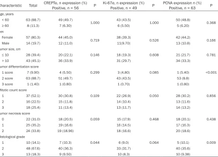
[image:3.612.94.523.73.433.2]Table 1. Correlations between Ki-67, CREPT and PCNA expression and clinicopathological characteris-tics
Characteristic Total CREPTe, n expression (%)Positive, n = 56 P Ki-67e, n expression (%) Positive, n = 49 P PCNA expression n (%) Positive, n = 63 P
Age, years
< 60 63 (88.7) 49 (49.7)
1.000 43 (43.5) 1.000 50 (48.8) 0.368
≥ 60 8 (11.3) 7 (6.30) 6 (5.50) 5 (6.20)
Sex
Female 57 (80.3) 44 (45.0)
0.719 38 (39.3) 0.526 42 (44.2) 0.166
Male 14 (19.7) 12 (11.0) 11(9.70) 13 (10.8)
Tumor size, cm
≤ 10 28 (39.4) 20 (22.1) 0.146 18 (19.3) 0.608 21 (21.7) 0.781
> 10 43 (45.1) 36 (33.9) 31 (29.7) 34 (33.3)
Tumor differentiation score
1 score 7 (9.90) 4 (5.50) 0.299 3 (4.80) 0.085 1 (5.40) <0.001
2 score 63 (88.7) 51 (49.7) 43 (43.5) 53 (8.8)
3 score 1 (1.40) 1 (0.80) 1 (0.70) 1 (0.80)
Mitotic count score
1 37 (52.1) 30 (30.8) 0.109 22 (26.9) 0.050 28 (30.2) 0.856
2 16 (22.5) 15 (11.8) 14 (10.4) 13 (11.6)
3 18 (25.4) 11 (13.4) 13 (11.7) 14 (13.2)
Tumor necrosis score
0 22 (31.0) 18 (20.5) 0.059 15 (17.9) 0.468 18 (20.1) 0.438
1 25 (35.2) 19 (16.6) 16 (14.5) 17 (16.3)
2 24 (33.8) 19 (18.96) 18 (16.6) 20 (18.6)
Histological grade
1 10 (14.1) 7 (10.3) 0.044 6 (9.0) 0.064 5 (10.1) 0.009
2 48 (67.6) 40 (36.3) 33 (31.7) 40 (35.6)
3 13 (18.3) 9 (9.50) 10 (8.3) 10 (9.38)
Statistical analyses were performed using the Pearson χ2 test.
0.05 was considered to be statistically signi-
ficant.
Results
Expression of CREPT, Ki-67, and PCNA in ret-roperitoneal LMS samples
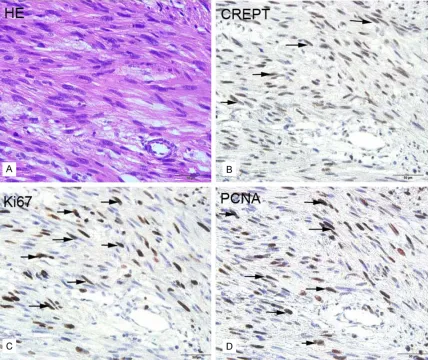
Histopathological examination showed that the retroperitoneal LMS tumor tissues consisted of spindle cells that formed sheets or fascicles [10]. These cells contained oval nuclei and an elongated hyperchromatic and abundant
eosinophilic cytoplasm (H&E, × 400, Figure 1A). To investigate the potential roles of CREPT, Ki-67 and PCNA in retroperitoneal LMS, we examined the expression of CREPT, Ki-67 and
PCNA in paraffin-embedded sections from 71
patients via immunohistochemistry. We detect-ed the expression of CREPT, Ki-67 and PCNA only in the nucleus (Figure 1B-D). A total of 56 patients (78.9%) displayed positive CREPT expression. Positive expression of Ki-67 was detected in 49 cases (69.0%), and positive
PCNA expression was detected in 63 cases
(88.7%). There was a significant association
between the expression of CREPT and the mitotic count, the histological grade, Ki-67 expression and PCNA expression (P = 0.046, P = 0.044, P = 0.002 and P < 0 .001, respec-tively, Table 1).
Univariate and multivariate analysis for prog-nostic factors
A total of 64 retroperitoneal LMS patients with available follow-up data were selected to gener-ate the Kaplan-Meier survival curves. The OS rangeds from 5.0 to 74.0 (mean 34.0) months.
The five-year OS rate of the retroperitoneal LMS
patients was 28.1%. Univariate analysis revealed that the expression levels of CREPT,
Ki-67 and PCNA were significant prognostic
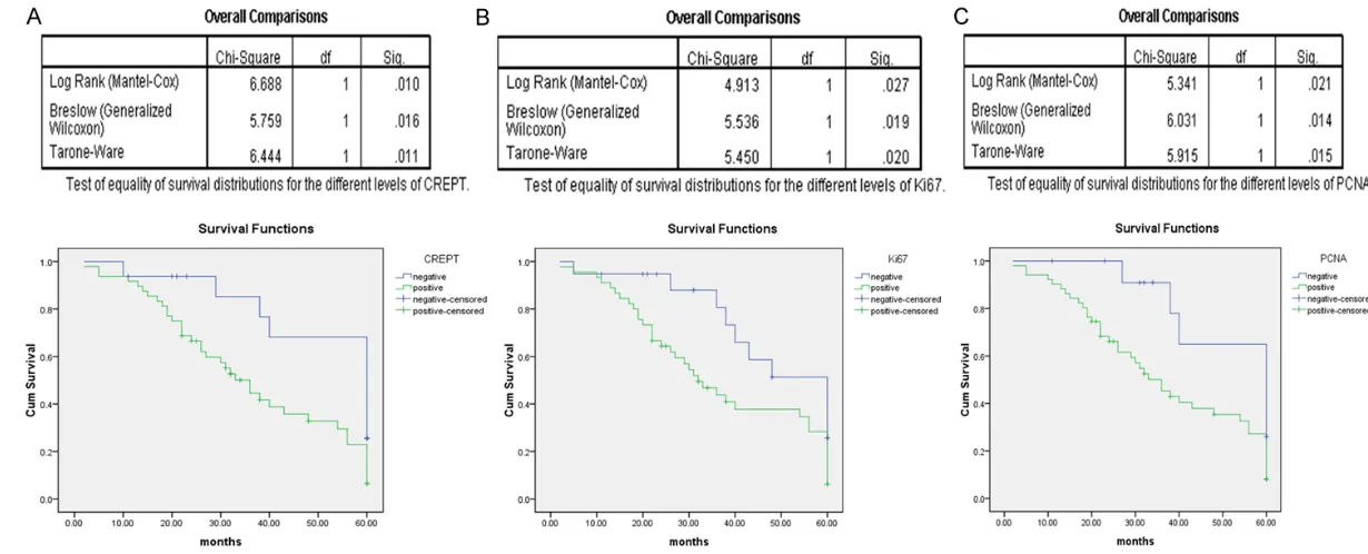
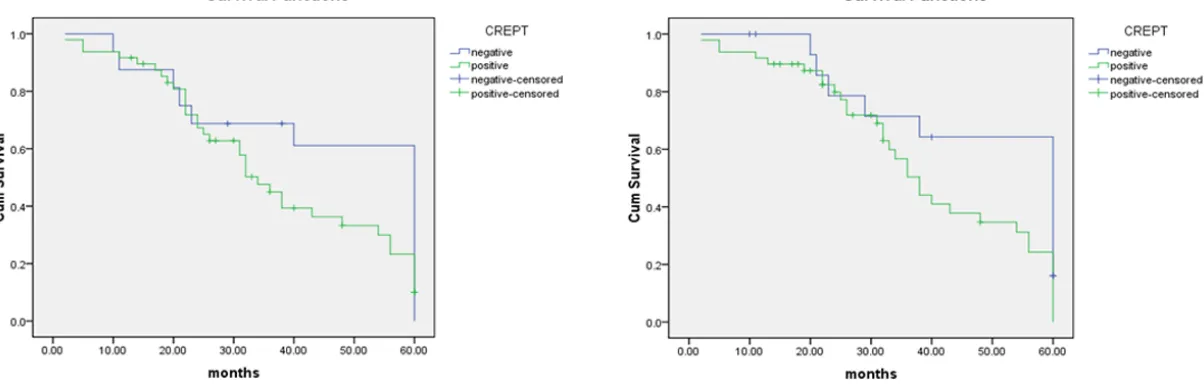
of CREPT, Ki-67 and PCNA correlated with poor OS (Figure 2; Table 2). Furthermore, a multivari-ate analysis revealed that CREPT expression and the histological grade are independent prognostic factors of OS (Table 2; P = 0.012 and P = 0.011, respectively). Taken together, these results suggested that CREPT is strongly correlated with the prognosis of retroperitoneal LMS patients.
Comparison of the expression of CREPT, Ki-67, and PCNA in retroperitoneal LMS tumors and adjacent noncancerous tissues via WB.
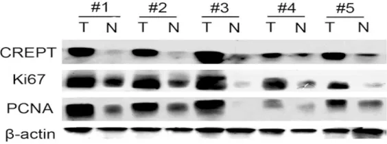
Five paired fresh frozen samples, which includ-ed retroperitoneal LMS tumor and adjacent noncancerous tissues, were collected for CREPT, Ki-67 and PCNA protein expression analysis (Figure 3). The results showed that CREPT was highly expressed in retroperitoneal LMS tumor tissues (Figure 3, top panel). In con-trast, low levels of CREPT expression were detected in the adjacent normal tissues (Figure 3, top panel). Additionally, we found that the expression levels of Ki-67 and PCNA in the
ret-roperitoneal LMS tumor tissue were significant -ly higher than those in the adjacent noncancer-ous tissues (Figure 3, middle panel). These results suggested that CREPT expression is correlated with the tumorigenesis of retroperi-toneal LMS.
Correlation between CREPT expression and initial recurrence
A total of 18 patients survived for the median follow-up duration of 41 (range, 1-127) months. Based on univariate analysis, histological
grade, tumor size and CREPT expression were
significant prognostic factors (Table 2). Clinical follow-up was performed, and the survival anal-ysis suggested a close correlation between CREPT expression and initial recurrence of
ret-roperitoneal LMS. The 5-year disease-specific
survival (DSS) was 34% for primary LMS. Among the 45 patients experiencing recurrence, the pattern of recurrence was predominately dis-tant recurrence (DR), which occurred in 19 patients (42.2%). Local recurrence (LR) occurred in only 16 patients (35.6%); 10 patients (22.2%) exhibited both LR and DR. As shown in Figure 4, CREPT expression was a prognostic factor for distant primary LMS recur-rence (P = 0.04).
Discussion
Soft tissue sarcomas constitute 0.7% of adult malignancies [11]. Ten to twenty percent of soft tissue sarcomas occur in the retroperitoneum,
and LMS, liposarcoma and fibrosarcoma are
the most common histological types [12-16]. The retroperitoneum provides a broadly expandable anatomic location for tumor growth, and these tumors often attain a large size before symptoms manifest [1, 17]. Relevant symptoms are often indistinguishable from those of a mass in the abdominal viscera or reproductive organs. LMSs are relatively insen-sitive to radiotherapy and chemotherapy, and broad surgical excision is the preferred treat-ment. Retroperitoneal LMSs display relatively late presentation and are often proximal to sev-eral vital structures in the retroperitoneal space [18, 19]. Although the standard treatment Table 2. Univariate and multivariate survival analyses of 71 patients with retroperitoneal LMS
Univariate analysis Multivariate analysis
RR P Value 95.0% CI for Exp (B) RR P Value 95.0% CI for Exp (B)
Age 1.130 0.780 0.479-2.669
Sex 1.804 0.116 0.846-3.766
Size 1.878 0.038 1.036-3.405
Tumor differentiation 1.330 0.540 0.535-3.308
Mitotic count 1.293 0.127 0.930-1.800
Tumor necrosis 1.142 0.422 0.826-1.579
Histological grade 1.719 0.024 1.074-2.751 1.858 0.011 1.153-2.995
Ki-67 expression 2.041 0.047 1.011-4.123
CREPT expression 2.381 0.022 1.132-5.006 2.606 0.012 1.236-5.494
remains margin-negative surgical resection, the 5-year survival rate after surgical resection remains low, ranging from 29% to 67% [20, 21]. However, the exact molecular mechanisms of the progression and recurrence of retroperito-neal LMSs are unclear, and there is a lack of valid and reliable biomarkers to predict retro-peritoneal LMS recurrence.
Ki-67, a nuclear antigen present during the G1, S, G2, and M phases of all proliferating human cells, is a biomarker of tumor proliferation [22]. Patients displaying increased Ki-67 expression
exhibit a lower cancer-specific survival rate
[23]. The level of Ki-67 expression has been used in clinical analyses to predict the survival rate in a scenario with several neoplasms.
Previous studies have identified Ki-67 as a tool
in determining the malignancy of smooth
mus-cle neoplasms (exemplified by myxoid LMS of
the uterus) [24, 25]. PCNA is central to many essential cellular processes, such as DNA repli-cation, DNA damage repair, chromatin struc-ture maintenance and cell cycle progression [26]. PCNA has been widely used in studies assessing the growth rate of human
malignan-cies. These previous studies indicated a signifi -cant expression level of PCNA in low- and inter-mediate-grade LMS [27].
CREPT has been reported to be a highly expressed oncogene in a variety of tumors [7]. CREPT expression accelerates malignant cell growth and tumorigenesis. Lu D et al. revealed that CREPT mRNA expression was up-regulated in various human malignant tumors and that the CREPT protein expression level was closely associated with the degree of differentiation and the clinical stage of the tumor [7].
In this report, we first detected the protein
tissues. The expression of CREPT in the
retro-peritoneal LMS tumor tissue was significantly
stronger than that in the adjacent noncancer-ous tissue. Therefore, we speculated that CREPT may play an important role in retroperi-toneal LMS.
Our statistical analysis showed that the expres-sion of CREPT, Ki-67 and PCNA closely corre-lated with 5-year overall survival and the dis-tant recurrence of retroperitoneal LMS. Patients displaying high CREPT, Ki-67 or PCNA expression exhibited relatively short recur-rence-free survival and overall survival; the patients displaying low CREPT expression exhibited relatively longer survival (P = 0.010, P
= 0.027 and P = 0.021, respectively).
Univariate analyses identified tumor size, histo -logical grade, and the expression level of
CREPT, Ki-67 and PCNA as significant prognos -tic factors (P = 0.038, P = 0.024, P = 0.022, P
= 0.047 and P = 0.042, respectively), and mul-tivariate Cox regression analysis revealed that the histological grade and expression levels of CREPT were independent prognostic factors of poor overall survival (OS) among retroperitone-al LMS patients (P = 0.011 and P = 0.012, respectively). The retroperitoneal LMS patients in our study experienced a higher incidence of DR (42.2% at 5 years) than LR. With respect to DR, we found that CREPT expression is a prog-nostic factor for the distant recurrence of retro-peritoneal LMS (P = 0.04, Figure 4). The above results strongly suggested that CREPT plays an important role in retroperitoneal LMS progres-sion and that CREPT represents a valuable bio-marker in predicting the prognosis of patients with retroperitoneal LMS.
[image:7.612.91.373.73.178.2]CREPT has been suggested to display unique
Figure 3. Western blot analysis of CREPT, Ki-67 and PCNA expression in ret-roperitoneal LMS tumor and normal adjacent tissues. T refers to the tumor tissue, and N refers to the paired normal tissue from the same patient.
tumor tissue. Immunohistoch- emical staining results sho- wed that CREPT, Ki-67 and PCNA were positively expre- ssed in more than half of the retroperitoneal LMS patients. Further investigation showed that the expression of CREPT correlated with the expression
distant recurrence of retroperitoneal LMS com-pared with conventional biomarkers such as Ki-67 and PCNA. Therefore, CREPT serves as a candidate biomarker that could be used in combination with the conventional clinical bio-markers mentioned above. In clinical studies, accurate and reliable prognostic markers are crucial for providing comprehensive prognostic
information and an accurate basis for
treat-ment decisions. In this study, we identified the
novel potential biomarker CREPT to be a prog-nostic factor in cancer progression and distant recurrence among patients with retroperitoneal LMS.
Acknowledgements
We would like to thank Lixin Wei of The Department of Pathology at the Chinese PLA General Hospital for providing assistance with
the paraffin sections. We thank B. S.M.T.
Junzhen Fan for his assistance with sample preparation in this study. This work was sup-ported by grants from The 973 Project (2011- CB910502, 2011ZX08011-006), The NSFC (81372167, 30871286, 31071225, 310300- 40), The Tsinghua Science Foundation (2012- 1080018), and The 863 project (2012AA0- 21703) of China.
Disclosure of conflict of interest
None.
Address correspondence to: Dr. Peiyu Li, Depart- ment of General Surgery, Chinese PLA General Hos- pital, Beijing 100853, China. E-mail: peiyuli301@ 163.com
References
[1] Strauss DC, Hayes AJ and Thomas JM. Retro-peritoneal tumours: review of management. Ann R Coll Surg Engl 2011; 93: 275-280. [2] Strauss DC, Hayes AJ, Thway K, Moskovic EC,
Fisher C and Thomas JM. Surgical manage-ment of primary retroperitoneal sarcoma. Br J Surg 2010; 97: 698-706.
[3] Jenkins MP, Alvaranga JC and Thomas JM. The management of retroperitoneal soft tissue sar-comas. Eur J Cancer 1996; 32A: 622-626. [4] Neville A and Herts BR. CT characteristics of
primary retroperitoneal neoplasms. Crit Rev Comput Tomogr 2004; 45: 247-270.
[5] Karakousis CP and Perez RP. Soft tissue sarco-mas in adults. CA Cancer J Clin 1994; 44: 200-210.
[6] Alvarenga JC, Ball AB, Fisher C, Fryatt I, Jones L
treatment of retroperitoneal sarcoma. Br J Surg 1991; 78: 912-916.
[7] Lu D, Wu Y, Wang Y, Ren F, Wang D, Su F, Zhang Y, Yang X, Jin G, Hao X, He D, Zhai Y, Irwin DM, Hu J, Sung JJ, Yu J, Jia B and Chang Z. CREPT accelerates tumorigenesis by regulating the transcription of cell-cycle-related genes. Can-cer Cell 2012; 21: 92-104.
[8] Pautier P, Floquet A, Penel N, Piperno-Neu-mann S, Isambert N, Rey A, Bompas E, Cioffi A, Delcambre C, Cupissol D, Collin F, Blay JY, Jimenez M and Duffaud F. Randomized multi-center and stratified phase II study of gem-citabine alone versus gemgem-citabine and docetaxel in patients with metastatic or re-lapsed leiomyosarcomas: a Federation Natio-nale des Centres de Lutte Contre le Cancer (FNCLCC) French Sarcoma Group Study (TAXO-GEM study). Oncologist 2012; 17: 1213-1220. [9] Esposito V, Baldi A, De Luca A, Tonini G, Vin-cenzi B, Santini D, Persichetti P, Mancini A, Citro G and Baldi F. Cell cycle related proteins as prognostic parameters in radically resected non-small cell lung cancer. J Clin Pathol 2005; 58: 734-739.
[10] Weiss SW, Goldblum JR and Enzinger FM. Enz-inger and Weiss’s Soft Tissue Tumors. St. Lou-is: Mosby; 2001.
[11] Todd CS, Michael H and Sutton G. Retroperito-neal leiomyosarcoma: eight cases and a litera-ture review. Gynecol Oncol 1995; 59: 333-337. [12] Adam YG, Oland J, Halevy A and Reif R. Primary
retroperitoneal soft-tissue sarcomas. J Surg Oncol 1984; 25: 8-11.
[13] Cody HS 3rd, Turnbull AD, Fortner JG and Haj-du SI. The continuing challenge of retroperito-neal sarcomas. Cancer 1981; 47: 2147-2152. [14] McGrath PC, Neifeld JP, Lawrence W Jr, DeMay
RM, Kay S, Horsley JS 3rd and Parker GA. Im-proved survival following complete excision of retroperitoneal sarcomas. Ann Surg 1984; 200: 200-204.
[15] Catton CN, O’Sullivan B, Kotwall C, Cummings B, Hao Y and Fornasier V. Outcome and prog-nosis in retroperitoneal soft tissue sarcoma. Int J Radiat Oncol Biol Phys 1994; 29: 1005-1010.
[16] Hassan I, Park SZ, Donohue JH, Nagorney DM, Kay PA, Nasciemento AG, Schleck CD and Il-strup DM. Operative management of primary retroperitoneal sarcomas: a reappraisal of an institutional experience. Ann Surg 2004; 239: 244-250.
[17] Ludwig JA and Weinstein JN. Biomarkers in cancer staging, prognosis and treatment se-lection. Nat Rev Cancer 2005; 5: 845-856. [18] Wang LD, Hong JY, Qiu SL, Gao H and Yang CS.
[19] Liles JS, Tzeng CW, Short JJ, Kulesza P and Heslin MJ. Retroperitoneal and intra-abdomi-nal sarcoma. Curr Probl Surg 2009; 46: 445-503.
[20] Clary BM, DeMatteo RP, Lewis JJ, Leung D and Brennan MF. Gastrointestinal stromal tumors and leiomyosarcoma of the abdomen and ret-roperitoneum: a clinical comparison. Ann Surg Oncol 2001; 8: 290-299.
[21] Gladdy RA, Qin LX, Moraco N, Agaram NP, Brennan MF and Singer S. Predictors of sur-vival and recurrence in primary leiomyosarco-ma. Ann Surg Oncol 2013; 20: 1851-1857. [22] Gerdes J, Lemke H, Baisch H, Wacker HH,
Schwab U and Stein H. Cell cycle analysis of a cell proliferation-associated human nuclear antigen defined by the monoclonal antibody Ki-67. J Immunol 1984; 133: 1710-1715. [23] Rioux-Leclercq N, Turlin B, Bansard JY, Patard
JJ, Manunta A, Moulinoux JP, Guillé F, Ramée MP and Lobel B. Value of immunohistochemi-cal Ki-67 and p53 determinations as predic-tive factors of outcome in renal cell carcinoma. Urology 2000; 55: 501-505.
[24] Mittal K and Demopoulos RI. MIB-1 (Ki-67), p53, estrogen receptor, and progesterone re-ceptor expression in uterine smooth muscle tumors. Hum Pathol 2001; 32: 984-987. [25] Sprogoe-Jakobsen S and Holund B.
Immuno-histochemistry (Ki-67 and p53) as a tool in de-termining malignancy in smooth muscle neo-plasms (exemplified by a myxoid leiomyosar- coma of the uterus). APMIS 1996; 104: 705-708.
[26] Moldovan GL, Pfander B and Jentsch S. PCNA, the maestro of the replication fork. Cell 2007; 129: 665-679.