INTRODUCTION
The velvet belly lantern shark (Etmopterus spinax) is a common deepwater shark of the East Atlantic and the Mediterranean Sea (Coehlo and Erzini, 2008). Like other members of the Etmopteridae family, it is able to emit a visible chemical light from thousands of tiny epidermal organs called photophores (Compagno et al., 2005; Claes and Mallefet, 2008). These organs are organised in nine different luminous zones forming a complex luminous pattern (Claes and Mallefet, 2008). Although few studies have focused on the function of this bioluminescence, it is probably used for various purposes including camouflage by counter-illumination and intraspecific behaviours such as schooling and sexual signalling (Claes and Mallefet, 2009a).
Recently, photophores of this shark, unlike those of any other luminescent animal, have been shown to be under hormonal control: (i) application of classical neurotransmitters [(nor)adrenaline, GABA, serotonin and acetylcholine] on E. spinax photophore preparations does not induce a light reaction (Claes and Mallefet, 2009b), which contrasts with the neural luminescence control mechanism (mostly adrenergic) of teleost fish intrinsic (non-bacterial) photophores (Baguet, 1975; Baguet and Marechal, 1978; Baguet and Christophe, 1983), and (ii) melatonin (MT) and prolactin (PRL) respectively trigger a slow and a fast light emission, which are both inhibited by a prior application of a-melanocyte stimulating hormone (a-MSH) (Claes and Mallefet, 2009b). Hormonal luminescent responses, however, vary greatly between investigated shark specimens (Claes and Mallefet, 2009b) and a recent study highlighted a differential sensitivity to both hormones between the different luminous zones of the pattern and, in the case of PRL, between sexes for several luminous zones probably involved in mating (Claes and Mallefet, 2010). These results suggest therefore
that an additional control mechanism might be present to modulate the hormonally induced luminescence in E. spinax.
Nitric oxide (NO) is a small gaseous signalling molecule that plays a key role in invertebrate as well as in vertebrate biological functions (Jacklet, 1997). Its effects are mediated either by the production of cyclic guanosine monophosphate (cGMP) or by the inhibition of mitochondrial respiration in the target cells (Brown, 1995; Jacklet, 1997). Since several recent studies demonstrated the involvement of NO as a neuromodulator of neurally induced luminescence from distantly related marine organisms, such as the northern krill
Meganyctiphanes norvegica(Krönström et al., 2007) and teleost fish species including the hatchetfish Argyropelecus hemigymnus
(Krönström et al., 2005) and the midshipman fish Porichthys notatus
(Krönström, 2009; Krönström and Mallefet, 2009), we investigated the possible role of NO in the modulation of hormonally induced luminescence in the cartilaginous fish E. spinax.
In this study, therefore, this hypothesis was investigated by a combination of immunohistochemical and pharmacological methods in order to (i) detect the presence of nitric oxide synthase (NOS) in photophore structures, (ii) highlight photophore innervations, if any, (iii) investigate the putative role of NO in the luminescence control mechanism of E. spinaxand, if any, (iv) determine the intracellular pathway used by this substance.
MATERIALS AND METHODS Fish collection
Twenty-eight adult velvet belly lantern sharks Etmopterus spinax
(Linnaeus 1758) (30–51.5 cm total length, TL) were caught during three field sessions (in February 2009, June 2009 and December 2009) by longlines lowered in a deep area (depth ≥200 m) of the
The Journal of Experimental Biology 213, 3005-3011 © 2010. Published by The Company of Biologists Ltd doi:10.1242/jeb.040410
Nitric oxide in the control of luminescence from lantern shark (Etmopterus spinax)
photophores
Julien M. Claes
1,*, Jenny Krönström
2, Susanne Holmgren
2and Jérôme Mallefet
11Laboratory of Marine Biology, Earth and Life Institute, Université catholique de Louvain, B-1348 Louvain-la-Neuve, Belgium and
2Department of Zoophysiology, University of Gothenburg, Box 463, SE 405 30 Göteborg, Sweden
*Author for correspondence (julien.m.claes@uclouvain.be)
Accepted 11 May 2010
SUMMARY
Photophores (photogenic organs) of the lantern shark Etmopterus spinaxare under hormonal control, with prolactin (PRL) and melatonin (MT) triggering the light emission. Differential sensitivity to these hormones in adult individuals suggests, however, that the luminescence of this shark is controlled by an additional mechanism. In this study, different techniques were used to investigate a potential modulator ofE. spinaxluminescence – nitric oxide (NO). NO synthase (NOS)-like immunoreactivity (IR) was found in the photocytes (photogenic cells) of the photophores. In addition, acetylated tubulin IR also supported the presence of nerves running through the photogenic tissue and innervating different structural elements of the photophores: photocytes, pigmented cells from the iris-like structure and lens cells. Pharmacological experiments confirmed a modulatory action of NO on the hormonally induced luminescence: NO donors sodium nitroprusside (SNP) and hydroxylamine decreased the time to reach the maximum amplitude (TLmax) of MT-induced luminescence while these substances decreased the maximum amplitude of PRL-induced luminescence (and also the TLmax in the case of SNP). The small impact of the NOS inhibitor L-NAME on hormonally induced luminescence suggests that NO is only produced on demand. The cGMP analogue 8BrcGMP mimicked the effects of NO donors suggesting that the effects of NO are mediated by cGMP.
Raunefjord, Norway. Living specimens were transferred to two large (1 m3) tanks in a dark cold (6°C) room at Espeland Marine Station
(Espegrend, Norway) where they were kept until use. Captive animals were finally killed, before experimentation, by a blow to the head, following the local rules for experimental fish care.
Immunohistochemistry
Ventral skin patches containing photophores were taken from 8 different individuals (6 adults and 2 embryos from a pregnant female) and used for immunohistochemistry. Each tissue was fixed for 48 h in 3% formaldehyde (in saline) then transferred to phosphate-buffered saline (PBS) with sodium azide (NaN3), and stored overnight in PBS
with 30% sucrose before embedding. The tissue was embedded in OCT (Sakura, Zoeterwude, The Netherlands) and quickly frozen in isopentane chilled with liquid nitrogen.
A cryostat microtome (Zeiss Microm International GmbH, Walldorf, Germany) was used to cut 10mm sections collected on chrome alum gelatine-coated slides and left overnight to dry.
Fluorescence histochemistry
Sections were preincubated with normal donkey serum (10%) for 30 min to prevent non-specific staining. Sections were incubated for 48 h after the application of primary antibodies (Table 1). They were rinsed (3⫻5 min in PBS with 2% NaCl) and incubated for 60 min after the application of the secondary antibodies (Table 1). All incubations occurred in a humid chamber at room temperature (20°C). Sections were rinsed anew with the same procedure and mounted in Vectashield mounting medium (Vector Laboratories, Burlingame, CA, USA). Sections were examined using a Nikon eclipse E1000 microscope (Nikon, Tokyo, Japan) equipped with a Nikon DMX1200 digital camera.
In order to confirm the specificity of the secondary antibody, several control preparations were performed with omission of the primary antibody. These control experiments also allowed us to highlight potential autofluorescence from the tissue.
Avidin–biotin histochemistry
After preincubation with normal donkey serum, sections were first incubated with the primary and the secondary antibodies (Table 1) following the procedure described above (endogenous peroxidase activity was blocked by H2O2 preincubation; 0.3% or 3% for
5–10 min). Sections were then incubated with the avidin-biotinylated peroxidase complex (ABC Elite PK 6100 standard, Vector Laboratories) for 30min and developed in Vector Nova red substrate kit (SK 4800, Vector Laboratories) for 3 min. Sections were then mounted in 50% glycerol and 50% carbonate buffer (pH 8.5) and examined as described above.
In order to confirm the specificity of the secondary antibody, several control preparations were performed with omission of the primary antibody.
NADPH-diaphorase activity
In addition to the different control experiments mentioned above, we also performed control experiments testing the NADPH-diaphorase activity in the photogenic tissue of E. spinax, in order to confirm the staining obtained with antibodies raised against universal NOS (uNOS).
Ventral skin patches containing photophores were taken from 6 different adult individuals, fixed in formaldehyde (3.7% in phosphate buffer) for 2 h, and rinsed for at least 1 h in PBS. The preparations were embedded, frozen and sectioned. They were then preincubated in a solution containing 0.2% b-NADPH (Sigma Chemical Company, St Louis, MO, USA), 0.025% nitro-blue tertrazolium (Sigma), 0.5% methanol, 0.5% dimethylformamide, 0.01% DMSO and 0.3% Triton X in PBS for 1h at 37°C. The sections were rinsed for 4⫻5 min in PBS and mounted in carbonate-buffered glycerol (50% glycerol and 50% carbonate buffer, pH 8.5).
In order to confirm the specificity of the staining, we included several control skin patches that received a similar solution but without the b-NADPH.
Pharmacology
Luminometry
Photophore preparations used in this work consisted of circular ventral skin patches (0.25 cm2) containing photophores dissected
out from adult shark specimens with a metal cap driller and placed in a shark saline (Bernal et al., 2005) following the method of Claes and Mallefet (Claes and Mallefet, 2009b). These preparations will hereafter be called ‘ventral skin patches’ (VSP).
VSP were transferred to a 96-wells plate, each well containing 100ml saline, which was inserted in a luminometer (Berthold Orion, Pforzheim, Germany) calibrated with a standard 470 nm light source (Beta light, Saunders Technology, Hayes, UK). The luminescent responses were recorded for 1 h after the stimulation and were characterised using different light parameters (Fig. 1): the maximum intensity of light emission (Lmax, in megaquanta per
second, Mq s–1), the total quantity of light emitted during the
experiment (Ltot, in teraquanta per hour, Tq h–1), and the time to
reach Lmax from the stimulation time (TLmax, in seconds). Light
parameters were standardised by skin surface area (cm–2).
Chemicals
Chemical agents used in this study were the followings: 8-bromoguanosine 3⬘,5⬘-cyclic monophosphate sodium salt
Table 1. Antibodiesand peptidesused for immunohistochemistry
Antibody Host
Working
dilution Antigen (peptide sequence) Source Primary
uNOS PA1-039 Acetylated tubulin T-6793
Secondary
DaM-FITC 715-095-150 DaR-biotin 711-065-152 DaR-Cy3 711-165-152
Rabbit Mouse
Donkey Donkey Donkey
1:100 1:1000
1:100 1:400 1:800
Murine iNOSand nNOS C-terminal (1113–1122)
Seaurchin acetylated tubulin
Transduction Laboratories1
Sigma2
Jackson ImmunoResearch Laboratories3
Jackson ImmunoResearch Laboratories
Jackson ImmunoResearch Laboratories
Antibodiesagainst nitric oxide synthase (NOS) are polyclonals while those against acetylated tubulin are monoclonal. DaM, donkey anti-mouse; DaR, donkey anti-rabbit; uNOS, universal NOS, i.e. antibody reacting to all NOS isoforms.
1
San Jose, CA, USA; 2
St Louis, MO, USA; 3
monohydrate (8BrcGMP; Sigma), hydroxylamine hydrochloride (Sigma), melatonin (Sigma), NG-nitro-L-arginine methyl ester (L
-NAME; Sigma), 1H-[1,2,4]oxadiazolo[4,3-a]quinoxaline-1-one (ODQ; Tocris Cookson Inc., Ellisville, MS, USA), prolactin (Sigma), and sodium nitroprusside (SNP; Sigma). All chemicals were dissolved just before use and diluted to the proper concentration in a shark saline of the following composition: 292 mmol l–1NaCl, 3.2 mmol l–1KCl,
5 mmol l–1 CaCl
2, 0.6 mmol l–1 MgSO4, 1.6 mmol l–1 Na2SO4,
300 mmoll–1urea, 150mmoll–1trimethylamine N-oxide, 10mmoll–1
glucose, 6mmoll–1NaHCO
3, adjusted to pH7.7 (Bernal et al., 2005).
Methods
This section aimed to investigate a potential modulatory action of NO on hormonally induced luminescence from E. spinaxphotophores, and, if this role was confirmed, the intracellular pathway used by this substance to mediate its effects. For this purpose, we first checked whether nitrergic drugs (NO donors, NOS inhibitor, cGMP analogue and soluble guanylyl cyclase inhibitor) were able to trigger light per se, and then tested the effects of a 20 min pre-treatment with nitrergic drugs on the light parameters of the luminescence induced by MT or PRL (both also applied alone as a control). The experimental procedure was performed at room temperature.
Statistics
Statistical analyses (Student’s t-test and linear regressions) were performed with the software SAS/STAT (SAS Institute Inc., 1990, Cary, NC, USA). Two regression slopes were only considered to be significantly different if there was no overlap between their 95% slope intervals. Since luminescence responses showed a high variability in amplitude and kinetics between individuals, we expressed the light parameters of the luminescence from treated patches in relative units, i.e. as a percentage (%) of the control response of the same animal. Each mean value was expressed with its standard error (mean ± s.e.m.) and Nis the number of skin patches used for a specific treatment (which actually corresponds to the number of shark specimens tested, as one skin patch from each individual was used in each treatment).
RESULTS Immunohistochemistry
Etmopterus spinaxphotophores are relatively simple in structure: they consist of a pigmented cup containing 6–10 photogenic cells (photocytes) covered by one or several lens cell(s) (Fig. 2A, inset). Blood sinuses are present in the pigmented cup and probably provide a route for photophore luminescence-controlling hormones to reach their target, which could be the iris-like structure (ILS) present between photogenic and lens cells, since this structure is thought to regulate the amount of light emitted by photocytes (Ohshima, 1911; Claes and Mallefet, 2009b). Control experiments (with omission of the primary antibody) showed that endogeneous fluorescence is only found in E. spinax’s photocytes (Fig.2A); this has already been demonstrated for embryos of this species (Claes and Mallefet, 2008).
Antibodies raised against acetylated tubulin supported the presence of nerve fibres in the connective tissues underlying E. spinax’s photophores, in the epidermis and also inside the photophores (Fig.2B,C). Nerve-like bundles entered the pigmented cup of the photophores at different locations, reaching the photocytes directly as well as other functional cell types including pigmented cells of the ILS and lens cells (Fig. 2B,C). Nerve-like structures were also observed to link different photophores suggesting the presence of a nervous network (Fig. 2C).
NOS-like immunoreactivity (IR) was observed in the cytoplasm of photocytes by fluorescence histochemistry (Fig.2B,C) as well as by avidin–biotin histochemistry (Fig. 2D,E). The specificity of this staining was confirmed by the strong NADPH-diaphorase activity also present in the cytoplasm of the photocytes as shown by the intense blue staining of these cells. Furthermore, control preparations lacking the primary antibody (immunohistochemistry; Fig. 2D) or b-NADPH (NADPH-diaphorase activity; Fig.2F) did not show any unspecific straining.
Pharmacology
Since an identical relationship was found between the light parameters
Ltotand Lmax(Table2) after injections of MT or PRL, with or without
pre-treatment with a nitrergic drug, only the light parameters Lmax
and TLmaxwere considered in the following analyses.
Control responses
Addition of PRL or MT at a final concentration of 10–6moll–1always
evoked a slow and sustained luminescence from photophores of control VSP. In these experiments the mean Lmaxinduced by MT
on 14 VSP (15.89±2.58 Mq s–1cm–2) was attained in 42.88±
4.71 min (TLmax) while 20 control preparations stimulated by PRL
gave a higher Lmax(23.14±5.16Mqs–1cm–2) that was reached earlier,
in 20.46±0.96 min (TLmax).
NO donors SNP
The effects of the NO donor SNP were tested. Application of SNP (10–6 to 10–3mol l–1; N=8) for 1 h on VSP did not induce light
emission per se. SNP (10–6 to 10–3mol l–1) had no effect on the
amplitude (Lmax) of luminescence induced by MT but reduced the
TLmaxof this light emission (Fig. 3A). SNP (10–5to 10–3mol l–1)
decreased the amplitude (Lmax) of PRL-induced luminescence, and
also affected the TLmaxof this light emission (Fig. 3B). Similar to
what was found in the hatchetfish A. hemigymnus(Krönström et al., 2005), the inhibitory effect of SNP on the Lmaxof PRL-induced
luminescence was not constant but instead directly dependent on the Lmax of the control photophores, i.e. on the capacity of the
photophore to luminesce in response to PRL (Fig. 4).
Time (s)
L
u
mine
s
cence (
q
s
–1
)
Ltot
Lm
a
x
[image:3.612.46.237.65.261.2]TLmax
Fig. 1. Typical luminescence emission (in quanta per second, q s–1) curve
from photophores of Etmopterus spinax showing the different light parameters. Lmax, maximum intensity of light emission; Ltottotal quantity of
light emitted during the experiment (shaded area); TLmax, time from
Hydroxylamine
Hydroxylamine is converted into NO in organs having a catalase activity (DeMaster et al., 1989; Krönström et al., 2005). Application of hydroxylamine (10–3mol l–1; N=8) for 1 h on VSP did not induce
light emission per se. Hydroxylamine (10–3mol l–1) decreased the
TLmaxof MT-induced luminescence but had no effect on its Lmax
(Fig.5A,B). Conversely, hydroxylamine (10–3moll–1) decreased the
Lmaxof PRL-induced luminescence but had no effect on its TLmax
(Fig. 5A,B).
NOS inhibitors
In order to investigate the level of endogenous NO production in
E. spinaxphotophores, we studied the effect of the NOS inhibitor
L-NAME. Application of L-NAME (10–6to 10–3mol l–1; N=6) for
1 h on VSP did not induce luminescence per se. L-NAME (10–6to
10–3mol l–1) had no effects on the light parameters (L
maxor TLmax)
of the MT-induced luminescence (Fig. 6A). On the other hand, L
-NAME treatment unexpectedly decreased the TLmaxof PRL-induced
luminescence at 10–4and 10–3mol l–1(Fig. 6B).
Effects on the cGMP pathway
In order to determine whether the effect of NO on hormonally induced luminescence of lantern shark photophores was mediated through the production of cGMP, we tested the effects of a membrane-permeable cGMP analogue (8BrcGMP) and a selective inhibitor of NO-sensitive guanylyl cyclase (ODQ) on E. spinaxlight emission. None of these substances induced light emission per se
in E. spinaxwhen applied on VSP for 1 h (at 10–3and 10–5mol l–1
for 8BrcGMP and ODQ, respectively). 8BrcGMP (10–4mol l–1) decreased the TL
maxof the MT-induced
luminescence but had no effects on its Lmax (Fig. 7A,B). No
[image:4.612.51.565.66.436.2]significant effect of 8BrcGMP was detected on PRL-induced luminescence (Fig. 7A,B). However, there was a strong tendency towards a decrease in Lmaxafter 8BrcGMP application (P=0.06).
Control and ODQ (10–5moll–1)-treated photophores show similar
hormonally induced luminescence curves, with values of light parameters Lmaxand TLmaxthat are not significantly different (Fig.
8A,B).
DISCUSSION
This study shows for the first time the presence of nerve fibres and NOS-like IR associated with E. spinaxphotophores, suggesting a role for NO and nerves in the control of luminescence in this shark. Pharmacological results confirm the involvement of NO in the control of E. spinax’s luminescence. However, rather than having a direct effect on the luminescence control mechanism, NO appears to act more as a modulator of the effects of MT and PRL: NO donors applied onto photophore preparations failed to trigger luminescence
per sebut modulated the parameters of luminescence induced by MT or PRL.
Control of hormone-induced luminescence
Contrary to earlier views (Johann, 1899), this study suggests that photophores of the lantern shark E. spinaxare innervated. Various attempts to trigger luminescence in this species using classical neurotransmitters [acetylcholine, (nor)adrenaline, serotonin and GABA] have failed (Claes and Mallefet, 2009b), which suggests that
either these nerves do not contain classical neurotransmitters or they do not have a stimulatory role in the light production/emission in this shark. However, since the nerves reach the photophores through the underlying connective tissue and enter these photogenic organs to reach several structural components (photocytes, ILS, lens cells), they must be involved somehow in the control mechanism of E. spinax
luminescence. Moreover, since they sometimes branch into different photophores, they may also serve to coordinate the luminescence activity of these organs. However, further investigations are needed to determine whether the nerves are spinal autonomic or some other type of neuron (Nilsson, 1983).
In the hatchetfish (A. hemigymnus), the NO modulation of adrenaline-induced luminescence appears to occur vianerves reaching the photocytes, and other luminescent bony fish harbour NO-containing nerves inside their photogenic organs (Krönström et al., 2005; Krönström and Mallefet, 2009). No clear evidence of such NO-containing nerves was found in the photophores of the cartilaginous
E. spinax. We found, however, strong NOS-like IR and NADPH-diaphorase activity in the photocytes of this species, suggesting a role for NO in the luminescence control mechanism of E. spinax.
Pharmacological results clearly supported the involvement of NO in the modulation of luminescence induced by MT as well as PRL. However, the effects of NO on these hormonal light responses were not similar: NO only had an effect on the kinetics of MT-induced luminescence but affected the kinetics and the amplitude of PRL-induced luminescence. This result is not surprising: MT and PRL have different intracellular transmission pathways (Claes and Mallefet, 2009b), and one can therefore assume that NO will act at
Table 2. Relationship between Lmax and Ltot for control photophore preparationsand nitrergic drugs*
N Slope R2 P
Melatonin Control (10–6
moll–1 ) +SNP (10–3 moll–1)
+Hydroxylamine (10–3 moll–1) +8BrcGMP (10–4 moll–1) +L-NAME (10–3 moll–1)
Prolactin
Control (10–6 moll–1) +SNP (10–3
moll–1 ) +Hydroxylamine (10–3
moll–1 ) +8BrcGMP (10–4 moll–1) +L-NAME (10–3 moll–1)
13 8 8 8
6
14
8 8 8
6
0.38±0.03
0.39±0.02 0.37±0.03
0.38±0.02 0.35±0.04
0.61±0.05 0.63±0.05 0.68±0.08
0.64±0.12 0.81±0.25
0.99 1.00 0.99 1.00 0.99
0.98
0.99 0.98
0.96 0.93
<0.0001 <0.0001 <0.0001 <0.0001 <0.0001
<0.0001 <0.0001 <0.0001 <0.0001 0.0004
*Lmax (maximum intensity of light emission) is in Mqs –1
cm–2
, Ltot (total quantity of light emitted) is in Tq h –1
cm–2
(where q isquanta). L-NAME, NG
-nitro-L-arginine methyl ester (NO inhibitor); SNP, sodium nitroprusside (NO donor).
[SNP] (mol l–1) Lm
a
x
/
TL
m
a
x
(% of control)
0 50 100 150 200 250
10–6 10–5 10–4 10–3
A
*
**
**
***
10–6 10–5 10–4 10–3
B
*
[image:5.612.48.569.79.218.2]**
**
**
*
*
Fig. 3. Effect of the NO donor sodium nitroprusside (SNP) on hormonally induced luminescence from E. spinax photophores. (A) Dose-dependent effect of SNP on melatonin (MT; 10–6mol l–1)-induced luminescence. SNP
had no effect on Lmax(circles) but significantly decreased TLmax(triangles)
of this light emission. (B) Dose-dependent effect of SNP on prolactin (PRL; 10–6mol l–1)-induced luminescence. SNP decreased L
max(circles) but also
had an effect on TLmax(triangles) of this light emission. Values are
expressed as a percentage of values obtained in control photophore preparations, i.e. the ventral skin patches (VSP) stimulated with MT or PRL alone.N=8 for each experiment. Asterisks indicate significant differences between control and treated ventral skin patches: *P<0.05, **P<0.01, ***P<0.001.
Lmax (Mqs–1 cm–2)
Δ
Lm
a
x
(M
q
s
–1
cm
–2
)
–25 –20 –15 –10 –5 0
50 10 20 30
ΔLmax=–1.05Lmax+2.64
Fig. 4. Relationship between Lmaxof control photophores and DLmax
(difference between Lmaxvalues of SNP-treated and control VSP), for
[image:5.612.51.289.524.623.2] [image:5.612.364.563.578.693.2]different levels when modulating the action of these two hormones on E. spinaxphotophores.
The effect of the NO donors used in this study (SNP and hydroxylamine) on PRL-induced luminescence in E. spinaxcan be compared with the effect of NO on the hatchetfish and the northern krill (M. norvegica), which is predominantly inhibitory (Krönström et al., 2005; Krönström et al., 2007). Similar to what was found in hatchetfish, the inhibitory effect of NO on the amplitude of the luminescent response was not constant but directly depends on the luminous capability of the photophore preparation: the inhibition was maximal in VSP that produced a high PRL-induced luminescence and minimal in VSP that exhibited a low response to this hormone.
On the other hand, both NO donors speeded up the luminous response from E. spinax photophores after MT application. This suggests that NO affects a mechanism controlling the rate of light emission in E. spinaxphotophores. A similar modulation of the luminescence time course by NO has already been observed in other organisms including the hatchetfish, the northern krill and the midshipman fish (P. notatus) (Krönström et al., 2005; Krönström et al., 2007; Krönström, 2009).
Hydroxylamine is a precursor of NO in biological systems that possess a catalase activity (DeMaster et al., 1989). Since the effects of hydroxylamine on hormonally induced luminescence of E. spinax are similar to the effect of a NO donor, this suggests the presence of endogenous catalase activity in this shark’s photophores, as previously demonstrated in the photophores of the hatchetfish (Krönström et al., 2005).
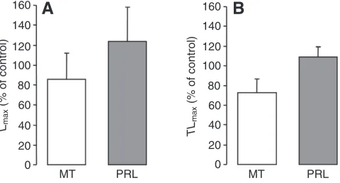
The NOS inhibitor L-NAME only had a small impact on the
timing of PRL-induced luminescence and did not affect the light
parameters of MT-induced luminescence from E. spinax
photophores. This supports the idea that NO is generated only at certain times, when changes occurring in the environment require the shark to quickly modulate the expression of its luminescence. The effects of this molecule on hormonally induced luminescence are therefore mainly transitory rather than permanent, similarly to what has been suggested for hatchetfish (Krönström et al., 2005).
In physiological processes, the effects of NO are mediated either by cGMP induced after guanylyl cyclase activation (Katsuki et al., 1977), or by a cGMP-independent pathway involving the inhibition of mitochondrial respiration (Brown, 1995). In our study, the cGMP analogue 8BrcGMP mimicked the effect of the NO donors SNP and hydroxylamine. This suggests that the NO–cGMP pathway is important in the luminescence control mechanism of E. spinax, contrary to what was found in other species in which NO appears
PRL MT
Lm
a
x
(% of control)
0 20 40 60 80 100 120 140
A
*
PRL MT TL m a x(% of control)
0 20 40 60 80 100 120 140
B
***
[L-NAME] (mol l–1) Lm a x / TL m a x
(% of control)
0 50 100 150 200 250 300 350
10–6 10–5 10–4 10–3
A
10–6 10–5 10–4 10–3
B
*
*
PRL MT Lm a x(% of control)
0 20 40 60 80 100 120 † PRL MT TL m a x
(% of control)
0 20 40 60 80 100 120
B
A
*
PRL MT Lm a x(% of control)
0 20 40 60 80 100 120 140 160 0 20 40 60 80 100 120 140 160 PRL MT TL m a x
(% of control)
[image:6.612.319.563.67.168.2]B
A
Fig. 5. Effects of the NO donor hydroxylamine (10–3mol l–1) on (A) L maxand
(B) TLmaxof hormonally (10–6mol l–1) induced luminescence from E. spinax
[image:6.612.50.294.69.182.2]photophores. Values are expressed as a percentage of values obtained in control photophore preparations, i.e. VSP stimulated with MT or PRL alone. N=8 for each experiment. Asterisks indicate significant differences between control and treated VSP: *P<0.05, ***P<0.001.
Fig. 7. Effects of the cGMP analogue 8BrcGMP (10–4mol l–1) on (A) L max
and (B) TLmaxof the hormonally (10–6mol l–1) induced luminescence from
E. spinax photophores. Values are expressed as a percentage of values obtained in control photophore preparations, i.e. VSP stimulated with MT or PRL alone.N=8 for each experiment. Asterisks indicate significant differences between control and treated VSP: *P<0.05, †P=0.06.
Fig. 6. Effect of the NOS inhibitor L-NAME on the hormonally induced luminescence from E. spinax photophores. (A) Dose-dependent effect of L -NAME on MT (10–6mol l–1)-induced luminescence. L-NAME had no effect
on the light parameters (Lmax, circles; TLmax, triangles) of this light emission.
(B) Dose-dependent effect of L-NAME on PRL (10–6mol l–1)-induced
luminescence. L-NAME had no effect on Lmax (circles) of the PRL
(10–6mol l–1)-induced luminescence but significantly reduced TL
max(triangles)
of this light emission at 10–4and 10–3mol l–1. Values are expressed as a
percentage of values obtained in control photophore preparations, i.e. VSP stimulated with MT or PRL alone.N=6 for each experiment. Asterisks indicate significant differences between control and treated VSP: *P<0.05.
Fig. 8. Effects of the guanylyl cyclase inhibitor ODQ (10–5mol l–1) on (A)
Lmaxand (B) TLmaxof the hormonally (10–6mol l–1) induced luminescence
[image:6.612.50.293.509.633.2] [image:6.612.323.563.553.682.2]to regulate light emission using a cGMP-independent pathway (Trimmer et al., 2001; Krönström et al., 2005; Krönström et al., 2007). The absence of effect of the guanylyl cyclase inhibitor ODQ on MT-induced luminescence is consistent with our hypothesis that the effect of NO is transient.
Function of NO in luminescence control
Luminescent behaviours generally necessitate rapid, precise timing and coordination to be ecologically successful (Buck, 1978; Denton et al., 1985; Branham and Greenfield, 1996; Haddock et al., 2010). The rapid action and short half-life of NO make it a perfect mediator for quick adjustments of luminescence properties. In a recent study, it was suggested that NO could be involved in the control of light production/emission of many luminescent teleost fish species (Krönström and Mallefet, 2009). Our results confirm this idea and show that cartilaginous luminescent fish also evolved a NO regulation system for their luminescence. It is not clear, however, at this time whether nerves reaching photocytes in the lumen of E. spinax photophores are NO containing or not. The presence of these nerves nevertheless confirms the idea that hormonal inputs need an additional mechanism(s) for the efficient tuning of E. spinaxphotophore luminescence.
Various adaptive advantages have been suggested for the luminescence produced by the velvet belly lantern shark, including camouflage through counterillumination and intra-specific communication (Claes and Mallefet, 2008; Claes and Mallefet, 2009a). These different functions appear to be achieved by different zones of the luminous pattern: the ventral zone (investigated in the present study) is believed to be restricted to camouflage by counterillumination while luminous zones of the pelvic area are probably used for sexual signalling (Claes and Mallefet, 2009a). With this in mind, NO could finely tune the intensity of the luminescence from glowing E. spinaxphotophores to allow this shark to counterilluminate efficiently in the mesopelagic zone, the light properties of which change with time and depth (Johnsen, 2003), similar to what was suggested in the hatchetfish (Krönström et al., 2005).
The early appearance of NOS inside the photocytes of embryonic shark photophores underlines the importance of NO modulation in
E. spinax’s life, from the very beginning. The presence of NOS in the caudal organ of the myctophid M. punctatum(Krönström and Mallefet, 2009), which is probably used for sexual communication (Herring, 2007), supports the idea that the effects of NO on luminescence are not restricted to the counterilluminating behaviour in fishes. It is therefore possible that NO modulation also exists in
luminous zones of E. spinax that are not involved in
counterillumination, but this needs further analysis.
LIST OF ABBREVIATIONS AcT acetylated tubulin
8BrcGMP 8-bromo cyclic guanosine monophosphate cGMP cyclic guanosine monophosphate 5-HT 5-hydroxytryptamine (serotonin) ILS iris-like structure
IR immunoreactivity
Lmax maximum intensity of light emission
DLmax Lmax (control preparations)–Lmax (treated preparations)
Ltot total light emission
Mq s–1cm–2 megaquanta per second per square centimetre (of skin)
a-MSH a-melanocyte stimulating hormone MT melatonin
L-NAME NG-nitro-L-arginine methyl ester
NO nitric oxide NOS nitric oxide synthase
ODQ 1H-[1,2,4]oxadiazolo[4,3-a]quinoxalin-1-one PRL prolactin
TLmax time between stimulation and maximum intensity of light
emission
Tq h–1cm–2 teraquanta per hour per square centimeter (of skin) uNOS universal nitric oxide synthase
VSP ventral skin patch
ACKNOWLEDGEMENTS
This work was supported by grants from the F.N.R.S. to J.M.C. and J.M. (FRFC 2.4516.01) and by grants from the Swedish Research Council to S.H. We would like to acknowledge A. Aadnesen, manager of Espeland Marine Biological station, T. Sørlie for his skilful help during field collections, and the two anonymous reviewers who provided valuable comments on the manuscript. J.M.C. is a postdoctoral researcher of F.N.R.S. J.M. is a research associate of FRS-F.N.R.S. Contribution to the Biodiversity Research Centre (BDIV) and to the Centre Interuniversitaire de Biologie Marine (CIBIM).
REFERENCES
Baguet, F.(1975). Excitation and control of isolated photophores of luminous fishes.
Progr. Neurobiol. 5, 97-125.
Baguet, F. and Christophe, B.(1983). Adrenergic stimulation of isolated photophores
of Maurolicus muelleri. Comp. Biochem. Physiol. 75, 79-84.
Baguet, F. and Marechal, G.(1978). The stimulation of isolated photophores
(Argyropelecus) by epinephrine and norepinephrine. Comp. Biochem. Physiol. 60, 137-143.
Bernal, D., Donley, J. M., Shadwick, R. E. and Syme, D. A.(2005). Mammal-like
muscle power swimming in a cold-water shark. Nature437, 1349-1352.
Branham, M. A. and Greenfield, M. D.(1996). Flashing males win male success.
Nature381, 745-746.
Brown, G. C.(1995). Nitric oxide regulates mitochondrial respiration and cell functions
by inhibiting cytochrome oxidase. FEBS Lett. 369, 136-139.
Buck, J. B.(1978). Functions and evolution of bioluminescence. In Bioluminescence in
Action (ed. P. J. Herring), pp. 419-460. New York: Academic Press.
Claes, J. M. and Mallefet, J.(2008). Early development of bioluminescence suggests
counter-illumination in the velvet belly lantern shark Etmopterus spinax (Squaloidea: Etmopteridae). J. Fish Biol. 73, 1337-1350.
Claes, J. M. and Mallefet, J.(2009a). Ontogeny of photophore pattern in the velvet
belly lantern shark, Etmopterus spinax. Zoology12, 433-441.
Claes, J. M. and Mallefet, J.(2009b). Hormonal control of luminescence from lantern
shark (Etmopterus spinax) photophores. J. Exp. Biol. 212, 3684-3692. Claes, J. M. and Mallefet, J. (2010). Functional physiology of lantern shark
(Etmopterus spinax) luminescent pattern: differential hormonal regulation of luminous zones. J. Exp. Biol. 213, 1852-1858.
Coehlo, R. and Erzini, K.(2008). Life history of a wide-ranging deepwater lantern
shark in the north-east Atlantic, Etmopterus spinax (Chondrichthyes: Etmopteridae), with implications for conservation. J. Fish Biol.73, 1419-1443.
Compagno, L., Dando, M. and Fowler, S.(2005). Sharks of the World. 480 pp.
London: HarperCollins.
DeMaster, E. G., Raij, L., Archer, S. L. and Weir, E. K.(1989). Hydroxylamine is a
vasorelaxant and a possible intermediate in the oxidative conversion of L-arginine to nitric oxide. Biochem. Biophys. Res. Commun. 163, 527-533.
Denton, E. J., Herring, P. J., Widder, E. A., Latz, M. F. and Case, J. F.(1985). The
roles of filters in the photophores of oceanic animals and their relation to vision in the oceanic environment. Proc. R. Soc. Lond. B. Biol. Sci. 225, 63-97.
Haddock, S. H. D., Moline, M. A. and Case, J. F.(2010). Bioluminescence in the
sea. Annu. Rev. Mar. Sci. 2, 443-493.
Herring, P. J.(2007). Sex with the lights on? A review of bioluminescent dimorphism
in the sea. J. Mar. Biol. Assoc. UK87, 829-842.
Jacklet, J. W.(1997). Nitric oxide signaling in invertebrates. Invert. Neurosci. 3, 1-14.
Johann, L.(1899). Uber eigentiumliche epitheliale gebilde (leuchtorgane) bei Spinax
niger. Z. Wiss. Zool. 66, 136-160.
Johnsen, S.(2003). Lifting the cloak of invisibility: the effect of changing optical
conditions on pelagic crypsis. Integr. Comp. Biol. 43, 580-590.
Katsuki, S., Arnold, W., Mittal, C. and Murad, F.(1977). Stimulation of guanylate
cyclase by sodium nitroprusside, nitroglycerin and nitric oxide in various tissue preparations and comparison to the effects of sodium azide and hydroxylamine. J. Cyclic Nucleotide Res. 3, 23-25.
Krönström, J.(2009). Control of bioluminescence: operating the light switch in
photophores from marine animals. PhD thesis, University of Gothenburg.
Krönström, J. and Mallefet, J.(2009). Evidence for a widespread involvement of NO
in control of photogenesis in bioluminescent fish. Acta Zool. doi: 10.1111/j.1463-6395.2009.00438.x.
Krönström, J., Holmgren, S., Baguet, F., Salpietro, L. and Mallefet, J.(2005). Nitric
oxide in control of luminescence from hatchetfish (Argyropelecus hemigymnus) photophores. J. Exp. Biol. 208, 2951-2961.
Krönström, J., Dupont, S., Mallefet, J., Thorndyke, M. and Holmgren, S.(2007).
Serotonin and nitric oxide interaction in the control of bioluminescence in northern krill, Meganyctiphanes norvegica (M. Sars). J. Exp. Biol. 210, 3179-3187.
Nilsson, S.(1983). Autonomic Nerve Function in the Vertebrates, 253 pp. New York:
Springer-Verlag.
Ohshima, H.(1911). Some observations on the luminous organs of fishes. J. Coll. Sci.
Imp. Univ. Tokyo.27, 1-25.
Trimmer, B. A., Aprille, J. R., Dudzinski, D. M., Lagace, C. J., Lewis, S. M., Michel,
T., Qazi, S. and Zayas, R. M.(2001). Nitric oxide and the control of firefly flashing.