Original Article
Protective effects of adipose-derived stem cells
secretome on human dermal fibroblasts
from ageing damages
Ting Wang1, Shu Guo1, Xuehui Liu2, Nan Xv1, Shuangyi Zhang1
1Department of Plastic Surgery, The First Hospital of China Medical University, Shenyang, China; 2Department of Hand and Foot Microsurgery, Second Hospital of Dalian Medical University, Dalian, China
Received October 4, 2015; Accepted November 22, 2015; Epub December 1, 2015; Published December 15, 2015
Abstract: Background: Combined effects of intrinsic and extrinsic ageing factors on skin tissue and the therapies have been rarely studied before. ADSCs have gained popularity in anti-ageing field, which may provide promising methods to fight against skin ageing. Objective: To find out the fate of HDFs exposed to intrinsic or extrinsic ageing factors or both of them and further examine the impacts of ADSC-CM on the damaged HDFs. Methods: We irradi-ated HDFs with UVB at different senescent levels, and then treirradi-ated them with ADSC-CM. After 48 h, we detected cellular proliferative activity, morphology, SA-β-Gal expression, apoptosis, mRNA expression of collagen I, collagen III and elastin. Results: Intrinsic ageing factors inhibited cellular proliferation, increased senescent ratio and reduced mRNA expression of collagen I, collagen III and elastin, so did UVB, except for its induction of elastin mRNA expres-sion. Furthermore, ADSC-CM treatment can slightly or significantly improve cellular proliferative activity and restore functions both in irradiated and non-irradiated HDFs. Besides, ADSC-CM treatment decreased cellular apoptosis and senescence induced by UVB but had no obvious effect on cellular senescence induced by intrinsic ageing fac-tors. The results were similar in three generations of HDFs, yet in different degrees. Conclusions: The results sug-gest that ADSCs secretome protect HDFs from ageing damages but with some limitations.
Keywords: Adipose-derived stem cells, secretory factors, anti-ageing, human dermal fibroblasts
Introduction
Skin ageing, which is mostly characterized by loss of elasticity and generation of wrinkles, confuses human for centuries and inspires us to make great efforts to fight against it, yet still remains a conundrum. As exposed directly to the outside environment, skin undergoes ex- trinsic ageing except for intrinsic ageing; both of them are associated with structural and functional changes in cutaneous tissue [1, 2]. In the present study, we focus on fibroblasts because they are the main source of dermal extracellular matrix (ECM) proteins, mainly are collagens and elastins that are processed to assemble fibers conferring tensile strength and resilience to skin, thus maintains the homeo-stasis and juvenscence of skin [3, 4]. Ageing damages lead to decline of the amount, life span and function of fibroblasts in dermis and finally result in ageing appearance, which indi-cates the significance of fibroblasts in
research-ing on mechanisms and further therapies of skin ageing [5, 6]. In vitro studies, fibroblasts can be serially passaged until they reach repli-cative senescence to simulate intrinsic ageing [7, 8]. On the other hand, it is universally accept-ed that ultraviolet rays serve as a major extrin-sic factor (approximately 80%) in skin ageing and ultraviolet radiation B (UVB)-irradiated fibroblasts have been established as an experi-mental model applied in various studies related to skin ageing [9, 10].
or extrinsic ageing separately thus failed to establish a skin ageing model accorded with actual situation and better explore the underly-ing mechanisms. In addition, few researches focused on effects of ADSCs on intrinsic ageing damages. In our group, human dermal fibro-blasts (HDFs) at different senescent levels were exposed to UVB then cultured in condi-tioned medium of ADSCs (ADSC-CM) that con-tains the whole raw secretome of and takes advantages of easier storage and safer applica-tion over ADSCs. Afterwards we detected relat-ed biochemical indexes, aiming at more com-prehensively observing the ageing characte- ristics of HDFs as well as evaluating the anti-ageing effects of ADSCs secretome.
Materials and methods
Isolation and culture of human ADSCs (hAD-SCs)
Human subcutaneous adipose tissue was har-vested from a 20-year-old woman who under-went liposuction surgery with informed patient consent at the approval of the Research Ethical Committee of the First Hospital of China Medical University. The isolation procedures were based on the published methods with minor modifications [24]. Briefly, 150 ml human lipoaspirate was washed in phosphate buffered saline (PBS; Hyclone, Logan, UT, USA) and minced finely, then digested in 0.075% collage-nase I (Gibco, New York, NY, USA) at 37.0°C with constant vibration for about 45 min. Digestion was terminated by adding Dulbecco’s Modified Eagle Medium/F12 (DMEM; Hyclone) containing 10% Fetal Bovine Serum (FBS; Gibco), then the digested tissue was centri-fuged (1200 rpm, 25°C, 5 min). After that, the supernanant was discarded and the pellet con-taining cells at the bottom was washed in PBS then centrifuged (1000 rpm, 25°C, 5 min) for twice. Cells were resuspended in common cul-ture media (DMEM containing 10% FBS and 1% penicillin-streptomycin) and counted, then
inoc-incubated with FITC-conjugated antibodies against CD13, CD29, CD34 (Santa Cruz Bio- technology, Inc., Santa Cruz, CA, USA) at 37.0°C for 30 min in the dark, followed by being washed and resuspended in PBS then detected by flow cytometer (BD Biosciences, San Jose, CA, USA).
Preparation of ADSC-CM
When hADSCs (Passage 4) reached 80% con-fluence, the culture medium was changed into DMEM. ADSC-CM was collected after 72 h, then processed by being centrifuged at 300 × g for 5 min and being filtered with a 0.22 μm syringe filter (Jet Bio-Filtration, Guangzhou, China).
Culture, UVB radiation and ADSC-CM treat-ment of HDFs
On the basis of what Yeo et al. reported previ-ously [25, 26] and our preliminary experiments, we picked out cells at passage 5, 15 and 28 as young, intermediate and senescent generation respectively. HDFs isolated from dermis (pas-sage 3) were offered by Department of Der- matology, the First Hospital of China Medical University and cultured in their corresponding dishes and cultured until reaching 70% conflu-ence, then washed once with PBS and im- mersed in a thin-layer PBS to be irradiated with a single dose (200 mJ/cm2) of UVB by using a
UV lamp (Waldmann, Villingen-Schwenningen, Germany). Once irradiation completed, medium of both irradiated and non-irradiated HDFs was replaced with control medium (DMEM contain-ing 10% FBS) or 100% ADSC-CM. Cells were then cultured in incubator for 48 h.
Cell proliferative activity detection
HDFs were seeded (2×10³ per well) into 96-well
opti-cal density (OD) values were measured at 450 nm with a microplate reader (BioTek Instru- ments, Inc., Winooski, VT, USA).
Cellular senescence detection
HDFs were seeded (5×10⁴ per well) into 6-well
plates and treated with above procedure, then cells were fixed and stained with senescence associated β-galactosidase (SA-β-Gal) Staining Kit solution (Beyotime). After being incubated in a humidified, without CO2 injection, constant
temperature (37.0°C) environment overnight, the cellular morphology and staining condition were observed and photographed using a light microscopy (OLYMPUS, Tokyo, Japan) at magni-fication of 100 and 200, senescent cells were counted and their ratio was figured out.
Cellular apoptosis detection
HDFs were seeded (5×10⁴ per well) into 6-well
plates and treated with above procedure, then cells were fixed and stained with Hoechst stain-ing solution (Beyotime). After discardstain-ing stain solution, cells were washed twice with PBS then observed and photographed using a fluo-rescence microscopy (OLYMPUS) at magnifica-tion of 100 and 200, apoptotic cells were counted and their ratio was figured out.
Quantitive real time polymerase chain reaction (qRT-PCR)
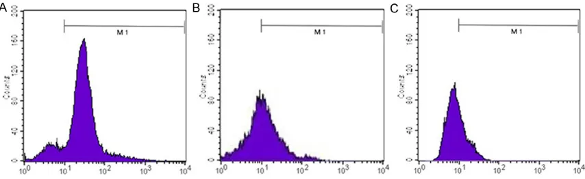
[image:3.629.102.529.80.211.2]Relative expression of type I collagen, type III collagen and elastin were measured by qRT-Figure 1. Cell surface markers expression. A. CD13: 87.37%; B. CD29: 86.57%; C. CD34: 2.51%.
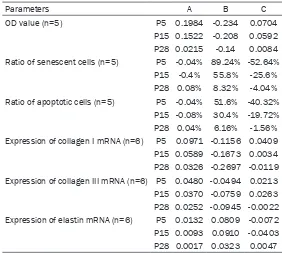
Table 1. OD value, senescent ratio, apoptotic ratio and relative mRNA expression of collagen I, col-lagen III and elastin of HDFs at different senescent levels under different treatments
Parameters Con Con+ADSC-CM UVB UVB+ADSC-CM
OD value (n=5) P5 0.5670 ± 0.0318 0.7654 ± 0.0482** 0.3330 ± 0.0396** 0.4034 ± 0.0297#
P15 0.5250 ± 0.0400 0.6772 ± 0.0502** 0.3170 ± 0.0404** 0.3762 ± 0.0356#
P28 0.4496 ± 0.0440 0.4711 ± 0.0301 0.3096 ± 0.0390** 0.3180 ± 0.0310
Ratio of senescent cells (n=5) P5 0.0008 ± 0.0011 0.0004 ± 0.0009 0.8932 ± 0.0353** 0.3668 ± 0.0204##
P15 0.3980 ± 0.0231 0.3940 ± 0.0183 0.9560 ± 0.0113** 0.7000 ± 0.0271##
P28 0.8908 ± 0.0213 0.8916 ± 0.0155 0.9740 ± 0.0134** 0.9336 ± 0.0385
Ratio of apoptotic cells (n=5) P5 0.0004 ± 0.0009 0.0000 ± 0.0000 0.5164 ± 0.0392** 0.1132 ± 0.0178##
P15 0.0012 ± 0.0018 0.0004 ± 0.0009 0.3052 ± 0.0263** 0.1080 ± 0.0191##
P28 0.0000 ± 0.0000 0.0004 ± 0.0009 0.0616 ± 0.0096** 0.0460 ± 0.0084#
Expression of collagen I mRNA (n=6) P5 1.0260 ± 0.0400 1.1231 ± 0.0423** 0.9104 ± 0.0138** 0.9513 ± 0.0342#
P15 0.9713 ± 0.0181 1.0302 ± 0.0358** 0.8040 ± 0.0362** 0.8074 ± 0.0152
P28 0.9807 ± 0.0160 1.0132 ± 0.0114** 0.7110 ± 0.0088** 0.6991 ± 0.0117
Expression of collagen III mRNA (n=6) P5 1.0293 ± 0.0329 1.0774 ± 0.0402* 0.9799 ± 0.0167* 1.0012 ± 0.0144#
P15 0.9646 ± 0.0196 1.0016 ± 0.0334* 0.8888 ± 0.0444** 0.9150 ± 0.0173
P28 0.9821 ± 0.0142 1.0074 ± 0.0111** 0.8876 ± 0.0192** 0.8855 ± 0.0283
Expression of elastin mRNA (n=6) P5 1.0111 ± 0.0238 1.0243 ± 0.0349 1.0920 ± 0.0221** 1.0847 ± 0.0527
P15 0.9661 ± 0.0203 0.9755 ± 0.0203 1.0571 ± 0.0523** 1.0168 ± 0.0173
P28 1.0354 ± 0.0203 1.0371 ± 0.0243 1.0677 ± 0.0111** 1.0724 ± 0.0246
[image:3.629.103.531.279.499.2]PCR. HDFs were seeded (5×10⁴ per well) into
6-well plates and treated with above proce-dure, then total RNA was extracted using the RNAiso Plus (TaKaRa, Shanghai, China) and quantitated by spectrophotometer (NanoDrop products, Wilmington, DE, USA). Afterwards, the obtained RNA was converted to cDNA using PrimeScriptTM RT Master Mix (TaKaRa) by PCR thermal cycler (Applied Biosystems, Darmstadt, Germany). The qRT-PCR assay was performed using SYBR Premix Ex TaqTM II (TaKaRa) by LightCycler 480 System (F. Hoffmann-La Ro- che, Ltd, Basel, Switzerland). Sequences of Primers used in this study were as followed: Human GAPDH (Forward: 5’-ACATCGCTCAGAC- ACCATG-3’; Reverse: 5’-TGTAGTTGAGGTCAAT- GAAGGG-3’); human type I collagen (Forward: 5’-CCCCTGGAAAGAATGGAGATG-3’; Reverse: 5’- TCCAAACCACTGAAACCTCTG-3’); human type III collagen (Forward: 5’-AAGTCAAGGAGAAAGTG- GTCG-3’; Reverse: 5’-CTCGTTCTCCATTCTTACC- AGG-3’); human elastin (Forward: 5’-CCTGGCT- TCGGATTGTCTC-3’; Reverse: 5’-CAA AGGGTT- TACATTCTCCACC-3’).
group (P < 0.01); ADSC-CM treatment groups exhibited higher OD values than common media treatment groups in non-irradiated (P < 0.01) and irradiated (P < 0.05) HDFs. Interme- diate HDFs (P15) displayed similar trends to young HDFs. In senescent HDFs (P28) culture, OD values of UVB group were obviously lower than that of control group (P < 0.01); ADSC-CM treatment had no obvious impacts both in non-irradiated and non-irradiated HDFs (P > 0.05) (Tables 1, 2 and Figure 2A).
Comparison of cellular senescence
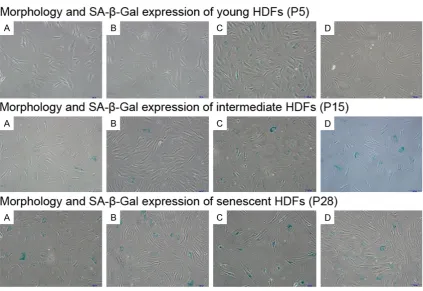
In young HDFs culture, senescent HDFs ratio of UVB group was obviously higher than control group (P < 0.01); Compared with common me- dia, ADSC-CM treatment had no obvious effect on non-irradiated HDFs (P > 0.05), but can obvi-ously decrease senescent ratio in irradiated HDFs (P < 0.01). Intermediate and senescent HDFs exhibited similar trends to young HDFs, except for the slight reduction of senescent ratio in irradiated senescent HDFs (P28) treat-ed with ADSC-CM (P>0.05) (Tables 1, 2 and
Figures 2B, 3).
Ratio of apoptotic cells (n=5) P5 -0.04% 51.6% -40.32%
P15 -0.08% 30.4% -19.72%
P28 0.04% 6.16% -1.56%
Expression of collagen I mRNA (n=6) P5 0.0971 -0.1156 0.0409
P15 0.0589 -0.1673 0.0034 P28 0.0326 -0.2697 -0.0119 Expression of collagen III mRNA (n=6) P5 0.0480 -0.0494 0.0213 P15 0.0370 -0.0759 0.0263 P28 0.0252 -0.0945 -0.0022
Expression of elastin mRNA (n=6) P5 0.0132 0.0809 -0.0072
P15 0.0093 0.0910 -0.0403 P28 0.0017 0.0323 0.0047
Differences of average values of detected indexes between groups (A. Control group and con+ADSC-CM group; B. Control group and UVB group; C. UVB group and UVB+ADSC-CM group) at different senescent levels (Negative value means inhibitory effects of the treatment factor, positive value means promoting effects of the treatment factor).
Results
Characterization and identifica-tion of hADSC
At passage 3, hADSCs dis-played CD13 and CD29 (both are stromal stem cell surface markers) positive expression, whereas CD34 (hematopoietic stem cell marker) negative expression (Figure 1).
Comparison of cellular prolif-erative activity
[image:4.629.96.378.103.356.2]Figure 2. Analysis of cellular proliferation, senes-cence and apoptosis. OD value (A), senescent ra-tio (B) and apoptotic rara-tio (C) of HDFs at different senescent levels under different treatments. c: Control group; c+A: ADSC-CM treated non-irradi-ated HDFs group; U: UVB group; U+A: ADSC-CM treated irradiated HDFs group. (**P < 0.01 com-pared with con group; #P < 0.05 and ##P < 0.01 compared with UVB group).
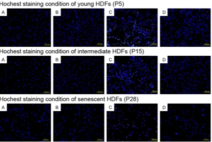
[image:5.629.103.527.422.713.2]Comparison of cellular apoptosis
In young HDFs culture, apoptotic HDFs ratio of UVB group was obviously higher than control group (P < 0.01); Compared with common media, ADSC-CM treatment had no obvious effect on non-irradiated HDFs (P > 0.05), but can obviously decrease apoptotic HDFs ratio in irradiated HDFs (P < 0.01). Intermediate and senescent HDFs shared similar trends with young HDFs, except for the moderate reduction of apoptosis in irradiated senescent HDFs (P28) treated with ADSC-CM (P < 0.05) (Tables 1, 2 and Figures 2C, 4).
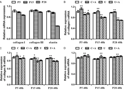
Comparison of relative expression type I colla-gen, type III collagen and elastin at mRNA level
In young HDFs culture, relative expression of type I collagen mRNA in UVB group obviously decreased compared with control group (P < 0.01); ADSC-CM treatment can increase its expression of either non-irradiated (P < 0.01) or irradiated HDFs (P < 0.05) compared with com-mon media. Intermediate HDFs displayed simi-lar changing tendency to the young generation,
except for its slight improvement in irradiated HDFs treated with ADSC-CM (P > 0.05). Senescent HDFs shared the similar trends with intermediate HDFs (Tables 1, 2 and Figure 5). In young HDFs culture, relative expression of type III collagen mRNA in UVB group decreased compared with control group (P < 0.05); ADSC-CM treatment can increase its expression of either non-irradiated or irradiated HDFs com-pared with common media (P < 0.05). In inter-mediate HDFs culture, type III collagen mRNA in UVB group obviously decreased compared with control group (P < 0.01); ADSC-CM treatment can increase its expression in non-irradiated HDFs compared with common media (P < 0.05), but had no obvious effect on irradiated HDFs (P > 0.05). Senescent HDFs shared simi-lar trends with intermediate HDFs (Tables 1, 2
and Figure 5).
[image:6.629.104.529.82.367.2]irradiated HDFs compared with common media (P > 0.05). Intermediate and senescent HDFs displayed similar changing tendency to the young generation (Tables 1, 2 and Figure 5).
Discussion
ADSCs stand out as an upstart in the field of anti-ageing mostly owing to their secretome that have been identified by using proteomics, including transforming growth factor-beta (TGF-β), platelet derived growth factor (PDGF), insu-lin-like growth factor (IGF), hepatocyte growth factor (HGF), pigment epithelium-derived factor (PEDF), interleukins and many antioxidants such as insulin-like growth factor-binding pro-teins (IGFBPs), Superoxide Dismutase2 (SOD2) and so on [14-17]. ADSCs have been proven to communicate with and modify the surrounding cells through a paracrine mechanism, thus ADSC-CM, which not only contains the whole bioactive components secreted by ADSCs but
also takes advantages of easier storage and safer application over ADSCs, has received considerable attention. Numerous studies have verified ADSCs and their secretome can fight against photoageing. Kim et al. demonstrated subcutaneous injection of ADSCs can improve UVB-induced wrinkles and increase dermal thickness in mice [19]. Song et al. reported ADSC-CM, better than ADSCs co-culture, im- proved proliferation and converted necrotic or late apoptotic cells to early apoptotic cells in UVB-irradiated HDFs [21]. These results were consistent with that of the present study: ADSC-CM increased proliferative activity but decreased apoptotic cell ratio in UVB-irradiated HDFs. In addition, our team reported ADSC-CM decreased UVB-irradiated cellular senescence in HDFs for the first time. As Xu and Kim et al.
[image:7.629.104.526.83.390.2]been confirmed as a reliable biomarker of cel-lular senescence, both its mRNA and protein expression increase significantly in senescent cells, as does its enzymatic activity. Shen X et al. presumed ADSCs co-culture may accelerate fibroblast senescence [28], which to some extent deviated from our results: ADSC-CM treatment had no impact on SA-β-Gal activity in the serially passaged HDFs in vitro, this may imply the existing and accumulated damages at genetic level can hardly be reversed. We sup-pose the inconsistence in these results indi-cates that ADSCs don’t simply act on HDFs via paracrine factors, there exists a feedback between HDFs and ADSCs through cell-to-cell cross talk. We are looking forward to interpret-ing the differences and underlyinterpret-ing mechanisms in subsequent experiments.
Results of the present study showed that UVB irradiation inhibited proliferation, induced apoptosis in HDFs. On the other hand, along with the passage of HDFs, cellular proliferative activity declined but cellular apoptosis remained low and what interested us was, the proliferation-inhibition and apoptosis-induction effect of UVB declined. These suggest UVB interacts with intrinsic ageing factors rather than simply superimposes on them to result in fibroblast senescence, this finding verifies the necessity and significance of our ageing model establishment. UVB-induced cellular apoptosis decreased in senescent HDFs, which, accord-ing to Yeo et al., is probably ascribed to dimin-ished activation of SAPK/JNK, moreover, p21waf1 protein, whose reduction was related to cellular apoptosis induced by DNA-damage, was certified to increase in senescent cells [26]. After all, these results just involve a tiny part of the whole ageing process of fibroblasts, much need to be revealed. In spite of the differ-ences in action patterns and approaches, intrinsic ageing and photoageing share some important molecular features, among which the
ed to manage a potent activity [31, 32]. On the other hand, we found the protective effects of ADSC-CM declined with the passage of HDFs, probably due to damages accumulating and weaker response of senescent HDFs.
Remodeling of collagens and elastins, the most important components among ECM proteins, is a key feature of both intrinsic ageing and photo-ageing [3, 4] and has become the target of most anti-ageing researches pertaining to the skin. Previous researches displayed diverse or even controversial results about expression of these proteins. Our team found that along with the passage of HDFs, type I collagen and elas-tin mRNA expression gradually decreased, type III collagen mRNA expression changed slightly, this reveals the attenuation of HDFs function. Besides, UVB-induced reduction in type I and III collagen mRNA expression aggravated and increasing of elastin mRNA expression caused by UVB firstly enhanced slightly and then atten-uated significantly. The differences in changing tendency of the three indexes reflect their role and significance in maintaining skin youth. Slightly or significantly, ADSC-CM treatment can promote their expression both in irradiated and non-irradiated HDFs, but the synthesis-promoting effect of ADSC-CM decreased with the passage of HDFs. In addition to the anti-oxidative activity of ADSC-CM, TGF-β1 may play a dominant role in stimulating their mRNA syn-thesis through its following cascade reactions, among which the restoration of Wnt/β-catenin pathway may hold sway [20, 32-34].
Acknowledgements
This work was financially supported by National Natural Science Foundation of China (grant No. 51272286).
Disclosure of conflict of interest
None.
Address correspondence to: Dr. Shu Guo, Depart- ment of Plastic Surgery, The First Hospital of China Medical University, 155 North Nanjing Street, Shenyang 110001, China. Tel: +86 1804 0097887; E-mail: guoshu67@sohu.com
References
[1] Montagna W, Kirchner S, Carlisle K. Histology of sun-damaged human-skin. J Am Acad Der-matol 1989; 21: 907-18.
[2] Warren R, Gartstein V, Kligman AM, Montagna W, Allendorf RA, Ridder GM. Age, sunlight, and facial skin: a histologic and quantitative study. J Am Acad Dermatol 1991; 25: 751-60. [3] Langton AK, Sherratt MJ, Griffiths CE, Watson
RE. A new wrinkle on old skin: the role of elas-tic fibres in skin ageing. Int J Cosmet Sci 2010; 32: 330-9.
[4] Bailey AJ. Molecular mechanisms of ageing in connective tissues. Mech Ageing Dev 2001; 122: 735-55.
[5] Champion RH, Burton JL, Burns DA, et al.
Rook/Wilson/Ebling Textbook of Dermatology. 3rd edition. Oxford: Blackwell; 1998.
[6] Cavaillon JM. Les cytokines. 2nd edition. Mas-son; 1996.
[7] Yeo EJ, Jang IS, Lim HK, Ha KS, Park SC. Ago-nist specific differential changes of cellular sig-nal transduction pathways in senescent hu-man diploid fibroblasts. Exp Gerontol 2002; 37: 871-83.
[8] Wang E. Senescent human fibroblasts resist programmed cell death, and failure to sup-press bcl2 is involved. Cancer Res 1995; 55: 2284-92.
[9] Straface E, Vona R, Ascione B, Matarrese P, Strudthoff T, Franconi F, Malorni W. Single ex-posure of human fibroblasts (WI-38) to a sub-cytotoxic dose of UVB induces premature se-nescence. FEBS Lett 2007; 581: 4342-8. [10] Debacq-Chainiaux F, Borlon C, Pascal T, Royer
V, Eliaers F, Ninane N, Carrard G, Friguet B, de Longueville F, Boffe S, Remacle J, Toussaint O. Repeated exposure of human skin fibroblasts to UVB at subcytotoxic level triggers premature senescence through the TGF-beta1 signaling pathway. J Cell Sci 2005; 118: 743-58. [11] Bourin P, Bunnell BA, Casteilla L, Dominici M,
Katz AJ, March KL, Redl H, Rubin JP, Yoshimura
K, Gimble JM. Stromal cells from the adipose tissue- derived stromal vascular fraction and culture expanded adipose tissue-derived stro-mal/stem cells: a joint statement of the Inter-national Federation for Adipose Therapeutics and Science (IFATS) and the International Soci-ety for Cellular Therapy (ISCT). Cytotherapy 2013; 15: 641-8.
[12] Zuk PA, Zhu M, Mizuno H, Huang J, Futrell JW, Katz AJ, Benhaim P, Lorenz HP, Hedrick MH. Multilineage cells from human adipose tissue: implications for cell-based therapies. Tissue Eng 2001; 7: 211-28.
[13] Nakagami H, Morishita R, Maeda K, Kikuchi Y, Ogihara T, Kaneda Y. Adipose tissue-derived stromal cells as a novel option for regenerative cell therapy. J Atheroscler Thromb 2006; 13: 77-81.
[14] Skalnikova H, Motlik J, Gadher SJ, Kovarova H. Mapping of the secretome of primary isolates of mammalian cells, stem cells and derived cell lines. Proteomics 2011; 11: 691-708. [15] Kilroy GE, Foster SJ, Wu X, Ruiz J, Sherwood S,
Heifetz A, Ludlow JW, Stricker DM, Potiny S, Green P, Halvorsen YD, Cheatham B, Storms RW, Gimble JM. Cytokine profile of human adi-pose-derived stem cells: expression of angio-genic, hematopoietic, and pro-inflammatory factors. J Cell Physiol 2007; 212: 702-9. [16] Peinado JR, Pardo M, de la Rosa O, Malagón
MM. Proteomic characterization of adipose tis-sue constituents, a necessary step for under-standing adipose tissue complexity. Pro-teomics 2012; 12: 607-20.
[17] Kim WS, Park BS, Kim HK, Park JS, Kim KJ, Choi JS, Chung SJ, Kim DD, Sung JH. Evidence supporting antioxidant action of adipose-de-rived stem cells: protection of human dermal fibroblasts from oxidative stress. J Dermatol Sci 2008; 49:133-42.
[18] Kim W, Park B, Sung J. Protective role of adi-pose-derived stem cells and their soluble fac-tors in photoaging. Arch Dermatol Res 2009; 301: 329-36.
[19] Kim WS, Park BS, Park SH, Kim HK, Sung JH. Antiwrinkle effect of adipose derived stem cell: activation of dermal fibroblast by secretory fac-tors. J Dermatol Sci 2009; 53: 96-102. [20] Xu X, Wang HY, Zhang Y, Liu Y, Li YQ, Tao K, Wu
CT, Jin JD, Liu XY. Adipose-derived stem cells cooperate with fractional carbon dioxide laser in antagonizing photoaging: a potential role of Wnt and β-catenin signaling. Cell Biosci 2014; 4: 24.
nipulated adipose-derived stem cells for more rapid and safe stem cell therapy. Plast Recon-str Surg 2014; 133: 1406-9.
[25] Yeo EJ, Hwang YC, Kang CM, Kim IH, Kim DI, Parka JS, Choy HE, Park WY, Park SC. Senes-cence like changes induced by hydroxyurea in human diploid fibroblasts. Exp Gerontol 2000; 35: 553-71.
[26] Yeo EJ, Hwang YC, Kang CM, Choy HE, Park SC. Reduction of UV-induced cell death in the hu-man senescent fibroblasts. Mol Cells 2000; 10: 415-22.
[27] Ressler S, Bartkova J, Niederegger H, Bartek J, Scharffetter-Kochanek K, Jansen-Dürr P, Wlas-chek M. p16INK4A is a robust in vivo biomark-er of cellular aging in human skin. Aging Cell 2006; 5: 379-89.
[28] Shen X, Du Y, Shen W, Xue B, Zhao Y. Adipose-derived stem cells promote human dermal fi-broblast function and increase senescence-associated β-galactosidase mRNA expression through paracrine effects. Mol Med Rep 2014; 10: 3068-72.
Pathol Biol (Paris) 2003; 51: 569-73.
[32] Cho JW, Kang MC, Lee KS. TGF-β1-treated AD-SCs-CM promotes expression of type I collagen and MMP-1, migration of human skin fibro-blasts, and wound healing in vitro and in vivo. Int J Mol Med 2010; 26: 901-6.
[33] Gao R, Ball DK, Perbal B, Brigstock DR. Con-nective tissue growth factor induces c-fos gene activation and cell proliferation through p44/42 MAP kinase in primary rat hepatic stellate cells. J Hepatol 2004; 40: 431-8. [34] Huang HC, Yang M, Li JZ, Wang HY. Connective