Original Article
Expression and functional role of miR-29b in renal cell
carcinoma
Yi Xu, Jianyong Zhu, Zhangming Lei, Lijun Wan, Xiuwen Zhu, Feng Ye, Yanyue Tong Department of Urology, Quzhou People’s Hospital, Quzhou 324000, Zhejiang, China
Received September 25, 2015; Accepted October 27, 2015; Epub November 1, 2015; Published November 15, 2015
Abstract: Objectives: microRNAs (miRNAs) play essential roles in many tumors, including renal cell carcinoma (RCC). The aim of the present study was to investigate the expression and functional role of miR-29b in RCC and to identify its target genes. Methods: We determined the expression of miR-29b in clear cell RCC (ccRCC) tissues and RCC cell lines (786-O, A498, and SN12-PM6) using quantitative real-time PCR (qRT-PCR). The associations between miR-29b expression and clinical pathological parameters and prognosis were explored. Besides, the role of miR-29b in the SN12-PM6 cells proliferation, apoptosis, cycle, and invasion were investigated after transduction with lentivirus vec-tors. The kines in family member 1B (KIF1B), possible miR-29b target genes, were predicted using bioinformatics approaches, as well as the role in the pathogenesis of RCC. Results: Elevated expression of miR-29b was found in both tumor tissues and cell lines. High expression of miR-29b was significantly associated with tumor-node-metas-tasis (TNM) stage (P = 0.026) and the overall survival (P = 0.009) in the ccRCC. Inhibition of miR-29b expression could promote apoptosis, and inhibit proliferation and invasion ability in SN12-PM6 cells. Also, we confirmed that miR-29b could directly regulate the expression of KIF1B at the post transcriptional level. Conclusion: These data suggest that miR-29b acts as an oncomiR, promoting proliferation and invasion ability through KIF1B suppression, and it might be a potential marker for prognosis of RCC.
Keywords: Renal cell carcinoma, miR-29b, kinesin family member 1B
Introduction
It has been reported that renal cell cancer (RCC) accounts for approximately 2-3% of all human tumors, and the incidence is still increasing at a rate of 2% per year [1, 2]. Among the genitourinary cancers, RCC has the highest mortality rate [3]. It mainly affects older sub-jects (at the age of 50-70 years), and men have higher incidence. Most infections are asymp-tomatic and not clinically evident in early stag-es. But RCC presents longitudinal clinical effec-tiveness by surgery if detected early. However, more than 50% RCC are found incidentally, and approximately 25-30% patients who are diag-nosed as RCC develop a metastatic recurrence [4]. Although the 5-year disease-specific sur-vival rate is more than 60%, patients with met-astatic recurrence still have a poor prognosis [5, 6]. Therefore, it is important to search new molecular markers for early detection and prognosis.
OSCC [18]. Therefore, we speculated that miR-29b might also play a significant role in RCC. This study was focused on the expression and functional role of miR-29b in RCC. The correla-tion between the expression of miR-29b and clinical pathological parameters was explored. Besides, we investigated the role of miR-29b in the development of RCC including cell pro- liferation, apoptosis, cycle, and invasion. Using bioinformatics approaches, we predicted re- lated target genes of miR-29b and explored their roles in the pathogenesis of RCC. Our study may provide a theoretical basis for fur-ther study on the pathogenesis and new treat-ment for RCC.
Materials and methods
Patients and samples
The study was approved by the local Ethics Committee and informed consents were obtained from all patients. Between April 2008 and May 2012, a total of 45 patients with ccRCC who underwent partial or radical nephrectomy at our hospital were enrolled into the retrospective study. Of the patients, 30 were men and 15 were women. The ages of the patients ranged from 50 to 75 years (mean, 62.4 years). The diagnosis was based on the clinical pathological examination. Patients received non-preoperative special treatment. The original tumors were staged according to the clinical tumor-node-metastasis (TNM) clas-sification [19] and graded according to Fuhrman. The carcinomas tissues and matched adjacent normal tissues (located 3 cm away from the tumor margin) were harvested from surgical specimens. All specimens were imme-diately snap-frozen at -80°C after operation. A fully complete follow up for survival was obtained from all patients.
Cell lines and cultures
The clear cell renal cell carcinoma (ccRCC) cell lines 786-O and A498 were purchased from Shanghai cell biochemical institute, China Academy of Science (Shanghai, China). SN12-PM6 cell line was supplied by MD Anderson Cancer Center (Houston, TX). Additionally, human 293T cell line was purchased from American Type Culture Collection. The 786-O and A498 cells were maintained in RPMI (GIBCO Invitrogen GmbH, Karlsruhe, Germany).
The 293T cells were cultured in Dulbecco’s modified Eagle’s medium (DMEM, Invitrogen). The SN12-PM6 cells and were maintained in DMEM with 1% minimum essential medium (MEM) Vitamin Solution (Invitrogen) and 1% MEM non-essential amino acid solution (Invi- trogen). Each culture medium was supplement-ed with 10% fetal bovine serum (FBS) (Life Technologies, Inc.), 100 U/mL penicillin (Invitro- gen), and 100 U/mL streptomycin (Invitrogen). All cell lines were maintained at 37°C in humidi-fied atmosphere of 5% CO2.
Lentivirus production and transduction
The 3rd generation of lentivirus vector systems
containing enhanced green fluorescent protein (EGFP) (GeneCopoeia Inc. Guangzhou, China) was used to construct the hsa-miR-29b lentivi-rus expression vector pEGFP-antagomiR-29b as well as the lentivirus vector pEGFP-negative as a negative control. The plasmid and vector were transfected into cells using lipofectamine 2000. Briefly, 293T cells were seeded in 24-well plates. When the cells density reached 70%, they were transduced with recombinant lentivi-rus vectors at a multiplicity of infection (MOI) of 30. The cell line stably expressing miR-29b was termed SN12-PM6-AmiR-29b, and while the negative control cell line was termed SN12-PM6-ctr. After 48 h post-transfection, lucifer-ase activity was assessed using the Dual-Luciferase Reporter Assay System (Promega). Renilla luciferase activity was normalized to firefly luciferase activity. Thereafter, the cells were then collected for further analysis includ-ing quantitative real-time PCR (qRT-PCR), west-ern blotting analysis, cell proliferation analysis, cell cycle, cell apoptosis, and Transwell inva-sion assays.
Analysis of miRNAs
expres-sion levels of miRNAs were measured using TaqMan® MicroRNA assays (Applied Biosy- stems) and then calculated by the comparative 2-ΔΔCT methods. U6 snRNA was used as a
load-ing control. The primers used in RT-PCR were listed as below: miRNA29b, forward 5’-ACAC- TCCAGCTGGGUAGCACCAUUUGA A AUC-3’, Reverse 5’-TGGTGTCGTGGAGTCG-3’; U6, for-ward 5’-CTCGCTTCGGCAGCACA-3’, reverse 5’-CTCGCTTCGGCAGCACA-3’.
Cell proliferation assay
After transient transfection with pEGFP-miRNA, cell viability was measured by 3-(4, 5-dimethyl-thiazol-2-yl)-2, 5-diphenyltetrazolium bromide (MTT) colorimetric assay. In brief, SN12-PM6 cells were placed in 96-well plate at a final con-centration of 1 × 10⁴ per mL in an assay
medi-um, and incubated at 37°C in a 5% CO2 incuba-tor. At different time points (24 h, 48 h, 72 h, 96 h, and 120 h), 10 μl MTT was added to each well, and then incubated at 37°C for another 4 h. The absorbance at 570 nm was read on a BioTek synergy 4 multi-mode microplate reader (BioTek Instruments, Winooski, VT, USA). Cell cycle and apoptosis
Cell cycle status and cell apoptosis were detect-ed by flow cytometry (FCM) and analyzdetect-ed by CellQuest software (Becton Dickinson, Bedford, MA). The transfected cells (1 × 10⁶ to 2 × 10⁶
cells) were collected, washed three times with phosphate buffered saline (PBS), and then re-suspended in PBS. Thereafter, the cells were fixed in ice-cold 70% ethanol at 4°C for 24 h, washed and re-suspended with PBS, stained with 0.25% propidium iodide (PI, Sigma-Aldrich) for 30 min and digested with RNase (500 U/ mL, Sigma-Aldrich) in a 37°C water bath for 30 minutes. The cells were then analyzed by FCM with a FACS LSR II (BD Biosciences). For the cell apoptosis, 1 × 10⁶ cells were processed for
labeling with Annexin V/7AAD according to the PE Annexin V apoptosis detection kit, BD Biosciences, Franklin Lakes, NJ, USA). The results were analyzed by CellQuest software. Transwell invasion assay
Cell invasion ability was analyzed by Transwell chamber assay (Costar, Cambridge, MA). Briefly, the transfected cells (1 × 10⁵ cells/mL) were
harvested and suspended in serum-free 100
μL DMEM media on 24-Transwell membranes (8 μm pores). The upper chamber was coated with 50 μL Matrigel, and the lower chamber was filled with 10% FBS. Cell suspensions were added to the upper side, and then were disas-sembled after incubation at 37°C for 48 h. The membranes were then fixed with75% ethyl alco-hol, stained with 0.5% crystal violet reagent, and calculated in eight random microscopic fields.
Prediction of miRNA-targeting genes
We used TargetScan 6.2, miRanda, and Pictarto search for putative miR-29b targets. A 3’-Un- translated region (UTR) of wild-type (WT) kine-sin family member 1 B (KIF1B) containing the miR-29b-binding sites were cloned into the psi-CHECK-2 vector (Promega, Madison, WI, USA) downstream of the Renilla luciferase gene. Besides, the mutant-type (MT) 3’ UTR plasmids were also constructed as a control. The regula-tory effect of miR29b on KIF1B was verified by luciferase reporter system.
Real-time PCR
We extracted total RNA from SN12-PM6 cells with Trizol RNA isolation kit (Life Technologies) according to the manufacturer’s instructions. RNA samples (1 μg) were reverse transcribed into cDNA, amplified, and quantified using qRT-PCR with SYBR green PCR master mix (Invitrogen) on LightCycler 480 (Roche Applied Science). Primers for KIF1B were designed by Invitrogen. GAPDH gene was used as a reference.
Western blotting
Statistical analysis
The experiment was repeated three times. All data were showed as mean ± standard devia-tion (SD). The intergroup differences were
[image:4.629.102.516.80.219.2]com-parameters and prognosis (Table 1). Our analy-sis demonstrated that high expression of miR-29b was significantly associated with TNM stage (P = 0.026) and the overall survival (P = 0.009) in the ccRCC, whereas no statistically Figure 1. Expression of miR-29b in ccRCC tumor tissues and high expression correlates with a lower cancer patient survival rate. A. Expression of miR-29b in ccRCC tumor tissues; B. Survival curve of cancer patient. ccRCC, clear cell renal cell carcinoma.
Table 1. Clinical characteristics and the correlation between the expression of miR-29b and clinical pathological parameters and prognosis in RCC
Characteristics Number High level (n) % Low level (n) % P value Gender
Male 30 (66.7%) 23 (76.7) 7 (23.3) 0.368 Female 15 (33.3%) 11 (73.3) 4 (26.7)
Ages (years)
< 60 26 (57.8%) 15 (57.7) 11 (42.3) 0.569 ≥60 19 (42.2%) 11(57.9) 8 (42.1)
Recurrence
Yes 29 (64.4) 16 (55.2) 13 (44.8) 0.654 No 16 (35.6%) 9 (56.25) 7 (43.75) TNM stage
Stage I 7 (15.6%) 1 (14.3) 6 (85.7) 0.026 Stage II 23 (51.1%) 19 (82.6%) 4 (17.3)
Stage III 15 (33.3%) 11 (73.3) 4 (26.7)
Lymph node metastasis 0.387
Yes 28 (62.2%) 19 (67.9) 9 (32.1)
No 17 (37.8%) 11 (64.7) 6 (35.3)
Tumor size (cm)
D < 3 12 (26.7%) 6 (50) 6 (50) 0.698 D > 3 33 (73.3%) 16 (48.5) 17 (51.5) Overall survival
Yes 20 (44.4%) 4 (20) 16 (80) 0.009
No 25 (55.6%) 21 (84) 4 (16)
TNM, Tumor node metastasis; D, diameter, RCC, renal cell carcinoma.
pared by using a Student’s t test or Chi-squared test. Kaplan-Me- ier method was used to deter-mine univariate survival analysis, and log-rank test was perfor- med to analyze the differences between the survival curves. All the statistical analysis was ana-lyzed by the SPSS software ver-sion 19.0 (SPSS, Chicago, IL). P < 0.05 was considered statistically significant.
Results
Association between miR-29b expression and clinical patholog-ical parameters and prognosis
[image:4.629.100.368.318.629.2]significant correlation was found between miR-29b expression and gender (P = 0.368), ages (P = 0.569), recurrence (P = 0.654), lymph node metastasis (P = 0.387), and tumor size (P = 0.698). In addition, the survival curves were obtained from Kaplan-Meier method (Figure 1B). The results demonstrated that high miR-29b expression in ccRCC cells could be used as a potential marker for prognosis.
Upregulation of miR-29b in RCC cell lines
We performed qRT-PCR to confirm the expres-sion of miR-29b in RCC cell lines (786-O, A498 and SN12-PM6). As shown in Figure 2, in con-trast to the levels of miR-29b in the matched adjacent normal tissues, the relative levels of miR-29b were all significantly increased in the three cell lines, especially in the SN12-PM6 cells (P < 0.05).
Establishment of stably transduced in SN12-PM6 cells
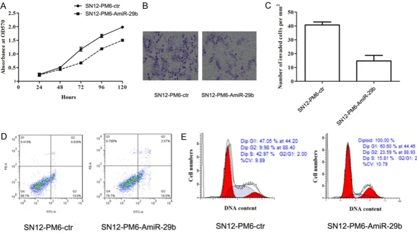
To investigate the effect of miR-29b on tumor cells, SN12-PM6 cells were transduced with pEGFP-hsa-miR-29b to generate cell lines sta-bly expressing miR-29b. The transduction effi-ciency was assessed by fluorescence micro-scope at 8 random fields for each condition. The transduction efficiency was more than 85% in SN12-PM6-ctr and SN12-PM6-AmiR-29b (Figure 3A). The results of qRT-PCR showed that compared to the PM6-ctr, SN12-PM6-AmiR-29b cells had lower miR-29b expres-sion (P < 0.05), indicating that the expression
of miR-29b was successfully inhibited (Figure 3B).
Effect of miR-29b dysregulation on SN12-PM6 cells
To validate the contribution of miR-29b dysreg-ulation to proliferation, invasion, and apopto-sis, functional analysis was performed to con-firm the effects of miR-29b. In the proliferation test, the results showed that inhibition of miR-29b expression could significantly reduce the cell viability compared with SN12-PM6-ctr group (P < 0.05) (Figure 4A). In the invasion test, the mean number of invaded cells express-ing miR-29b per field was 40, which was signifi-cantly higher than that in the control group (15 per mm2, P < 0.05) (Figure 4B and 4C). In the
apoptosis test, apoptosis rate was significantly higher by inhibition of miR-29b expression compared with the control group (P < 0.05) (Figure 4D). Furthermore, we examined the effect of miR-29b on cell cycle regulation by FCM. As shown in Figure 4E, knockdown of miR-29b promoted cell cycle arrest in the G0-G1 phase. These results indicated that in- hibition of miR-29b expression could promote apoptosis, and inhibit proliferation and inva-sion ability in SN12-PM6 cells.
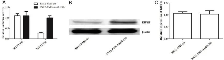
Regulation of miR-29b on KIF1B
To predict that KIF1B gene mRNA 3’-UTR might contain a hsa-miR-29b binding site, the Tar- getScan 6.2, miRanda, and Pictar was used to confirm the prediction. We constructed a WT psiCHECK3’ UTR KIF1B plasmid (WT 3’UTR), which could bind with miR-29b and degrade fluorescence activity. Also, we constructed a mutant plasmid (MT3’UTR) as a control, which could not be combined with miR-29b, and the fluorescence activity was unchanged. As shown in Figure 5A, the fluorescence activity was sig-nificantly increased by dysregulation of miR-29b (P < 0.05), suggesting that KIF1B could be directly regulated by miR-29b. To further con-firm how hsa-miR-29b regulates the expression of KIF1B, we determined the protein and mRNA levels of KIF1B in SN12-PM6 cells after dysreg-ulation of miR-29b. We found that there were no significant differences in the mRNA levels of KIF1B, but the protein levels were markedly higher in the SN12-PM6-AmiR-29b group than that in the SN12-PM6-ctr group (Figure 5B and
5C). These results indicated that miR-29b could regulate the expression of KIF1B at the post transcriptional level.
Discussion
Prognosis for ccRCC patients with metastatic recurrence is still extremely worse. Thus, there
[image:6.629.100.528.79.352.2]is a strong need to develop new strategies that can provide robust biomarkers to predict short-and long-term survival in ccRCC. In this present study, we show that miR-29b expression is Figure 3. The transduction efficiency and the expression of miR-29b after transduction. A. The transduction effi-ciency; B. The relative expression of miR-29b after transduction.
[image:6.629.101.526.402.640.2]increased in RCC cell lines and tissues com-pared to matched normal tissues. Moreover, we found that miR-29b promotes cell prolifera-tion, invasion, and apoptosis resistance by repression of KIF1B expression. Additionally, miR-29b is strongly associated with survival rate and might be a potential molecular prog-nostic marker for RCC and disease progression.
MiRNAs are an abundant class of 20-22-nt non-coding single-strand RNA and play significant roles in various physiological and pathological processes, such as organogenesis, cell prolif-eration, apoptosis, and differentiation [20-22]. A great deal of miRNAs has been shown the association with tumorigenesis [23, 24]. They show their functional abilities by directly bind-ing to target genes, leadbind-ing to translational repression of messenger RNAs. To date, a num-ber of studies have identified several miRNAs that exhibit aberrant expression patterns in RCC tumors compared to normal tissue [25-27]. In spite of a role of miRNAs in RCC has been proposed, the possible molecular mecha-nisms involved in modulation tumor metasta-ses are still covered. In our study, we focused on the expression and functional role of miR-29b in RCC.
MiR-29b is one of members of miR-29b family. Previous studies have shown the expression and functional role of miR-29b in many tumors. However, there are some contradictions. Some researchers suggested that miR-29 family has a potential tumor suppressive function. For example, Wang et al. suggested that miR-29a/ b/c significantly repressed lung cancer [28], and Fabbri et al. showed that miR-29 family had suppressed rhabdomyosarcoma cell lines to
[image:7.629.97.531.82.198.2]G0-G1 phase. Therefore, we demonstrated that regulation of miR-29b expression could alter the proliferation and invasion abilities of SN12-PM6 cells, indicating that miR-29b plays signifi-cant roles in RCC cells.
We then identified miR-29b as a promoter of RCC through direct targeting of KIF1B, which is a well-known tumor suppressor gene. KIF1B is one of the important microtubule-dependent monomeric motors and functions as a regulator of transporting membranous organelles, such as mitochondria and synaptic vesicle [32]. Recent studies have showed that abnormal expression of KIF1B is involved in some malig-nant diseases such as neuroblastoma and gas-trointestinal stromal tumors [33-35]. Moreover, KIF1B is involved in tumor development by reg-ulating cell apoptosis [36]. In our study, we con-firmed that KIF1B is one of miR-29b targeted genes by qRT-PCR, Western blotting and lucifer-ase report assay system. Our results showed that the fluorescence activity was significantly increased by dysregulation of miR-29b, indicat-ing that miR-29b could directly regulate KIF1B. The mRNA and protein levels of KIF1B indicat-ed that miR-29b could regulate the expression of KIF1B at the post transcriptional level, not at the transcriptional level.
In conclusion, we provide evidence for the con-cept that miR-29b acts as an oncomiR and might be a potential marker for prognosis of RCC. In addition, miR-29b promotes SN12-PM6 cell proliferation and invasion ability, and in- hibits the cell apoptosis by directly suppressing the expression of KIF1B. Our research might provide important theoretical support for prognosis evaluation and development of new treatment for RCC. However, further study should be performed to confirm the results. Disclosure of conflict of interest
None.
Address correspondence to: Yanyue Tong, Depart- ment of Urology, Quzhou People’s Hospital, 2 Zhongloudi, Kecheng District, Quzhou 324000, Zhejiang, China. E-mail: tongyanyue85_22@126. com
References
[1] Ljungberg B, Cowan NC, Hanbury DC, Hora M, Kuczyk MA, Merseburger AS, Patard JJ,
Mul-ders PF, Sinescu IC; European Association of Urology Guideline Group. EAU guidelines on renal cell carcinoma: the 2010 update. Eur Urol 2010; 58: 398-406.
[2] Rini BI, Campbell SC and Escudier B. Renal cell carcinoma. Lancet 2009; 373: 1119-1132. [3] Cairns P. Renal cell carcinoma. Cancer
Bio-marker 2011; 9: 461-473.
[4] Pantuck AJ, Zisman A and Belldegrun AS. The changing natural history of renal cell carcino-ma. J Urol 2001; 166: 1611-1623.
[5] Jemal A, Siegel R, Ward E, Hao Y, Xu J and Thun MJ. Cancer statistics, 2009. CA C J Clin 2009; 59: 225-249.
[6] Cohen HT and McGovern FJ. Renal-cell carci-noma. N Engl J Med 2005; 353: 2477-2490. [7] Nicoloso MS, Spizzo R, Shimizu M, Rossi S and
Calin GA. MicroRNAs-the micro steering wheel of tumour metastases. Nat Rev Cancer 2009; 9: 293-302.
[8] Chandrasekaran K, Karolina DS, Sepramani-am S, ArmugSepramani-am A, Wintour EM, BertrSepramani-am JF and Jeyaseelan K. Role of microRNAs in kidney homeostasis and disease. Kidney Int 2012; 81: 617-627.
[9] Volinia S, Calin GA, Liu CG, Ambs S, Cimmino A, Petrocca F, Visone R, Iorio M, Roldo C, Ferracin M, Prueitt RL, Yanaihara N, Lanza G, Scarpa A, Vecchione A, Negrini M, Harris CC and Croce CM. A microRNA expression signature of hu-man solid tumors defines cancer gene targets. Proc Natl Acad Sci U S A 2006; 103: 2257-2261.
[10] Heinzelmann J, Henning B, Sanjmyatav J, Posorski N, Steiner T, Wunderlich H, Gajda MR and Junker K. Specific miRNA signatures are associated with metastasis and poor progno-sis in clear cell renal cell carcinoma. World J Urol 2011; 29: 367-373.
[11] White NM, Bao TT, Grigull J, Youssef YM, Girgis A, Diamandis M, Fatoohi E, Metias M, Honey RJ, Stewart R, Pace KT, Bjarnason GA and Yousef GM. miRNA profiling for clear cell renal cell carcinoma: biomarker discovery and iden-tification of potential controls and conse-quences of miRNA dysregulation. J Urol 2011; 186: 1077-1083.
[12] Slaby O, Redova M, Poprach A, Nekvindova J, Iliev R, Radova L, Lakomy R, Svoboda M and Vyzula R. Identification of MicroRNAs associat-ed with early relapse after nephrectomy in re-nal cell carcinoma patients. Genes Chromo-somes Cancer 2012; 51: 707-716.
[13] Osanto S, Qin Y, Buermans HP, Berkers J, Lerut E, Goeman JJ and van Poppel H. Genome-wide microRNA expression analysis of clear cell re-nal cell carcinoma by next generation deep se-quencing. PLoS One 2012; 7: e38298. [14] Hidaka H, Seki N, Yoshino H, Yamasaki T,
Enokida H. Tumor suppressive microRNA-1285 regulates novel molecular targets: aberrant ex-pression and functional significance in renal cell carcinoma. Oncotarget 2012; 3: 44-57. [15] Wang X, Chen X, Wang R, Xiao P, Xu Z, Chen L,
Hang W, Ruan A, Yang H and Zhang X. microR-NA-200c modulates the epithelial-to-mesen-chymal transition in human renal cell carcino-ma metastasis. Oncol Rep 2013; 30: 643-650. [16] Wulfken LM, Moritz R, Ohlmann C, Holdenrie-der S, Jung V, Becker F, Herrmann E, Walgen-bach-Brunagel G, von Ruecker A, Muller SC and Ellinger J. MicroRNAs in renal cell carcino-ma: diagnostic implications of serum miR-1233 levels. PLoS One 2011; 6: e25787. [17] Hauser S, Wulfken LM, Holdenrieder S, Moritz
R, Ohlmann CH, Jung V, Becker F, Herrmann E, Walgenbach-Brunagel G, von Ruecker A, Muller SC and Ellinger J. Analysis of serum microRNAs (26a-2*, 191, 337-3p and miR-378) as potential biomarkers in renal cell car-cinoma. Cancer Epidemiol 2012; 36: 391-394. [18] Yang CN, Deng YT, Tang JY, Cheng SJ, Chen ST,
Li YJ, Wu TS, Yang MH, Lin BR, Kuo MY, Ko JY and Chang CC. MicroRNA-29b regulates migra-tion in oral squamous cell carcinoma and its clinical significance. Oral Oncol 2015; 51: 170-177.
[19] Novara G, Ficarra V, Antonelli A, Artibani W, Bertini R, Carini M, Cosciani Cunico S, Imbim-bo C, Longo N, Martignoni G, Martorana G, Mi-nervini A, Mirone V, Montorsi F, Schiavina R, Simeone C, Serni S, Simonato A, Siracusano S, Volpe A, Carmignani G; SATURN Project-LUNA Foundation. Validation of the 2009 TNM ver-sion in a large multi-institutional cohort of pa-tients treated for renal cell carcinoma: are fur-ther improvements needed? Eur Urol 2010; 58: 588-595.
[20] Filipowicz W, Bhattacharyya SN and Sonen-berg N. Mechanisms of post-transcriptional regulation by microRNAs: are the answers in sight? Nat Rev Genet 2008; 9: 102-114. [21] Stefani G and Slack FJ. Small non-coding RNAs
in animal development. Nat Rev Mol Cell Biol 2008; 9: 219-230.
[22] Boren T, Xiong Y, Hakam A, Wenham R, Apte S, Wei Z, Kamath S, Chen DT, Dressman H and Lancaster JM. MicroRNAs and their target messenger RNAs associated with endometrial carcinogenesis. Gynecol Oncol 2008; 110: 206-215.
[23] Ma L and Weinberg RA. MicroRNAs in malig-nant progression. Cell Cycle 2008; 7: 570-572. [24] Croce CM. Oncogenes and cancer. N Engl J
Med 2008; 358: 502-511.
[25] Slaby O, Jancovicova J, Lakomy R, Svoboda M, Poprach A, Fabian P, Kren L, Michalek J and Vyzula R. Expression of miRNA-106b in
con-ventional renal cell carcinoma is a potential marker for prediction of early metastasis after nephrectomy. J Exp Clin Cancer Res 2010; 29: 90.
[26] Yi Z, Fu Y, Zhao S, Zhang X and Ma C. Differen-tial expression of miRNA patterns in renal cell carcinoma and nontumorous tissues. J Cancer Res Clin Oncol 2010; 136: 855-862.
[27] Weng L, Wu X, Gao H, Mu B, Li X, Wang JH, Guo C, Jin JM, Chen Z, Covarrubias M, Yuan YC, Weiss LM and Wu H. MicroRNA profiling of clear cell renal cell carcinoma by whole-ge-nome small RNA deep sequencing of paired frozen and formalin-fixed, paraffin-embedded tissue specimens. J Pathol 2010; 222: 41-51. [28] Wang H, Garzon R, Sun H, Ladner KJ, Singh R,
Dahlman J, Cheng A, Hall BM, Qualman SJ, Chandler DS, Croce CM and Guttridge DC. NF-kappaB-YY1-miR-29 regulatory circuitry in skel-etal myogenesis and rhabdomyosarcoma. Can-cer Cell 2008; 14: 369-381.
[29] Fabbri M, Garzon R, Cimmino A, Liu Z, Zanesi N, Callegari E, Liu S, Alder H, Costinean S, Fer-nandez-Cymering C, Volinia S, Guler G, Morri-son CD, Chan KK, Marcucci G, Calin GA, Hueb-ner K and Croce CM. MicroRNA-29 family reverts aberrant methylation in lung cancer by targeting DNA methyltransferases 3A and 3B. Proc Natl Acad Sci U S A 2007; 104: 15805-15810.
[30] Xiong Y, Fang JH, Yun JP, Yang J, Zhang Y, Jia WH and Zhuang SM. Effects of microRNA-29 on apoptosis, tumorigenicity, and prognosis of hepatocellular carcinoma. Hepatology 2010; 51: 836-845.
[31] Wang C, Bian Z, Wei D and Zhang JG. miR-29b regulates migration of human breast cancer cells. Mol Cell Biochem 2011; 352: 197-207. [32] Nangaku M, Sato-Yoshitake R, Okada Y, Noda
Y, Takemura R, Yamazaki H and Hirokawa N. KIF1B, a novel microtubule plus end-directed monomeric motor protein for transport of mito-chondria. Cell 1994; 79: 1209-1220.
[33] Yang HW, Chen YZ, Takita J, Soeda E, Piao HY and Hayashi Y. Genomic structure and muta-tional analysis of the human KIF1B gene which is homozygously deleted in neuroblastoma at chromosome 1p36.2. Oncogene 2001; 20: 5075-5083.
[34] Caren H, Ejeskar K, Fransson S, Hesson L, Latif F, Sjoberg RM, Krona C and Martinsson T. A cluster of genes located in 1p36 are down-regulated in neuroblastomas with poor progno-sis, but not due to CpG island methylation. Mol Cancer 2005; 4: 10.
of gastrointestinal stromal tumors: an integra-tive analysis of gene expression profiling and high-resolution genomic copy number. Lab In-vest 2010; 90: 1285-1294.
[36] Munirajan AK, Ando K, Mukai A, Takahashi M, Suenaga Y, Ohira M, Koda T, Hirota T, Ozaki T and Nakagawara A. KIF1Bbeta functions as a