Original Article
MiR-451 inhibits synovial fibroblasts proliferation
and inflammatory cytokines secretion in rheumatoid
arthritis through mediating p38MAPK signaling pathway
Zhan-Chao Wang1, Hua Lu2, Qiang Zhou1, Si-Ming Yu1, Yu-Lun Mao1, Hao-Jie Zhang1, Ping-Chao Zhang1,
Wang-Jun Yan3
1Department of Orthopedics, Xinhua Hospital (Chongming), Shanghai Jiaotong University, Shanghai 202150,
China; 2Department of Orthopedics, Xinhua Hospital, Shanghai Jiaotong University; Shanghai 200092, China; 3Department of Orthopedics, Changzheng Hospital, Second Military Medical University, Shanghai 200061, China
Received May 21, 2015; Accepted June 28, 2015; Epub November 1, 2015; Published November 15, 2015
Abstract: Rheumatoid arthritis is an autoimmune disease characterized as joint synovial inflammation. MicroRNA is a group of small noncoding RNA molecules discovered in recent years that can posttranscriptional regulate mRNA expression and involved in a variety processes of immune cell activation and differentiation. There is still lack of study about the role of miR-451 in rheumatoid arthritis. Synovial fibroblasts isolated from rheumatoid arthritis pa-tients were cultured in vitro. Chemical synthesized miR-451 was lipo-transfected, real-time RT-PCR was applied to detect miR-451 expression level, and MTT method was used to detect the effect of miR-451 on synovial fibroblasts proliferation. Enzyme-linked immunosorbent assay was used to detect tumor necrosis factor TNF-α, IL-1β, and IL-6 level in the supernatant. Western blot was applied to test target protein p38 MAPK expression level. Our study found that synovial fibroblasts expressed higher miR-451 mRNA level in miR-451 treatment group. MiR-451 treatment significantly decreased cell proliferation ability (P < 0.05). Compared with the control, p38 MAPK protein expression reduced obviously in the miR-451 treatment group (P < 0.05). MiR-451 transfected synovial fibroblasts secreted lower levels of TNF-α (198 ± 12 pg/ml vs 124 ± 13 pg/ml, P < 0.01), IL-1β (352 ± 43 pg/ml vs 165 ± 87 pg/ml, P < 0.01), and IL-6 (487 ± 84 pg/ml vs 257 ± 92 pg/ml, P < 0.01). The results proved that miR-451 can down-regulate p38 MAPK protein expression, and reduce synovial fibroblasts proliferation and cytokine expression level.
Keywords: miR-451, rheumatoid arthritis, synovial fibroblast, cell proliferation
Introduction
Rheumatoid arthritis (RA) is a chronic systemic autoimmune disease characterized by multiple joints’ synovial chronic inflammation in clinic. Immune function abnormality is common among RA patients. Further elucidating the role of immune factors in RA incidence is beneficial to RA prevention and treatment [1-4].
MicroRNA (miRNA) is a noncoding single-stranded RNA molecule composed of 22 nucle-otides. It is encoded by endogenous gene and involved in gene expression regulation [5]. It plays an important role in various kinds of ph- ysiological processes [6-8]. In recent years, following the rapid development of molecular biology technology, numerous clinical and basic
scholars have reported a variety of microRNA abnormally expressed in RA patients and ani-mal models [9-11].
is not only related to cell apoptosis, cell mo- vement, and a variety of pathophysiological process in stress [15], and but also play an important role in the pathological process in mediating inflammation and cytokine gener- ation. p38MAPK pathway is thought to be an important regulating target during normal and disease states of RA.
Thus, this experiment mainly studied the effect and mechanism of miRNA-451 on synovial fibroblasts proliferation and inflammatory fac-tors secretion in RA patients.
Materials and methods
The clinical trial was approved by the ethics committee in our hospital. All enrolled patients had signed the informed consent. Articular synovial tissue was collected from the RA patients in the department of joint surgery receiving joint replacement. RA diagnosis was performed according to the American rheu- matism association standard. Patients were between 16 and 60 years old with no other complications and received no immune inhibi-tors or similar drugs.
Materials, reagents and instruments
Real time PCR, confocal microscope (Bio-Rad), ECL chemiluminescence reagent kit (Amer- sham), CO2 cell incubator and flow cytometry (BD), etc. RNA extraction kit was obtained from Takara; miR-451 was bought from Genepharma (Shanghai, China); TaqMan miRNA real-time RT-PCR Assays kit and TaqMan miRNA Reverse Transcription kit were got from Applied Biosystems (USA); Enzyme-linked immunosor-bent Assay kit was purchased from BD compa-ny (USA); Lipofectamine 2000 was bought from Invitrogen (USA); Primary antibodies for p38 MAPK and β-actin were from Santa Cruz (USA).
Synovial fibroblasts culture
Knee joint synovial tissue was collected from the RA patient receiving joint replacement in the department of joint surgery. The tissue was cut into pieces in the sterile condition and digested by trypsin under the condition of 37°C for two hours. The synovial fibroblasts were col-lected after centrifugation and maintained in DMEM medium containing 5 mmol/L glucose and containing 10% fetal bovine serum in a humid atmosphere containing 5% CO2 at 37°C. The cells were passaged when they grow to
fusion. 4-6th generation of cells were used for
the experiment.
Cell grouping and transfection
Synovial fibroblasts were divided into the miR-451 treatment group and control group when grow to 80%. MiR-451 or control was transfect-ed to the synovial fibroblasts mtransfect-ediattransfect-ed by Lipofectamine 2000, respectively and cultured for 6 hours. After changing the DMEM medium and continue cultured for 48 h, the synovial fibroblasts were harvested.
RT-PCR
Total RNA was extracted from the synovial fibro-blasts and reverse transcripted to cDNA for PCR amplification. The PCR primers were designed by Primer 5.0 software as follows: miR-451 sense 5’-AAACCGUUACCAUUACUG- AGUU-3’ anti-sense 5’-UAGUAAUGGUAAUGGU- UCUC-3’ β-actin sense 5’-GGTGTGATGGTGGG- TATGGGT-3’ anti-sense 5’-CTGGGTCATCTTTT- CACGGT-3’. The cycling conditions consisted of an initial, single cycle of 5 min at 94°C, followed by 30 cycles of 60 s at 94°C, 60 s at 60°C, and 3 min at 72°C. PCR products were tested by agarose gel electrophoresis and analyzed by an optimized comparative Ct (ΔΔCt) value method.
MTT
Cells were seeded in 96-well plates at a density of 2 × 10⁴ cells/well and incubated overnight at
37°C. The cells were incubated with miR-451 for 48 h at 37°C. After addition of 20 µL of MTT solution (5 g/L) to each well, plates were incu-bated for 4 h at 37°C. After removing the medi-um, 150 μL DMSO was added and oscillated for 10 min. Absorbance of each well at 570 nm was read using a spectrophotometer.
Western blot
Total protein was extracted and separated by denaturing 10% SDS-polyacrylamide gel elec-trophoresis. Detection was performed with ECL chemiluminescence system. Antibody dilutions were 1:200 for p38 MAPK, 1:500 for IL-1β, and 1:2000 for β-actin. Protein levels were normal-ized to β-actin and changes were determined.
Elisa
accord-ing to the specification in three duplicates for each sample.
Statistical analysis
All statistical analyses were performed using SPSS17.0 software (Chicago, IL). Numerical data were presented as means and standard deviation (± SD). Differences between means were analyzed using one-way ANOVA or paired t test when necessary. P < 0.05 was considered to indicate a statistically significant result.
Results
MiR-451 RNA expression in synovial fibroblasts
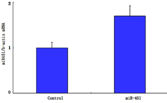
miR-451 level was 2.06 ± 0.12 and 0.93 ± 0.06 in the miR-451 treatment group and control, respectively. MiR-451 expression level was significantly higher in the treatment group
Discussion
[image:3.629.100.390.83.262.2]RA is an autoimmune disease with the patho-logical features of joint synovial inflammation. Studies have shown that there are many immune cells and cytokines in the synovial tis-sue and synovial fluid of RA patients. MiRNA, on the other hand, is a set of noncoding small RNA molecules found in recent years [16] that can posttranscriptional regulate mRNA expression and involve in a variety of immune cell activa-tion and differentiaactiva-tion processes [17]. It has been reported that miRNA has influence on synovial fibroblasts proliferation [18]. However, there is still lack of report about the role of miR-451 in autoimmune diseases, especially in RA. Our results showed that miR-451 may affect RA occurrence through inhibiting synovial fibro-blasts proliferation. As miRNA performed its biological function generally by inhibiting target
Figure 1. miR-451 expression level in two groups.
The effect of miR-451 on synovial fibroblasts prolifera-tion MTT test results show- ed that compared with the control group, miR-451 treat- ment group grew obviously slower than in the 2nd and 3rd
day (P < 0. 05). It suggested that overexpressed miR-451 significantly decreased cell proliferation ability (P < 0.05) (Figure 2).
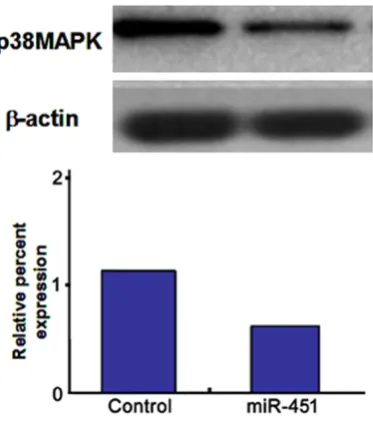
P38 MAPK expression
Compared with the control, p38 MAPK protein expres-sion reduced obviously in the miR-451 treatment group (P
< 0.05, Figure 3).
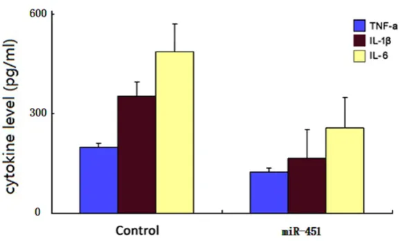
MiR-451 down-regulated cytokines expression in sy-novial fibroblasts
ELISA results showed that compared with control, miR-451 transfected synovial fibroblasts secreted lower levels of TNF-α (198 ± 12 pg/ml vs 124 ± 13 pg/ml, P
< 0.01), IL-1β (352 ± 43 pg/ ml vs 165 ± 87 pg/ml, P < 0.01), and IL-6 (487 ± 84 pg/ ml vs 257 ± 92 pg/ml, P < 0.01, Figure 4).
[image:3.629.100.390.295.461.2]may be the target gene of miR-451. MAPK is an important signaling pathway in mediating cell reaction, of which p38 MAPK is an important member of the MAPK family [19]. It can regu-late cell growth, development and intercellular function by cytoskeleton related protein, tran-scription factors phosphorylation, and enz- ymes. Also, it plays an important role in the pro-cess of cell-mediated inflammatory response and cytokines generation. Thus, we thought p38MAPK might be an important target [15, 20]. We transfected miR-451 to synovial fibro-blasts and found that, compared with the con-trol group, miR-451 can obviously reduce p38 MAPK protein expression in synovial fibro-blasts. Therefore, we speculated that p38 MAPK activity is associated with miR-451 regu-lation, and involved in intracellular signal trans-duction process of miR-451.
In recent years, the role of miR-451 in autoim-mune disease began to be attentioned by researchers [21]. For example, it was reported that miR-451 expression level in the diseased tissue is correlated with helper T lymphocytes 17 (Th17) subgroup expression changes in the viral myocarditis mice model. The author thought miR-451 involved in the pathogenesis of viral myocarditis, and related to Th17su- bgroup. Another experiment proved that miRNA-451 may inhibit glomerular mesangial cell proliferation in the diabetic nephropathy model, and can reduce p38 MAPK level. We
also confirmed similar findings in RA synovial fibroblasts, and provided new evidence in investigating the role of miR-451 in autoim-mune disease.
A large number of in vitro cells experiment and in vivo animal experiments found that cytokines is closely related to RA development. Currently, the cytokines thought to be associated with RA includes TNF-α, IL-1β, and IL-6. They are impor-tant inflammatory cytokines in RA. These cyto-kines can stimulate synovial fibroblasts hyper-plasia resulting in chemokines and cytokines secretion and joint damage. In addition, there are some cytokines in synovial tissue and syno-vial fluid of RA, and these cytokines mainly pro-duced by activated macrophages, monocytes, and dendritic cells. These cytokines, especially TNF-α, IL-1β, and IL-6, participate in the occur-rence and development of the disease by induc-ing extensive chemokine expression and upreg-ulating adhesion molecules in endothelial cells [18]. In this study, we found that miR-451 can down-regulate TNF-α, IL-1β, and IL-6 level in synovial fibroblasts. Our research proved that the key role of p38 MAPK in the incidence of RA. MiR-451 could inhibit p38 MAPK expres-sion to suppress synovial fibroblasts prolife- ration and down-regulate corresponding cyt- okines levels, and potentially improve the disease clinical manifestation.
Up to now, though there is still no direct report about the role of miRNA-451 in the synovial fibroblasts of RA, it is reported that the miRNA-451 expression level is lower in the peripheral neutrophils of RA patients than normal control [22]. Further in vitro experiments proved that miR-451 can reduce the infiltration capacity of neutrophil in local inflammation model, while overexpressed miR-451 can obviously inhibit neutrophil activity [22]. Our results suggested that the miRNA-451 could inhibit synovial fibro-blasts proliferation and cytokines production in RA. Thus, combined with the existing reports, miRNA-451 may have a variety of ways in RA by reducing the neutrophils infiltrating ability and inhibits synovial fibroblasts proliferation. Future study can further investigate the possible effect of miRNA-451 in animal model or clinical RA patients.
[image:4.629.104.291.80.291.2]Our studies demonstrated that the expression of cytokines, including TNF-α, IL-1β, and IL-6,
can be inhibited by miR-451. Previous in vitro experiments also proved that these cytokines can be regulated by different miRNAs. For example, miR-203 can regulate IL-6 production in synovial fibroblasts from RA through NF-κB signaling pathway. Other studies found miR-146a overexpressed in the peripheral blood mononuclear cells from RA, and its expression is associated with TNF-α expression level. The abovementioned results and our study showed that a number of different miRNAs can regulate different cytokines expression, while these cytokines actively involved in the occurrence and development of RA. The synergy of multiple miRNAs inhibited a variety of cytokines expres-sion, and also provides a new strategy for RA even other autoimmune disease treatment. Furthermore, a variety of miRNAs were found abnormally expressed in the peripheral blood or synovial tissue of the RA patients, and some of miRNAs are closely related to the clinical symptoms and disease activity of RA. Some clinical scholars suggested that miRNA may be used for RA early diagnosis and disease activity judgment. On the other hand, as the great ther-apeutic effect and rare inflammation of miRNA in tumor, its role in RA treatment is also a new therapeutic direction [21, 23, 24]. Our experi-ments also suggested the potential treatment effect of miR-451 in RA.
Above all, miR-451 plays an important role in the pathogenesis of RA. It can both inhibit syno-vial fibroblasts proliferation and regulate p38 MAPK expression, resulting in corresponding cytokines downregulation. MiR-451 might be a
References
[1] McInnes IB, Schett G. Cytokines in the patho-genesis of rheumatoid arthritis. Nat Rev Immunol 2007; 7: 429-442.
[2] Zhao S, Wang Y, Liang Y, Zhao M, Long H, Ding S, Yin H, Lu Q. MicroRNA-126 regulates DNA methylation in CD4+ T cells and contributes to systemic lupus erythematosus by targeting DNA methyltransferase 1. Arthritis Rheum 2011; 63: 1376-1386.
[3] Alevizos I, Alexander S, Turner RJ, Illei GG. MicroRNA expression profiles as biomarkers of minor salivary gland inflammation and dys-function in Sjogren’s syndrome. Arthritis Rheum 2011; 63: 535-544.
[4] Semaan N, Frenzel L, Alsaleh G, Suffert G, Gottenberg JE, Sibilia J, Pfeffer S, Wachsmann D. miR-346 controls release of TNF-alpha pro-tein and stability of its mRNA in rheumatoid arthritis via tristetraprolin stabilization. PLoS One 2011; 6: e19827.
[5] Filipowicz W, Bhattacharyya SN, Sonenberg N. Mechanisms of post-transcriptional regulation by microRNAs: are the answers in sight? Nat Rev Genet 2008; 9: 102-114.
[6] Davalos V, Esteller M. MicroRNAs and cancer epigenetics: a macrorevolution. Curr Opin Oncol 2010; 22: 35-45.
[7] Yang BF, Lu YJ, Wang ZG. MicroRNAs and apop-tosis: implications in the molecular therapy of human disease. Clin Exp Pharmacol Physiol 2009; 36: 951-960.
[8] Wang Y, Blelloch R. Cell cycle regulation by mi-croRNAs in stem cells. Results Probl Cell Differ 2011; 53: 459-472.
[image:5.629.99.392.79.253.2][9] Wang H, Peng W, Ouyang X, Li W, Dai Y. Circulating microRNAs as candidate biomark-ers in patients with systemic lupus erythema-tosus. Transl Res 2012; 160: 198-206.
Figure 4. Cytokines secretion.
new therapeutic drug for RA. Its specific mechanism still remains further research.
Disclosure of conflict of in-terest
None.
[10] Gallo A, Tandon M, Illei G, Alevizos I. Discovery and validation of novel microRNAs in Sjogren’s syndrome salivary glands. Clin Exp Rheumatol 2014; 32: 761-762.
[11] Chatzikyriakidou A, Voulgari PV, Georgiou I, Drosos AA. miRNAs and related polymor-phisms in rheumatoid arthritis susceptibility. Autoimmun Rev 2012; 11: 636-641.
[12] Kirschner MB, Kao SC, Edelman JJ, Armstrong NJ, Vallely MP, van Zandwijk N, Reid G. Haemolysis during sample preparation alters microRNA content of plasma. PLoS One 2011; 6: e24145.
[13] Nan Y, Han L, Zhang A, Wang G, Jia Z, Yang Y, Yue X, Pu P, Zhong Y, Kang C. MiRNA-451 plays a role as tumor suppressor in human glioma cells. Brain Res 2010; 1359: 14-21.
[14] Rosenberger CM, Podyminogin RL, Navarro G, Zhao GW, Askovich PS, Weiss MJ, Aderem A. miR-451 regulates dendritic cell cytokine re-sponses to influenza infection. J Immunol 2012; 189: 5965-5975.
[15] Nakamachi Y, Kawano S, Takenokuchi M, Nishimura K, Sakai Y, Chin T, Saura R, Kurosaka M, Kumagai S. MicroRNA-124a is a key regulator of proliferation and monocyte chemoattractant protein 1 secretion in fibro-blast-like synoviocytes from patients with rheu-matoid arthritis. Arthritis Rheum 2009; 60: 1294-1304.
[16] Stanczyk J, Ospelt C, Karouzakis E, Filer A, Raza K, Kolling C, Gay R, Buckley CD, Tak PP, Gay S, Kyburz D. Altered expression of microR-NA-203 in rheumatoid arthritis synovial fibro-blasts and its role in fibroblast activation. Arthritis Rheum 2011; 63: 373-381.
[17] Shahrara S, Pickens SR, Dorfleutner A, Pope RM. IL-17 induces monocyte migration in rheu-matoid arthritis. J Immunol 2009; 182: 3884-3891.
[18] Berzat A, Hall A. Cellular responses to extracel-lular guidance cues. EMBO J 2010; 29: 2734-2745.
[19] Ondrouskova E, Slovackova J, Pelkova V, Prochazkova J, Soucek K, Benes P, Smarda J. Heavy metals induce phosphorylation of the Bcl-2 protein by Jun N-terminal kinase. Biol Chem 2009; 390: 49-58.
[20] Li J, Wan Y, Guo Q, Zou L, Zhang J, Fang Y, Fu X, Liu H, Lu L, Wu Y. Altered microRNA expression profile with miR-146a upregulation in CD4+ T cells from patients with rheumatoid arthritis. Arthritis Res Ther 2010; 12: R81.
[21] Duroux-Richard I, Presumey J, Courties G, Gay S, Gordeladze J, Jorgensen C, Kyburz D, Apparailly F. MicroRNAs as new player in rheu-matoid arthritis. Joint Bone Spine 2011; 78: 17-22.
[22] Murata K, Yoshitomi H, Furu M, Ishikawa M, Shibuya H, Ito H, Matsuda S. MicroRNA-451 down-regulates neutrophil chemotaxis via p38 MAPK. Arthritis Rheumatol 2014; 66: 549-559.
[23] Long L, Yu P, Liu Y, Wang S, Li R, Shi J, Zhang X, Li Y, Sun X, Zhou B, Cui L, Li Z. Upregulated microRNA-155 expression in peripheral blood mononuclear cells and fibroblast-like synovio-cytes in rheumatoid arthritis. Clin Dev Immunol 2013; 2013: 296139.