Comparable reductions in hyperpnoea-induced bronchoconstriction and
markers of airway in
fl
ammation after supplementation with 6
·
2 and 3
·
1 g/d
of long-chain
n-3 PUFA in adults with asthma
Neil C. Williams
1*, Kirsty A. Hunter
1, Dominick E. Shaw
2, Kim G. Jackson
3, Graham R. Sharpe
1and
Michael A. Johnson
11Exercise and Health Research Group, Department of Sport Science, Sport, Health and Performance Enhancement (SHAPE)
Research Centre, Nottingham Trent University, Nottingham NG11 8NS, UK
2Respiratory Research Unit, University of Nottingham, Nottingham NG5 1PB, UK
3Hugh Sinclair Unit of Human Nutrition, Department of Food and Nutritional Sciences, University of Reading,
Reading RG6 6AP, UK
(Submitted 27 October 2016–Final revision received 2 May 2017–Accepted 3 May 2017)
Abstract
Although high dose n-3 PUFA supplementation reduces exercise- and hyperpnoea-induced bronchoconstriction (EIB/HIB), there are concurrent issues with cost, compliance and gastrointestinal discomfort. It is thus pertinent to establish the efficacy of lowern-3 PUFA doses. Eight male adults with asthma and HIB and eight controls without asthma were randomly supplemented with twon-3 PUFA doses (6·2 g/d (3·7 g EPA and 2·5 g DHA) and 3·1 g/d (1·8 g EPA and 1·3 g DHA)) and a placebo, each for 21 d followed by 14 d washout. A eucapnic voluntary hyperpnoea (EVH) challenge was performed before and after treatments. Outcome measures remained unchanged in the control group. In the HIB group, the peak fall in forced expiratory volume in 1 s (FEV1) after EVH at day 0 (−1005 (SD520) ml,−30 (SD18) %) was unchanged after placebo. The peak fall in FEV1 was similarly reduced from day 0 to day 21 of 6·2 g/d n-3 PUFA (−1000 (SD 460) ml,
−29 (SD17) %v.−690 (SD460) ml,−20 (SD15) %) and 3·1 g/dn-3 PUFA (−970 (SD480) ml,−28 (SD18) %v.−700 (SD420) ml,−21 (SD15) %) (P<0·001). Baseline fraction of exhaled nitric oxide was reduced by 24 % (P=0·020) and 31 % (P=0·018) after 6·2 and 3·1 g/dn-3 PUFA, respectively. Peak increases in 9α, 11βPGF2after EVH were reduced by 65 % (P=0·009) and 56 % (P=0·041) after 6·2 and 3·1 g/dn-3 PUFA, respectively. In conclusion, 3·1 g/dn-3 PUFA supplementation attenuated HIB and markers of airway inflammation to a similar extent as a higher dose. Lower doses ofn-3 PUFA thus represent a potentially beneficial adjunct treatment for adults with asthma and EIB.
Key words:n-3 PUFA: Asthma: Exercise: Inflammation
Exercise-induced bronchoconstriction (EIB) is a prominent asthma phenotype affecting an estimated 90 % of asthma
patients and up to 50 % of elite athlete populations(1). EIB is
characterised by transient airway narrowing during and/or
after exercise(2) and is ascribed to airway drying leading to
degranulation of inflammatory cells and release of inflammatory
mediators(3,4). Inhaled corticosteroids and short- and
long-acting β2-agonists are effective therapies, but they are not
curative and do not modify disease progression(5). Furthermore,
inhaled corticosteroids adherence is notoriously poor and may
have undesirable side effects, whereas chronic β2-agonist use
results in tolerance(5,6). Development of therapies that modulate
asthma immunopathology without adverse side effects is therefore desirable.
One potential candidate therapy involves the long-chain
n-3 PUFA, EPA and DHA(7). Dietary supplementation of n-3
PUFA increases cell membrane EPA and DHA content and
reduces n-6 arachidonic acid content(8). This subsequently
reduces the synthesis of the pro-inflammatory arachidonic
acid-derived eicosanoids cysteinyl leukotrienes and PG. EPA and DHA may also increase synthesis of resolvin and protectin compounds through the cyclo-oxygenase and lipoxygenase
pathways which are involved in the resolution of inflammation.
These mechanisms are thought to be central to the
well-established anti-inflammatory effects ofn-3 PUFA(8), which
provide a rationale for the use of n-3 PUFA in asthma(7). To
date, however, the role of EPA and DHA in the management of
asthma and EIB remains uncertain(9–14). Early research showed
Abbreviations: EIB, exercise-induced bronchoconstriction; EVH, eucapnic voluntary hyperpnoea; FENO, fraction of exhaled nitric oxide; FEV1, forced expiratory volume in 1 s; FVC, forced vital capacity; HIB, hyperpnoea-induced bronchoconstriction; ppb, parts per billion.
*Corresponding author:Dr N. C. Williams, email [email protected]
that supplementation of 5·4 g/d n-3 PUFA (3·2 g/d EPA and
2·2 g/d DHA) for 10 weeks in adults with asthma reduced
leukotriene generation and neutrophil chemotactic responsive-ness, but did not reduce bronchoconstriction after cycling
exercise and a histamine challenge(9). Similarly, supplementation
of 6·0 g/dn-3 PUFA (4·0 g/d EPA and 2·0 g/d DHA) for 3 weeks
in adults with asthma did not attenuate bronchoconstriction
or markers of airway inflammation in response to a mannitol
challenge test(12). Conversely, supplementation of 5·4 g/d n-3
PUFA (3·2 g/d EPA and 2·2 g/d DHA) for 3 weeks in elite
athletes(10)and physically active asthmatic males(11,15)abolished
EIB. Subsequent studies showed that supplementation of n-3
PUFA (3·2 g/d EPA and 2·0 g/d DHA) reduced
bronchoconstric-tion and markers of airway inflammation after eucapnic
voluntary hyperpnoea (EVH)(16)and thatn-3 PUFA 3·2 g/d EPA
and 2·0 g/d DHA) was as effective as a leukotriene modifier(17).
Given the lack of reported risk in taking n-3 PUFA, the
American Thoracic Society/European Respiratory Society
(ATS/ERS) guidelines suggest that the latter studies offer some
support for the consumption ofn-3 PUFA by interested individuals
with EIB(10,11,17). In contrast however, recent practice parameter(14)
cautions the recommendation forn-3 PUFA use in EIB based on
more recentfindings(12,18). When considering the practicalities
associated with n-3 PUFA supplementation for asthma
man-agement, previous studies have used doses of 5·2–6·0 g/d. Such
doses require individuals to ingest between eight and twenty
capsules of commercial fish oil daily, which has implications
for cost, compliance and gastrointestinal discomfort(19,20). In a
single-blind study an alternative flavoured beverage delivery
was investigated (3·0 g/d EPA, 3·0 g/d DHA and 30µg of vitamin
D3) but failed to show any attenuation in hyperpnoea-induced
bronchoconstriction (HIB) or markers of airway inflammation
(fraction of exhaled nitric oxide (FENO), urinary 9α, 11βPGF2
and cysteinyl leukotriene E4)(18). However, no measure of
compliance to the treatment was made and participants inclu-ded had mild asthma or did not have a physician diagnosis of
asthma. It is therefore pertinent to establish if lower doses ofn-3
PUFA are effective in reducing EIB. With respect to the simu-lation of EIB under laboratory conditions, an EVH challenge causes a highly reproducible HIB (a surrogate for EIB) in adults
with asthma(21)which makes this an attractive challenge test to
evaluate the effects ofn-3 PUFA treatments on airway
hyper-responsiveness. Thus, the aim of the current study was to
compare the effects of a 6·2 g/dn-3 PUFA dose with a half dose
of 3·1 g/dn-3 PUFA on HIB and markers of airway infl
amma-tion in adults with asthma. Provisional data from the study was
previously part published in abstract form(22).
Methods
Participants
In all, sixteen non-smoking, recreationally active men
(com-pleting≥6 h of endurance exercise per week) provided written,
informed consent to participate in the study (Table 1). Eight participants formed a HIB group and eight formed a control group. Inclusion criteria for the HIB group were: physician diagnosis of asthma, a baseline forced expiratory volume in 1 s
(FEV1)>65 % of predicted
(23)
and a≥10 % fall in FEV1following
initial EVH screening(24,25). The HIB group were on steps 1–3
of the stepwise approach to asthma control, indicating well-controlled asthma using either reliever medication (short-acting
β2-agonsit) alone or in combination with controller medication
(low-dose inhaled corticosteroid and/or long-actingβ2-agonist(26)
(Table 1). Inclusion criteria for the control group were: a baseline
FEV1>65 % of predicted and a<10 % fall in FEV1following initial
EVH screening. Participants avoided exercise for 24 h before an EVH test, and the HIB group ceased their medication as
previously described(21,24). On EVH test days, participants
abstained from caffeine and alcohol and arrived at the laboratory
>2 h post-prandial(27,28). Participants were instructed to abstain
from consumption ofn-3 PUFA supplements and eat no more
than two oilyfish meals per week for 3 weeks before the study
and throughout the study(11). Participants were free from acute
upper respiratory tract infections throughout the study.
Experimental design and protocol
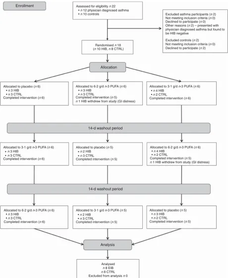
This study was conducted in accordance with the Declaration of Helsinki and all procedures were approved by the Nottingham Trent University Human Ethics Committee (approval no. 186; Clinical trial no. ISRCTN80857707). The study adopted a counter balanced, double-blind, placebo-controlled crossover design over 14 consecutive weeks (Fig. 1).
Participants were randomised (block randomisation) to receive three 21 d treatments each separated by a 14-d washout period. All treatments involved daily oral consumption of eight capsules (Croda International Plc). Four capsules were taken in the morning and four in the afternoon with a recommendation
to take with food. The three treatments were: 6·2 g/dn-3 PUFA
(3·7 g EPA and 2·5 g DHA), 3·1 g/d n-3 PUFA (1·8 g EPA and
1·3 g DHA) (INCROMEGATMTG4030; Croda International Plc)
and placebo (CRODAMOLTMGTCC medium chain TAG; Croda
International Plc) (Table 2). CRODAMOLTMGTCC was chosen
as it is readily oxidised in the liver so has little impact on human
health-related biomarkers(29,30). Measurements were taken at
days 0 and 21 of each treatment period. The 6·2 g/dn-3 PUFA
dose was comprised of fourn-3 PUFA capsules in the morning
and four n-3 PUFA capsules in the afternoon. To ensure an
equal number of capsules were taken the 3·1 g/d n-3 PUFA
comprised of twon-3 PUFA capsules and two placebo capsules
in the morning and two n-3 PUFA capsules and two placebo
capsules in the afternoon. All capsules (placebo andn-3 PUFA)
were identical in appearance.
Measurement of pulmonary function, eucapnic voluntary hyperpnoea and fraction of exhaled nitric oxide
The EVH test was undertaken at days 0 and 21 of each treatment and comprised 6 min of breathing dry gas at a target minute
ventilation (V_E) of 85 % of the predicted maximal voluntary
ventilation (MVV) (30×baseline FEV1). Pulmonary function
(forced vital capacity (FVC), FEV1, peak expiratory flow (PEF)
and forced expiratory flow 25–75 % (FEF25–75 %)) was assessed
and in duplicate at 3, 6, 16, 20 and 30 min after EVH, as previously described. The highest values recorded were used for
analyses. Baseline FENO was measured (NIOX MINO; Aerocrine)
according to ATS/ERS guidelines(32)in the HIB group only, as it is
elevated in asthma patients but not in healthy controls(33).
Urinary 9α, 11β-PGF2analysis
Participants provided a urine sample at baseline and at 12, 60
and 90 min after EVH. Urinary concentration of 9α, 11β-PGF2,
a metabolite of PGD2,was subsequently determined by ELISA
(Caymen Chemicals) and standardised for urinary creatinine concentration (ABX Pentra 400; Horiba) based on a kinetic
method using alkaline picrate (Jaffe method)(34). The corrected
9α, 11β-PGF2was expressed as ng/mmol creatinine. The
inter-and intra-assay CV was <15 %. Two control participants and
one HIB participant had samples at day 0 below the limit
of detection for urinary 9α, 11β-PGF2 (5 pg/ml) and were
subsequently excluded from analysis.
Neutrophil phospholipid fatty acid analysis
The neutrophil phospholipid fatty acid composition was assessed as a measure of compliance to the treatments in addition to count of capsules returned after each treatment. Neutrophil cells were isolated from 20 ml of whole venous blood (drawn at baseline from an antecubital vein) through a
three-step purification protocol that consisted of dextran
sedimentation (Fisher Scientific), hypotonic lysis and Ficoll–
Paque sedimentation (GE Healthcare)(35). This method
achieved up to 98 % of pure neutrophils which were stored at
−80°C under N until extraction of phospholipids using
pre-viously described methods(36). Fatty acid composition was
analysed by GC as previously described(37). To identify the fatty
acid methyl esters (FAME), retention times were compared against known standards, Supelco 37 component FAME mix and PUFA-3 menhaden oil (Sigma). EPA, DHA, arachidonic acid and linoleic acid were expressed as a percentage of total fatty acids.
Statistical analysis
The average minimum perceptible improvement in FEV1 in
adults with asthma is 230 ml(38), whereas the within participant
SD for the fall in FEV1 after EVH is 100 ml(21).A priori sample
size calculation revealed that with power=0·90 and α=0·05,
a sample size of seven in the HIB group would be required to
detect a 230 ml improvement in the fall in FEV1after EVH.
Data were analysed using SPSS. Following assessment for nor-mality (Shapiro–Wilk test, skewness and kurtosis), data were analysed using repeated measures ANOVA and Bonferroni
adjusted pairedttests. Statistical significance was set atP<0·05.
Data presented are mean values and standard deviations unless otherwise stated. Within treatment percentage differences (from
day 0 to day 21) for the fall in FEV1following EVH were calculated
as: ((% fall day 0−% fall day 21)/% fall day 0)×100. For the HIB
group, the individual percentage protection afforded by then-3
PUFA treatments compared with placebo was calculated for the
% fall in FEV1following EVH as: ((% fall day 21 placebo−% fall
day 21 treatment)/% fall day 21 placebo)×100. In the HIB group,
the overall severity of HIB was determined by calculating the AUC
[image:3.595.80.516.88.332.2]for % fall in FEV1after EVH (AUC0–30) using the trapezoidal rule.
Table 1.Individual anthropometric data, baseline pulmonary function and medication (Individual values; mean values and standard deviations; percentage predicted)
FVC (litres) FEV1(litres)
Age (years) Height (cm) Body mass (kg) Volume % Volume % Medications
HIB
1 21 177 75 5·54 106 4·54 103 S
2 44 178 87 4·22 94 3·17 87 S, BUD + FORM
3 21 173 58 3·78 76 2·77 65 S, BUD + FORM
4 38 180 77 4·62 95 3·80 95 S
5 38 173 83 4·43 91 3·49 90 S
6 28 177 68 4·91 96 4·31 100 S
7 22 181 82 5·70 107 5·10 114 S
8 31 173 75 4·10 91 2·84 76 S, BEC
Mean 30 177 76 4·66 95 3·75* 91
SD 9 3 9 0·68 10 0·84 16
Control
1 30 183 80 5·30 98 4·15 92
2 20 184 80 5·93 106 5·18 110
3 20 178 69 4·61 88 4·49 101
4 28 170 84 4·87 103 4·18 104
5 26 184 70 5·23 93 4·90 104
6 23 189 92 5·34 91 5·00 102
7 27 177 69 5·31 103 4·45 103
8 27 181 88 5·46 101 4·69 104
Mean 25 181 79 5·26 98 4·63 103
SD 4 6 9 0·39 7 0·38 7
FVC, forced vital capacity; FEV1, forced expiratory volume in 1 s; HIB, hyperpnoea-induced bronchoconstriction; S, salbutamol; BUD, budesonide; FORM, formoterol; BEC, beclomethasone.
Results
Pulmonary function and ventilation rate during eucapnic voluntary hyperpnoea
Baseline FEV1was lower in the HIB group (3·75 (SD0·81) litres)
than the control group (4·63 (SD0·37) litres) (P=0·017) and FVC
tended to be lower in the HIB group (4·66 (SD0·06) litres) than
the control group (5·26 (SD0·39) litres) (P=0·051). There were
no within-group differences in FEV1or FVC measured at day 0
between the three treatments.
As expected, at day 0 there was a greater peak fall in FEV1
after EVH in the HIB group (pooled data:−29 (SD17) %) than
Enrollment Assessed for eligibility n 22
Randomised n 18
(n 10 HIB, n 8 CTRL)
Excluded asthma participants (n 2)
Not meeting inclusion criteria (n 0)
Declined to participate (n 0)
Other reasons (n 2) – presented with
physician diagnosed asthma but found to be HIB negative
Excluded controls (n 2)
Not meeting inclusion criteria (n 0)
Declined to participate (n 2)
Allocation
Allocated to placebo (n 6)
Completed intervention (n 6) Completed intervention (n 5)
n 1 HIB withdrew from study (GI distress) Completed intervention (n 6)
Allocated to 6.2 g/d n-3 PUFA (n 6) Allocated to 3.1 g/d n-3 PUFA (n 6)
n 3 HIB
n 3 CTRL
Completed intervention (n 6)
Allocated to 3.1 g/d n-3 PUFA (n 6)
Completed intervention (n 5)
Allocated to 3.1 g/d n-3 PUFA (n 5)
Completed intervention (n 6)
Allocated to 6.2 g/d n-3 PUFA (n 6)
n 2 HIB
n 3 CTRL
Completed intervention (n 5)
Allocated to placebo (n 5)
Completed intervention (n 5)
Allocated to placebo (n 5)
Completed intervention (n 5)
n 1 HIB withdrew from study (GI distress)
Allocated to 6.2 g/d n-3 PUFA (n 6)
14-d washout period
14-d washout period
Analysis
Analysed
n 8 EIB
n 8 CTRL
Excluded from analysis n 0
• n 12 physician diagnosed asthma
• n 10 controls
• n 3 HIB
• n 3 CTRL
• n 3 HIB
• n 3 CTRL
• n 4 HIB
• n 2 CTRL
• n 4 HIB
• n 2 CTRL
• • •
•
• n 3 HIB
• n 3 CTRL
• n 2 HIB
• n 3 CTRL
• n 3 HIB
[image:4.595.67.528.63.624.2]• n 2 CTRL
the control group (pooled data:−3 (SD2) %) (P=0·001). In the control group, there was no effect of treatment or day on the
peak fall in FEV1 after EVH. In the HIB group, there was a
treatment×day interaction for the peak fall in FEV1after EVH
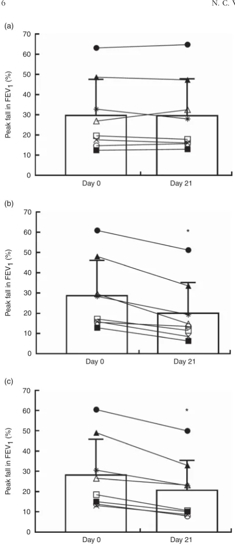
(P=0·011). Further analyses revealed an effect of treatment for
the peak fall in FEV1after EVH at day 21 (P=0·001). Specifically,
the peak fall in FEV1 after EVH was reduced by 34 (SD 14) %
(−690 (SD460) ml) after 6·2 g/dn-3 PUFA (mean difference=310
(SD150) ml, 95 % CI 185, 432 ml,P=0·001, effect size=0·70), and
by 30 (SD11) % (−700 (SD420) ml) after 3·1 g/dn-3 PUFA (mean
difference=270 (SD120) ml, 95 % CI 170, 377 ml,P=0·001, effect
size=0·58). The reduced peak falls in FEV1after 6·2 and 3·1 g/d
n-3 PUFA were not different (P=0·834) (Fig. 2, Table 3).
The percentage protection afforded by then-3 PUFA treatments
compared with placebo is shown in Table 3. The peak fall in
FEV1was unchanged from day 0 to day 21 of placebo.
Fig. 3 shows the percentage change in FEV1during 30 min
recovery after EVH in the HIB group. At day 0, the percentage
change in FEV1 during recovery was not different between
treatments. At day 21, the percentage change in FEV1 was
reduced for up to 20 and 30 min recovery after 6·2 and 3·1 g/d
n-3 PUFA, respectively. Furthermore, compared with placebo
at day 21, the percentage change in FEV1was lower for up to
6 and 20 min recovery after 6·2 and 3·1 g/d n-3 PUFA,
respectively.
In the HIB group, there was a treatment×day interaction for
AUC0–30(P=0·004). Further analysis revealed that at day 21 the
AUC0–30 was reduced after 6·2 g/d (−415 (SD 382), P=0·002)
and 3·1 g/d (−398 (SD399),P=0·001)n-3 PUFA compared with
placebo (−595 (SD 424)). The AUC0–30 at day 21 of 6·2 and
3·1 g/dn-3 PUFA was not different (P=0·751).
Consistent with our previous findings(21,39)the peak fall in
FEV1 after EVH in the HIB group was reproducible with no
differences occurring between day 0 of the three treatments
(within participant CV=7 (SD4) %; measurement error=84 ml;
reproducibility=231 ml; smallest meaningful change=115 ml).
The peak fall in FVC showed similar outcomes. At day 0 of the three treatments the peak fall in FVC was greater in the HIB
group (pooled data:−992 (SD604) ml;−21 (SD15) %) than the
control group (pooled data: −140 (SD 90) ml; −3 (SD 2) %)
(P=0·004). In the control group, the peak fall in FVC was
unchanged after placebo and both doses of n-3 PUFA. The
peak fall in FVC after EVH at day 0 was reduced by 30 (SD21) %
after 6·2 g/d n-3 PUFA and by 29 (SD 24) % after 3·1 g/d n-3
PUFA but was unchanged after placebo in the HIB group. The
reduced peak fall in FVC after 6·2 and 3·1 g/dn-3 PUFA was not
different (P=0·847). In the HIB group, the peak fall in PEF was
unchanged after placebo and both n-3 PUFA treatments
(pooled data:−2·52 (SD1·68) litre/s). In the HIB group, the peak
fall in FEF25–75 %was reduced from day 0 (−1·55 (SD0·37) litre/s)
to day 21 (−1·24 (SD 0·45) litre/s) of 6·2 g/dn-3 PUFA (mean
difference=0·31 (SD 0·23) litre/s, 95 % CI 0·12, 0·50 litre/s,
P=0·006, effect size=0·77). In the HIB group, the peak fall
in FEF25–75 % was unchanged after 3·1 g/d n-3 PUFA and
placebo in the HIB group, and after all treatments in the
control group (P>0·05).
Minute ventilation achieved during each of the six
EVH trials did not differ in the HIB group (P>0·05; between
trial CV=0·23) (pooled data: 109·5 (SD 21·4) litre/min;
78 (SD 18) % of MVV target) which was less compared
with the control group (P=0·001; between trial CV=0·09)
(pooled data: 133·4 (SD 10·8) litre/min; 60 (SD 6) % of MVV
target).
Fraction of exhaled nitric oxide
In the HIB group, there was a treatment×day interaction for
FENO (P=0·004). After 6·2 g/dn-3 PUFA, FENO was reduced
by 24 % from day 0 (48 (SD33) parts per billion (ppb)) to day 21
(35 (SD 28) ppb) (mean difference=13 (SD 12) ppb, 95 % CI 3,
24 ppb,P=0·020, effect size=0·41). Similarly, after 3·1 g/dn-3
PUFA, FENO was reduced by 31 % from day 0 (49 (SD33) ppb)
to day 21 (34 (SD 28) ppb) (mean difference=15 (SD 14) ppb,
95 % CI 4, 27 ppb, P=0·018, effect size=0·46) (Fig. 4). The
reduced FENO after 6·2 and 3·1 g/dn-3 PUFA was not different
(P=0·491) (Fig. 4). FENO remained unchanged from day 0
(47 (SD26) ppb) to day 21 (47 (SD27) ppb) of placebo.
Urinary 9α, 11βPGF2
Baseline urinary 9α, 11β PGF2 did not differ between the
[image:5.595.89.515.90.224.2]HIB group (day 0 pooled data: 40·85 (SD 21·49) ng/mmol
Table 2.Fatty acid composition (% total fatty acids) of then-3 PUFA (INCROMEGATMTG4030) and placebo (CRODAMOLTMGTCC, medium-chain TAG) treatments*
% Total fatty acids
Fatty acids Name n-3 PUFA (INCROMEGA™TG4030) Placebo (CRODAMOLTMGTCC)
8 : 0 Caprylic acid 56
10 : 0 Capric acid 43
18 : 4n-3 Octadecatetraenoic acid 3
20 : 4n-6 Arachidonic acid 3
20 : 4n-3 Eicosatetraenoic acid 2
20 : 5n-3 Eicosapentaenoic acid 45
21 : 5n-3 Heneicosapentaenoic acid 2
22 : 5n-3 Docosapentaenoic acid 6
22 : 6n-3 Docosahexaenoic acid 31
Total 92 99
creatinine) and the control group (day 0 pooled data: 27·05
(SD 14·01) ng/mmol creatinine) (P=0·133). There were no
within-group differences in urinary 9α, 11β PGF2 measured
before EVH at days 0 and 21 of the three treatments.
At day 0, urinary 9α, 11β PGF2 increased by 31·53
(SD 22·77) ng/mmol creatinine (pooled data) after EVH in the
HIB group (P=0·030) but not in the control group (pooled
data: 11·11 (SD16·30) ng/mmol creatinine). At day 0, changes in
9α, 11β PGF2 after EVH were different between groups
(P=0·003). In the HIB group, subsequent analyses revealed an
effect of treatment on the peak increase in 9α, 11βPGF2after
EVH at day 21 (P=0·014). Specifically, the peak increase in 9α,
11βPGF2after EVH was reduced after 6·2 g/dn-3 PUFA (mean
difference=11·75 (SD 7·42) ng/mmol creatinine, 95 % CI 5·54,
17·96 ng/mmol creatinine, P=0·009, effect size=0·50) and
3·1 g/dn-3 PUFA (mean difference=12·86 (SD14·90) ng/mmol
creatinine, 95 % CI 0·40, 25·33 ng/mmol creatinine, P=0·041,
effect size=0·40). The reduced peak increase in 9α, 11βPGF2
after EVH following 6·2 and 3·1 g/dn-3 PUFA was not different
(P=0·377) (Fig. 5). There was no effect of placebo on the
increase in urinary 9α, 11βPGF2after EVH.
Compliance assessment–neutrophil phospholipid fatty acid content, and capsule counts
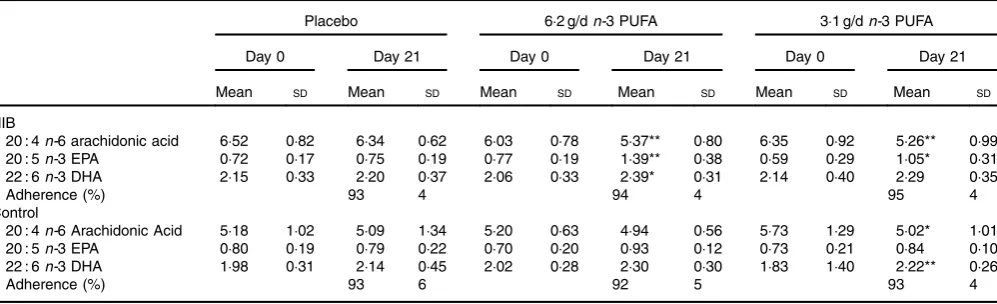
Good compliance to all treatments was shown from the capsule counts in both the HIB and control groups (Table 4). Further compliance evidence was provided from the neutrophil
phos-pholipid fatty acid content. In the HIB group, 6·2 g/dn-3 PUFA
increased EPA (P=0·002) and DHA (P=0·045) content, and
reduced arachidonic acid (P=0·005). In the HIB group, 3·1 g/d
n-3 PUFA increased EPA content (P=0·018) and reduced
ara-chidonic acid (P=0·009); there was a trend for an increase in
DHA content (P=0·074). In the HIB group, linoleic acid content
was unchanged after 6·2 and 3·1 g/dn-3 PUFA. Phospholipid
fatty acid content was unchanged after placebo in both groups.
In the control group, 6·2 g/dn-3 PUFA tended to increase EPA
(P=0·054) and DHA content (P=0·087), whereas 3·1 g/dn-3
PUFA increased DHA content (P=0·007) and reduced
arachi-donic acid (P=0·038); EPA content was unchanged (Table 4).
Discussion
Previous research has shown that high doses of n-3 PUFA
(5·2–5·4 g/d) reduce the severity of HIB and EIB(10,11,15–17). The
present study demonstrates that a lower dose of 3·1 g/d n-3
PUFA is equally effective in reducing HIB in adult men with
asthma. The 6·2 and 3·1 g/dn-3 PUFA treatments also resulted
in similar reductions in baseline FENO, and comparable
sup-pression of urinary 9α, 11β-PGF2 after EVH. These findings
suggest that 3·1 g/d n-3 PUFA could be used as an adjunct
therapy for physically active adults with asthma and EIB. The
percentage protection afforded by 6·2 g/d n-3 PUFA (35
(SD 14) %) and 3·1 g/dn-3 PUFA (33 (SD 12) %) highlights the
efficacy of n-3 PUFA as an adjunct therapy. The percentage
protection afforded byn-3 PUFA is comparable with montelukast
(50 mg twice daily)(40) but less than the 60–70 % protection
afforded by short-actingβ2-agonists procaterol (10 micrograms/
inhalation) and albuterol (90 micrograms/inhalation)(41).
Long-actingβ2-agonists also provide effective treatment for EIB(42–44),
although chronic treatment with both long- and short-acting
70
60
50
40
30
20
10
0
Day 0 Day 21
P
eak f
all in FEV
1
(%)
(a)
70
60
50
40
30
20
10
0
Day 0 Day 21
P
eak f
all in FEV
1
(%)
(b)
70
60
50
40
30
20
10
0
Day 0 Day 21
P
eak f
all in FEV
1
(%)
(c)
*
[image:6.595.44.285.44.604.2]*
β2-agonists can result in tolerance (45,46)
. Furthermore, compliance
to the lower 3·1 g/dn-3 PUFA dose may be improved due to
reduced capsule numbers, reduce cost and risk of gastrointestinal distress, which is commonly associated with bloating, and stomach upset. Indeed, two participants withdrew from the study
when on the 6·2 g/dn-3 PUFA treatment due to gastrointestinal
distress, whereas full compliance and no participant withdrawals
(capsule counts) was observed with 3·1 g/dn-3 PUFA.
The EVH test is an indirect bronchial provocation test that is a
suitable objective surrogate for identifying EIB(24). We have
previously reported that the EVH protocol used in the present
study elicits a highly reproducible fall in FEV1
( 21,39)
which is
crucial when assessing treatment efficacy. The approximately
290 ml (32 %) decrease in the post-EVH fall in FEV1 after 6·2
and 3·1 g/d n-3 PUFA supplementation exceeds the minimum
perceptible change of 230 ml(38) and is therefore clinically
relevant. Our findings are in agreement with previous studies
showing an attenuation of HIB(17)and EIB(11) aftern-3 PUFA
supplementation in adults with asthma. These findings
collec-tively support the ATS/ERS guidance(25) suggesting n-3 PUFA
supplementation could be of benefit in individuals with EIB. Our
findings are also in broad agreement with work on n-3 PUFA
supplementation in elite athletes with EIB(10), and asthmatic
patients with EIB and HIB(11,15–17). An interesting difference is
that previous work showed abolition of EIB after n-3 PUFA
supplementation(10), whereas in the present study only three
participants became non-diagnostic (Fig. 2). This may be partly due to inter-study differences in the severity of EIB: the peak fall
in FEV1 in the present study (−30 %) was approximately 50 %
greater than that reported in previousn-3 PUFA supplementation
studies demonstrating a reduction in EIB/HIB(11,15–17).
Unlike the rather consistent finding that pharmacological
therapy reduces EIB(42–44), the efficacy of n-3 PUFA remains
controversial. Our findings contrast those of Arm et al.(9) and
Brannan et al.(12) who despite using relatively high n-3 PUFA
doses (5·4–6·0 g/d), reported no change in bronchial
hyperre-sponsiveness to inhaled histamine and mannitol, respectively. Armet al.(9)reported no change in airway resistance (fall in FEV1
was not measured) after a cycling exercise challenge, although their data are confounded by: (1) the suggestion that low-grade,
non-pharmaceutical n-3 PUFA was used(47); (2) low statistical
power due to the small number of participants (n6) performing
the exercise; (3) exercise being performed at ambient
tempera-ture and humidity, which may result in insufficient environmental
stress; and (4) the low exercise intensity (60–130 W) which
likely (data not measured/reported) elicited only a modest
increase inV_E. The efficacy ofn-3 PUFA supplementation may
also partly depend on the choice of bronchial provocation test. Direct bronchial provocation tests such as inhaled histamine act directly on receptors on the airway smooth muscle causing
contraction(48)and may not, therefore, fully reveal the potential
anti-inflammatory effects of n-3 PUFA. Indirect bronchial
[image:7.595.69.513.90.242.2]provocation tests such as mannitol and EVH both alter the tonicity and volume of the airway surface liquid thereby Table 3.Individual peak % fall in forced expiratory volume in 1 s (FEV1) at days 0 and 21 of each treatment and percentage protection
afforded byn-3 PUFA in the hyperpnoea-induced bronchoconstriction (HIB) group
Peak % fall in FEV1
Percentage protection afforded byn-3 PUFA Placebo 6·2 g/dn-3 PUFA 3·1 g/dn-3 PUFA treatments for peak % fall in FEV1*
HIB Day 0 Day 21 Day 0 Day 21 Day 0 Day 21 6·2 g/dn-3 PUFA 3·1 g/dn-3 PUFA
1 −12·58 −13·10 −13·02 −6·54 −14·95 −9·98 50 24
2 −63·22 −64·81 −61·04 −51·36 −60·32 −49·84 21 23
3 −48·73 −47·40 −48·20 −33·70 −48·92 −32·83 29 31
4 −32·90 −28·10 −28·50 −19·54 −30·45 −22·91 30 18
5 −27·02 −32·64 −29·71 −14·98 −26·44 −23·12 54 29
6 −19·72 −18·01 −17·33 −11·67 −18·41 −10·50 35 42
7 −17·74 −16·21 −16·05 −8·64 −13·04 −8·63 47 47
8 −14·83 −15·82 −15·71 −13·67 −13·78 −7·90 14 50
Mean −30 −30 −29 −20 −28 −21 35 33
SD 18 18 17 15 18 15 14 12
* Percentage protection afforded by then-3 PUFA treatments calculated as: ((% fall day 21 placebo−% fall day 21 treatment)/% fall day 21 placebo) × 100.
10
0
–10
–20
–30
–40
–50
0 3 6 16 20 30
Minutes after EVH
P
ercentage change in FEV
1
a* a**
b* c* d*
b c* d b b*
c* c**
d** d d
Fig. 3. Percentage change in forced expiratory volume in 1 s (FEV1) after eucapnic voluntary hyperpnoea (EVH) in participants with hyperpnoea-induced bronchoconstriction (HIB). A 10 % fall in FEV1 (shown by the ) is diagnostic of HIB. , Day 0 of placebo treatment; , day 21 of placebo treatment; , day 0 of 6·2 g/dn-3 PUFA treatment; , day 21 of 6·2 g/dn-3 PUFA; , day 0 of 3·1 g/d n-3 PUFA; , day 21 of 3·1 g/d n-3 PUFA. Mean values with unlike letters were significantly different:a, 6·2 g/dn-3 PUFA
[image:7.595.45.283.285.447.2]promoting the release of bronchoconstrictive inflammatory
mediators(49,50). However, mannitol and EVH may exert
differ-ent levels of osmotic and mechanical stress on the airways.
Specifically, mannitol induces dehydration stress primarily in
the proximal airways(50,51), whereas the high V_E during EVH
results in the dehydration stress being extended to peripheral
airways(52)which are increasingly recruited to heat and
humi-dify inspired air. Furthermore, EVH also elicits airflow-induced
shear stress and high transepithelial pressure gradients(53).
These differences may predispose the airway epithelium to greater damage after EVH than mannitol; indeed, EVH tends to result in greater increases in urinary Clara cell protein 16
con-centration, a marker of airway epithelial damage(54,55). This is
significant becausen-3 PUFA supplementation attenuates the
increase in urinary Clara cell protein concentration after EVH, which suggests that reduced HIB may be partly due to reduced
airway epithelial damage(56). Differences in the intensity of the
stimulus to the airways may thus explain the divergent effects of
n-3 PUFA supplementation on bronchial hyperresponsiveness
to EVH and mannitol.
Recently, Price et al.(18)reported no change in the severity
of HIB or markers of airway inflammation (FENO, urinary 9α,
11βPGF2and cysteinyl leukotriene E4) in recreationally active
adults supplemented daily with a combined treatment of
30µg vitamin D3 with n-3 PUFA (3·0 g EPA and 2·0 g DHA).
Unfortunately, objective treatment compliance measures
(e.g. neutrophil phospholipid DHA and EPA content) were not
taken in this study. The fall in FEV1after EVH was also modest
(approximately 16 %) compared with the approximately 30 % fall observed at day 0 in the present study, and six from ten of the participants did not have physician diagnosed asthma (and consequently no asthma medication). Two participants in Price et al.(18)also became non-diagnostic for HIB after the placebo
treatment. It may be that n-3 PUFA supplementation is thus
more effective in individuals with diagnosed asthma and more severe HIB, as observed in the present study.
The increase in urinary 9α, 11β-PGF2after EVH at day 0 in the
HIB group is similar in magnitude to that reported previously
in adults with asthma performing exercise or EVH(10,57). The
reduced increase in 9α, 11β-PGF2 after EVH was comparable
after 6·2 and 3·1 g/d n-3 PUFA supplementation. Previous
work also reports a reduced increase in 9α, 11β-PGF2 after
EVH following n-3 PUFA supplementation(10). The increased
EPA and DHA cell membrane content following n-3 PUFA
110
100
90
80
70
60
50
40
30
20
10
0 FE
NO (ppb)
(a)
110
100
90
80
70
60
50
40
30
20
10
0
Day 0 Day 21
Day 0 Day 21
FE
NO (ppb)
(b)
*
110
100
90
80
70
60
50
40
30
20
10
0
Day 0 Day 21
FE
NO (ppb)
(c)
[image:8.595.45.286.53.600.2]*
Fig. 4.Baseline fraction of exhaled nitric oxide (FENO) before and after placebo (a), 6·2 g/dn-3 PUFA (b) and 3·1 g/dn-3 PUFA (c) in the hyperpnoea-induced bronchoconstriction (HIB) group (n 8). Values are means, and standard deviations represented by vertical bars and identical symbols represent the same HIB participant. * Day 0v. day 21 (P<0·05) ppb, Parts per billion.
P
eak increase in 9
, 11
PGF
2
(ng/mmol creatinine)
after EVH
Placebo 6.2 g/d 3.1 g/d
70
60
50
40
30
20
10
0
[image:8.595.313.547.70.225.2]* *
supplementation is likely to result in dual inhibition of arachidonic acid-dependent cyclo-oxygenase and 5-lipoxygenase
pathways which are responsible for eicosanoid generation(7).
This may explain the decrease in 9α, 11β-PGF2, a metabolite of
the arachidonic acid-derived pro-inflammatory eicosanoid
PGD2. The osmotic changes in the airways following EVH
results in mast cell activation and release of PGD2 during
bronchoconstriction(49,50). Suppression of mast cell activation, and
reduced eicosanoid generation, may thus partially explain reduced
HIB aftern-3 PUFA supplementation.
The comparable reduction in baseline FENO after 6·2 and
3·1 g/d n-3 PUFA supplementation concurs with previous
work(17,58) and is indicative of reduced baseline eosinophilic
airway inflammation. Increased FENO can also result from
elevated expression of inducible nitric oxide synthase in T-cells,
macrophages, airway epithelial cells, and other inflammatory
cells within the airways(59,60). Elevated expression of inducible
nitric oxide synthase can be induced by certain pro-inflammatory
cytokines such as TNF-α(59,60) probably via the activation of
NF-κB transcription factor(61). Then-3 andn-6 PUFA modulate
NF-κB transcription factor activation(62)and presumably therefore
TNF-αexpression, which may explain the observed fall in FENO
aftern-3 PUFA supplementation in the present study. Support for
this argument comes from work showing that 5·4 g/dn-3 PUFA
suppresses circulating plasma TNF-αin athletes with EIB(10). The
comparable reduction in both baseline FENO and the post-EVH
increase in 9α, 11β-PGF2 after 6·2 and 3·1 g/d n-3 PUFA
supplementation suggests that both doses increased the n-3
PUFA content of the phospholipid bilayer of cell membranes
to influence the inflammatory response. Furthermore, and
con-sistent with previous reports(17,56), the lower FENO observed after
n-3 PUFA supplementation in the present study may have
resulted from an increase in airway pH, which is considered a
determinant of FENO and airway inflammation(63).
Limitations of the current study include no run-in period to
assess habitualn-3 PUFA intake, the exclusion of females, the
comparatively low participant numbers, and the wide range of asthma phenotypes suggested by the heterogeneous fall in
FEV1 and large variation in inflammatory markers after EVH.
However, our cohort provides evidence for the use ofn-3 PUFA
in more severe HIB than previous literature(10,11). Future work
may explore whether particular asthma phenotypes and
HIB/EIB severity respond preferentially to n-3 PUFA
supple-mentation. Furthermore, given the existing inter-study
differ-ences regarding the efficacy ofn-3 PUFA for the management
of EIB, further studies using larger sample sizes are warranted.
In conclusion, 3·1 g/dn-3 PUFA supplementation effectively
reduced HIB in men with asthma to a similar extent as a higher
dose. Lower doses of n-3 PUFA thus represent a potentially
beneficial adjunct treatment for adults with asthma and EIB,
whilst also reducing the burden of cost, compliance and potential gastrointestinal distress. Further studies are needed to elucidate if the bronchial provocation test used and the severity of asthma and HIB/EIB affect the degree of protection afforded
byn-3 PUFA supplementation.
Acknowledgements
In-kind support of then-3 PUFA and placebo supplements was
provided by Croda Healthcare Ltd (Croda International Plc, Cowick Hall, Snaith, Goole, East Yorkshire, DN14 9AA, UK). The company was not involved in the design or implementation of the study.
N. C. W., K. A. H., D. E. S., G. R. S. and M. A. J. designed the research; N. C. W., K. A. H., G. R. S. and M. A. J. conducted research; N. C. W. and K. G. J. provided essential reagents and conducted analysis of urine and blood samples; N. C. W., G. R. S. and M. A. J analysed data. N. C. W., K. A. H., G. R. S. and M. A. J. wrote paper; N. C. W., K. A. H., D. E. S., K. G. J., G. R. S. and M. A. J.
contributed to reviewing and approval of thefinal manuscript.
None of the authors has any conflicts of interest to declare.
References
[image:9.595.50.549.99.251.2]1. Carlsen K, Anderson S, Bjermer L, et al. (2008) Exercise‐ induced asthma, respiratory and allergic disorders in elite athletes: epidemiology, mechanisms and diagnosis: part I of the report from the Joint Task Force of the European Respiratory Table 4.Treatment adherence (%) based on capsule count, and fatty acid composition of neutrophil extracts (%total fatty acids) at days 0 and 21 of each treatment in hyperpnoea-induced bronchoconstriction (HIB) and control groups
(Mean values and standard deviations)
Placebo 6·2 g/dn-3 PUFA 3·1 g/dn-3 PUFA
Day 0 Day 21 Day 0 Day 21 Day 0 Day 21
Mean SD Mean SD Mean SD Mean SD Mean SD Mean SD
HIB
20 : 4n-6 arachidonic acid 6·52 0·82 6·34 0·62 6·03 0·78 5·37** 0·80 6·35 0·92 5·26** 0·99 20 : 5n-3 EPA 0·72 0·17 0·75 0·19 0·77 0·19 1·39** 0·38 0·59 0·29 1·05* 0·31 22 : 6n-3 DHA 2·15 0·33 2·20 0·37 2·06 0·33 2·39* 0·31 2·14 0·40 2·29 0·35
Adherence (%) 93 4 94 4 95 4
Control
20 : 4n-6 Arachidonic Acid 5·18 1·02 5·09 1·34 5·20 0·63 4·94 0·56 5·73 1·29 5·02* 1·01 20 : 5n-3 EPA 0·80 0·19 0·79 0·22 0·70 0·20 0·93 0·12 0·73 0·21 0·84 0·10 22 : 6n-3 DHA 1·98 0·31 2·14 0·45 2·02 0·28 2·30 0·30 1·83 1·40 2·22** 0·26
Adherence (%) 93 6 92 5 93 4
Society (ERS) and the European Academy of Allergy and Clinical Immunology (EAACI) in cooperation with GA2LEN. Allergy63, 387–403.
2. Weiler JM, Anderson SD, Randolph C, et al. (2010) Pathogenesis, prevalence, diagnosis, and management of exercise-induced bronchoconstriction: a practice parameter. Ann Allergy Asthma Immunol105, S1–S47.
3. Hallstrand TS (2012) New insights into pathogenesis of exercise-induced bronchoconstriction.Curr Opin Allergy Clin Immunol12, 42–48.
4. Hallstrand TS, Altemeier WA, Aitken ML,et al. (2013) Role of cells and mediators in exercise-induced bronchoconstriction. Immunol Allergy Clin North Am33, 313–328.
5. Barnes PJ (2010) New therapies for asthma: is there any progress?Trends Pharmacol Sci31, 335–343.
6. Ramage L, Lipworth B, Ingram C, et al. (1994) Reduced protection against exercise induced bronchoconstriction after chronic dosing with salmeterol.Respir Med88, 363–368. 7. Kumar A, Mastana SS & Lindley MR (2016) n-3 Fatty acids
and asthma.Nutr Research Rev29, 1–16.
8. Calder PC (2010) Omega-3 fatty acids and inflammatory processes.Nutrients2, 355–374.
9. Arm JP, Horton CE, Mencia-Huerta JM,et al. (1988) Effect of dietary supplementation with fish oil lipids on mild asthma. Thorax43, 84–92.
10. Mickleborough TD, Murray RL, Ionescu AA,et al. (2003) Fish oil supplementation reduces severity of exercise-induced bronchoconstriction in elite athletes. Am J Respir Crit Care Med168, 1181–1189.
11. Mickleborough TD, Lindley MR, Ionescu AA, et al. (2006) Protective effect of fish oil supplementation on exercise-induced bronchoconstriction in asthma.Chest129, 39–49. 12. Brannan JD, Bood J, Alkhabaz A, et al. (2015) The effect of
omega-3 fatty acids on bronchial hyperresponsiveness, sputum eosinophilia, and mast cell mediators in asthma.Chest
147, 397–405.
13. Reisman J, Schachter H, Dales R, et al. (2006) Treating asthma with omega-3 fatty acids: where is the evidence? A systematic review.BMC complement Altern Med6, 1. 14. Weiler JM, Brannan JD, Randolph CC,et al. (2016)
Exercise-induced bronchoconstriction update – 2016. J Allergy Clin Immunol138, 1292–1295. e36.
15. Kumar A, Mastana SS & Lindley MR (2016) EPA/DHA dietary supplementation attenuates exercise-induced bronchocon-striction in physically active asthmatic males. Cogent Med3, 1172696.
16. Mickleborough T & Lindley M (2014) The effect of combining
fish oil and vitamin C on airway inflammation and hyperpnea-induced bronchoconstriction in asthma.J Allergy Ther5, 2. 17. Tecklenburg-Lund S, Mickleborough TD, Turner LA, et al.
(2010) Randomized controlled trial offish oil and montelukast and their combination on airway inflammation and hyperpnea-induced bronchoconstriction.PLoS ONE5, e13487.
18. Price OJ, Hull JH, Howatson G, et al. (2015) Vitamin D and omega-3 polyunsaturated fatty acid supplementation in athletes with exercise-induced bronchoconstriction: a pilot study.Expert Rev Respir Med9, 369–378.
19. Burns CP, Halabi S, Clamon G,et al. (2004) Phase II study of high‐dose fish oil capsules for patients with cancer‐related cachexia.Cancer101, 370–378.
20. Walser B & Stebbins CL (2008) Omega-3 fatty acid supple-mentation enhances stroke volume and cardiac output during dynamic exercise.Eur J Appl Physiol104, 455–461.
21. Williams NC, Johnson MA, Hunter KA, et al. (2015) Repro-ducibility of the bronchoconstrictive response to eucapnic voluntary hyperpnoea.Respir Med109, 1262–1267.
22. Williams N, Hunter K, Johnson M,et al. (2013) A randomised placebo controlled trial to compare the effects of two dosages of omega-3 PUFA on exercise-induced bronchoconstriction (EIB). Br J Sports Med47, e4–e4.
23. Argyros GJ, Roach JM, Hurwitz KM, et al. (1996) Eucapnic voluntary hyperventilation as a bronchoprovocation techni-que: development of a standardized dosing schedule in asthmatics.Chest109, 1520–1524.
24. Anderson SD, Argyros GJ, Magnussen H,et al. (2001) Provo-cation by eucapnic voluntary hyperpnoea to identify exercise induced bronchoconstriction.Br J Sports Med35, 344–347. 25. Parsons JP, Hallstrand TS, Mastronarde JG, et al. (2013) An
official American Thoracic Society clinical practice guideline: exercise-induced bronchoconstriction.Am J Respir Crit Care Med187, 1016–1027.
26. Boulet LP, FitzGerald JM & Reddel HK (2015) The revised 2014 GINA strategy report: opportunities for change. Curr Opin Pulm Med21, 1–7.
27. Duffy P & Phillips Y (1991) Caffeine consumption decreases the response to bronchoprovocation challenge with dry gas hyperventilation.Chest99, 1374–1377.
28. Vally H & Thompson P (2002) Alcoholic drinks and asthma. Clin Exp Allergy32, 186–191.
29. Yusof HM, Miles EA & Calder P (2008) Influence of very long-chain n-3 fatty acids on plasma markers of inflammation in middle-aged men. Prostaglandins Leukot Essent Fatty Acids
78, 219–228.
30. Bahal SM, Romansky JM & Alvarez FJ (2003) Medium chain triglycerides as vehicle for palatable oral liquids: TECHNICAL NOTE.Pharm Dev Technol8, 111–115. 31. Miller MR, Hankinson J, Brusasco V,et al. (2005)
Standardi-sation of spirometry.Eur Respir J26, 319–338.
32. Dweik RA, Boggs PB, Erzurum SC,et al. (2011) An official ATS clinical practice guideline: interpretation of exhaled nitric oxide levels (FENO) for clinical applications.Am J Respir Crit Care Med184, 602–615.
33. Alving K, Weitzberg E & Lundberg JM (1993) Increased amount of nitric oxide in exhaled air of asthmatics.Eur Respir J6, 1368–1370.
34. Junge W, Wilke B, Halabi A, et al. (2004) Determination of reference intervals for serum creatinine, creatinine excretion and creatinine clearance with an enzymatic and a modified Jaffe method.Clin Chim Acta344, 137–148.
35. Nauseef WM (2007) Isolation of human neutrophils from venous blood.Methods Mol Biol,412, 15–20.
36. Bligh EG & Dyer WJ (1959) A rapid method of total lipid extraction and purification.Can J Biochem Physiol37, 911–917.
37. Jackson KG, Bateman PA, Yaqoob P, et al. (2009) Impact of saturated, polyunsaturated and monounsaturated fatty acid-rich micelles on lipoprotein synthesis and secretion in Caco-2 cells.Lipids44, 1081–1089.
38. Santanello N, Zhang J, Seidenberg B,et al. (1999) What are minimal important changes for asthma measures in a clinical trial?Eur Respir J14, 23–27.
39. Williams NC, Johnson MA, Shaw DE,et al. (2016) A prebiotic galactooligosaccharide mixture reduces severity of hyperpnoea-induced bronchoconstriction and markers of airway inflammation.Br J Nutr116, 798–804.
40. Reiss TF, Hill JB, Harman E, et al. (1997) Increased urinary excretion of LTE4 after exercise and attenuation of exercise-induced bronchospasm by montelukast, a cysteinyl leuko-triene receptor antagonist.Thorax52, 1030–1035.
42. Nelson JA, Strauss L, Skowronski M, et al. (1998) Effect of long-term salmeterol treatment on exercise-induced asthma. N Engl J Med339, 141–146.
43. Anderson S, Rodwell L, Du Toit J, et al. (1991) Duration of protection by inhaled salmeterol in exercise-induced asthma. Chest100, 1254–1260.
44. Bronsky EA, Yegen Ü, Yeh CM, et al. (2002) Formoterol provides long-lasting protection against exercise-induced bronchospasm.Ann Allergy Asthma Immunol89, 407–412. 45. Cheung D, Timmers MC, Zwinderman AH, et al. (1992)
Long-term effects of a long-acting β2-adrenoceptor agonist, salmeterol, on airway hyperresponsiveness in patients with mild asthma.N Engl J Med327, 1198–1203.
46. Simons FE, Gerstner TV & Cheang MS (1997) Tolerance to the bronchoprotective effect of salmeterol in adolescents with exercise-induced asthma using concurrent inhaled glucocorticoid treatment.Pediatrics99, 655–659.
47. Mickleborough T & Rundell K (2005) Dietary polyunsaturated fatty acids in asthma-and exercise-induced bronchoconstric-tion.Eur J Clin Nutr59, 1335–1346.
48. Brannan JD & Lougheed MD (2012) Airway hyperrespon-siveness in asthma: mechanisms, clinical significance, and treatment.Front Physiol3, 460.
49. O’Sullivan S, Roquet A, Dahlen B,et al. (1998) Evidence for mast cell activation during exercise-induced bronchocon-striction.Eur Respir J12, 345–350.
50. Brannan JD, Gulliksson M, Anderson SD, et al. (2003) Evidence of mast cell activation and leukotriene release after mannitol inhalation.Eur Respir J22, 491–496.
51. Daviskas E, Anderson SD, Brannan JD,et al. (1997) Inhalation of dry-powder mannitol increases mucociliary clearance.Eur Respir J10, 2449–2454.
52. Daviskas E, Anderson SD, Gonda I, et al. (1995) Changes in mucociliary clearance during and after isocapnic hyperventi-lation in asthmatic and healthy subjects.Eur Respir J8, 742–751.
53. Kippelen P, Fitch KD, Anderson SD,et al. (2012) Respiratory health of elite athletes – preventing airway injury: a critical review.Br J Sports Med46, 471–476.
54. Kippelen P, Tufvesson E, Ali L, et al. (2013) Urinary CC16 after challenge with dry air hyperpnoea and mannitol in recreational summer athletes. Respir Med 107, 1837–1844.
55. Romberg K, Bjermer L & Tufvesson E (2011) Exercise but not mannitol provocation increases urinary Clara cell protein (CC16) in elite swimmers.Respir Med105, 31–36.
56. Mickleborough TD, Vaughn CL, Shei R,et al. (2013) Marine lipid fraction PCSO-524™(lyprinol®/omega XL®) of the New Zealand green lipped mussel attenuates hyperpnea-induced bronchoconstriction in asthma. Respir Med 107, 1152–1163.
57. Simpson AJ, Bood JR, Anderson SD,et al. (2016) A standard, single dose of inhaled terbutaline attenuates hyperpnea-induced bronchoconstriction and mast cell activation in athletes.J Appl Physiol (1985)120, 1011–1017.
58. Schubert R, Kitz R, Beermann C, et al. (2009) Effect ofn-3 polyunsaturated fatty acids in asthma after low-dose allergen challenge.Int Arch Allergy Immunol148, 321–329.
59. Hamid Q, Springall DR, Polak J,et al. (1993) Induction of nitric oxide synthase in asthma.Lancet342, 1510–1513.
60. Robbins R, Springall D, Warren J,et al. (1994) Inducible nitric oxide synthase is increased in murine lung epithelial cells by cytokine stimulation. Biochem Biophys Res Commun 198, 835–843.
61. Barnes PJ (1995) Nitric oxide and airway disease. Ann Med
27, 389–393.
62. Schmitz G & Ecker J (2008) The opposing effects ofn-3 and n-6 fatty acids.Prog Lipid Res47, 147–155.