Developmental cell-cell communication pathways in the
cephalochordate amphioxus: actors and functions
STEPHANIE BERTRAND*
,1, YANN LE PETILLON
1,
ILDIKÓ M.L. SOMORJAI
2,3and HECTOR ESCRIVA*
,11Sorbonne Universités, UPMC Univ Paris 06, CNRS, Biologie Intégrative des Organismes Marins (BIOM),
Observatoire Océanologique, Banyuls/Mer, France, 2Gatty Marine Laboratory, Scottish Oceans Institute, East Sands,
University of St Andrews, St Andrews, Scotland, UK and 3Biomedical Sciences Research Complex, North Haugh,
University of St Andrews, St Andrews, Scotland, UK
ABSTRACT During embryonic development, cells of metazoan embryos need to communicate in order to construct the correct bodyplan. To do so, they use several signals that usually act through interactions between ligands and receptors. Interestingly, only a few pathways are known to be fundamental during animal development, and they are usually found in all the major metazoan clades, raising the following question: how have evolution of the actors and of the functions of these pathways participated in the appearance of the current diversity of animal morphologies? The chordate lineage comprises vertebrates, their sister group the urochordates, and the cephalochor-dates (i.e. amphioxus). Urochorcephalochor-dates are quite derived relative to the chordate ancestor, whereas cephalochordates and vertebrates share many morphological traits. Thus, comparing embryonic development between vertebrates and cephalochordates should give us some insight into the an-cestral characters present in chordates and into the morphological evolution in this clade. However, while much is known about the function of different signalling pathways in vertebrates, data are still scarce in the literature for cephalochordates. In this review, we summarize the current state of the field concerning the expression of actors and the function of the major cell-cell communication pathways, including Hedgehog (Hh), Notch, Nuclear Receptor (NR), Receptor Tyrosine Kinase (RTK), Transforming Growth Factor-b (TGF-b) and Wingless/Int (Wnt), in amphioxus.
KEY WORDS:
chordates, evolution, development, signalling pathways
Introduction
Vertebrates belong to the chordate group together with the urochordates, their sister group, and the cephalochordates (i.e. amphioxus). Although urochordates are more closely related to vertebrates, they show derived features making difficult the inference of ancestral characters by comparing their traits with those of the other clades (Delsuc et al., 2006). On the other hand, amphioxus seem to have retained many ancestral chordate characters at both developmental and genomic levels, making these small marine animals a good model for chordate evo-devo studies (Bertrand and Escriva, 2011). Most cephalochordates live in the sand in shallow waters and are filter feeders. They are gonochoric, and reproduc-tion occurs through external fertilizareproduc-tion in the sea water column after males and females have released their gametes at sunset
Int. J. Dev. Biol. 61: 697-722 (2017)
doi: 10.1387/ijdb.170202sb
www.intjdevbiol.com
*Address correspondence to: Hector Escriva. BIOM, UMR7232 CNRS/UPMC, Observatoire Océanologique de Banyuls sur Mer, Avenue Pierre Favre, 66650, Banyuls sur Mer, France. Tel: +33(0)468887390. Fax: +33(0)468887393. E-mail:[email protected] http://orcid.org/0000-0001-7577-5028 or
Stéphanie Bertrand, BIOM, UMR7232 CNRS/UPMC, Observatoire Océanologique de Banyuls sur Mer, Avenue Pierre Favre, 66650, Banyuls sur Mer, France. Tel: +33(0)430192405. Fax: +33(0)468887393. E-mail:[email protected] http://orcid.org/0000-0002-0689-0126
Submitted: 13 August, 2017; Accepted: 22 September, 2017.
ISSN: Online 1696-3547, Print 0214-6282
© 2017 UPV/EHU Press (Bilbao, Spain) and Creative Commons CC-BY. This is an open access article distributed under the terms of the Creative Commons Attribution License (http://creative-commons.org/licenses/), which permits you to Share (copy and redistribute the material in any medium or format) and Adapt (remix, transform, and build upon the material for any purpose, even commercially), providing you give appropriate credit, provide a link to the license, and indicate if changes were made. You may do so in any reasonable manner, but not in any way that suggests the licensor endorses you or your use. Printed in Spain
Abbreviations used in this paper: EPH, erythropoietin-producing human hepatocellular
receptor; FGFR, fibroblast growth factor receptor; Hh, hedgehog; INSR, insulin receptor; NR, nuclear receptor; PDGFR, platelet-derived growth factor receptor; RTK, receptor tyrosine kinase; TGF, transforming growth factor; TRK, tropomyosin receptor kinase; VEGFR, vascular endothelial growth factor receptor.
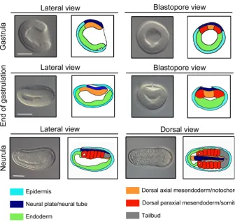
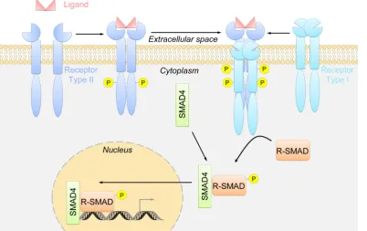
notochord, whereas the dorsal paraxial mesendoderm gives rise to the somites. Finally, the ventral mesendoderm will form the embryonic gut. At the end of gastrulation, the epidermis that has detached from the neural plate covers the whole embryo. During neurulation, the neural plate curves to form the neural tube, which is enlarged in the most anterior region, forming the so-called cere-bral vesicle (Fig. 2). The somites start to form by enterocoely from the dorsal paraxial mesendoderm while the embryo elongates. In the late neurula stage embryo, several regions of the anterior endoderm begin to form different structures. Two diverticula are established in the most anterior region, the Hatschek’s left and right diverticulum, which will later develop into the pre-oral pit and the rostral coelom, respectively. On the right side, the future club-shaped gland and the region where the first gill slit will open can be detected, whereas the endostyle starts to develop from the ventral anterior endoderm. Finally, at the larval stage, the forma-tion of these structures is achieved and the mouth opens on the left side (Fig. 2).
Many questions remain as to how cell-cell communication medi-ates the processes taking place in these different developmental stages. In metazoans, few intercellular signals are known, and deciphering their specific functions in each lineage is crucial for our understanding of morphological evolution in animals. In this review, we will therefore briefly present the mode of action for the main signalling pathways (Hedgehog (Hh), Notch, Nuclear
Receptor (NR), Receptor Tyrosine Kinase (RTK), Transforming Growth Factor-b (TGF-b), Wingless/Int (Wnt) pathways), as well as
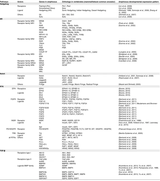
describe the embryonic expression of known actors in amphioxus. We will then detail available information on the embryonic function of each signal. Table 1 summarizes the bibliography concerning gene expression patterns, whereas Table 2 presents the main results obtained using pharmacological treatments to modulate these different pathways.
Hedgehog pathway
How does it work?
[image:2.609.45.382.364.682.2]The role of the Hedgehog (Hh) signal during embryogenesis was first discovered in Drosophila. The name was derived from the phenotype of null mutants for Hh, which presented hair-bristles resembling the spines of the hedgehog (Nusslein-Volhard and Wieschaus, 1980). This signalling pathway is very complex and requires the participation of many actors in addition to the ligands and receptors. The Hedgehog ligand proteins contain two regions. The N-terminal region is the active peptide whereas the C-terminal region, which shows some similarity with inteins, possesses auto-catalytic activity. After autocleavage of Hh, the N-terminal peptide is linked to a cholesterol molecule and to a palmitic acid and is recruited at the plasma membrane. From there it can be released in different ways into the extracellular space in order to spread
Fig. 1. Early embryonic development in amphioxus. Three early developmental stages are presented, from gastrula to neurula. For each, two pictures of the same fixed embryo are shown together with small schemes indicating the different presumptive, or already formed, territories/structures. Scale bar, 50 mm.
through tissues (Briscoe and Therond, 2013, Lee et al., 2016) (Fig. 3). The first identified receptor of Hh is a 12-pass transmembrane protein called Patched (Ptc), which acts as a constitutive repressor of the pathway (Hooper and Scott, 1989, Nakano et al., 1989). In the absence of Hh, Ptc represses Smoothened (Smo), a GPCR family protein, resulting in the proteolysis of the zinc-finger domain tran-scription factor Gli (or Cubitus interruptus (Ci) in Drosophila), which is processed to its repressor form (Fig. 3). Following Hh binding to Ptc, Smo is activated and Gli translocates into the nucleus in its active form, leading to the transcription of target genes (Briscoe and Therond, 2013, Lee
et al., 2016) (Fig. 3).
Receptors and ligands in amphioxus
Amphioxus possesses a unique Hh gene, orthologous to vertebrate Sonic,
Indian and Desert Hedgehog (Shimeld,
1999). Its expression pattern has been described in three amphioxus species,
Branchiostoma floridae, B. belcheri and B. lanceolatum (Shimeld, 1999, Shimeld et al., 2007, Somorjai et al., 2008, Zhang et al., 2002). Hh transcripts are first
Cell-cell communication pathways in amphioxus
699
TABLE 1
AMPHIOXUS ACTORS OF THE DIFFERENT SIGNALLING PATHWAYS CITED IN THIS REVIEW AND REFERENCES OF ARTICLES IN WHICH THEIR EXPRESSION PATTERN HAS BEEN DESCRIBED
Pathways Actors Genes in amphioxus Orthologs in vertebrates (mammal/teleost common ancestor) Amphioxus developmental expression pattern Hedgehog
Receptors Patched (Ptc) Ptc1, Ptc2 (Lin et al., 2009)
Smoothened (Smo) Smo (Lin et al., 2009)
Ligand Hedgehog (Hh) Sonic Hedgehog, Indian Hedgehog, Desert Hedgehog (Shimeld, 1999, Somorjai et al., 2008, Zhang et al., 2002)
Others Gli Gli1, Gli2, Gli3 (Shimeld et al., 2007)
Sufu Sufu (Lin et al., 2009)
NR
Receptor family NR0 NR0B DAX1, SHP
Receptor family NR1 TR TRα, TRβ (Paris et al., 2008)
RAR RARα, RARβ, RARγ (Escriva et al., 2002)
PPAR PPARα, PPARβ, PPARγ
REV-ERB REV-ERBα, REV-ERBβ, REV-ERBγ
ROR RORα, RORβ, RORγ
NR1H1-10 LXRα, LXRβ, FXRα, FXRβ
NR1I/J/K VDR, PXR, CAR
Receptor family NR2 HNF4 HNF4α, HNF4β, HNF4γ
RXR RXRα, RXRβ, RXRγ (Escriva et al., 2002)
TR2/4 TR2, TR4 (Escriva et al., 2002)
TLX TLX
PNR PNR
NR2E
COUP-TF COUP-TFα, COUP-TFβ, COUP-TFγ, EAR2 (Langlois et al., 2000)
Receptor family NR3 ER ERα, ERβ (Bridgham et al., 2008)
ERR ERRα, ERRβ, ERRδ, ERRγ (Bardet et al., 2005)
SR AR, GR, MR, PR (Bridgham et al., 2008)
Receptor family NR4 NR4A NGFI-B, NURR1, NOR1 (Candiani et al., 2009)
Receptor family NR5A NR5A SF1, LRH1
Receptor family NR6 GCNF GCNF
Others NRa
NRb NRc Notch
Receptor Notch Notch1, Notch2, Notch3, (Notch4?) (Holland et al., 2001, Somorjai et al., 2008)
Ligands Delta DLK1, DLK2, DLL3 (Rasmussen et al., 2007)
JaggedA JAG1, JAG2
JaggedB JAG1, JAG2
Others Fringe Lunatic Fringe, Manic Fringe, Radical Fringe (Mazet and Shimeld, 2003) RTK
EPH Receptors EPH1 EPHA1-10, EPHB1-6 (Bosne, 2010)
EPH2 EPHA1-10, EPHB1-6 (Bosne, 2010)
Ligands Efn1 EFNA1-5, EFNB1-3 (Bosne, 2010)
Efn2 EFNA1-5, EFNB1-3 (Bosne, 2010)
Efn3 EFNA1-5, EFNB1-3
FGFR Receptor FGFR FGFR1, FGFR2, FGFR3, FGFR4 (Bertrand et al., 2011)
Ligands FGF1/2 FGF1, FGF2 (Bertrand et al., 2011)
FGF8/17/18 FGF8, FGF17, FGF18, FGF24 (Bertrand et al., 2011; Meulemans and
Bronner-Fraser, 2007)
FGF9/16/20 FGF9, FGF16, FGF20 (Bertrand et al., 2011)
FGFA (FGF3, FGF7, FGF10, FGF22?) (Bertrand et al., 2011)
FGFB (FGF4, FGF5, FGF6?) (Bertrand et al., 2011)
FGFC (FGF19, FGF21, FGF23?) (Bertrand et al., 2011)
FGFD (Bertrand et al., 2011)
FGFE (Bertrand et al., 2011)
INSR Receptor INSR INSR, INSRR, IGF1R (Pashmforoush et al., 1996)
Ligands Ilp Insulin, IGF1, IGF2 (Guo et al., 2009, Holland et al., 1997, Lecroisey
et al., 2015)
Ilp2 Insulin, IGF1, IGF2
PD/VEGFR Receptor PD/VEGFR PDGFRA, PDGFRB, FLT3, CSF1R, KIT, VEGFR1, VEGFR2,
VEGFR3 (Pascual-Anaya et al., 2013)
TRK Receptor Trk NTRK1, NTRK2, NTRK3 (Benito-Gutierrez et al., 2005)
Ligand NT NGF, BDNF, NT-3, NT-4
all RTKs Others AKT AKT1, AKT2, AKT3 (Bertrand et al., 2009)
PDK PDK (Bertrand et al., 2009)
PLCγ PLCγ1, PLCγ2 (Bertrand et al., 2009)
PKCα/β/γ PKCα, PKCβ, PKCδ (Bertrand et al., 2009)
RAF RAF1, ARAF, BRAF (Bertrand et al., 2009)
H/K/NRASa HRAS, NRAS, KRAS (Bertrand et al., 2009)
TGF-β
Receptors type I Alk1/2 Alk1, Alk2
Alk3/6 Alk3, Alk6
Alk4/5/7 Alk4, Alk5, Alk7 Receptors type II TGF-βRII TGF-βRII
ActRII ActRII, ActRIIB
BMPRII BMPRII, AMHR2
Ligands BMP family ADMP ADMP (Kozmikova et al., 2013, Yu et al., 2007)
BMP2/4 BMP2, BMP4 (Kozmikova et al., 2013, Panopoulou et al., 1998,
Yu et al., 2007)
BMP3/3b BMP3, GDF10 (Sun et al., 2010)
BMP5/8 BMP5, BMP6, BMP7, BMP8 (Kozmikova et al., 2013, Yu et al., 2007)
BMP9/10 BMP9, BMP10
GDF5/6/7 GDF5, GDF6, GDF7
Ligands Activin/Inhibin 1 (Inhibinβ-A, Inhibinβ-B, Inhibinβ-C, Inhibinβ-E ?) TGFβ/Nodal/Activin family Activin/Inhibin 2 (Inhibinβ-A, Inhibinβ-B, Inhibinβ-C, Inhibinβ-E?)
Lefty Lefty1, Lefty2 (Morov et al., 2016, Onai et al., 2010, Yu et al.,
2007) Myostatin Myostatin, GDF11
Nodal Nodal (Morov et al., 2016, Onai et al., 2010, Yu et al.,
2002, Yu et al., 2007)
TGF-β TGF-β TGF-β TGF-β
Vg1 GDF1 (Onai et al., 2010)
Others BAMBI BAMBI (Yu et al., 2007)
Chordin Chordin (Somorjai et al., 2008, Yu et al., 2007)
Cerberus Cerberus, DAND5 (Le Petillon et al., 2013, Onai et al., 2010)
Gremlin Gremlin1, Gremlin2 (Le Petillon et al., 2013)
NBL1 NBL1 (Le Petillon et al., 2013)
Tolloid BMP1, Tolloid like 1, Tolloid like 2 (Yu et al., 2007)
Tsg Tsg (Yu et al., 2007)
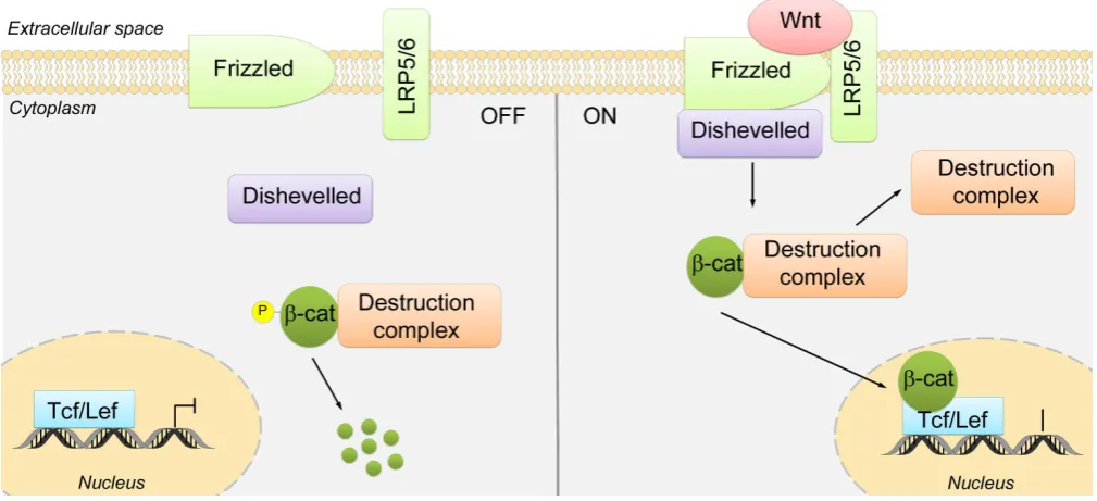
Wnt
Receptors Frizzled 1/2/7 Fzd1, Fzd2 (Qian et al., 2013)
Frizzled 4 Fzd4 (Qian et al., 2013)
Frizzled 5/8 Fzd5, Fzd8 (Qian et al., 2013)
Frizzled 9/10 Fzd9, Fzd10 (Qian et al., 2013)
Antagonists sFRP1/2/5 SFRP1, SFRP2/sizzled, SFRP3/FrzB, sFRP5 (Kong et al., 2012, Yu et al., 2007)*
sFRP3/4 sFRP4 (Yu et al., 2007)
Dkk1/2/4 Dkk1 Dkk2, Dkk4 (Yu et al., 2007, Zhang and Mao, 2010)
Dkk3 Dkk3 (Yu et al., 2007, Zhang and Mao, 2010)
Co-Receptors LRP5/6 LRP5, LRP6 (Wang et al.)
Kremen1, Kremen2,
Kremen3, Kremen4 Kremen1, Kremen2 (Zhang and Mao, 2010)
Ligands Wnt1 Wnt1 (Holland et al., 2000)
Wnt2 Wnt2, Wnt2b/Wnt13 (Somorjai et al., in prep)
Wnt3 Wnt3, Wnt3a (Albuixech-Crespo et al., 2017, Schubert et al.,
2001, Somorjai et al., 2008, Yu et al., 2007)
Wnt4 Wnt4 (Schubert et al., 2000b)
Wnt5 Wnt5a, Wnt5b (Albuixech-Crespo et al., 2017, Schubert et al.,
2001, Somorjai et al., 2008, Somorjai et al., 2012)
Wnt6 Wnt6 (Schubert et al., 2001, Somorjai et al., 2008)
Wnt7 Wnt7a, Wnt7b (Albuixech-Crespo et al., 2017, Schubert et al.,
2000b, Somorjai et al., 2008)
Wnt8 Wnt8a, Wnt8b (Albuixech-Crespo et al., 2017, Morov et al., 2016,
Schubert et al., 2000c, Yu et al., 2007)
Wnt9 Wnt9a, Wnt9b (Somorjai et al., in prep)
Wnt10 Wnt10a, Wnt10b (Somorjai et al., in prep)
Wnt11 Wnt11 (Schubert et al., 2000a, Yu et al., 2007)
Wnt16 Wnt16 (Somorjai et al., in prep)
Others Dsh Dvl1, Dvl2, Dvl3 (Wang et al., 2016)
Axin Axin1, Axin2 (Beaster-Jones et al., 2008, Wang et al., 2016)
GSK3β GSK3β (Wang et al., 2016)
β-catenin β-catenin, Plakoglobin
TCF TCF1, TCF3, TCF4, LEF1 (Lin et al., 2006)
CK1α CK1α (Wang et al., 2016)
CK1δ CK1δ (Wang et al., 2016)
Groucho Groucho (Wang et al., 2016)
700
S. Bertrand et al.
and in two endodermal regions adjacent to the paraxial mesoderm (Shimeld, 1999, Somorjai et al., 2008). In neurula stage embryos and in larvae, Hh is expressed in the notochord, the tailbud, the endostyle and the pre-oral pit (Shimeld, 1999, Shimeld et al., 2007, Somorjai et al., 2008, Zhang et al., 2002), as well as transiently in some ventral neural tube cells (Shimeld, 1999). The expression of both Ptc, orthologous to Ptc1 and Ptc2 in vertebrates, and Smo was analyzed in B. belcheri (Lin et al., 2009). The Ptc transcripts are first detected in the dorsal region of gastrula stage embryos. In neurulae, Ptc is expressed in newly formed somites and in the anterior endoderm (Lin et al., 2009). As the embryo elongates, expression is also detected in the notochord, the presumptive gill slit and the pre-oral pit. Finally, at the larval stage, Ptc is expressed in the cerebral vesicle, the somites, the gill slits, the pre-oral pit, the club-shaped gland and in the gut and pharynx epithelium (Lin et al., 2009). Smo has a similar expression pattern. It is first expressed later than Ptc in the newly formed somites and anterior endoderm in early neurula stage embryos (Lin et al., 2009). Expression is
observed at later stages in the somites, the gut and the anterior notochord (Lin et al., 2009).
Other actors
The expression of two other major players of the Hh pathway has been described in amphioxus. A single Gli gene, orthologous to
Gli1, 2 and 3 in vertebrates, was found in the amphioxus genome
(Shimeld et al., 2007). This gene produces at least two splice vari-ants and the presence of transcripts was first analyzed by using a probe able to recognize both (Shimeld et al., 2007). Gli expres-sion is first detected in the dorsal ectoderm and mesendoderm of late gastrula embryos. In neurulae, Gli is expressed in the lateral neural plate and in the paraxial mesoderm, in cells alongside the
Hh expressing regions. Expression then fades in these territories
and is observed in the cerebral vesicle, in the Hatschek’s left diverticulum and in the forming gill slits. In the larva, labelling is observed in the cerebral vesicle, the pre-oral pit, the club-shaped gland as well as in the gill slits. Using a probe only recognizing the Ligand Hedgehog (Hh) Sonic Hedgehog, Indian Hedgehog, Desert Hedgehog (Shimeld, 1999, Somorjai et al., 2008, Zhang et
al., 2002)
Others Gli Gli1, Gli2, Gli3 (Shimeld et al., 2007)
Sufu Sufu (Lin et al., 2009)
NR
Receptor family NR0 NR0B DAX1, SHP
Receptor family NR1 TR TRα, TRβ (Paris et al., 2008)
RAR RARα, RARβ, RARγ (Escriva et al., 2002)
PPAR PPARα, PPARβ, PPARγ
REV-ERB REV-ERBα, REV-ERBβ, REV-ERBγ
ROR RORα, RORβ, RORγ
NR1H1-10 LXRα, LXRβ, FXRα, FXRβ
NR1I/J/K VDR, PXR, CAR
Receptor family NR2 HNF4 HNF4α, HNF4β, HNF4γ
RXR RXRα, RXRβ, RXRγ (Escriva et al., 2002)
TR2/4 TR2, TR4 (Escriva et al., 2002)
TLX TLX
PNR PNR
NR2E
COUP-TF COUP-TFα, COUP-TFβ, COUP-TFγ, EAR2 (Langlois et al., 2000)
Receptor family NR3 ER ERα, ERβ (Bridgham et al., 2008)
ERR ERRα, ERRβ, ERRδ, ERRγ (Bardet et al., 2005)
SR AR, GR, MR, PR (Bridgham et al., 2008)
Receptor family NR4 NR4A NGFI-B, NURR1, NOR1 (Candiani et al., 2009)
Receptor family NR5A NR5A SF1, LRH1
Receptor family NR6 GCNF GCNF
Others NRa
NRb NRc Notch
Receptor Notch Notch1, Notch2, Notch3, (Notch4?) (Holland et al., 2001, Somorjai et al., 2008)
Ligands Delta DLK1, DLK2, DLL3 (Rasmussen et al., 2007)
JaggedA JAG1, JAG2
JaggedB JAG1, JAG2
Others Fringe Lunatic Fringe, Manic Fringe, Radical Fringe (Mazet and Shimeld, 2003) RTK
EPH Receptors EPH1 EPHA1-10, EPHB1-6 (Bosne, 2010)
EPH2 EPHA1-10, EPHB1-6 (Bosne, 2010)
Ligands Efn1 EFNA1-5, EFNB1-3 (Bosne, 2010)
Efn2 EFNA1-5, EFNB1-3 (Bosne, 2010)
Efn3 EFNA1-5, EFNB1-3
FGFR Receptor FGFR FGFR1, FGFR2, FGFR3, FGFR4 (Bertrand et al., 2011)
Ligands FGF1/2 FGF1, FGF2 (Bertrand et al., 2011)
FGF8/17/18 FGF8, FGF17, FGF18, FGF24 (Bertrand et al., 2011; Meulemans and
Bronner-Fraser, 2007)
FGF9/16/20 FGF9, FGF16, FGF20 (Bertrand et al., 2011)
FGFA (FGF3, FGF7, FGF10, FGF22?) (Bertrand et al., 2011)
FGFB (FGF4, FGF5, FGF6?) (Bertrand et al., 2011)
FGFC (FGF19, FGF21, FGF23?) (Bertrand et al., 2011)
FGFD (Bertrand et al., 2011)
FGFE (Bertrand et al., 2011)
INSR Receptor INSR INSR, INSRR, IGF1R (Pashmforoush et al., 1996)
Ligands Ilp Insulin, IGF1, IGF2 (Guo et al., 2009, Holland et al., 1997, Lecroisey
et al., 2015)
Ilp2 Insulin, IGF1, IGF2
PD/VEGFR Receptor PD/VEGFR PDGFRA, PDGFRB, FLT3, CSF1R, KIT, VEGFR1, VEGFR2,
VEGFR3 (Pascual-Anaya et al., 2013)
TRK Receptor Trk NTRK1, NTRK2, NTRK3 (Benito-Gutierrez et al., 2005)
Ligand NT NGF, BDNF, NT-3, NT-4
all RTKs Others AKT AKT1, AKT2, AKT3 (Bertrand et al., 2009)
PDK PDK (Bertrand et al., 2009)
PLCγ PLCγ1, PLCγ2 (Bertrand et al., 2009)
PKCα/β/γ PKCα, PKCβ, PKCδ (Bertrand et al., 2009)
RAF RAF1, ARAF, BRAF (Bertrand et al., 2009)
H/K/NRASa HRAS, NRAS, KRAS (Bertrand et al., 2009)
TGF-β
Receptors type I Alk1/2 Alk1, Alk2
Alk3/6 Alk3, Alk6
Alk4/5/7 Alk4, Alk5, Alk7 Receptors type II TGF-βRII TGF-βRII
ActRII ActRII, ActRIIB
BMPRII BMPRII, AMHR2
Ligands BMP family ADMP ADMP (Kozmikova et al., 2013, Yu et al., 2007)
BMP2/4 BMP2, BMP4 (Kozmikova et al., 2013, Panopoulou et al., 1998,
Yu et al., 2007)
BMP3/3b BMP3, GDF10 (Sun et al., 2010)
BMP5/8 BMP5, BMP6, BMP7, BMP8 (Kozmikova et al., 2013, Yu et al., 2007)
BMP9/10 BMP9, BMP10
GDF5/6/7 GDF5, GDF6, GDF7
Ligands Activin/Inhibin 1 (Inhibinβ-A, Inhibinβ-B, Inhibinβ-C, Inhibinβ-E ?) TGFβ/Nodal/Activin family Activin/Inhibin 2 (Inhibinβ-A, Inhibinβ-B, Inhibinβ-C, Inhibinβ-E?)
Lefty Lefty1, Lefty2 (Morov et al., 2016, Onai et al., 2010, Yu et al.,
2007) Myostatin Myostatin, GDF11
Nodal Nodal (Morov et al., 2016, Onai et al., 2010, Yu et al.,
2002, Yu et al., 2007)
TGF-β TGF-β TGF-β TGF-β
Vg1 GDF1 (Onai et al., 2010)
Others BAMBI BAMBI (Yu et al., 2007)
Chordin Chordin (Somorjai et al., 2008, Yu et al., 2007)
Cerberus Cerberus, DAND5 (Le Petillon et al., 2013, Onai et al., 2010)
Gremlin Gremlin1, Gremlin2 (Le Petillon et al., 2013)
NBL1 NBL1 (Le Petillon et al., 2013)
Tolloid BMP1, Tolloid like 1, Tolloid like 2 (Yu et al., 2007)
Tsg Tsg (Yu et al., 2007)
Wnt
Receptors Frizzled 1/2/7 Fzd1, Fzd2 (Qian et al., 2013)
Frizzled 4 Fzd4 (Qian et al., 2013)
Frizzled 5/8 Fzd5, Fzd8 (Qian et al., 2013)
Frizzled 9/10 Fzd9, Fzd10 (Qian et al., 2013)
Antagonists sFRP1/2/5 SFRP1, SFRP2/sizzled, SFRP3/FrzB, sFRP5 (Kong et al., 2012, Yu et al., 2007)*
sFRP3/4 sFRP4 (Yu et al., 2007)
Dkk1/2/4 Dkk1 Dkk2, Dkk4 (Yu et al., 2007, Zhang and Mao, 2010)
Dkk3 Dkk3 (Yu et al., 2007, Zhang and Mao, 2010)
Co-Receptors LRP5/6 LRP5, LRP6 (Wang et al.)
Kremen1, Kremen2,
Kremen3, Kremen4 Kremen1, Kremen2 (Zhang and Mao, 2010)
Ligands Wnt1 Wnt1 (Holland et al., 2000)
Wnt2 Wnt2, Wnt2b/Wnt13 (Somorjai et al., in prep)
Wnt3 Wnt3, Wnt3a (Albuixech-Crespo et al., 2017, Schubert et al.,
2001, Somorjai et al., 2008, Yu et al., 2007)
Wnt4 Wnt4 (Schubert et al., 2000b)
Wnt5 Wnt5a, Wnt5b (Albuixech-Crespo et al., 2017, Schubert et al.,
2001, Somorjai et al., 2008, Somorjai et al., 2012)
Wnt6 Wnt6 (Schubert et al., 2001, Somorjai et al., 2008)
Wnt7 Wnt7a, Wnt7b (Albuixech-Crespo et al., 2017, Schubert et al.,
2000b, Somorjai et al., 2008)
Wnt8 Wnt8a, Wnt8b (Albuixech-Crespo et al., 2017, Morov et al., 2016,
Schubert et al., 2000c, Yu et al., 2007)
Wnt9 Wnt9a, Wnt9b (Somorjai et al., in prep)
Wnt10 Wnt10a, Wnt10b (Somorjai et al., in prep)
Wnt11 Wnt11 (Schubert et al., 2000a, Yu et al., 2007)
Wnt16 Wnt16 (Somorjai et al., in prep)
Others Dsh Dvl1, Dvl2, Dvl3 (Wang et al., 2016)
Axin Axin1, Axin2 (Beaster-Jones et al., 2008, Wang et al., 2016)
GSK3β GSK3β (Wang et al., 2016)
β-catenin β-catenin, Plakoglobin
TCF TCF1, TCF3, TCF4, LEF1 (Lin et al., 2006)
CK1α CK1α (Wang et al., 2016)
CK1δ CK1δ (Wang et al., 2016)
Groucho Groucho (Wang et al., 2016)
APC APC (Wang et al., 2016)
Pathways Actors Genes in amphioxus Orthologs in vertebrates (mammal/teleost common ancestor) Amphioxus developmental expression pattern TABLE 1 (CONTINUED)
Cell-cell communication pathways in amphioxus
701
large splicing isoform, the authors showed that it is present only in neurulating embryos in the somitic mesoderm and in the neural plate (Shimeld et al., 2007).
Another actor for which embryonic expression has been de-scribed is Sufu (Suppressor of Fused homolog). Sufu is a negative regulator of Hh signalling (Fig. 1). It binds to Gli or Ci and impedes its translocation into the nucleus (Briscoe and Therond, 2013, Lee
et al., 2016). In B. belcheri, Sufu is expressed in the same regions
as Smo (Lin et al., 2009).
Functional studies
Amphioxus mutants for the Hh gene were generated using TALEN (Wang et al., 2015). 25% of the F1 embryos showed deformities with a curled tail, no mouth and a ventral endostyle and gill slits, suggesting a role of Hh pathway in the control of L/R asymmetry in amphioxus (Wang et al., 2015). Purmorphamine (Sinha and Chen, 2006) and Cyclopamine (Chen et al., 2002) are molecules that are known to activate and inhibit the Hh pathway, respectively, by interacting with Smo. However, up to now there is no study
report-ing the effects of such molecules in amphioxus.
Functional studies have been undertaken in heterologous assays to test for the activity of the two Gli isoforms. Using different approaches such as mammalian cell transfection, chick neural tube electroporation and expression of both isoforms in Drosophila imaginal discs, the authors showed that the long isoform is an activator of Hh signalling, whereas the small isoform acts as a repressor (Shimeld et al., 2007).
Notch pathway
How does it work?
The first Notch gene sequence was described in the 80’s in Drosophila (Kidd et al., 1986, Wharton et al., 1985). The Notch receptor is a one-pass transmembrane protein with extracellular EGF repeats, LIN-12-Notch repeats (LNR) and an intracellular specific domain called NICD (Notch IntraCellular Domain) containing a RBP-jk association module (RAM), nuclear localization signals, ankyrin repeats and PEST motifs (proline, glutamic acid, serine, threonine) (Wang, 2011). Canonical ligands of Notch are likewise trans-membrane proteins forming the Delta/Serrate/lag-2 (DSL) family, which also present EGF repeats in their extracel-lular region as well as a conserved so-called DSL domain (Wang, 2011). The fact that both receptors and ligands are transmembrane proteins implies that the Notch signal can only be achieved between contacting cells. Following ligand binding, the Notch receptor is first cleaved by metalloprote-ases releasing the ectodomain, and then by a g-secretase
[image:5.609.42.323.88.423.2]complex that frees the NICD, which can translocate into the cell nucleus and interact with the DNA-binding protein CSL (CBF1/RBPjκ/Su(H)/Lag-1) (Kopan and Ilagan, 2009) (Fig. 4). Together, NICD and CSL recruit the coactivator Mastermind/Lag-3, leading to transcriptional activation of target genes (Kopan and Ilagan, 2009) (Fig. 4). Although a panel of genes coding for regulators of the Notch pathway exists in the amphioxus genome (Gazave et al., 2009), we will mainly focus our attention on genes for which expres-sion patterns have been described.
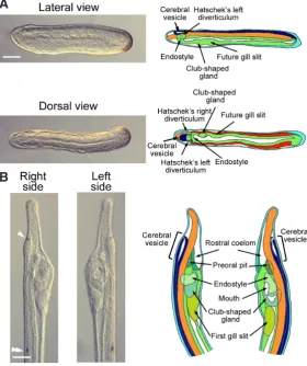
Fig. 2. Late embryonic development in amphioxus.(A) Pictures of lateral and dorsal view of the same late neurula stage embryo are shown together with small schemes presenting the different presumptive structures that are found in the anterior region. Scale bar, 50mm. (B) Pictures of the anterior region of a larva on its left and right side are shown together with schemes highlighting the different pharyngeal structures that are formed at this stage. The frontal eye is indicated by a white arrowhead and the first pigment spot by a double arrowhead. Scale bar, 50mm. Colour code is according to Fig. 1.
Receptors and ligands in amphioxus
In vertebrates, four receptors are known, at least in mammals. In amphioxus, one Notch receptor gene has been described in B.
floridae, which is orthologous to vertebrate Notch1/2/3/4 (Holland et al., 2001). The Notch protein contains 36 EGF repeats, 3 LNR,
a transmembrane region, a RAM domain, 6 ankyrin repeats and several sites for proteolytic cleavage, which are required for Notch signalling pathway activation. Transcripts are first detected by in
situ hybridization at the gastrula stage, in a ring of presumptive
mesendoderm (Holland et al., 2001). Subsequently, a gradient in the dorsal mesendoderm is observed with higher expression in the posterior region (Holland et al., 2001). In the neurulating embryo, strong Notch expression is detected in the posterior mesoderm and in the dorsal part of the three most anterior somites (Holland
et al., 2001). While the embryo is elongating, Notch transcripts are
expressed in the second-youngest somites and in the neural plate and forming notochord (Holland et al., 2001). Thereafter, expres-sion in the somites gets more intense in their posterior part, and becomes stronger in the forming neural tube (Holland et al., 2001).
B
TABLE 2
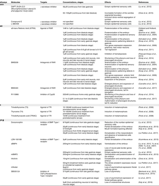
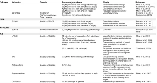
SUMMARY OF PHENOTYPES OBTAINED AFTER PHARMACOLOGICAL TREATMENTS INTERFERING WITH DIFFERENT SIGNALLING PATHWAYS
Pathways Molecules Targets Concentrations, stages Effects References
Notch
DAPT
(N-[N-(3,5- difluorophenacetyl)-l-alanyl]-S-phenylglycine t-butyl ester)
γ-secretase inhibitor 50µM (continuous from late gastrula) Ectopic epidermal sensosry cells (Lu et al., 2012)
50–100µM (continuous from late blastula) Incomplete formation of the somites segmental boundary
Incorrect dorso-ventral segregation of somites
(Onai et al., 2015)
Compound E γ-secretase inhibitor not specified Ectopic epidermal sensosry cells (Lu et al., 2012)
L-685458 γ-secretase inhibitor not specified Ectopic epidermal sensosry cells (Lu et al., 2012)
RAR
all-trans Retinoic Acid (ATRA) Agonist of RAR 1nM to 1µM (continuous from blastula stage) Posteriorization of the embryo (Holland and Holland, 1996)
1µM (continuous from blastula stage) Posteriorization of the embryo (Escriva et al., 2002)
1µM (continuous from blastula stage) Posteriorization of epidermal sensory
neurons (Schubert et al., 2004)
1µM (continuous from blastula stage) Posteriorization of the pharynx (Schubert et al., 2005)
1µM (continuous from early gastrula stage) Anteroposterior patterning defects in
gastrula (Koop et al., 2010)
1µM (continuous from blastula stage) Hox genes expression expansion
anteriorly, less motor neurons. (Schubert et al., 2006) 1µM (continuous from 6-8 gill slit larvae to 8-10
gill slit larvae) Tail regression (Koop et al., 2011)
1µM (continuous from early gastrula stage) Inhibition of hematopoiesis (Pascual-Anaya et al.,
2013) 1µM (continuous from early-mid neurula,
mid-neurula and late mid-neurula to larval stage) Shortening of the pharynx and loss of pharyngeal structures (Koop et al., 2014) BMS009 Antagonist of RAR 1.5µM (continuous from blastula stage) Anteriorization of the embryo (Escriva et al., 2002)
1µM (continuous from blastula stage) Anteriorization of epidermal sensory
neurons (Schubert et al., 2004)
1µM (continuous from blastula stage) Anteriorization of the pharynx (Schubert et al., 2005)
1µM (continuous from early gastrula stage) Anteroposterior patterning defects in
gastrula (Koop et al., 2010)
1µM (continuous from blastula stage) Hox genes expression anterior limit
shifted posteriorly, more motor neurons (Schubert et al., 2006) 2µM (continuous from early-mid neurula,
mid-neurula and late mid-neurula to larval stage) Expansion of the pharynx (Koop et al., 2014) 1µM (continuous from late blastula stage) Enlarged pharynx and expansion of
pharyngeal structures (Carvalho et al., 2017) BMS493 Antagonist of RAR 1µM (continuous from late blastula stage) Enlarged pharynx and expansion of
pharyngeal structures, tail fin elongation
(Carvalho et al., 2017)
R115866 Inhibitor of Cyp26 500nM (continuous frome eraly gastrula stage) Shortening of the pharynx and loss of
pharyngeal structures (Koop et al., 2014) 0,1 to 0,5µM (continuous from late blastula
stage) Shortening of the pharynx and malformation of pharyngeal structures, tail fin reduction
(Carvalho et al., 2017)
TR
Triiodothyronine (T3) Agonist of TR 10-100nM (continuous treatment from
premetamorphic larval stage) Induction of metamorphosis (Paris et al., 2008) Thyroxine (T4) Agonist of TR 10-100nM (continuous treatment from
premetamorphic larval stage) Induction of metamorphosis (Paris et al., 2008) Triiodothyroacetic acid (TRIAC) Agonist of TR 10nM (continuous treatment from
premetamorphic larvae) Induction of metamorphosis (Paris et al., 2008) TGF-β
Dorsomorphin Inhibitor of BMP Type I
receptor 8/16µM (continuous from late gastrula stage) Reduction of the number epidermal sensosry cells (Lu et al., 2012) 20µM (continuous from early blastula stage) Partial dorsalization of the embryo (Kozmikova et al., 2013)
5/10/25µM
(continuous from Knife shaped larval stage) Mouth formation/opening affected (Kaji et al., 2016) 35µM (continuous from early blastula stage) Dorsalization of the mesendoderm
and absence of ectodermal cells specification
(Le Petillon et al., 2017)
LDN-193189 Inhibitor of BMP Type I
receptor 3µM (continuous from early blastula stage) Partial dorsalization of the embryo (Kozmikova et al., 2013) zBMP4 250ng/ml (continuous from early blastula stage) Ventralization of the embryo (Lu et al., 2012, Yu et al.,
2008, Yu et al., 2007)
Loss of neural pate border genes
expression (Yu et al., 2008)
250ng/ml (continuous from late gastrula stage) Ectopic epidermal sensosry cells (Lu et al., 2012)
mBMP2 250ng/ml (continuous from early blastula stage) Ventralization and posteriorization of
the embryo (Kozmikova et al., 2013) hActivin 10ng/ml (continuous from early blastula stage) Dorsalization and anteriorization of the
embryo (Onai et al., 2010)
50ng/ml (treatment before early gastrula stage
on ectodermal explants) The whole ectoderm expresses neural markers (Le Petillon et al., 2017)
mNodal 8µg/ml
(continuous from early gastrula stage) Loss of asymmetrical expression Nodal pathway genes (Li et al., 2017)
SB505124 Inhibitor of
TGF-β/Nodal/Activin Type I receptor
5/10µM (continuous from late gastrula stage) Loss of asymmetry (Bertrand et al., 2015,
Soukup et al., 2015)
50µM (continuous from early gastrula stage) Loss of asymmetrical expression of
Nodal pathway genes (Li et al., 2017) 5µM (from prehatching neurula to hatching
neurula stage) Loss of orobranchial structures (Kaji et al., 2016) 50µM (continuous from early gastrula stage) Ventralization of the embryo (Onai et al., 2010)
50µM (continuous from early blastula stage) Ventralization of the embryo (Kozmikova et al., 2013)
1µM (continuous from early gastrula stage) Loss of dorsal structures (Onai et al., 2012)
50µM (continuous from one cell stage) Reduced blastopore diameter (Morov et al., 2016)
SB431532 Inhibitor of
TGF-β/Nodal/Activin Type I receptor
5/10/20µM
(continuous from late gastrula stage) Loss of asymmetry (BertrandSoukup et al., 2015) et al., 2015,
FGFR
SU5402 Inhibitor of FGFR 50µM (continuous from 8-cell stage) Gastrulation defects (Bertrand et al., 2011)
50µM (continuous from blastula stage) Absence of anterior somites (Bertrand et al., 2011)
50µM (continuous from late neurula stage) Malformation of the notochord (Bertrand et al., 2011)
PD/VEGFR
SU5416 Inhibitor of PD/VEGFR 0,1-20µM (continuous from early gastrula stage) Curved tail (Pascual-Anaya et al.,
2013) Wnt
Inhibitor of GSK3-β Loss of anterior markers expression;
forebrain truncation, posteriorization (Onai et al., 2009) 400mM (30 min from early blastula stage) Exogastrula (Holland et al., 2005)
Various concentrations (from early blastula
stage) Posteriorization and lack of neural plate; severity is concentration dependent
(Holland et al., 2005)
50 to 100mM (1-128 cell stage) Early effect: abnormal cell divisions and apparent loss of polarity; late effect: gastrulation and elongation defects
(Yasui et al., 2002)
BIO Inhibitor of GSK3-β 0.5 µM for 30min at early gastrula stage Disruption of early polarity, posteriorization and elongation defects, downregulation of anterior ectoderm and CNS markers
(Onai et al., 2012)
Alsterpaullone Inhibitor of GSK3-β 0.75-1.5µM Reduction of forebrain and pharynx as assessed by Otx expression; more
severe elongation defects than with lithium
(Onai et al., 2009)
Azakenpaullone Inhibitor of GSK3-β 10 µM (continuous from late gastrula to early
neurula or larva) Loss of of Bra2 posterior expression; loss of Sp5 expression and expansion
anterior-most structures by larval stages
(Dailey et al., 2017)
CHIR99021 Inhibitor of GSK3-β 10 µM; cleavage to gastrula Increase of Sp5 expression (Dailey et al., 2017)
Li+/LiCl 30 min at onset of gastrulation; Na+substituted
Cell-cell communication pathways in amphioxus
703
Pathways Molecules Targets Concentrations, stages Effects References
Pathways Molecules Targets Concentrations, stages Effects References
Notch
DAPT
(N-[N-(3,5- difluorophenacetyl)-l-alanyl]-S-phenylglycine t-butyl ester)
γ-secretase inhibitor 50µM (continuous from late gastrula) Ectopic epidermal sensosry cells (Lu et al., 2012)
50–100µM (continuous from late blastula) Incomplete formation of the somites segmental boundary
Incorrect dorso-ventral segregation of somites
(Onai et al., 2015)
Compound E γ-secretase inhibitor not specified Ectopic epidermal sensosry cells (Lu et al., 2012)
L-685458 γ-secretase inhibitor not specified Ectopic epidermal sensosry cells (Lu et al., 2012)
RAR
all-trans Retinoic Acid (ATRA) Agonist of RAR 1nM to 1µM (continuous from blastula stage) Posteriorization of the embryo (Holland and Holland, 1996)
1µM (continuous from blastula stage) Posteriorization of the embryo (Escriva et al., 2002)
1µM (continuous from blastula stage) Posteriorization of epidermal sensory
neurons (Schubert et al., 2004)
1µM (continuous from blastula stage) Posteriorization of the pharynx (Schubert et al., 2005)
1µM (continuous from early gastrula stage) Anteroposterior patterning defects in
gastrula (Koop et al., 2010)
1µM (continuous from blastula stage) Hox genes expression expansion
anteriorly, less motor neurons. (Schubert et al., 2006) 1µM (continuous from 6-8 gill slit larvae to 8-10
gill slit larvae) Tail regression (Koop et al., 2011)
1µM (continuous from early gastrula stage) Inhibition of hematopoiesis (Pascual-Anaya et al.,
2013) 1µM (continuous from early-mid neurula,
mid-neurula and late mid-neurula to larval stage) Shortening of the pharynx and loss of pharyngeal structures (Koop et al., 2014) BMS009 Antagonist of RAR 1.5µM (continuous from blastula stage) Anteriorization of the embryo (Escriva et al., 2002)
1µM (continuous from blastula stage) Anteriorization of epidermal sensory
neurons (Schubert et al., 2004)
1µM (continuous from blastula stage) Anteriorization of the pharynx (Schubert et al., 2005)
1µM (continuous from early gastrula stage) Anteroposterior patterning defects in
gastrula (Koop et al., 2010)
1µM (continuous from blastula stage) Hox genes expression anterior limit
shifted posteriorly, more motor neurons (Schubert et al., 2006) 2µM (continuous from early-mid neurula,
mid-neurula and late mid-neurula to larval stage) Expansion of the pharynx (Koop et al., 2014) 1µM (continuous from late blastula stage) Enlarged pharynx and expansion of
pharyngeal structures (Carvalho et al., 2017) BMS493 Antagonist of RAR 1µM (continuous from late blastula stage) Enlarged pharynx and expansion of
pharyngeal structures, tail fin elongation
(Carvalho et al., 2017)
R115866 Inhibitor of Cyp26 500nM (continuous frome eraly gastrula stage) Shortening of the pharynx and loss of
pharyngeal structures (Koop et al., 2014) 0,1 to 0,5µM (continuous from late blastula
stage) Shortening of the pharynx and malformation of pharyngeal structures, tail fin reduction
(Carvalho et al., 2017)
TR
Triiodothyronine (T3) Agonist of TR 10-100nM (continuous treatment from
premetamorphic larval stage) Induction of metamorphosis (Paris et al., 2008) Thyroxine (T4) Agonist of TR 10-100nM (continuous treatment from
premetamorphic larval stage) Induction of metamorphosis (Paris et al., 2008) Triiodothyroacetic acid (TRIAC) Agonist of TR 10nM (continuous treatment from
premetamorphic larvae) Induction of metamorphosis (Paris et al., 2008) TGF-β
Dorsomorphin Inhibitor of BMP Type I
receptor 8/16µM (continuous from late gastrula stage) Reduction of the number epidermal sensosry cells (Lu et al., 2012) 20µM (continuous from early blastula stage) Partial dorsalization of the embryo (Kozmikova et al., 2013)
5/10/25µM
(continuous from Knife shaped larval stage) Mouth formation/opening affected (Kaji et al., 2016) 35µM (continuous from early blastula stage) Dorsalization of the mesendoderm
and absence of ectodermal cells specification
(Le Petillon et al., 2017)
LDN-193189 Inhibitor of BMP Type I
receptor 3µM (continuous from early blastula stage) Partial dorsalization of the embryo (Kozmikova et al., 2013) zBMP4 250ng/ml (continuous from early blastula stage) Ventralization of the embryo (Lu et al., 2012, Yu et al.,
2008, Yu et al., 2007)
Loss of neural pate border genes
expression (Yu et al., 2008)
250ng/ml (continuous from late gastrula stage) Ectopic epidermal sensosry cells (Lu et al., 2012)
mBMP2 250ng/ml (continuous from early blastula stage) Ventralization and posteriorization of
the embryo (Kozmikova et al., 2013) hActivin 10ng/ml (continuous from early blastula stage) Dorsalization and anteriorization of the
embryo (Onai et al., 2010)
50ng/ml (treatment before early gastrula stage
on ectodermal explants) The whole ectoderm expresses neural markers (Le Petillon et al., 2017)
mNodal 8µg/ml
(continuous from early gastrula stage) Loss of asymmetrical expression Nodal pathway genes (Li et al., 2017)
SB505124 Inhibitor of
TGF-β/Nodal/Activin Type I receptor
5/10µM (continuous from late gastrula stage) Loss of asymmetry (Bertrand et al., 2015,
Soukup et al., 2015)
50µM (continuous from early gastrula stage) Loss of asymmetrical expression of
Nodal pathway genes (Li et al., 2017) 5µM (from prehatching neurula to hatching
neurula stage) Loss of orobranchial structures (Kaji et al., 2016) 50µM (continuous from early gastrula stage) Ventralization of the embryo (Onai et al., 2010)
50µM (continuous from early blastula stage) Ventralization of the embryo (Kozmikova et al., 2013)
1µM (continuous from early gastrula stage) Loss of dorsal structures (Onai et al., 2012)
50µM (continuous from one cell stage) Reduced blastopore diameter (Morov et al., 2016)
SB431532 Inhibitor of
TGF-β/Nodal/Activin Type I receptor
5/10/20µM
(continuous from late gastrula stage) Loss of asymmetry (BertrandSoukup et al., 2015) et al., 2015,
FGFR
SU5402 Inhibitor of FGFR 50µM (continuous from 8-cell stage) Gastrulation defects (Bertrand et al., 2011)
50µM (continuous from blastula stage) Absence of anterior somites (Bertrand et al., 2011)
50µM (continuous from late neurula stage) Malformation of the notochord (Bertrand et al., 2011)
PD/VEGFR
SU5416 Inhibitor of PD/VEGFR 0,1-20µM (continuous from early gastrula stage) Curved tail (Pascual-Anaya et al.,
2013) Wnt
Inhibitor of GSK3-β Loss of anterior markers expression;
forebrain truncation, posteriorization (Onai et al., 2009) 400mM (30 min from early blastula stage) Exogastrula (Holland et al., 2005)
Various concentrations (from early blastula
stage) Posteriorization and lack of neural plate; severity is concentration dependent
(Holland et al., 2005)
50 to 100mM (1-128 cell stage) Early effect: abnormal cell divisions and apparent loss of polarity; late effect: gastrulation and elongation defects
(Yasui et al., 2002)
BIO Inhibitor of GSK3-β 0.5 µM for 30min at early gastrula stage Disruption of early polarity, posteriorization and elongation defects, downregulation of anterior ectoderm and CNS markers
(Onai et al., 2012)
Alsterpaullone Inhibitor of GSK3-β 0.75-1.5µM Reduction of forebrain and pharynx as assessed by Otx expression; more
severe elongation defects than with lithium
(Onai et al., 2009)
Azakenpaullone Inhibitor of GSK3-β 10 µM (continuous from late gastrula to early
neurula or larva) Loss of of Bra2 posterior expression; loss of Sp5 expression and expansion
anterior-most structures by larval stages
(Dailey et al., 2017)
CHIR99021 Inhibitor of GSK3-β 10 µM; cleavage to gastrula Increase of Sp5 expression (Dailey et al., 2017)
Li+/LiCl 30 min at onset of gastrulation; Na+substituted
for Li+in seawater
TABLE 2 (CONTINUED)
[image:7.609.44.571.129.408.2]SUMMARY OF PHENOTYPES OBTAINED AFTER PHARMACOLOGICAL TREATMENTS INTERFERING WITH DIFFERENT SIGNALLING PATHWAYS
At the late neurula stage, a new expression domain is observed in the anterior pharyngeal endoderm (Holland et al., 2001). At later stages, Notch is expressed in the tailbud, the cerebral vesicle and Hatcheck’s left diverticulum (Holland et al., 2001). The expression of Notch has also been analyzed in B. lanceolatum, where it was shown to be quite similar to that in the Florida species. However, expression in the pharyngeal endoderm and cerebral vesicle can also be detected at an earlier developmental stage (mid-neurula) in the European species (Somorjai et al., 2008).
In vertebrates at least 5 ligands are known: Jagged1 and 2 and Delta-like 1, 3, and 4 (Wang, 2011). Although two genes for ligands of the Jagged family have been found in the B. floridae genome (Gazave et al., 2009), only cloning and expression of the single
Delta gene, orthologous to vertebrate Delta-like genes (Gazave et al., 2009), was reported (Rasmussen et al., 2007). The Delta
protein of amphioxus contains a signal peptide, a DSL domain and nine EGF repeats (Rasmussen et al., 2007). Delta expression, as for Notch, is first observed at the gastrula stage, in the dorsal mesendoderm region (Rasmussen et al., 2007). Then, expression becomes restricted to the paraxial mesoderm and is observed after hatching in the two first pairs of somites before they begin to form, and in some cells of the neural plate (Rasmussen et al., 2007). As development proceeds, transcripts are also detected in some epidermal cells in the ventral region (Rasmussen et al., 2007). At the late neurula stage, Delta is expressed in the posterior part of somites, in some neural plate cells, in ectodermal cells on both sides of the embryo, and in the pharyngeal endoderm (Rasmus-sen et al., 2007). In the larva, expression is mainly observed in the forming somites budding from the tailbud, in some cells of the club-shaped gland and in the most anterior pharyngeal cells (Rasmussen et al., 2007).
It is interesting to note that the expression patterns of Notch and Delta are not fully overlapping. Concerning the expression of
Notch in territories in which Delta is not expressed, we might expect
another ligand coding gene (from the Jagged family possibly) to be transcribed, although we cannot exclude Notch activation through a non-canonical pathway (Andersen et al., 2012) in such regions.
Other actors
The only other actor of the Notch pathway for which expression data are available is Fringe (Mazet and Shimeld, 2003), the ohnolog of lunatic fringe, radical fringe and manic fringe in vertebrates. Fringe proteins are glucosaminyltransferases that have been shown to modulate Notch activity by initiating elongation of O-linked fucose residues attached to the EGF repeats of its ectodomain (Bruckner
et al., 2000, Moloney et al., 2000). In a first study, amphioxus Fringe expression was detected at the late gastrula stage in the
anterior neural plate (Mazet and Shimeld, 2003). Then, neural expression becomes segmented as somites start to form, and
Fringe transcripts are detected in the posterior endoderm and in
the endodermal cells that are fated to become the dorsal midline of the embryonic gut (Mazet and Shimeld, 2003). In late neurula stage embryos, Fringe is expressed in the cerebral vesicle and in the anterior and posterior endoderm (Mazet and Shimeld, 2003). In a more recent study, Fringe expression was detected earlier during gastrulation, in the ventral mesendoderm, with a dorsal boundary just adjacent to the Delta expressing paraxial mesoderm territory (Onai et al., 2015).
Functional studies
Notch pathway function has mainly been studied using treatments with DAPT (N-[N-(3,5-difluorophenacetyl)-l-alanyl]-S-phenylglycine t-butyl ester), a g-secretase inhibitor (Dovey et al., 2001) that
in-hibits Notch receptor cleavage, thereby impeding Notch pathway activation by any ligand. Amphioxus possesses a dorsal central nervous system and a peripheral nervous system composed of several populations of epidermal sensory neurons (ESNs) (Wicht and Lacalli, 2005). Among these cells, the type I solitary neurons are the most abundant, and their progenitors express Delta, to-gether with the neural bHLH factor achaete-scute homologue at the neurula stage (Lu et al., 2012). Lu and colleagues showed that inhibiting the Notch pathway using DAPT, and other g-secretase
[image:8.609.175.572.507.736.2]inhibitors, induces the formation of more ESN progenitors, which are then grouped in clusters containing more cells than in control embryos (Lu et al., 2012). This experiment suggests that Delta/
Cell-cell communication pathways in amphioxus
705
Notch-mediated lateral inhibition would be required in amphioxus for the specification of individual sensory neurons, as is the case in other animals like Drosophila (Lu et al., 2012). In vertebrates, the Delta/Notch pathway is implicated in the segmentation of the paraxial mesoderm, which forms somites. Onai and colleagues stud-ied the role of Notch signalling during somitogenesis in amphioxus (Onai et al., 2015). They showed that after DAPT treatments, the enterocoelic somites, which normally form from the paraxial dorsal mesendoderm, are not completely separated from the archenteron roof, and that the boundaries between somites are not apparent (Onai et al., 2015). The authors suggest that in amphioxus, the Notch signal would be important for the correct dorso-ventral segregation of somites as well as for antero-posterior boundary formation between somites (Onai et al., 2015).
Nuclear receptors
How do they work?
Nuclear receptors (NRs) are ligand-activated transcription factors that consist of a modular structure comprising two major functional domains. These include the N-terminal domain (i.e. DNA binding domain, DBD), which is responsible for the interaction between the NRs and the response elements present in the promoter re-gion of their target genes, and the C-terminal domain (i.e. ligand binding domain, LBD), which interacts with the ligands and with co-regulators to control gene expression (Gronemeyer et al., 2004). Thus, NRs occupy a special position in gene regulation since they provide a direct link between the ligand, which they bind, and the target genes, whose expression they regulate. However, not all NRs have a ligand and some of them, the so-called orphan recep-tors, either function through the binding of an unknown ligand, or control expression of their target genes without binding any ligand (Benoit et al., 2006). Known ligands in vertebrates include many different lipophilic molecules of different chemical natures, such as steroids, thyroid hormones, retinoic acid, or even fatty acids (Robinson-Rechavi et al., 2003). Mammalian genomes contain 48-49 different NRs, grouped into 7 subfamilies (NR0-NR6) includ-ing 19-20 paralogy groups (Escriva Garcia et al., 2003). The B.
floridae genome contains 33 NRs, including members of all the
mammalian paralogy groups except two (NR1I/J/K and NR2F) (Schubert et al., 2008). However, a NR2F gene has been cloned and studied (COUP-TF), indicating that genome data are incomplete (Langlois et al., 2000). Moreover, amphioxus also has a member of the NR5B subfamily, which is not present in vertebrates and was only known in ecdysozoans; and a member of a completely new subfamily, NR7, which is not present in vertebrates but that has been found in echinoderms. Furthermore, amphioxus shows some lineage specific duplications for the NR1H and the NR2E subfamilies. Analyses of gene expression and functional studies have only been undertaken for a few of the amphioxus NRs. We will detail below expression and functional data for these receptors.
Expression patterns of nuclear receptors
Gene expression has been studied for only a few NRs in amphi-oxus, including NR1A (Paris et al., 2008a) and NR1B (Escriva et al., 2002) (thyroid hormone receptor – TR, and retinoic acid receptor – RAR, respectively), NR2B (RXR) (Escriva et al., 2002), NR2C (TR2/4) (Escriva et al., 2002), NR2F (COUP-TF) (Langlois et al., 2000), NR3A, B and C (ER, ERR and SR, respectively) (Bardet et
al., 2005, Bridgham et al., 2008) and NR4A (Nurr1/NGFIB/NOR1)
(Candiani et al., 2009).
The biological functions of retinoic acid (RA) are mediated by heterodimers of two NRs: RAR and RXR. RXR also acts as an heterodimer of other nuclear receptors in accordance with its broad and weak expression throughout all developmental stages in amphi-oxus (Escriva et al., 2002). In contrast, by the mid-gastrula stage,
RAR is expressed throughout the mesendoderm and more weakly
throughout the ectoderm. At the early neurula stage expression becomes downregulated anteriorly in the different tissues where it is expressed (i.e. the neural plate, the anterior endoderm and the non-neural ectoderm), but remains high in the posterior mesoderm, somites and in the posterior three-quarters of the neural plate and endoderm (Escriva et al., 2002). Later on, at the mid-neurula stage, expression is also downregulated in the posterior third of the en-doderm, and the only remaining strong expression is in the nerve cord, posterior to the cerebral vesicle, in the somites of the middle third of the embryo and in a small region of the endoderm. In late neurulae, RAR expression is limited to the nerve cord posterior to the cerebral vesicle and to the endoderm at low levels. Finally, in larvae, RAR is expressed only in the middle third of the nerve cord, somites and gut (Escriva et al., 2002).
TR2/4 shows an expression pattern complementary to RAR
(Escriva et al., 2002). Thus, expression is first detected at the early neurula stage in the anterior neuroectoderm, particularly in the future cerebral vesicle, and in the dorso-anterior endoderm. Later on, at the mid-neurula stage, expression is most intense posteriorly and anteriorly, especially in the posterior mesoderm, cerebral vesicle and Hatcheck’s left diverticulum, which is homologous to part of the vertebrate pituitary. In late neurulae, expression is strong in the cerebral vesicle, in Hatschek’s left diverticulum and in the primordia of the mouth and first gill slit. There is also weak expression in the gut. At the late neurula stage, before the mouth opens, expression persists in the tailbud, cerebral vesicle and in pharyngeal structures (in the forming mouth, first gill slit and Hatschek’s left diverticulum). This expression remains until the larval stage except in the tailbud (Escriva et al., 2002).
Gene expression of the amphioxus thyroid hormone receptor (TR) has only been studied quantitatively using RT-qPCR. The authors showed that TR expression is low during embryonic development, increases in pre-metamorphic larvae just before metamorphosis, and then decreases in juveniles and adults (Paris et al., 2008a).
The amphioxus orthologue of COUP-TF NRs is not expressed during early development and transcripts only start to be detected by in situ hybridization at the late larval stage (4-5 gill slit larvae) in the neural tube posterior to the cerebral vesicle, the homologue of the vertebrate hindbrain and spinal cord, and in dorsal and lateral groups of cells which form dorso-ventral stripes in the nerve cord (Langlois et al., 2000).
The estrogen related receptor gene (ERR) shows an interest-ing expression pattern durinterest-ing amphioxus embryonic development (Bardet et al., 2005). Transcripts are first detected at the neurula stage in the dorsal part of somites 2 to 6, and then in the newly formed somites when the neurula elongates. From the mid-neurula stage, ERR starts to be expressed in individual mesodermal and epidermal cells, but this expression disappears in later stages when
ERR starts to be weakly expressed in the pharyngeal endoderm.
ERR is expressed from this developmental stage in paired cells in
the neural tube, from the border between somites 1 and 2 up to somite 4, and this pattern is extended in later stages until labelling is observed in six pairs of cells. This segmented pattern remains until the larval stage when a new expression domain appears in the posterior part of the cerebral vesicle (Bardet et al., 2005).
Expression studies for other NRs of subfamily 3 have also been published, but only in adults. Thus, while estrogen receptor (ER) and steroid receptor (SR) genes are co-expressed in the cyto-plasm of the oocytes in the female gonads, only ER is expressed weakly in the gills. In males, SR is expressed broadly throughout the testes, at all stages of spermatogenesis, and ER is expressed in the germinal epithelium of the testis in a narrow band of cells that are likely to be early spermatogonia (Bridgham et al., 2008).
The last NR for which gene expression has been documented is the orthologue of Nurr1/NGFIB/NOR1, which shows a restricted expression pattern in late neurula stage embryos in the ventro-medial wall of the Hatschek’s left diverticulum and in the posterior region of the pre-oral pit of larvae (Candiani et al., 2009).
Other actors
Nuclear receptor-mediated transactivation of target genes implies direct contact between the receptor and co-regulators (co-activators or co-repressors). Although orthologues of vertebrate co-regulators are present in the B. floridae genome (Schubert et al., 2008), no expression or functional data have been published. How-ever, several studies have shown that mammalian co-regulators are able to interact with several amphioxus NRs. Thus, amphioxus TR in the presence of its ligand TRIAC (3,5,3’-triiodothyroacetic acid) interacts with the co-activator SRC-1 (Steroid Receptor Coactivator 1) (Paris et al., 2008a), amphioxus RXR interacts with the co-activator hTIF2 (human Transcriptional Intermediary Factor 2) in the presence of its ligand 9-cis RA (Tocchini-Valentini et al., 2009) and amphioxus ERRs (there are two isoforms of this receptor in amphioxus, long and short ERR), interact with the mammalian SRC-1, GRIP1 (Glutamate Receptor Interacting Protein 1), TIF1 (Transcriptional Intermediary Factor 1) and RIP140 (Receptor-Interacting Protein 140) (Horard et al., 2004).
Other actors playing important roles in NR signalling pathways are enzymes implicated in ligand anabolism and catabolism. However, while the presence of many genes for these enzymes has been described in amphioxus (e.g. enzymes implicated in steroid metabolism such as cytochrome P450 (CYP11A, CYP17, and CYP19) and the 17b-hydroxysteroid dehydrogenase) (Castro et al., 2005, Mizuta and Kubokawa, 2007), gene expression and/
or function has only been characterized for enzymes involved in the synthesis and degradation of RA, and for CYP19 aromatase. Using semi-quantitative RT-PCR, it was shown that the CYP19 is expressed in the middle third of amphioxus adults and preferentially in females (Callard et al., 2011, Castro et al., 2005). Other enzymes for which the expression pattern has been characterized include the retinoic acid degradation enzyme CYP26. Three CYP26 genes (CYP26 1-3), which originated by tandem duplication, are found in amphioxus (Carvalho et al., 2017). The expression profiles of
CYP26-1 and CYP26-3 are quite similar and weak, starting at
the mid-gastrula stage in the lateral anterior mesoderm. Then, in mid-neurulae, expression is detected in the anterior somites and in the anterior central nervous system. A slight difference is then found in the larvae where CYP26-1 is more strongly expressed in
central and posterior regions, whereas CYP26-3 labelling is more intense in central and anterior regions. The expression of CYP26-2 starts at the mid-gastrula stage around the blastopore and, later, an expression domain appears in the presumptive lateral mesoderm and anterior neuroectoderm. At the mid-neurula stage, expression is detected in the anterior somites and neuroectoderm, but also in the most anterior and posterior tips of the ectoderm and endoderm and in the tailbud. In late neurulae, before the mouth opens, it is expressed in all anterior germ layers and at the posterior end in the ectoderm. Finally, in larvae, the overall domains of CYP26-2 expression are maintained, with conspicuous labelling anteriorly and posteriorly and a weaker signal in the centre (Carvalho et
al., 2017). RA producing enzyme (aldehyde dehydrogenases,
ALDHs) expression patterns have also been studied in amphioxus (Sobreira et al., 2011), which possesses 6 ALDH1 genes and one ALDH2 (Canestro et al., 2006). Amphioxus ALDH1a is expressed caudally close to the developing tailbud with a sharp anterior boundary at the mid-neurula stage (Sobreira et al., 2011). Later, it is detectable in the posterior gut endoderm of late neurulae. The expression of ALDH1d overlaps with that of ALDH1a at the mid-neurula stage, but in late neurulae, expression is broad and slightly stronger in the posterior gut (Sobreira et al., 2011). Other ALDH1 genes, such as ALDH1b, ALDH1c, ALDH1e, and ALDH1f are weakly expressed in posterior domains overlapping with that of ALDH1a in mid-neurula stage embryos (Sobreira et al., 2011). However, by the late neurula, they are expressed diffusely and weakly throughout the amphioxus embryo with a weak to moderate concentration of the signal for ALDH1b, ALDH1c, and ALDH1e in the posterior gut (Sobreira et al., 2011). ALDH2 expression is restricted to posterior mesendodermal tissues at the mid-neurula stage and, subsequently, spreads throughout the embryo in late neurulae (Sobreira et al., 2011).
Functional studies
Pharmacological treatments with molecules that are agonist or antagonist ligands of NRs have been used to study the function of NRs during embryonic development in amphioxus. In this context, the most studied function has been the role of RA during embryonic development using all-trans RA itself as well as RA antagonists (BMS009, BMS493). An excess of RA induces a posteriorization of the embryo, whereas the use of antagonists of RAR induces anteriorization (Escriva et al., 2002). This role of RA in antero-posterior patterning starts at the early gastrula stage (Koop et
al., 2010) and affects different tissues, such as the ectoderm,
through the control of antero-posterior patterning of epidermal sensory neurons (Schubert et al., 2004); or the endoderm, where RA establishes the posterior limit of the pharynx through control of Hox1 expression (Schubert et al., 2005). Besides this role in patterning, RA also controls pharynx segmentation (Koop et al., 2014), neuronal specification (Schubert et al., 2006), or even tis-sue remodelling in the amphioxus tail (Koop et al., 2011) and tail fin formation (Carvalho et al., 2017). Finally, treatment of neurula stage embryos with RA was shown to inhibit hematopoiesis in amphioxus (Pascual-Anaya et al., 2013).
Another receptor for which the developmental function has been studied is TR. It has been shown that amphioxus metamorphosis is controlled by a thyroid hormone, the 3,5,3’-triiodothyroacetic acid (TRIAC), through its binding to TR and its activation (Paris
Cell-cell communication pathways in amphioxus
707
Other functional studies on amphioxus NRs concern their mo-lecular capacities to bind DNA and to homo- or heterodimerize. Using in vitro EMSA (Electro Mobility Shift Assay) approaches,TR has been shown to bind both as homo- or heterodimer with RXR to DR4 (Direct repeat 4) and HREpal (Hormone Response Element palindrome) elements (Paris et al., 2008a). RAR heterodimerizes with RXR and binds classical DR5 response elements, as well as an IR7 (Inverted Repeat 7) element found in the 5’ non coding region of TR2/4. This receptor, TR2/4, also binds to this IR7 and to DR5 elements (Escriva et al., 2002). In addition, RXR binds as a heterodimer with TR and RAR to DR4 and DR5 elements, respectively, and as a homodimer to DR1 elements (Escriva et
al., 2002). COUP-TF can bind elements ranging from DR0 to DR5
with different affinities (Langlois et al., 2000). Finally, receptors of the NR3 subfamily (i.e.; ERR, ER, SR) bind ERE (Estrogen Re-sponse Element). Thus, ERR, which is expressed as two isoforms (a short and a long isoform), binds ERE as homo- or heterodimers formed by the two isoforms, or to SFRE (Steroidogenic Factor Binding Element) as a homodimer for the short isoform, and as monomer for the long isoform (Horard et al., 2004). ER and SR also bind to ERE as homodimers (Bridgham et al., 2008, Paris
et al., 2008a). These results show that, globally, amphioxus NRs
function in a similar way as their vertebrate orthologues, except for SR, which can bind ERE, in contrast to its vertebrate ortho-logues AR (Androgen Receptor), MR (Mineralocorticoid Receptor), PR (Progesterone Receptor) and GR (Glucocorticoid Receptor) (Bridgham et al., 2008).
[image:11.609.56.555.333.631.2]The ligand binding capacity of different NRs has also been tested either directly (using limited proteolysis assays or mass spectrometry) or indirectly (using a transactivation assay after transient transfection). Thus, RAR binds all-trans and 9-cis RA (Escriva et al., 2006), similarly to its vertebrate orthologues with similar affinities, but TR does not bind vertebrate thyroid hor-mones (triiodothyronine (T3) or thyroxine (T4)). However, a new ligand was discovered in amphioxus which is a thyroid hormone derivative, TRIAC, that binds with high affinity to the amphioxus TR (Paris et al., 2008a). Other receptors for which ligand binding has been studied are RXR, which binds several molecules such as 9-cis RA, DHA (DocosaHexaenoic Acid) or oleic acid (with lower affinities than vertebrate RXRs) (Tocchini-Valentini et al., 2009). TR2/4 does not bind any ligand but is able to transactivate expression of a reporter gene controlled by an IR7 element and also to inhibit the transactivation of the RAR-RXR heterodimer by competing for the DR5 response element (Escriva et al., 2002).
Fig. 5. Simplified schematic view of the receptor tyrosine kinase (RTK) canonical signalling pathway. RTK are transmembrane receptors and interaction of dimers with their ligands induces a transphosphorylation on tyrosine residues of the intracellular domain. The phosphorylated cytoplasmic domain interacts with the adaptor protein Grb2 (Growth factor receptor-bound protein 2), which is associated with the Ras-guanine exchange factor Sos (Son of sevenless) that transforms Ras-GDP into Ras-GTP. This is the first step of activation of the MAPK (Mitogen-activated protein kinase) cascade, which ends with the activation of target gene transcription. Phosphorylated receptors also interact with PI3K (phosphatidylinositide 3-kinase) and PLCg. Active PI3K then activates Akt whereas PLCg (Phospholipase C-g) catalyzes the formation of two secondary messengers, inositol 1,4,5-trisphosphate (IP3) and diacylglycerol (DAG), from phosphatidylinositol 4,5-bisphosphate (PIP2). IP3 diffuses in the cytoplasm and, through interaction with its recep-tor, induces the release of Ca2+ from the endoplasmic reticulum resulting in an increase of Ca2+ cytoplasmic concentration. DAG activates PKC (Protein