1
Copyright WILEY-VCH Verlag GmbH & Co. KGaA, 69469 Weinheim, Germany, 2016.
Supporting Information
Synthetic Light-Curable Polymeric Materials Provide a Supportive Niche for Dental Pulp Stem Cells
Kyle H. Vining, Jacob C. Scherba, Alaina M. Bever, Morgan R. Alexander, Adam D. Celiz*, and David J. Mooney*
Supplementary Experimental Methods –
Time-of-flight secondary-ion mass spectrometry (ToF-SIMS) analysis –
ToF-SIMS measurements were conducted using a ToF-SIMS 4 (IONTOF GmbH) instrument operated using a 25 kV Bi3+ primary ion source exhibiting a pulsed target current of ∼1pA.
Samples were scanned at a pixel density of 100 pixels per mm, with 8 shots per pixel over a given area. An ion dose of 2.45×10¹¹ ions per cm2 was applied to each sample area ensuring
static conditions were maintained throughout. Both positive and negative secondary ion spectra were collected (mass resolution of >7000), over an acquisition period of 15 scans (the data from which were added together). Owing to the non-conductive nature of the samples, charge compensation was applied in the form of a low energy (20eV) electron floodgun.
Multivariate analysis - Principal component analysis (PCA) –
The ToF-SIMS spectral data were analyzed using principal component analysis (PCA) as previously described.[1] All positive and negative ion peak intensities in a ToF-SIMS spectrum
2 Atomic Force Microscopy (AFM) –
AFM of polymer samples was performed using a Dimension Icon FastScan instrument (Bruker) in PeakForce tapping quantitative nanomechanical mapping (QNM) mode using silicon nitride cantilevers. Force–indentation curves were fit using the DMT model with a pyramid indenter.
Preparation of primary human DPSCs and PDLFs –
DPSCs were acquired commercially from Lonza (Poietics human DPSCs, PT-5025) after 12 passages. According to the manufacturer, over 90% of cells tested positive for CD105, CD166, CD29, CD90, and CD73, and over 95% were negative for CD34, CD45, and CD133. The DPSCs were expanded in culture for 2 passages and then cryopreserved in complete growth media and 7.5% DMSO. DPSCs were thawed for subsequent experiments in aMEM (Gibco) complete media, containing 1% penicillin-streptomycin (P/S), 100 µM L-ascorbic acid, and 20% fetal bovine serum. Human periodontal ligament fibroplasts (PDLFs) were commercially obtained from extracted human teeth (Lonza, CC-7049) at passage 2 and cultured in stromal growth media (Lonza) for 2 additional passages prior to cryopreservation as previously described. Incubation conditions were 37C at 5% CO2.
Quantitave polymerase chain reaction (qPCR) measurements –
RNA was isolated with PureLink RNA Micro Kit (Invitrogen), quantified by NanoDrop spectrophotometer, and cDNA was reverse-transcribed by iScript Advanced Reverse
3
computed by the delta-delta Ct method, which compared Ct values to a control sample and reference gene (GAPDH). See Table S3 for list of individual gene primers.
Micro-computed tomography imaging (MicroCT)
MicroCT images were acquired at the Harvard Center for Nanoscale Systems with a tungsten filament in reflection mode (105 kV, 205 uA, 4 frames per projection). Following 3D
reconstruction on VG studio, the coronal pulp in each tooth was segmented to calculate the mineralized volume of reparative dentin.
Supplementary Reference
[image:4.612.107.523.61.539.2]
4
5
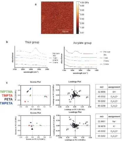
[image:5.612.98.445.225.361.2]diamond), TMPTA (red triangle), PETA (black square), and TMPETA (blue circle) was performed using principal component analysis (PCA) of the respective ToF-SIMS spectra. PCA revealed significant differences in surface chemistry between samples with principal component 1 represented the majority of chemical variance (53%) which was assigned to the substrate background poly(2-hydroxyethyl methacrylate) (pHEMA) moieties in the arrayed samples and thiol functionality present in the scaled-up polymers.
[image:6.612.110.515.69.317.2]
6
7
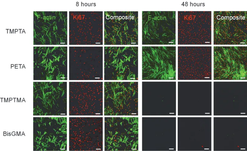
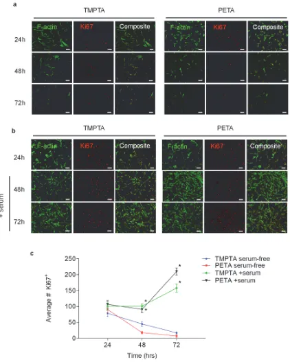
Figure S4. DPSCs maintain expression of proliferation marker Ki67 over 72 hours in serum-containing media. a) DPSCs seeded at subconfluent density (2.5 x10⁴ cells/cm2) on TMPTA
8
hours. DPSCs were adhered at low seeding density (2.5 x10⁴ cells/cm2) initially in serum-free
[image:8.612.100.491.224.367.2]media, which was replaced at 24 hours with media containing 10% FBS. Fluorescence imaging of Ki67 (red) and F-actin (green) staining show the growth and recovery of Ki67+ cells. Scale bar 100 µm. c) Quantification of average number of Ki67+ cells at 24, 48 and 72 hours on TMPTA and PETA, * statistically significant difference from TMPTA-serum free condition, p<0.05, n ≥8.
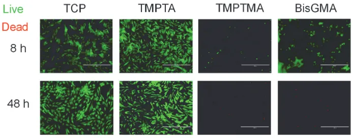
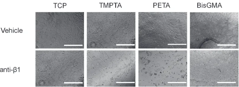
Figure S5. DPSCs adhere to and remain viable on triacrylates in an integrin-dependent manner. Phase images at 8 and 48 hours showed cells did not remain adhered on the
triacrylates TMPTA and PETA, as well as BisGMA, when treated with an anti-β1 antibody to block the integrin β1 receptor subunit, while it had no significant effect on the tissue culture
9
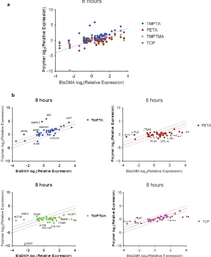
Figure S6. Gene expression analysis of DPSCs adhered after 8 hours to TCP, TMPTA, PETA, TMPTMA, TCP relative to BisGMA control. a) Differential expression of gene markers of β1
integrin signaling from a commercial gene array, using the BioRad PrimePCR Integrin Outside-In Signaling 384-well plate, normalized to GAPDH, n=2. Dotted line indicates unity
a
-4 -2 0 2 4
-10 -5 0 5 10
BisGMA log2(Relative Expression)
8 hours TMPTA MAPK3 SRC HRAS FLNA GRB2 MAP2K1 LEF1 COL1A1 P olymer log 2 (R ela tive E xpres si on)
-4 -2 0 2 4
-10 -5 0 5 10
BisGMA log2(Relative Expression)
10
[image:10.612.96.520.126.658.2]between polymer and BisGMA. b) Scatterplots used to identify differentially expressed genes (defined as above 2-fold threshold).
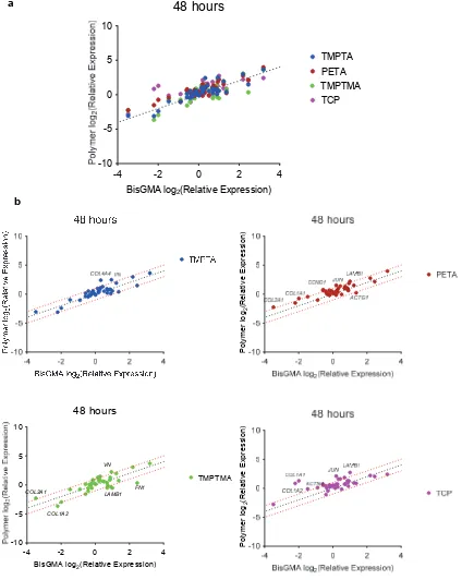
Figure S7. Gene expression analysis of DPSCs adhered after 48 hours to TCP, TMPTA, PETA, TMPTMA, and TCP relative to BisGMA control. a) Differential expression of gene
-4 -2 0 2 4
-10 -5 0 5 10
BisGMA log2(Relative Expression)
48 hours
TMPTA PETA TMPTMA TCP a Poly m er log 2 (R el at iv e E xpre ssi on )-4 -2 0 2 4
-10 -5 0 5 10
11
[image:11.612.98.509.175.605.2]markers of β1 integrin signaling from a commercial gene array, using the BioRad PrimePCR Integrin Outside-In Signaling 384-well plate, normalized to GAPDH, n=2. Dotted line indicates unity between polymer and BisGMA. b) Scatterplots used to identify specific differentially expressed genes (defined as above 2-fold threshold).
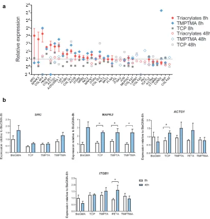
Figure S8. Candidate genes regulated by materials conditions. a) Ranked absolute relative gene expression of DPSCs adhered after 8 and 48 hours to TCP, triacrylates (mean of TMPTA and PETA), TMPTMA, and TCP, normalized to BisGMA control at 8 hours and GAPDH from the BioRad PrimePCR Integrin Outside-In Signaling gene array. b) Confirmation of
SRC MAPK 3 COL 4A4 HRA S COL 2A1 ACTG1 AC00 209 4.1 LEF 1 ITGB 1 COL1A 1 ACT B ITGA 5 GRB 2 AKMAT2PK
1 ITGA
3
COL 1A2AKT3
12
specific candidate genes by qPCR, in particular SRC, MAPK3, ACTG1, and ITGB1 expression in DPSCs on BisGMA, TCP, TMPTA, PETA, and TMPTMA after 8 and 48 hours,
[image:12.612.96.492.175.678.2]normalized to BisGMA control at 8 hours and GAPDH, * statistically significant difference, two-way ANOVA, p<0.05, n >3.
13
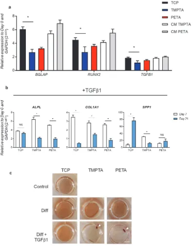
TMPTA and PETA after 21 days in serum-free media, as well as cells on TCP exposed to conditioned media from TMPTA and PETA (CM), * statistically significant difference, two-way ANOVA, p<0.05, n > 3. b) Relative expression of alkaline phosphatase (ALPL), collagen, type I (COL1A1), and osteopontin (SPP1) in DPSCs when cultured 7 and 21 days in
osteogenic media with the addition of TGFβ1 on TCP, TMPTA, and PETA, normalized by
[image:13.612.99.479.303.654.2]GAPDH and Day 0, *statistically significant difference, two-way ANOVA, p>0.05, n = 3. c) Alizarin red staining of DPSCs on TCP, TMPTA, and PETA at 14 days under osteogenic conditions with (Diff + TGFβ1) or without (Diff) growth factor TGFβ1. DPSCs in TGFβ 1-media contract into a cluster of cells on TMPTA and PETA (white arrow). Scale bar 5 mm.
14
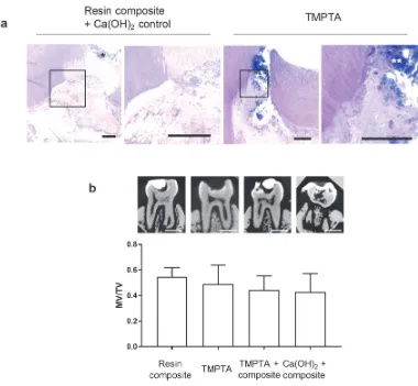
[image:14.612.204.402.203.320.2]analysis of dental pulp calcifications. MicroCT images and mineralized-volume to total-volume ratio, as measured at 8 weeks for treatment with commercially available dental resin composite, composite + calcium hydroxide paste, TMPTA, or TMPTA + commercial resin composite. Scale bar 1 mm. No statistically significant differences between groups, one-way ANOVA, p>0.05, n = 2.
Figure S11. Proposed clinical application of triacrylate-based dental material to treat dental pulp injury following partial resection of pulp tissue (red) in molars and application of regenerative therapy (arrow). Triacrylate biomaterial (green) is placed in direct contact with treated pulp tissue to restore the dentin-pulp interface, because it supports DPSC regenerative behavior, and then is light-cured using traditional visible-light irradiation. Proper dental form and function are restored with a traditional dental composite material (blue). Alternatively, triacrylate-based materials could be used to develop one-step bio-instructive dental composite filling materials that are compatible with the dental pulp cells, as well as restore the tooth.
15
Figure S12. Triacrylate polymers TMPTA and PETA promoted adhesion of primary human periodontal ligament fibroblasts similar to tissue culture control (TCP). Phase images show confluent monolayers, scale bar 1 mm. Quantification of cell number, relative to highest cell number, and cell viability after 48 hours in serum-free media.
Table S1. Monomer list for polymer microarray
Monomer chemical name
Glycidyl methacrylate N-(Butoxymethyl)acrylamide N-Hydroxyethyl acrylamide Benzyl 2-n-propyl acrylate Norbornyl methacrylate Isobornyl methacrylate Methylthioethyl methacrylate
16
Tetrahydrofurfuryl acrylate Hexafluoropent-1,5-diyl diacrylate Tert-butylcyclohexylacrylate
Tricyclodecane-dimethanol diacrylate Butanediol-1,3 diacrylate
Poly(ethylene glycol) methyl ether acrylate N-[2-(1H-indol-3-yl)ethyl]acrylamide N-[Tris(hydroxymethyl)methyl]acrylamide N-(3-Methoxypropyl)acrylamide
Ter-butyl methacrylate Stearyl methacrylate Phenyl methacrylate Isodecyl methacrylate
Acryloyloxy-β,β-dimethyl-γ-butyrolactone Poly(ethylene glycol) phenyl ether acrylate Hydroxypropyl acrylate
Dimethylamino-propyl acrylate Isobutyl acrylate
Hydroxypivalyl hydroxypivalate bis[6-(acryloyloxy)hexanoate] Tetra(ethylene glycol) diacrylate
Di(ethylene glycol) diacrylate Acrylamide
N-Phenylmethacrylamide
Tri(ethylene glycol) methyl ether methacrylate Glycerol dimethacrylate
Isobutyl methacrylate Lauryl methacrylate
t-Butyl cyclohexyl methacrylate
Caprolactone 2-(methacryloyloxy)ethyl ester Poly(propylene glycol) acrylate
Glycerol propoxylate triacrylate Isodecyl acrylate
Isooctyl acrylate
Trimethylolpropane benzoate diacrylate
3-Hydroxy-2,2-dimethylpropyl 3-hyrdoxy-2,2-dimethylpropionate diacrylate Ethylene glycol dimethacrylate
1,4-Bis(acryloyl)piperazine
N-(1,1,3,3-tetramethylbutyl)acrylamide Hydroxybutyl methacrylate
Acryloyloxy-2-hydroxypropyl methacrylate Decyl methacrylate
Ethylene glycol phenyl ether methacrylate Diethylaminoethyl methacrylate
17
Pentaerythritol triacrylate N-Methylmethacrylamide
Di(ethylene glycol) 2-ethylhexyl ether acrylate Lauryl acrylate
Hexanediol ethoxylate diacrylate Bisphenol A ethoxylate diacrylate Dimethylamino-ethyl acrylate N-(4-Hydroxyphenyl)methacrylamide Methacrylamide
N-[3-(Dimethylamino)propyl]acrylamide Vinyl methacrylate
Cyclohexyl methacrylate
Ethylene glycol methyl ether methacrylate Ethylhexyl methacrylate
Tridecafluoro-2-hydroxynonyl acrylate Poly(propylene glycol) methyl ether acrylate Glycidyl acrylate
Benzyl acrylate Epoxidized acrylate Ethylene glycol diacrylate Hexamethylene diacrylate Neopentyl glycol diacrylate N,N'-Ethylenebisacrylamide Sulfopropyl acrylate potassium salt
Di(ethylene glycol) methyl ether methacrylate Butyl methacrylate
Tetrahydrofurfuryl methacrylate
Poly(ethylene glycol) methyl ether methacrylate Poly(ethylene glycol) methacrylate
Hexyl methacrylate
Hydroxy-3-phenoxypropyl acrylate Hydroxy-3-phenoxypropyl acrylate Di(ethylene glycol) ethyl ether acrylate Hexyl acrylate
Glycerol 1,3-diglycerolate diacrylate Neopentyl glycol propoxylate diacrylate Ethylene glycol methyl ether acrylate Diacetone acrylamide
N-(Hydroxymethyl)acrylamide Butoxyethyl methacrylate tetraethyleneglycol dimethacrylate Furfuryl methacrylate
Tert-butylamino-ethyl methacrylate
18
Tri(ethylene glycol) dimethacrylate Butylamino carbonyl oxy ethyl acrylate Trimethylolpropane ethoxylate triacrylate
Poly(propylene glycol) 4-nonylphenyl ether acrylate Trimethylhexyl acrylate
Poly(propylene glycol) diacrylate
Trimethylolpropane ethoxylate methyl ether diacrylate
[2-(Methacryloyloxy)ethyl]dimethyl-(3-sulfopropyl) ammonium hydroxide N-tert-Butylacrylamide mono-2-(Methacryloyloxy)ethyl succinate N-[3-(Dimethylamino)propyl]methacrylamide Methyl 3-hydroxy-2-methylenebutyrate Methacryloyloxy)ethyl acetoacetate Benzyl methacrylate Ethyl-cis-B-cyano-acrylate Hydroxybutyl acrylate
Trimethylolpropane propoxylate triacrylate Isobornyl acrylate
Propargyl acrylate Butanediol diacrylate
[image:18.612.96.519.423.741.2]Methyl-1,2-ethanediyl bis[oxy(methyl-2,1-ethanediyl)]diacrylate
Table S2. BioRad PrimePCR_Integrin Outside-In Signaling H384 Plate
Gene Gene Name PrimePCR ID
ACTB actin, beta qHsaCED0036269
ACTG1 actin, gamma 1 qHsaCED0005010
ACTN1 actinin, alpha 1 qHsaCED0002731
ACTN4 actinin, alpha 4 qHsaCID0012278
CTNNB1 catenin (cadherin-associated protein), beta 1, 88kDa qHsaCID0010363
COL1A1 collagen, type I, alpha 1 qHsaCED0002181
COL1A2 collagen, type I, alpha 2 qHsaCED0003988
COL2A1 collagen, type II, alpha 1 qHsaCED0001057
COL4A1 collagen, type IV, alpha 1 qHsaCID0010223
COL4A2 collagen, type IV, alpha 2 qHsaCED0004426
COL4A3 collagen, type IV, alpha 3 (Goodpasture antigen) qHsaCID0013308
COL4A4 collagen, type IV, alpha 4 qHsaCID0016411
CCND1 cyclin D1 qHsaCID0013833
FN1 fibronectin 1 qHsaCID0012349
FLNA filamin A, alpha qHsaCID0010844
GAPDH glyceraldehyde-3-phosphate dehydrogenase qHsaCED0038674
GSK3B glycogen synthase kinase 3 beta qHsaCID0010097
GRB2 growth factor receptor-bound protein 2 qHsaCED0004795
HPRT1 hypoxanthine phosphoribosyltransferase 1 qHsaCID0016375
ITGA2 integrin, alpha 2 qHsaCID0016134
ITGA3 integrin, alpha 3 qHsaCID0010823
ITGA5 integrin, alpha 5 qHsaCID0021495
ITGAV integrin, alpha V qHsaCID0006233
ITGB1 integrin, beta 1 qHsaCED0005248
ILK integrin-linked kinase qHsaCED0038739
JUN jun proto-oncogene qHsaCED0018770
19
LEF1 lymphoid enhancer-binding factor 1 qHsaCED0001977
MAPK1 mitogen-activated protein kinase 1 qHsaCID0006818
MAPK3 mitogen-activated protein kinase 3 qHsaCID0010939
MAP2K1 mitogen-activated protein kinase kinase 1 qHsaCID0011553
gDNA PrimePCR DNA Contamination Control Assay qHsaCtlD0001004
PCR PrimePCR Positive Control Assay qHsaCtlD0001003
RT PrimePCR Reverse Transcription Control Assay qHsaCtlD0001001
RQ1 PrimePCR RNA Quality Assay qHsaCtlD0001002
RQ2 PrimePCR RNA Quality Assay qHsaCtlD0001002
PTK2 PTK2 protein tyrosine kinase 2 qHsaCED0001879
RAC1 ras-related C3 botulinum toxin substrate 1 (rho family, small GTP binding protein Rac1) qHsaCED0001330
TBP TATA box binding protein qHsaCID0007122
TCF7L2 transcription factor 7-like 2 (T-cell specific, HMG-box) qHsaCID0012510
AKT1 v-akt murine thymoma viral oncogene homolog 1 qHsaCID0011338
AKT2 v-akt murine thymoma viral oncogene homolog 2 qHsaCED0037964
AKT3 v-akt murine thymoma viral oncogene homolog 3 qHsaCID0036448
HRAS v-Ha-ras Harvey rat sarcoma viral oncogene homolog qHsaCED0023908
VCL vinculin qHsaCID0020885
AC002094.1 Vitronectin qHsaCED0001902
RAF1 v-raf-1 murine leukemia viral oncogene homolog 1 qHsaCED0001147
[image:19.612.91.519.355.599.2]SRC v-src sarcoma viral oncogene homolog (avian) qHsaCED0004489
Table S3. BioRad PrimePCR assays for qPCR
Gene Gene Name PrimePCR ID
AC002094.1 (VN) Vitronectin qHsaCED0001902
ACTA2 Smooth muscle actin qHsaCID0013300
ACTG1 Actin, gamma 1 qHsaCED0005010
ALPL Alkaline phosphatase qHsaCED0045991
BGLAP Osteocalcin qHsaCED0038437
CCND1 Cyclin D1 qHsaCID0013833
COL1A1 Collagen, type I, alpha 1 qHsaCED0043248
COL2A1 Collagen, type II, alpha 1 qHsaCED0001057
COL4A4 Collagen, type IV, alpha 4 qHsaCID0016411
DSPP Dentin sialophosphoprotein qHsaCED0002962
GAPDH Glyceraldehyde-3-phosphate dehydrogenase qHsaCED0038674
HRAS Harvey rat sarcoma viral oncogene homolog qHsaCED0023908
ITGB1 integrin, beta 1 qHsaCED0005248
LEF1 Lymphoid enhancer-binding factor 1 qHsaCED0001977
MAPK3 Mitogen-activated protein kinase 3 qHsaCID0010939
RUNX2 Runx-2 qHsaCED0044067
SPP1 Secreted phosphoprotein 1, osteopontin qHsaCED0057074
STC1 Staniocalcin qHsaCID0006115