Extracellular Matrix Hydrogels from Decellularized Tissues: Structure and
Function
Lindsey T. Saldina,b,* and Madeline C. Cramera,b,*, Sachin S. Velankarc, Lisa J. Whiteb,d, Stephen F. Badylaka,b,e
a Department of Bioengineering, University of Pittsburgh, 360B CNBIO, 300 Technology
Drive, Pittsburgh, PA 15219, USA b McGowan Institute for Regenerative Medicine,
Suite 300, 450 Technology Drive, University of Pittsburgh, Pittsburgh, PA 15219, USA c
Department of Chemical Engineering, University of Pittsburgh, 940 Benedum Hall,
Pittsburgh, PA 15261, USA d School of Pharmacy, University of Nottingham, University
Park, Nottingham, NG7 2RD, UK e Department of Surgery, University of Pittsburgh, 200
Lothrop Street, Pittsburgh PA 15213, USA * These authors contributed equally to this
work
Lindsey T. Saldin: [email protected]; Madeline C. Cramer: [email protected] ; Sachin S. Velankar: [email protected]; Lisa J. White: [email protected]; Stephen F. Badylak: [email protected]
Corresponding author: Dr. Stephen F. Badylak, Suite 300, 450 Technology Drive, Pittsburgh, PA 15219, Tel: +1 (412) 235-5253, Fax: +1(412) 235-5256,
Abstract
Extracellular matrix (ECM) bioscaffolds prepared from decellularized tissues have been
used to facilitate constructive and functional tissue remodeling in a variety of clinical
applications. The discovery that these ECM materials could be solubilized and
subsequently manipulated to form hydrogels expanded their potential in vitro and in vivo
utility; i.e. as culture substrates comparable to collagen or Matrigel, and as injectable
direct cell behavior and influence remodeling outcomes are only partially understood,
but likely include structural and biological signals retained from the native source tissue.
The present review describes the utility, formation, and physical and biological
characterization of ECM hydrogels. Two examples of clinical application are presented
to demonstrate in vivo utility of ECM hydrogels in different organ systems. Finally, new
research directions and clinical translation of ECM hydrogels are discussed.
Keywords: Extracellular matrix; Hydrogel; Decellularization; Naturally derived;
Injectable; Regenerative medicine; Biomaterial; Tissue engineering
1. Introduction
Hydrogels are defined as highly hydrated polymer materials (>30% water by weight),
which maintain structural integrity by physical and chemical crosslinks between polymer
chains [1]. The polymer chains can be synthetic [e.g., polyethylene oxide (PEO),
poly(vinyl alcohol) (PVA), poly(acrylic acid) (PAA), poly(propylenefumarate-co-ethylene
glycol) P(PF-co-EG)] or natural (e.g., alginate, chitosan, collagen, hyaluronic acid).
Synthetic and natural hydrogels have been widely used to fill space, deliver bioactive
molecules/drugs, and/or deliver cells to stimulate tissue growth [1].
Many hydrogels have been derived from components of the extracellular matrix
(ECM) such as collagen, hyaluronic acid and elastin or complex mixtures of ECM
proteins such as Matrigel. The focus of the present review is ECM hydrogels and
specifically, hydrogels that are 1) derived from decellularized mammalian tissue, and 2)
ECM materials that fulfill one of these criteria, such as decellularized tissues that are
“gel-like” but not further solubilized (for example decellularized human lipoaspirate [2] ,
intervertebral disc [3, 4], and devitalized cartilage [5, 6]) are beyond the scope of this
review. In contrast to hydrogels composed of individual ECM components, ECM
hydrogels retain the full biochemical complexity of the native tissue, and unlike Matrigel,
are not composed of a protein source that is a product of a tumorigenic cell line.
To date, ECM hydrogels have been primarily used as 3D organotypic culture models
and to stimulate tissue growth after injury. The present review describes the utility,
formation and physical and biological characterization of ECM hydrogels. Two examples
of clinical application in selected organ systems are presented. Finally, new research
directions and clinical translation of ECM hydrogels are discussed.
1.1. Why ECM?
The ECM consists of the structural and functional molecules secreted by the
resident cells of each tissue, hence the 3D organization and biochemical composition of
the ECM is distinctive for each tissue type. ECM has been influencing cell behavior,
dynamically and reciprocally [7] since single cell organisms evolved more than 600
million years ago, and likely played a central role in the transition from unicellular
organisms to multicellular organisms [8]. Mimicking aspects of the structure and
composition of the ECM has guided the rational design of biomaterials over the past
Although decellularization of tissue was first reported in 1973 as a technique to preserve
tissue intended to be used as a protective barrier for burn patients [10], the first reported
production of ECM by decellularization of a source tissue for subsequent use as a
bioscaffold for tissue reconstruction was the use of small intestinal submucosa (SIS) for
vascular applications [11-15]. These initial studies removed cellular material while
preserving the structural and functional proteins of the ECM such as
glycosaminoglycans (GAGs), proteoglycans, and growth factors [16]. When processed
appropriately, ECM materials harvested by such methods retain the biochemical
complexity, nanostructure, and bioinductive properties of the native matrix, and have
been shown to promote the in vivo creation of site-specific, functional tissue [17].
ECM-derived materials are FDA-allowed, can be preserved and used ‘off the shelf,’ have
been implanted in millions of patients to date; and have been extensively characterized
in both the 2D sheet and powder forms [17, 18].
The discovery that ECM bioscaffolds could be transformed into hydrogels expanded
their potential in vitro and in vivo utility [16]. For example, minimally invasive delivery
becomes possible wherein a pre-gel viscous fluid is injected with a catheter or syringe
and polymerizes at physiologic temperature into a hydrogel conforming to the shape of
any defect site. Compared to suspensions of ECM powders, ECM hydrogels can be
injected with a more homogenous concentration and with greater ease [19].
Hydrogels derived from SIS and urinary bladder matrix (UBM) have been shown
to retain the inherent bioactivity of the native matrix with the ability to promote
constructive remodeling in heterologous tissue applications [16, 20-26]. In the last
almost every organ system. The mechanisms by which the ECM hydrogel modulates
cell behavior are not fully understood but likely include release of bound growth factors
[27], cytokines, and chemokines [28], presentation of cryptic peptides [29-32], exposure
of bioactive motifs, and as recently reported, through bioactive matrix-bound
nanovesicles [33].
2. ECM Hydrogel Formation
ECM hydrogel formation is a collagen-based self-assembly process that is
regulated in part by the presence of glycosaminoglycans, proteoglycans, and ECM
proteins [34]. Therefore, polymerization kinetics will be influenced by the native
biochemical profile of the source tissue and of the proteins that remain after
decellularization and solubilization. It is important to achieve sufficient cell removal from
source tissues [35, 36] while maintaining ECM composition and ultrastructure. The
choice of solubilization protocol is crucial to not adversely affect the ability to
subsequently form an ECM hydrogel. Table 1 provides an overview of the many
methods used to decellularize source tissues and solubilize the remaining ECM. ECM
hydrogels are primarily derived from porcine tissue but some hydrogel types, e.g.,
adipose, tendon, umbilical cord are sourced from human tissue.
Formation of a hydrogel involves two key steps: 1) solubilization of the ECM
material into protein monomeric components, and 2) temperature- and/or pH-controlled
neutralization to induce spontaneous reformation of the intramolecular bonds of the
monomeric components into a homogeneous gel. The most prevalent method used to
form of ECM (also called “ECM digestion”). Pepsin is an enzyme derived from porcine
gastric juices that has been used since 1972 to solubilize a substantial portion (up to
99%) of acid-insoluble collagen [37, 38]. Pepsin cleaves the telopeptide bonds of the
collagen triple helix structure to unravel collagen fibril aggregates [39]. The ECM
material is first powdered and stirred in pepsin with dilute hydrochloric acid over 48
hours, as reported by Freytes et al. and designated herein as the “Freytes method” [20].
Another method involves the use of 0.5 M acetic acid instead of 0.1 M HCl as a base
medium for the pepsin enzyme (“Voytik-Harbin method”) [16]. Pepsin digestion or
solubilization is complete when the liquid is homogenous with no visible particles [20].
Different digestion times will produce a different profile of cryptic molecules, some of
which possess bioactive properties [31, 40], suggesting the preferred digestion period
will need to be tailored for each clinical application; times of 24 – 96 hours have been
reported (Table 1). The “solubilized ECM” or “ECM digest” forms a gel when the liquid is
neutralized to physiologic pH, salt concentration (“ECM pre-gel”) and temperature in
vitro (“ECM hydrogel”) in an entropy-driven process dominated by collagen kinetics.
Specifically, there is an increase in entropy when collagen monomers lose water, form
aggregates, and bury surface-exposed hydrophobic residues within the fibril in vitro, in a
self-assembly process [39, 41]. In practice, the “solubilized ECM” is neutralized to
physiologic pH and salt concentration and kept at a low temperature well-below 37C,
until the application of interest is identified for temperature-controlled gelation; e.g.,
injected by needle or catheter to gel in situ, or placed in an incubator for 3D cell culture.
Johnson et al. investigated the effect of changing a single neutralization
PBS) on the material properties of an ECM hydrogel, specifically myocardial ECM
hydrogel [42]. In brief, the gelation time could be modulated from ~ 20 minutes at
decreased salt concentration (0.5x PBS) or to > 8 hours at increased salt concentration
(1.5x PBS). Increasing the salt concentration also decreased the storage modulus by ~
2-3 fold. Interestingly, lowering the gelation temperature below 22C was shown to
inhibit gelation unlike pure collagen hydrogels that can gel between 4-37C. The impact
of gelation parameters on material properties underscores the importance of
understanding ECM hydrogel structure-function relationships.
Alternative methods for ECM digestion include an extraction process to solubilize
and form an ECM hydrogel from soft tissue [43, 44]. Proteins and glycoproteins can be
extracted using a homogenization process involving pestle and mortar or high speed
shear mixed within a high salt buffer that physically disrupts the ECM particles and
collagen fiber structure at physiologic pH [43-47]. Homogenization involves a dispase
enzymatic step that cleaves fibronectin, collagen IV, and collagen I and digests the
ECM, a urea extraction step which further disrupts the non-covalent bonding and
increases the solubility of the ECM proteins, and centrifugation that removes any
residual non-soluble ECM components. The resulting solubilized extracts form an ECM
hydrogel when increasing the temperature of the extract to 37C or by decreasing the
pH with acetic acid to pH 4.0 (“Uriel method”) [43]. The Uriel method is based on the
technique established to isolate commercial products Matrigel, Myogel, and Cartigel
[44]. Basement membrane complexes are believed to be formed by cells secreting a
surface to trigger laminin-111 arrangement; although the exact mechanism or
combination thereof of pH and temperature gelation has yet to be determined [44].
While collagen kinetics and basement membrane assembly have been used to
describe ECM hydrogel formation in vitro, the other components of the complex ECM
unavoidably influence the hydrogel formation process. Brightman et al. showed that
ECM hydrogels have distinct matrix assembly kinetics, fiber networks, and fibril
morphology compared to purified collagen I hydrogels [34]. Addition of GAGs (heparin)
or proteoglycans (decorin) to purified collagen I hydrogel show that the heparin moiety
causes the collagen to gel faster and form larger fibers that are less tightly packed,
while addition of decorin causes the collagen to gel faster but does not affect fibril
network. The results are consistent with the known role of heparin as a nucleation site
for collagen fibrillogenesis and for decorin as a known regulator of fibril self-assembly
[34, 39]. In addition to heparin and decorin, many other ECM proteins are known to
contribute to collagen polymerization: fibronectin is known to organize collagen fibers,
and minor collagens (collagen V and XI) are nucleation sites that must be present for
collagen fibrillogenesis in vivo [48]. The Brightman et al. study [34] shows ECM
glycoproteins and proteoglycans play a dynamic role in regulation of ECM hydrogel
fibrillogenesis, and therefore the importance of preserving the ECM proteins in their
stoichiometric ratios from the native tissues during the decellularization and
solubilization steps (Table 1).
Source tissue type and subsequent processing steps affect the topological,
biochemical, mechanical, and biological properties of an ECM hydrogel. These
properties have been well characterized for SIS and UBM hydrogels, as well as many
different tissue-derived hydrogels. Figure 1 provides an overview of methods that have
been used for various tissue types and is a general guide to the state of the field. Figure
1 is not a comprehensive list since hydrogels made from various species, tissues,
concentrations and processing methods have been classified only by the source tissue.
There are certain characteristics of ECM hydrogels that are widely conserved
regardless of source tissue; however, some properties vary markedly and are
influenced by many factors, including source tissue, source species, ECM
concentration, ECM processing method, method of sterilization, and even natural
variability among biologic samples.
3.1. Biochemical Composition
The ECM is composed of a complex mixture of both structural and functional
molecules that can be largely retained following the decellularization and solubilization
processes if appropriate methods are used. However, the enzymatic solubilization
process undoubtedly alters the proteins within the ECM hydrogel. Pouliot et al. directly
compared the protein profile of lung ECM powder and pepsin digested lung ECM
pre-gel with SDS-PAGE [49]. The protein profile shows a smear of smaller proteins in the
pre-gel solution, which must be due to fragmentation of larger proteins by the enzyme
solubilization process. The extent to which this protein fragmentation affects the
bioactivity of ECM hydrogels is currently unknown.
Even so, the biochemical composition of the hydrogel forms of SIS [34] and UBM [20,
23] are similar to that of the intact bioscaffolds with respect to collagen and sulfated
GAG (sGAG) content. Intact SIS scaffolds are composed mainly of collagen I with
lesser amounts of collagens III, IV, V, and VI [17]. SIS hydrogels are known to at least
contain collagens I, III, and IV and sGAGs [34]. Gel electrophoresis of UBM hydrogels
shows similar bands to SIS hydrogels and both show additional bands corresponding to
other ECM proteins [20]. Intact growth factors have also been confirmed in adipose [50],
colon [51], liver [52], and SIS [53] ECM hydrogels, although present in reduced amounts
compared to native tissue or ECM scaffolds. The impact of solubilization on cryptic
peptide and matrix-bound nanovesicle content or activity has yet to be evaluated.
In spite of the similarities, the composition of the ECM is distinctive for each tissue
and organ. For example, the soluble collagen content of brain ECM is significantly less
than UBM and spinal cord ECM [54], but that of dermis is significantly greater than UBM
[23]. Both spinal cord and dermal ECM have lower sGAG content than UBM [54].
Species-specific differences in the composition of the same tissue type ECM, such as
pericardium [55] and liver [56], have also been shown.
A commonly used technique to characterize the biochemical composition of ECM
hydrogels is mass spectroscopy. Reverse phase high-performance liquid
chromatography interfaced with tandem mass spectroscopy (LC-MS/MS) was used to
determine the proteomic profile of pepsin-solubilized hydrogels by comparing the
used to characterize liver [57], skeletal muscle [58], tendon [59], heart [55, 58, 60],
kidney [61], pancreas [62] and umbilical cord [63] ECM hydrogels.
3.2. Gel Ultrastructure
The native ECM structure is comprised of a 3D network of fibers with both tightly
and loosely associated proteoglycans and GAGs. Fiber diameter, pore size, and fiber
orientation can all influence cell behavior [44]. During the decellularization and
solubilization processes, the collagen fiber structure is disrupted, resulting in loss of the
native fiber network. The collagen monomers self-assemble into a fibrillar network which
does not exist in the pre-gel solution [64]. Scanning electron microscopy (SEM) is the
most common method of visualizing the topology of hydrogels, but transmission
electron microscopy (TEM) [44], atomic force microscopy (AFM) [65], and confocal
microscopy [34] have also been used. SEM images of fully-formed ECM hydrogels
generally show a loosely organized nanofibrous scaffold with interconnecting pores [20].
The nano-scale topography provides a high surface area to volume ratio that allows
increased area for integrin binding, and is small enough to be sensed and manipulated
by infiltrating cells [42, 60]. An algorithm has been developed to perform automated and
high-throughput analysis of SEM images with quantification of fiber diameter, pore size,
and fiber alignment of hydrogels [23, 56, 66]. UBM hydrogels show an average fiber
diameter of 74 nm [23]. Various source tissue ECMs showing an average fiber diameter
of approximately 100 nm have been reported (e.g. cardiac [42], SIS [53], adipose [67]).
As stated earlier, ECM hydrogels share many common features, but the tissue of
structure of these materials. For example, pore size and fiber diameter are independent
of concentration in UBM [23] and liver ECM gels [56], but vary with ECM concentration
in dermal ECM gels [23]. UBM hydrogels also show randomly organized fibers, whereas
more aligned fiber architecture has been observed in SIS hydrogels [53]. Qualitative
analysis of SEM images show easily recognizable differences in structure depending
upon the gelation mechanism (temperature- vs. pH-induced) used to create dermal
hydrogels [44]. Variation in structure with species source has also been reported for
liver hydrogels derived from human, rat, dog and pig [56].
Some structural characteristics of the native ECM are retained in ECM hydrogels.
For example the pore size, fiber diameter and primarily flocculent fiber structure of
dermal ECM hydrogels are comparable to the native basement membrane [44].
Additionally, periodic striations characteristic of the D-band morphology of native
collagen can be seen in fiber networks of liver [57] and tendon [59] hydrogels.
3.3. Viscoelastic Properties
Low viscosity of the pre-gel solution and application-appropriate gelation kinetics are
important criteria for minimally invasive delivery. Stated differently, sufficient time is
required for delivery of the pre-gel to selected anatomic sites before gelation is
complete. Substrate stiffness is also known to direct stem cell differentiation and
function in in vitro culture and also influences the remodeling outcome in vivo [68].
cell delivery or recruitment can be dependent upon pre-determined hydrogel properties.
Furthermore, all three of these properties (i.e. pre-gel viscosity, gelation kinetics and gel
stiffness) can affect whether the injected gel is retained within the defect site or instead
diffuses into the surrounding host tissue [21, 22]. Turbidimetric gelation kinetics and
rheology are the primary methods used to assess the viscoelastic properties of ECM
hydrogels. Other methods, such as indentation [69] and compression [46, 64, 70]
testing, AFM [65], and macroscopic rigidity [20, 23, 71] have been explored but will not
be further reviewed herein.
The turbidimetric gelation kinetics of UBM show a sigmoidal shape similar to that of
purified collagen I gels [20]. Sigmoidal gelation behavior is also observed with bone
[72], cartilage [70] and spinal cord ECM [54] hydrogels, whereas brain ECM hydrogels
[54] show exponential behavior. The lag phase (tlag) and the time to reach half of the
final turbidity (t1/2) is greater in UBM than collagen I gels, ostensibly due to the presence
of GAGs and other molecules that may modulate self-assembly [20]. The tlag and t1/2
vary with gelation mechanism [43, 44] and concentration [23, 71] in some cases, and
are concentration-independent in others [70].
Rheology is typically utilized to determine the storage modulus, or stiffness, of the
hydrogel following gelation, but can also provide the pre-gel viscosity and time to
gelation. ECM pre-gel solutions show low viscosity that increases with protein
concentration of the pre-gel [20, 22, 71]. Shear thinning behavior is also a common
the pre-gel with increasing shear rate [73]. This characteristic may be desirable for ECM
pre-gels intended for delivery through a catheter or syringe.
Upon increasing the temperature from storage of the pre-gel at 4oC to 37°C, gelation of the ECM pre-gel is initiated and the resulting change in properties can be measured.
The rate of gelation is greater with increasing concentration in UBM [23], bone [72], liver
[57] and dermal [23] ECM hydrogels. The gelation time determined by rheology is also
shorter than that determined by turbidimetric methods [20]. The final storage modulus is
related to the stiffness, and solid-like behavior of the gel is confirmed when the storage
modulus is greater than the loss modulus by approximately one order of magnitude, and
the storage modulus is largely independent of frequency [20]. An increase in storage
modulus occurs with increasing protein concentration for multiple source tissues
including UBM [20, 22, 23], lung [49], heart [42], bone [72], colon [71], and liver [57].
Frequency sweep analysis after gelation shows very little frequency dependence of the
storage modulus, indicative of a stable and uniform gel [22, 23, 57].
A substantial strain-dependence is observed in some ECM hydrogels, with an
increase in modulus occurring with increased strain [49, 72] and an irreversible change
in modulus above 5% [49]. The storage modulus of hydrogels has been determined for
gels formed directly on the rheometer, and for gels pre-formed in an incubator as long
as 24 hours prior to rheological testing. The influence of strain and gelation method on
observed modulus has yet to be studied, but the large variations could be partially due
to different testing methods used by each group [49].
Table 2 shows the concentration, testing parameters, and final storage modulus of
as determined by rheology are included where available. The dependence of storage
modulus on source tissue, concentration, testing parameters and natural variability
between samples is evident. The storage modulus of the ECM hydrogel is frequently
lower than the respective tissue from which the hydrogel is derived. The hydrogel
should be thought of, at least in part, as an inductive template to recruit cells that will
secrete de novo ECM comprising the stiffness of the new tissue. Though ECM
hydrogels derived only from porcine tissues are included in this table,
species-dependence of viscoelastic properties has also been noted [56].
Another important ECM hydrogel design criterion is injectability. While injectability
may be related to the viscoelastic properties (ECM pre-gel viscosity and gelation time),
injectability has been independently confirmed in vitro and/or in vivo for heart [55, 60,
74-81], spinal cord [82], small intestine [26, 51], umbilical cord [63], skeletal muscle [63,
64, 83], tendon [59, 84], dermal [23], lung [49], liver [57], cartilage [70], urinary bladder
[21, 22, 24, 82] and adipose [50, 67] ECM hydrogels with reported 18-27 gauge
syringes or catheters. For example, porcine myocardial gel (6 mg/mL) was confirmed to
be injectable through a 27 gauge catheter [75], and then confirmed to be injectable via
NOGA guided MyoSTAR catheter (27 gauge), which is the current gold standard
delivery device used in cellular cardiomyoplasty procedures [75]. The material remained
injectable for 1 hour at room temperature during injection, a clear advantage compared
to other natural materials such as collagen and fibrin that gel too quickly and cannot be
delivered by catheter [75].
The ECM represents, in large part, the microenvironmental niche of every cell. The
mechanism by which the native ECM influences cell behavior likely includes the
physical and mechanical properties of the ECM, embedded cytokines and chemokines,
cryptic peptides formed during ECM remodeling, and matrix-bound nanovesicle
mediated events, among others. The signaling mechanisms that are preserved during
production of an ECM hydrogel from a source tissue are only partially understood and
will obviously influence cell viability, proliferation, migration, morphology, differentiation
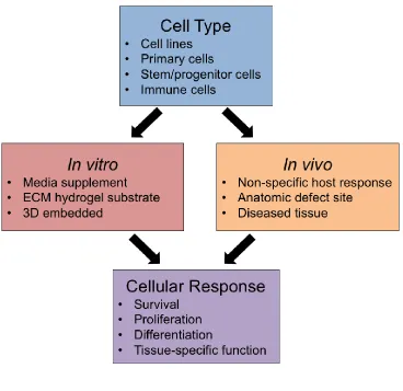
and phenotype. Established methods to evaluate the cellular response to ECM
hydrogels both in vitro and in vivo are summarized in Figure 2.
The viability of cells cultured on the surface of ECM hydrogels in vitro has been
consistently shown for cell lines [23, 54, 63, 64, 70, 71, 83], primary cells [57, 63, 69,
71, 75, 83, 85], and stem cells [44, 49, 50, 73, 82, 86]. In addition, the innate bioactivity
of soluble factors within the ECM has been demonstrated using in vitro culture with
media supplemented with solubilized ECM to remove the influence of hydrogel structure
on the function of cells.
Wolf et al. studied the response of 3T3 fibroblasts and C2C12 myoblast cells to
UBM and dermal ECM hydrogels by three different methods: cells seeded on the
surface of pre-formed gels (ECM hydrogel substrate), cells embedded within gels (3D
embedded), and gel placement in an anatomic defect site in vivo [23]. Almost 100%
viability of 3T3 fibroblasts and C2C12 myoblasts was observed after 7 days of culture
for all configurations investigated in vitro. C2C12 myoblast cells seeded on the surface
of the dermal ECM hydrogels fused into large diameter, multinucleated myotubes with
embedded within dermal ECM formed smaller elongated cell structures. Implantation of
the hydrogels within a rodent partial thickness abdominal wall defect produced a
significantly greater area of de novo muscle formation when the defects were treated
with UBM hydrogel compared to unrepaired defects. This result likely represents the
combination of microstructure, mechanical properties, and bioactivity. The collagen fiber
ultrastructure and low storage modulus of UBM hydrogels allows for cell infiltration and
fibroblast mediated contraction of the gel, two important aspects of wound healing [23].
4.1 Comparison to Collagen and/or Matrigel
Cell behavior in response to ECM hydrogels has consistently been shown to be
comparable to Matrigel and/or collagen substrate for liver [87, 88], skeletal muscle [58],
heart [58] and fat [43-45, 47, 67] applications. Uriel et al. [43] showed that primary rat
pre-adipocytes cultured on the surface of adipose ECM hydrogels (1 mg/mL) formed
colonies that were significantly larger compared to Matrigel (1 mg/mL) after 7 days
indicative of enhanced pre-adipocyte differentiation. Furthermore, the adipose ECM
hydrogels (1 mg/mL) that were formed by reducing pH to 4.0 showed significantly
greater adipose area compared to Matrigel (1 mg/mL) at 1, 3, and 6 weeks in vivo in an
epigastric pedicle model.
5. In Vivo Applications of ECM Hydrogels
Structure-function relationships of ECM hydrogels can provide a basis for predicting
the appropriate hydrogel formulation for given applications. Although in vitro
applications are largely unknown. There have been limited experiments with ECM
hydrogels in two anatomic locations: the heart and the brain.
5.1. Heart
Cardiac-derived gels are being investigated for cardiac reconstruction following
ischemic injury [42, 55, 58, 60, 75-78, 81]. Heterologous ECM hydrogels have been
evaluated in the heart but formed cartilaginous tissue suggesting that tissue-specific
cues may be necessary for appropriate cardiac tissue remodeling [75]. The Christman
laboratory has investigated different cardiac tissue types for cardiac application
including 1) the effect of species (porcine versus human) [60], and 2) the effect of
pericardium versus myocardium [55].
Both porcine and human source tissue has been evaluated for clinical translation.
Porcine cardiac tissue is more homogeneous for variables such as diet, age, and strain
unlike human cadaveric donor heart tissue which involves a range of ages, disease
states, and co-morbidities [60, 76]. Alternatively, a human ECM source tissue has been
cited as mitigating the risk for xenogeneic disease transfer [60], although there has not
been a reported case of zoonotic disease in the millions of patients that have received
porcine ECM scaffolds or porcine tissue (e.g., porcine heart valves) to date [89]. Both
porcine and human myocardial ECM formed similar hydrogel ultrastructure in vivo after
injection into the rat left ventricular myocardium [60]. However, perhaps most
importantly, over half of the human myocardial pre-gel solutions did not form gels even
allowing for the same DNA and lipid content. The differences may be attributed to the
lipid/DNA removal steps) required as a result of the increased ECM crosslinking and
adipose tissue of the human tissue (donor age of human tissue ranged from 41-69
years). Johnson et al. eventually recommended porcine myocardial ECM hydrogel as
the preferred source for clinical translation over human myocardial ECM hydrogel
because of the increased tissue availability, relatively more gentle decellularization
protocol, and more reliable gelation [60]. Human tissue was recommended as a useful
model system for in vitro study of the role of human ECM in cardiac disease.
Two different tissue types within the heart were evaluated for myocardial repair.
The pericardium is the fibrous sac surrounding the heart primarily composed of compact
collagen and elastin fibers. While not tissue specific, the pericardium was explored as a
potentially autologous therapy because the pericardium can be resected from the heart
without adverse effect on heart function and is currently FDA approved for structural
reinforcement in other body applications. The pericardial ECM hydrogel (6.6 mg/mL)
and myocardial ECM hydrogel (6 mg/mL) were evaluated in the non-diseased,
orthotopic location, and injected into the rat LV wall in separate studies. Both pericardial
ECM and myocardial ECM hydrogels supported vascular cell infiltration (endothelial
cells, smooth muscle cells) and almost identical arteriole formation within 2 weeks (51
+/- 42 vessels/mm2, 52+/- 20 arterioles/mm2 respectively) [55, 75]. In conclusion, it was suggested that pericardial ECM may be a candidate for same-patient ECM sourcing [55,
76], but myocardial ECM hydrogel was preferred for pre-clinical studies in the rat and
pig.
Porcine myocardial ECM hydrogel has been evaluated in both small and large
poses unique challenges such as the sustained release of pro-inflammatory cytokines
thought to promote cell apoptosis or necrosis, matrix metalloproteinase (MMP)
production that degrades the matrix, and an ischemic/hypoxic microenvironment.
Myocardial ECM preserved cardiac function in a rat model of MI while the saline treated
rats worsened 4 weeks after injection compared to baseline 1 week prior to injection.
Specifically, myocardial ECM showed an increased ejection fraction (EF) and a
relatively decreased percent change in end-systolic volume (ESV) and end-diastolic
volume (EDV) compared to saline treated control; however, none of the three markers
were significantly different compared to controls [79]. In an established large animal
model, the myocardial ECM was delivered by the clinical standard transendocardial
catheter two weeks after MI. After three months, myocardial ECM treated groups
showed significant improvement in three measures of cardiac function: 1)
echocardiography, 2) global wall motion index scoring, and 3) electromechanical NOGA
mapping [77]. Corroborating the functional improvement, myocardial ECM treated
animals promoted healthy muscle and blood vessel formation in infarcted areas: a
distinct band of muscle that stained positive for troponin T below the endocardium was
present in the myocardial ECM treated groups, and the muscle was significantly larger
than control muscle. The myocardial ECM treated group showed significantly reduced
fibrosis and neovascularization foci below the endocardium compared to controls.
Recently, Wassenaar et al. investigated the molecular mechanisms underlying
the ability of myocardial ECM to mitigate negative LV remodeling using whole
transcriptome analysis in the rat model of MI [81]. This was the first study to determine
compared to saline control after 1 week of treatment showed several significantly
altered pathways at the tissue level including: altered inflammatory response;
decreased cardiomyocyte apoptosis, altered myocardial metabolism, enhanced blood
vessel development, increased cardiac transcription factor expression, and increased
progenitor cell recruitment. Angiogenesis is one of the processes modulated by ECM
hydrogel treatment and a critically important process relevant to other in vivo
applications. Wassenaar et al. speculate the ECM hydrogel may directly recruit
endothelial progenitor cells through pro-angiogenic growth factors or matricryptic
peptides, provide a scaffold for blood vessel formation, or modulate the recruited
macrophages’ secretory profile [81].
5.2. Brain
While the use of homologous ECM has been investigated for cardiac
applications, the use of heterologous ECM, specifically UBM hydrogel, has been
evaluated in brain applications to treat traumatic brain injury (TBI) [24] and stroke [21,
22].
In a rat model of TBI [24], UBM hydrogel (5 mg/mL) was delivered one day after
controlled cortical impact injury. UBM mitigated adverse tissue damage with decreased
lesion volume, decreased white matter injury, and increased vestibulomotor function at
21 days. However, no cognitive improvement was shown by the Morris water maze
task. While the UBM hydrogel showed functional improvement in tissue repair, it has yet
type of clinical application which requires the addition of neural stem cells to the ECM
hydrogel, or other tailoring of ECM hydrogel properties.
ECM concentration-specific properties of UBM hydrogels were also used to
selectively affect the material retention [22] and the immune cell infiltrate [21] in a small
animal model of chronic stroke. Specifically, UBM hydrogel (1-8 mg/mL) was delivered
14 days after middle cerebral artery occlusion in the rat. UBM hydrogels < 3 mg/mL did
not form a gel within the stroke lesion and instead diffused into the surrounding brain
tissue as early as 24 hours, the earliest time point investigated [22]. In a follow-up study,
it was shown that with the use of UBM hydrogels < 3 mg/mL, the cells did not have a
medium through which to infiltrate the lesion and instead accumulated around the lesion
site [21]. UBM hydrogels > 3 mg/mL formed a hydrogel within the stroke cavity that
interfaced with the adjacent tissue [21, 22]. Because a distinct host/tissue interface was
formed, > 3 mg/mL treatment also showed extensive cell infiltration 1 day after delivery
[21]. Macrophages and microglia were accompanied by neural progenitor cells,
endothelial cells, oligodendrocytes, and astrocytes. An understanding of the cell
infiltrate based upon the viscoelastic properties of the hydrogel in the brain is crucial
since these cells will ultimately remodel the ECM and replace it with de novo matrix.
While this application would suggest that the > 3 mg/mL UBM hydrogels would be
preferred, other tissue applications may show improved outcomes if ECM signaling
molecules would be released and permeate the surrounding tissue.
For ECM hydrogels > 3 mg/mL that may be retained within the lesion and allow
for immune cell infiltration, there are several concentration-dependent properties that
hydrogels were tested in vitro as candidates for brain repair after stroke injury. Both 4
and 8 mg/mL hydrogels showed ideal properties of an injectable therapy: viscosities
ranging from that of water to honey (0.084 Pa*s and 0.443 Pa*s respectively), stably
formed gels (G’ > G’’ by ~ 10 fold), and 50% gelation times (~3 min) considered to be a
reasonable time frame in the operating room. The storage moduli or “stiffness” differed
more dramatically for the 4 and 8 mg/mL hydrogel, at 76 and 460 Pa respectively. Brain
tissue storage moduli has been reported between 200-500 Pa as a target moduli range
[22], however it is important to state again the recruited cells will ultimately remodel the
matrix.
5.3. Safety
The in vivo safety of an ECM hydrogel for any clinical application is obviously an
important consideration. ECM hydrogels were considered safe in the aforementioned
heart and brain in vivo applications. The ECM treated MI induced pigs did not show
arrhythmias, thromboembolism or ischemia 3 months after myocardial ECM injection
[77]. Hemocompatibility was further corroborated in vitro when the myocardial ECM gels
were tested at a physiologically relevant concentration and shown not to accelerate
coagulation.
Zhang et al. also showed that the UBM hydrogel (5 mg/mL) did not have a
deleterious effect when injected into the normal brain [24]. There was no reactive
astrocytosis (GFAP+), and no neuronal degeneration at 1, 3, and 7 days after UBM
days along the needle track and injection site, but was no different than PBS control;
and was resolved by 21 days.
The potential unintended presence of ECM hydrogels in peripheral organs was
evaluated in the studies of myocardial injection, and would be a safety concern relevant
to all ECM hydrogel applications. Myocardial ECM hydrogels were not found at 2 hours
in the pig lung, liver, spleen, kidney and brain [79], nor at 3 months [77]. Each clinical
application of ECM hydrogels would likely have a distinctive profile of safety measures.
5.4. In vivo Host Response
The clinical applications of ECM involving the heart and brain did not elicit an
adverse immune response. In general, ECM hydrogels have been well-tolerated in a
wide variety of in vivo applications. No adverse immune response was shown after ECM
hydrogels were injected in the heart [55, 60, 75-81], fat [43, 45, 47, 50, 67], liver [57],
brain [21, 22, 24] skeletal muscle [23, 63, 64, 83], tendon [26, 59, 84], spinal cord [82],
lung [49], cartilage [70], or colon [51, 71], and these studies included both homologous
and heterologous ECM hydrogels. The findings in vivo are consistent with in vitro
studies that have shown the pepsin-digested ECM (“pre-gel”) promotes a regulatory
(“M2-like”) macrophage activation state, which is associated with a constructive
remodeling response in vivo [71, 90, 91]. For example, macrophages activated toward
an M2-like phenotype with solubilized ECM promoted downstream effects such as
stimulating the migration and myogenesis of skeletal muscle progenitor cells [90]. In SIS
hydrogel treatment of ulcerative colitis in vivo, the ECM modulated the macrophage
response towards a predominately regulatory state by decreasing the number of
of M2-like macrophages [71]. This effect of altering the innate immune response by
shifting the M2:M1 ratio is observed in the host response to solid ECM scaffolds as well
[90].
5.5 Summary of In vivo Applications
Heart and brain were selected as two organ systems with a need for a minimally
invasive, injectable therapy. The heart showed safety and efficacy of myocardial ECM
hydrogel in small and large animal model of disease up to 3 months, and is currently
being evaluated in a Phase I clinical trial (ClinicalTrial.gov Identifier: NCT02305602)
[92]. The brain case study showed the importance of investigating multiple ECM
concentrations to determine preferred characteristics of an injectable therapy for central
nervous system (CNS) applications, including delivery, facilitation of the immune cell
infiltrate, and mitigation of the default response to injury. Future work in the brain will
likely identify the balance of factors required for cognitive improvement. Overall, each
new therapeutic application will need a thorough understanding of the ECM hydrogel
structure-function relationships for successful clinical translation. Relevant references to
other organ in vivo applications can be found in Figure 1.
6. Future Perspectives
With more than 70 papers published in the last decade it is evident that the
therapeutic potential of ECM hydrogels is recognized. Characterization of hydrogel
tissue and hydrogel formulation in selected body systems. However, the relationship
between in vitro structure-function and in vivo application is still largely unknown for
most other clinical applications.
The mechanisms by which ECM hydrogels mediate cell behavior are not fully
understood. Several hypotheses have been suggested including the possibility that the
architecture of the gelled hydrogel comprises a pore size and fiber diameter suitable for
endogenous cell infiltration [93]. Additionally, the bioinductive hydrogel provides
tissue-specific cues, likely through the release of bound growth factors [27], or the creation of
cryptic peptides or the exposure of bioactive motifs [29-32]. The recent report of
bioactive matrix-bound nanovesicles within biologic scaffolds [33] provides a new
possibility for study to determine the mechanisms contributing to the constructive tissue
remodeling facilitated by ECM hydrogels.
The use of ECM hydrogels as a delivery vehicle is an obvious area for future study.
Although a standalone ECM biomaterial therapy offers practical advantages by way of
reduced regulatory concerns, ease of manufacturing and route to market, combinations
of ECM hydrogels with growth factors and/or cells may provide significant mutual
enhancement. Recent studies have shown that sulfated GAGs within ECM hydrogels
bind to growth factors with prolonged release of basic fibroblast growth factor and
heparin-binding growth factor that enhances therapeutic effects [78, 94]. ECM hydrogels
have also been used as a delivery system for growth factor containing microparticles to
enhance skeletal tissue repair within an ex vivo chick femur defect model [95]. Cell
therapy for neurological conditions may require integration with an appropriate
system post implantation. Recent investigations of ECM hydrogels for CNS applications
have included the assessment of different source tissues to direct cell differentiation [96]
and the transplantation of human neural stem cells embedded within ECM hydrogels to
support the creation of de novo tissue [25]. Stem cells and primary cells have also been
embedded within lung [49], liver [57], spinal cord [82], and adipose [50] ECM hydrogels
to improve the tissue remodeling outcome.
In conclusion, the use of ECM hydrogels for a variety of clinical applications is in
its infancy, but has shown promise. The combination of in vitro and in vivo studies
designed to understand mechanical and material properties, the effects of processing
methods upon hydrogel performance, the mechanisms by which such hydrogels
influence cell behavior and tissue remodeling, and the safety of ECM hydrogels should
advance their clinical utility.
Acknowledgements
Funding Sources
LTS was supported by the National Institute of Biomedical Imaging and Bioengineering
of the National Institutes of Health (2T32 EB001026-11). LJW was funded by a Marie
Curie International Outgoing Fellowship under REA grant agreement no. 624841.
Tables & Figures
Source Tissue Decellularization Reagents Solubilization
Protocol Ref.
Adipose
Human (Lipoaspirate)
1% SDS, or 2.5 mM sodium
deoxycholate
2.5 mM sodium deoxycholate with 500
U lipase and 500 U colipase
Freytes
3200 IU pepsin
0.1 M HCl
[67]
0.5% SDS
Isopropanol
0.1% peracetic acid/4% ethanol
Voytik-Harbin
10 mg pepsin
RT, 48 hr
[73]
Rat
(Subcutaneous) 2 mL dispase/ g tissue Uriel
[43, 44, 47]
Porcine
10 mM Tris and 5 mM EDTA
99% isopropanol
HBSS with 10000 U DNase, 12.5 mg
RNase, 1000 U lipase
Freytes
37oC, 24 hr
[97]
Bone
Bovine (Cancellous Tibia)
0.5 M HCl
1:1 Chloroform:methanol
0.05% trypsin/0.02% EDTA
1% w/v pen/strep in PBS
Freytes
96 hr
[72, 86, 98] Cartilage Porcine (Articular)
10 mM Tris-HCl at pH 8
0.25% trypsin
1.5 M NaCl in 50 mM Tris-HCl at pH 7.6
50 U/mL DNase and 1 U/mL RNase in
10 mM Tris-HCl
1% Triton X-100
10 mM Tris-HCl
0.1% peracetic acid/4% ethanol
Voytik-Harbin
10 mg pepsin
RT, 48 hr
[73]
Porcine (Meniscus)
1% SDS
0.1% EDTA
Freytes
1.5 mg/mL pepsin
[70]
Central Nervous System
Porcine (Adult Brain, Spinal
Cord)
0.02% trypsin/0.05% EDTA
3% Triton X-100
1 M sucrose
4% deoxycholate
0.1% peracetic acid/4% ethanol
Freytes
Porcine (Fetal Brain)
0.05% trypsin-EDTA with 0.2% DNase I
3% Triton X-100 with 0.2% DNase I
1 M sucrose
1% sodium deoxycholate
0.2% peracetic acid in 4% ethanol
Freytes
24 hr
[100]
Colon
Porcine (Submucosa)
2:1 Chloroform:methanol
Graded ethanol (100%, 90%, 70%)
0.02% trypsin/0.05% EDTA
4% sodium deoxycholate
0.1% peracetic acid/4% ethanol
Freytes
0.1 M HCl
[71]
Cornea
Porcine 10 U/ml DNAse and 10 U/mL RNAse in
10 nM MgCL2
Freytes
0.1 M HCl
72 hr
[69]
Esophagus
Porcine (Mucosa/submucosa)
1% trypsin/0.05% EDTA
1 M sucrose
3% Triton X-100
10% deoxycholate
0.1% peracetic acid/4% ethanol
Freytes
[101]
Heart
Porcine, Rat (Ventricular Myocardium)
1% SDS
1% Triton X-100
Freytes
0.1 M HCl
[58, 74, 75, 77, 79, 81, 102] Porcine (Ventricular Myocardium)
1% SDS and 0.5% pen/strep Freytes
0.1 M HCl
[42, 60, 80, 103-106]
1% SDS
1% Triton X-100
0.1% peracetic acid/4% ethanol
Voytik-Harbin
10 mg pepsin
RT, 48 hr
[73, 107]
0.02% trypsin-EDTA
3% Tween-20
102 mM sodium deoxycholate Freytes
1% pen/strep
1% SDS
0.1% Triton X-100
Freytes
0.1 M HCl
12 hr
[109]
Perfusion
0.02% trypsin/0.05% EDTA
3% Triton X-100/0.05% EDTA
4% deoxycholic acid
0.1% peracetic acid
2:1 chloroform:methanol
100-70% ethanol
Freytes
2 mg/mL pepsin
[110]
Human (Ventricular Myocardium)
1% SDS and 0.5% pen/strep
Isopropyl alcohol
40 U/mL DNase and 1 U/mL RNase in
40 mM HCl, 6 mM MgCl2, 1 mM CaCl2, and 10 mM NaCl
1% SDS/0.5% pen/strep
0.001% Triton X-100
Freytes
[60]
10 mM Tris and 0.1% EDTA
0.5% SDS
100 U/mL pen/strep and nystatin in
DPBS
Fetal bovine serum
100 U/mL pen/strep and nystatin in
DPBS
Freytes
pH 1
37oC
Salts were not
neutralized
[111]
10 mM Tris and 0.1% EDTA
0.5% SDS
100 U/mL pen/strep and nystatin in
DPBS
Fetal bovine serum
100 U/mL pen/strep and nystatin in
DPBS
Freytes
pH 2
[111]
Goat (Ventricle)
0.1% peroxyacetic acid/4% ethanol
1% SDS
1% Triton X-100
Freytes
60-72 hr
[112]
Porcine, Human
(Pericardium) 1% SDS
Freytes
0.1 M HCl
[55, 76, 78, 94, 113] Kidney Human
(Cortex) 1% SDS Freytes
[61]
1% Triton X-100 and 0.1% ammonium hydroxide
10% (w/w) pepsin
0.1 M HCl
Rat, Porcine, Canine, Human
0.02% trypsin and 0.05% EGTA
3% Triton X-100
0.1% peracetic acid
Freytes
24-72 hr (until no
particulate)
[56]
Porcine
0.02% trypsin and 0.05% EDTA
3% Triton X-100
4% sodium deoxycholic acid
0.1% peracetic acid
Freytes
72 hr
[87, 88]
0.1% SDS
Freytes
3 mg/mL pepsin
0.1 M HCl
72 hr
[52]
Lung
Porcine
Perfusion
1x pen/strep
0.1% Triton X-100
2% sodium deoxycholate
DNase solution
NaCl
Freytes
[49]
Pancreas
Porcine
1.1% NaCl
0.7% NaCl
0.05% trypsin/0.02% EDTA, pH 8.2
1% Triton X-100/1% ammonium
hydroxide
70% ethanol
Freytes
5 mg/mL pepsin
0.1 M HCl
[62]
Skeletal Muscle
Porcine
(Intercostal, Hindleg) 1% SDS
Freytes
0.1 M HCl
[58, 83]
Porcine (Psoas)
1% SDS
1% SDS and 0.5% pen/strep
Isopropyl alcohol
Freytes
0.1 M HCl
[104]
1% SDS and 0.5% pen/strep
Isopropyl alcohol
0.001% Triton X-100
Freytes
0.1 M HCl
[63]
Porcine
0.2% trypsin/0.1% EDTA
0.5% Triton X-100
1% Triton X-100/ 0.2% sodium
deoxycholate
Isopropanol
5x10⁷ U/l DNase-I and 1x10⁶ U/l RNase
Freytes
Skin
Rat
(Dermis) 2 mL dispase/ g tissue Uriel
[43-46, 65]
Porcine (Dermis)
0.25% trypsin
70% ethanol
3% H2O2
1% Triton X-100 in 0.26% EDTA/0.69%
Tris
0.1% peracetic acid/4% ethanol
Freytes
72 hr
[23, 114-116] Small Intestine Porcine (Submucosa/muscularis mucosa/stratum compactum/lamina propria)
Mechanical delamination of other tissue layers only
Voytik-Harbin
Additional step:
centrifuged, dialyzed against 0.01 M acetic acid
[16, 34] Porcine (Submucosa/muscularis mucosa/stratum compactum)
0.1% peracetic acid/4% ethanol Freytes
72 hr
[26, 56, 90, 101]
0.1% peracetic acid/4% ethanol Freytes
0.5 mg/mL pepsin [53]
0.1% peracetic acid/4% ethanol Freytes
24 hr [51] Tendon Human (Flexor digitorum profudus, flexor digitorum superficialis,
flexor pollicic longus)
0.1% EDTA
0.1% SDS in 0.1% EDTA
Freytes
0.02 M HCl
24 hr
[59, 84]
Tooth
Human
(Dentin)
10% HCl
0.5% pen/strep
0.5M HCl
0.05% trypsin/0.025% EDTA
Freytes
84 hr
Human
1% SDS and 0.5% pen/step
0.001% Triton X-100
40 U/mL DNase and 1 U/mL RNase in
10 mM NaCl, 1 mM CaCl2 6 mM MgCl2, and 40 mM HCl
1% SDS and 0.5% pen/strep
0.001% Triton X-100
Freytes
0.1 M HCl
[63]
Urinary Bladder
Porcine (Basement membrane/lamina
propria)
0.1% peracetic acid/4% ethanol
Freytes
[20-24, 54, 56, 82, 91, 93, 99, 101, 116] Freytes:
1 mg/mL pepsin in 0.01 M HCl
Stir plate, RT, 48 hr
Neutralized to pH 7.4 and physiological salt with NaOH and 10x PBS
Uriel:
High salt buffer solution (0.05 M Tris pH 7.4, 3.4 M sodium chloride, 4 mM of
ethylenediaminete- traacetic acid, and 2 mM of N-ethylmaleimide) containing protease inhibitors (0.001mg/mL pepstatin, 0.01mg/mL aprotonin, 0.001mg/mL leupeptin, 2mM sodium orthova- nadate, and 1mM phenylmethylsulfonyl fluoride)
Homogenized with mortar and pestle
2 M urea buffer
Voytik-Harbin:
2 mg pepsin per 100 mg ECM in 0.5 M acetic acid
4C, 72 hr
Neutralized to pH 7.4 and physiological salt with NaOH and 10x PBS
Key
Table 2. Viscoelastic properties of porcine-derived ECM hydrogels. Italicized values
were estimated from representative images. Steady shear viscosities refer to the pre-gel
solution. “Pre-formed” indicates that gelation was induced in an incubator at 37oC prior
to rheologic testing. * indicates time to 50% gelation. “NR” indicates “not recorded.”
Tissue Conc. (mg/mL)
Protocol
(strain, frequency) G’ (Pa)
Steady Shear Viscosity
(Pa*s)
Gelation time (min)
Ref
Cartilage 30 2%, 1 rad/s 4000 3 [73]
Brain
4 5%, 1 rad/s 20.3 34.8 [54]
6 5%, 1 rad/s 49.9 2.4 [54]
8 5%, 1 rad/s 61.8 8.3 [54]
Colon 4 0.5%, 1 rad/s 9 0.75 [71]
8 0.5%, 1 rad/s 50 1.7 [71]
Heart
6
2.5%, 0.4 rad/s 11.3 Pre-formed [80]
2.5%, 1 rad/s 6.5 Pre-formed [78]
NR, 1 rad/s, 5.28 Pre-formed [42]
NR, 6.28 rad/s 6.08 Pre-formed [60]
8 2.5%, 0.5 rad/s 5.3 Pre-formed [74]
NR, 1 rad/s, 9.52 Pre-formed [42]
30 2%, 1 rad/s 800 33 [73]
Liver 8 0.5%, 1 rad/s 630 4.25 8.5 [56]
Lung
4 0.5%, 6.28 rad/s 15.3 [49]
6 0.5%, 6.28 rad/s 32.0 [49]
8 0.5%, 6.28 rad/s 59.0 [49]
Pancreas 16.7 2.5%, 1 rad/s 190 4.5 [62]
Skeletal
Muscle 6 NR, 1 rad/s 6.5 Pre-formed [83]
Skin
4 0.5%, 1 rad/s 110 2 [23]
6 0.5%, 1 rad/s 200 2 [23]
8 0.5%, 1 rad/s 466 7 [23]
Spinal cord
4 5%, 1 rad/s 138 11.7 [54]
6 5%, 1 rad/s 235 7 [54]
8 0.5%, 1 rad/s 757 28.9 [54]
Urinary Bladder
3 5%, 1 rad/s 6 10 [20]
76.6 0.084 3.2* [22]
5%, 1 rad/s 11.4 52.5 [54]
6
0.5%, 1 rad/s 40 0.9 [23]
5%, 1 rad/s 26 10 [20]
72.8 8.47 [54]
8 0.5%, 1 rad/s
182 0.9 [23]
460 0.443 3.0* [22]
Characterization Hydrogel Application
Biochemical Topological Mechanical References
Adipose [43, 44, 47, 67, 73, 97]
Bone [72, 86, 98]
Cartilage [70, 73]
CNS [54, 82, 91, 99, 100]
Colon [71]
Cornea [69]
Esophagus [101]
Heart
[42, 55, 58, 60, 73-81, 94, 102-113]
Kidney [61]
Liver [52, 56, 57, 87, 88]
Lung [49]
Pancreas [62]
Skeletal Muscle [58, 63, 64, 83, 104]
Skin [23, 43-46, 65, 114-116]
Small Intestine [16, 26, 34, 51, 53, 56, 90, 101]
Tendon [59, 84]
Umbilical Cord [63]
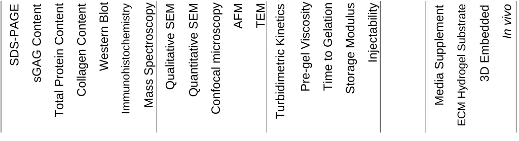
Figure 1. Overview of techniques used to characterize and to evaluate the cellular response to ECM hydrogels thus far.
ECM hydrogels derived from various species, concentrations and processing methods are categorized only by source
Figure 2. General approaches to assess cellular response to ECM hydrogels. The
response of various cell types in vitro or in vivo can be evaluated
REFERENCES
[2] Choi JS, Kim BS, Kim JD, Choi YC, Lee HY, Cho YW. In vitro cartilage tissue engineering using adipose-derived extracellular matrix scaffolds seeded with adipose-derived stem cells. Tissue Engineering Part A 2012;18:80-92.
[3] Mercuri JJ, Gill SS, Simionescu DT. Novel tissue-derived biomimetic scaffold for regenerating the human nucleus pulposus. J Biomed Mater Res A 2011;96:422-35.
[4] Mercuri JJ, Patnaik S, Dion G, Gill SS, Liao J, Simionescu DT. Regenerative potential of decellularized porcine nucleus pulposus hydrogel scaffolds: stem cell differentiation, matrix remodeling, and biocompatibility studies. Tissue Eng Part A 2013;19:952-66.
[5] Beck EC, Barragan M, Libeer TB, Kieweg SL, Converse GL, Hopkins RA, Berkland CJ, Detamore MS. Chondroinduction from Naturally Derived Cartilage Matrix: A Comparison Between
Devitalized and Decellularized Cartilage Encapsulated in Hydrogel Pastes. Tissue Eng Part A 2016;22:665-79.
[6] Beck EC, Barragan M, Tadros MH, Kiyotake EA, Acosta FM, Kieweg SL, Detamore MS. Chondroinductive Hydrogel Pastes Composed of Naturally Derived Devitalized Cartilage. Ann Biomed Eng 2016;44:1863-80.
[7] Bissell MJ, Hall HG, Parry G. How does the extracellular matrix direct gene expression? J Theor Biol 1982;99:31-68.
[8] Hynes RO. The evolution of metazoan extracellular matrix. J Cell Biol 2012;196:671-9. [9] Tibbitt MW, Anseth KS. Hydrogels as extracellular matrix mimics for 3D cell culture. Biotechnol Bioeng 2009;103:655-63.
[10] Elliott RA, Jr., Hoehn JG. Use of commercial porcine skin for wound dressings. Plast Reconstr Surg 1973;52:401-5.
[11] Badylak SF, Lantz GC, Coffey A, Geddes LA. Small intestinal submucosa as a large diameter vascular graft in the dog. J Surg Res 1989;47:74-80.
[12] Lantz GC, Badylak SF, Coffey AC, Geddes LA, Blevins WE. Small intestinal submucosa as a small-diameter arterial graft in the dog. J Invest Surg 1990;3:217-27.
[13] Lantz GC, Badylak SF, Coffey AC, Geddes LA, Sandusky GE. Small intestinal submucosa as a superior vena cava graft in the dog. J Surg Res 1992;53:175-81.
[14] Sandusky GE, Jr., Badylak SF, Morff RJ, Johnson WD, Lantz G. Histologic findings after in vivo placement of small intestine submucosal vascular grafts and saphenous vein grafts in the carotid artery in dogs. Am J Pathol 1992;140:317-24.
[15] Lantz GC, Badylak SF, Hiles MC, Coffey AC, Geddes LA, Kokini K, Sandusky GE, Morff RJ. Small intestinal submucosa as a vascular graft: a review. J Invest Surg 1993;6:297-310. [16] Voytik-Harbin SL, A.O. Brightman. Small intestinal submucosa: A tissue derived
extracellular matrix that promotes tissue-specific growth and differentiation of cells in vitro. Tissue Eng 1998;4:157-74.
[17] Badylak SF, Freytes DO, Gilbert TW. Extracellular matrix as a biological scaffold material: Structure and function. Acta Biomater 2009;5:1-13.
[18] Zantop T, Gilbert TW, Yoder MC, Badylak SF. Extracellular matrix scaffolds are repopulated by bone marrow-derived cells in a mouse model of Achilles tendon reconstruction. J Orthop Res 2006;24:1299-309.
[19] Gilbert TW, Stolz DB, Biancaniello F, Simmons-Byrd A, Badylak SF. Production and
[20] Freytes DO, Martin J, Velankar SS, Lee AS, Badylak SF. Preparation and rheological
characterization of a gel form of the porcine urinary bladder matrix. Biomaterials 2008;29:1630-7.
[21] Ghuman H, Massensini AR, Donnelly J, Kim SM, Medberry CJ, Badylak SF, Modo M. ECM hydrogel for the treatment of stroke: Characterization of the host cell infiltrate. Biomaterials 2016;91:166-81.
[22] Massensini AR, Ghuman H, Saldin LT, Medberry CJ, Keane TJ, Nicholls FJ, Velankar SS, Badylak SF, Modo M. Concentration-dependent rheological properties of ECM hydrogel for intracerebral delivery to a stroke cavity. Acta Biomater 2015;27:116-30.
[23] Wolf MT, Daly KA, Brennan-Pierce EP, Johnson SA, Carruthers CA, D'Amore A, Nagarkar SP, Velankar SS, Badylak SF. A hydrogel derived from decellularized dermal extracellular matrix. Biomaterials 2012;33:7028-38.
[24] Zhang L, Zhang F, Weng Z, Brown BN, Yan H, Ma XM, Vosler PS, Badylak SF, Dixon CE, Cui XT, Chen J. Effect of an inductive hydrogel composed of urinary bladder matrix upon functional recovery following traumatic brain injury. Tissue Eng Part A 2013;19:1909-18.
[25] Bible E, Dell'Acqua F, Solanky B, Balducci A, Crapo PM, Badylak SF, Ahrens ET, Modo M. Non-invasive imaging of transplanted human neural stem cells and ECM scaffold remodeling in the stroke-damaged rat brain by (19)F- and diffusion-MRI. Biomaterials 2012;33:2858-71. [26] Fisher MB, Liang R, Jung HJ, Kim KE, Zamarra G, Almarza AJ, McMahon PJ, Woo SL. Potential of healing a transected anterior cruciate ligament with genetically modified extracellular matrix bioscaffolds in a goat model. Knee Surg Sports Traumatol Arthrosc 2012;20:1357-65.
[27] Badylak SE. The extracellular matrix as a scaffold for tissue reconstruction. Seminars in Cell & Developmental Biology 2002;13:377-83.
[28] Londono R, Badylak SF. Biologic scaffolds for regenerative medicine: mechanisms of in vivo remodeling. Ann Biomed Eng 2015;43:577-92.
[29] Sarikaya A, Record R, Wu CC, Tullius B, Badylak S, Ladisch M. Antimicrobial activity associated with extracellular matrices. Tissue Eng 2002;8:63-71.
[30] Brennan EP, Reing J, Chew D, Myers-Irvin JM, Young EJ, Badylak SF. Antibacterial activity within degradation products of biological scaffolds composed of extracellular matrix. Tissue Eng 2006;12:2949-55.
[31] Agrawal V, Tottey S, Johnson SA, Freund JM, Siu BF, Badylak SF. Recruitment of progenitor cells by an extracellular matrix cryptic peptide in a mouse model of digit amputation. Tissue Eng Part A 2011;17:2435-43.
[32] Reing JE, Zhang L, Myers-Irvin J, Cordero KE, Freytes DO, Heber-Katz E, Bedelbaeva K, McIntosh D, Dewilde A, Braunhut SJ, Badylak SF. Degradation products of extracellular matrix affect cell migration and proliferation. Tissue Eng Part A 2009;15:605-14.
[33] Huleihel L, Hussey GS, Naranjo JD, Zhang L, Dziki JL, Turner NJ, Stolz DB, Badylak SF. Matrix-bound nanovesicles within ECM bioscaffolds. Sci Adv 2016;2:e1600502.
[34] Brightman AO, Rajwa BP, Sturgis JE, McCallister ME, Robinson JP, Voytik-Harbin SL. Time-lapse confocal reflection microscopy of collagen fibrillogenesis and extracellular matrix assembly in vitro. Biopolymers 2000;54:222-34.
[36] Keane TJ, Londono R, Turner NJ, Badylak SF. Consequences of ineffective decellularization of biologic scaffolds on the host response. Biomaterials 2012;33:1771-81.
[37] Drake MP, Davison PF, Bump S, Schmitt FO. Action of proteolytic enzymes on tropocollagen and insoluble collagen. Biochemistry 1966;5:301-12.
[38] Miller EJ. Structural studies on cartilage collagen employing limited cleavage and solubilization with pepsin. Biochemistry 1972;11:4903-9.
[39] Hulmes DJS. Collagen Diversity, Synthesis, and Assembly: Springer; 2008.
[40] Agrawal V, Kelly J, Tottey S, Daly KA, Johnson SA, Siu BF, Reing J, Badylak SF. An isolated cryptic peptide influences osteogenesis and bone remodeling in an adult mammalian model of digit amputation. Tissue Eng Part A 2011;17:3033-44.
[41] Parkinson J, Kadler KE, Brass A. Simple physical model of collagen fibrillogenesis based on diffusion limited aggregation. Journal of molecular biology 1995;247:823-31.
[42] Johnson TD, Lin SY, Christman KL. Tailoring material properties of a nanofibrous extracellular matrix derived hydrogel. Nanotechnology 2011;22:494015.
[43] Uriel S, Huang JJ, Moya ML, Francis ME, Wang R, Chang SY, Cheng MH, Brey EM. The role of adipose protein derived hydrogels in adipogenesis. Biomaterials 2008;29:3712-9.
[44] Uriel S, Labay E, Francis-Sedlak M, Moya ML, Weichselbaum R, Ervin N, Cankova Z, Brey EM. Extraction and Assembly of Tissue-Derived Gels for Cell Culture and Tissue Engineering. Tissue Eng Part C 2009;15.
[45] Cheng M-H, Uriel S, Moya ML, Francis-Sedlak M, Wang R, Huang J-J, Chang S-Y, Brey EM. Dermis-derived hydrogels support adipogenesis in vivo. Journal of Biomedical Materials Research Part A 2010;92A:852-8.
[46] Pilipchuk SP, Vaicik MK, Larson JC, Gazyakan E, Cheng M-H, Brey EM. Influence of
crosslinking on the stiffness and degradation of dermis-derived hydrogels. Journal of Biomedical Materials Research Part A 2013;101:2883-95.
[47] Poon CJ, Pereira E. Cotta MV, Sinha S, Palmer JA, Woods AA, Morrison WA, Abberton KM. Preparation of an adipogenic hydrogel from subcutaneous adipose tissue. Acta Biomaterialia 2013;9:5609-20.
[48] Kadler KE, Hill A, Canty-Laird EG. Collagen fibrillogenesis: fibronectin, integrins, and minor collagens as organizers and nucleators. Curr Opin Cell Biol 2008;20:495-501.
[49] Pouliot RA, Link PA, Mikhaiel NS, Schneck MB, Valentine MS, Kamga Gninzeko FJ, Herbert JA, Sakagami M, Heise RL. Development and characterization of a naturally derived lung extracellular matrix hydrogel. J Biomed Mater Res A 2016;104:1922-35.
[50] Kim EJ, Choi JS, Kim JS, Choi YC, Cho YW. Injectable and Thermosensitive Soluble Extracellular Matrix and Methylcellulose Hydrogels for Stem Cell Delivery in Skin Wounds. Biomacromolecules 2016;17:4-11.
[51] Keane TJ, Dziki J, Sobieski E, Smoulder A, Castleton A, Turner NJ, White LJ, Badylak S.