RESEARCH ARTICLE
EFFECT ON UNTREATED DIABETIC MACULAR EDEMA: POSTERIOR SUB-TENON
TRIAMCINOLONE VERSUS INTRAVITREAL BEVACIZUMAB
*Dr. Murali Mohan Gurram
Department of Ophthalmology, Kamineni Hospitals, Kamineni Academy of Medical Sciences and Research
Centre, L.B.Nagar, Hyderabad 500068, India
ARTICLE INFO ABSTRACT
Aim: To compare the anatomical and visual effects of posterior sub-tenon triamcinolone (PSTT) and intra-vitreal bevacizumab (IVB) when used as the primary treatment for diabetic macular edema (DME).
Methods: In a retrospective comparative study, 58 eyes of 47 patients which have received either PSTT or IVB were analyzed. Twenty-six eyes had received PSTT 40mg/1mL of preservative free Triamcinolone-acetonide (Group I) and 32 eyes had received intravitreal injection of 1.25mg/0.05 mL of avastin (Group II). Best corrected visual acuity (BCVA) and central macular thickness prior and six weeks post procedure were studied. Complications were studied.
Results: In group I, BCVA increased from 31.04±9.89 letters to 41.08±6.77 letters with an increase of 10.04±9.34
letters (P<0.001). The mean central macular thickness (CMT) in group I changed from 571.42±125.71µ to
274.73±116.76µ with a decrease of 296.7±182.34µ (P<0.001). In group II BCVA increased from 35.03±8.39
letters to 54.44±10.56 letters by 19.41±12.51 letters (P<0.001) and mean CMT decreased from 618.91±143.76µ to
216.56±76.14 with a change of 402.34±155.91µ (P<0.001). IVB group had superior effect both on BCVA and
CMT which was significant statistically too. P values for change in mean BCVA and change in CMT were 0.003 and 0.021. Two eyes in group I and 6 eyes in group II had subconjunctival hemorrhage. One eye in either group had raised intra ocular pressure (IOP) which was controlled medically. No major complications were noted in either group.
Conclusions: In treatment of DME, both PSTT and IVB are effective in increasing the BCVA and decreasing the CMT. IVB is superior to PSTT. IVB is costly and may be associated with serious complications, as it’s an intraocular procedure. So PSTT can be tried as a first line of management in cases of untreated DME. Long term studies with multiple injections are required to know the long term effect and complications.
Copyright © 2013 Dr. Murali Mohan Gurram,This is an open access article distributed under the Creative Commons Attribution License, which permits
unrestricted use, distribution, and reproduction in any medium, provided the original work is properly cited.
INTRODUCTION
Diabetes is the most common systemic disorder affecting retina and is a major cause of avoidable blindness. The prevalence of diabetic
retinopathy (DR) among diabetics was 34.6% as shown by a study[1].
A study from India has shown the prevalence of DR in as 28.2%[2].
Diabetic maculopathy is the most common cause for central defective
vision in diabetic retinopathy[3,4].Diabetic macular edema (DME) is
basically due to disruption of blood retinal barrier (BRB), which leads to accumulation of fluid and blood macromolecules in the retinal interstitial spaces. The exact pathogenesis of disruption of BRB is not known, but it is multifactorial with reasons including pericyte loss, endothelial cell loss, basement membrane thickening and capillary
occlusion[5,6,7]. Based on OCT features, the diabetic maculopathy can
be categorized into 5 groups which include diffuse retinal thickening (DRT), cystoid macular edema (CME), serous retinal detachment
(SRD), taut posterior hyaloid and traction retinal detachment[8].
Surgery is indicated in types 4 and 5. Many treatment modalities have been advocated for DME like macular laser photocoagulation,
intravitreal medications and vitrectomy[9-12]. Early treatment diabetic
retinopathy study (ETDRS) showed that focal grid-laser can reduce the risk of visual loss, but has few complications associated with it like scar formation which tends to increase in size. It also has limited effect in improving the visual effect, with only 17% of treated eyes
*Corresponding author:Murali Mohan Gurram, Department of
Ophthalmology, Kamineni Hospitals, L.B.Nagar, Hyderabad 500068, India
showing 3 lines or more of visual acuity improvement. It also has a
limited effect in diffuse DME[9]. Triamcinolone acetonide is a
commonly used corticosteroid which, apart from having anti-inflammatory effects, also causes down-regulation of vascular
endothelial growth factor (VEGF)[11]. ME was treated successfully
with intravitreal triamcinolone and subtenon triamcinolone[12].Due to
its intraocular nature, and the action of compound, Intravitreal
Triamcinolone may be associated with various complications like glaucoma, cataract, endophthalmitis, retinal detachment, scleritis
etc[13,14].
Recently some researches have revealed the role of VEGF in inducing vascular hyperpermeability and thus macular edema. Severity of macular edema is correlated with the level of VEGF in vitreous in
DME patients[15,16].Since then, anti-VEGF agents have been used in
DME. Bevacizumab which is humanized full-length monoclonal antibody, that inhibits all isoforms of VEGF, has been used for DME with good results in improvement of visual acuity and reduction of
macular edema[17].Ranibizumab and Pegaptanib are other anti-VEGF
molecules which are used. All these have similar efficacies[18,19].
Bevacizumab is most commonly used anti-VEGF agent due to its low cost. The operation theatre setup and the cost involved are the limiting factors of Intravitreal anti-VEGF agents. Due to its intraocular nature, it may be associated with severe complications
including endophthalmitis; retinal detachment etc[20]. Intra-ocular
anti-VEGF can also be associated with systemic complications[20].
Posterior subtenon space is the potential space beneath tenon’s
ISSN: 0975-833X
International Journal of Current Research
Vol. 5, Issue, 08, pp.2318-2322, August,2013
INTERNATIONAL JOURNAL
OF CURRENT RESEARCH
Article History:
Received 04th May, 2013
Received in revised form
26th June, 2013
Accepted 13th July, 2013
Published online 23rd August, 2013
Key words:
Macular edema, Avastin, Bevacizumab,
capsule behind the macular region. The steroid deposited in that area would transfuse through sclera and act on the macular edema. PSTT
acts with good results in DME[21]. PSTT is a cheap and simple OPD
procedure with no major complications. But is it as effective as IVB? Can it be used as an alternative to anti-VEGF agents? are the queries studied in this retrospective analysis. This study is aimed to compare the anatomical and visual effects of PSTT and that of IVB in untreated DME. To the best of our knowledge, this is the first ever study to do so.
MATERIAL AND METHODS
This is a retrospective comparative study done in ophthalmology department of a Tertiary Care hospital in Hyderabad, south India. All the records of macular edema associated with diabetic retinopathy, which received PSTT or IVB as first line of treatment, between March 2010 to September 2012, were analyzed. Procedures followed were in accordance with the Declaration of Helsinki. Inclusion
criteria for the treatment werediabetic macular edema as evidenced
by clinical and angiographic evaluation, decreased vision of duration less than three months, with vision less than 60 letters on ETDRS chart, CMT of >250µ, Type II diabetes and those who have records of one month followup. Criteria to exclude the cases from this study were: prior laser treatment, cataract which precludes the evaluation of macula, Vitreous hemorrhage, High risk PDR with membranes over macula, types 4 & 5 macular edema with VitreoMacular Traction (VMT), other retinal disorders like Vein occlusion, Ischemic heart disease, renal insufficiency, macular ischemia, iris neovascularization prior intravitreal injections, glaucoma and ocular hypertension. Other causes of macular edema were ruled out. All macular edema patients in the hospital would be first adviced the treatment with Intravitreal Avastin. Patients unwilling for the above would be given PSTT injection. Reasons for unwilling are cost, operation theater procedure, and the complications involved with intravitreal injections.
Materials
Medical records of all cases that fulfilled the above criteria were included in the analysis. 58 eyes of 47 patients were studied. All these patients had undergone basic pre-procedure eye examination including BCVA with 4 meter ETDRS chart (4m ETDRS chart model no.2121, Akriti Logistics) with 70 letters in 14 lines, thorough slit lamp examination, goldman applanation tonometry, contact lens biomicroscopy, indirect ophthalmoscopy, FFA, and OCT (Time domain OCT, Zeiss Stratus OCT). Demographics of the patients are given in Table 1.
Methods
All procedures were done by a single surgeon, the author. Intravitreal bevacizumab: all injections were given in operation theatre for its sterile nature. Eyes were painted and draped after instillation of Povidone Iodine drops. After placement of eye speculum, a point is selected in inferotemporal quadrant 3.5mm away from limbus in case of pseudophakics and aphakics and 4mm away from limbus in phakic
eyes. Conjunctiva is displaced and injection is given in a tunneled
incision technique[22].1.25mg of Avastin in 0.05 mL is used from a
multidose vial. (Avastin; Genentech Inc., California, USA). The
technique of PSTT was as described by Nozik[23]. All subtenon
injections were given as out-patient procedures. Patient was made to lie down comfortably on the treatment couch. Proparacaine 0.5% eye drops were instilled twice with 5min interval to anesthetize the eye. First drop is instilled in inferior cul-de-sac and the second drop is instilled over the superotemporal quadrant, after asking the patient to look inferonasally. 2mL syringe is loaded with 1mL (40mg) of preservative free triamcinolone acetonide (Aurocort, Aurolabs, India). Needle is replaced with a 26G half inch needle. Surgeon stands on the
opposite side e.g. he stands on right side of patient, for left eye
injection. Patient stares at his/her opposite shoulder (Inferonasal gaze). With left hand, surgeon retracts the upper lid upwards, exposing the superotemporal quadrant. Needle was passed through the bulbar conjunctiva and tenon’s capsule, at the posterior most visible area, with bevel facing towards globe. Needle is advanced keeping the needle as close to the globe as possible. Side to side movements of the needle was made and limbus is looked for any movement. Any movement of the limbus indicates the presence of needle in sclera. Needle was advanced till the hub is reached over the injection site. Aspiration was done to rule out any entry into blood vessel, and then the drug was injected with moderate force Post operatively all patients received oral acetazolamide and NSAID and topical medications which include Steroid-Antibiotic combination for five days and anti-glaucoma medications, usually timolol 0.5% for one month. Acetazolamide 250mg is given thrice daily for one day. NSAID is given for 2 days. Data from records was collected so that
the results after one month of procedure could be analyzed.
Statistical Analysis
Statistical Analysis was made with SPSS software (SPSS for Windows, version 13.0, SPSS Inc., Chicago, Illinois, USA). For the effect on BCVA and CMT in each group, Paired sample statistics was done with 95% confidence interval.
RESULTS
On 1st post op day, 5 patients in group 1 showed chemosis, 2 showed
sub-conjunctival hemorrhages and the rest showed no problems. In group II 5 patients had subconjunctival hemorrhage and 3 had chemosis. None of the patients had severe problems like, endophthalmitis, vitreous hemorrhage or retinal lesions. More than 5 letters improvement in BCVA was noted in 16 eyes (61.54%) in
[image:2.595.110.490.547.673.2]group I and 23 (88.46%) eyes in group II. All of them showed at least some amount of reduction in CMT. None of them had any major complications. The pre-op and post-op measurements are charted in Figure 1. In group I the preop Visual acuity was 31.04±9.89 letters (Mean±Standard deviation) which improved to 41.08±6.77 letters. There was increase in the BCVA by 10.04±9.34 letters. Visual acuity in group II increased from 35.03±8.39 letters to 54.44±10.56 letters by 19.41±12.51 letters.
Table 1. Patient Demographics
GROUP I (PSTT)
Mean ± SD *
GROUP II (Intravitreal Avastin) Mean ± SD
P value
Age 62.15±10.25 59.16±8.22 0.22
Male 13(50%) 17(53.1%)
Female 13(50%) 15(46.9%)
NPDR † 24(92.3%) 27(84.4%)
PDR ‡ 2(7.7%) 5(15.6%)
Visual Acuity (number of letters ETDRS chart) 31.04±9.89 35.03±8.39 0.102
Central Macular Thickness (Microns) 571.42±125.71 618.91±143.76 0.191
Type of macular edema Type I: Spongy thickening Type II: CME
Type III: sub foveal detachment
16(61.5%) 9(34.6%) 1(3.8%)
20(62.5%) 11(34.4%) 1(3.1%)
Chart 1. Change In Mean BCVA
Changes in Central Macular Thickness
The Central Macular thickness in group I changed from
571.42±125.71µ to 274.73±116.76µ with a decrease of
296.7±182.34µ. Central macular thickness in group II decreased from pro op value of 618.91±143.76 µ to 216.56±76.14 with a change of 402.34±155.91µ. The changes in CMT are depicted in Figure 2.
Chart 2. Change In Mean CMT
Two eyes in group I and 6 eyes in group II had developed sub-conjunctival hemorrhage which resolved by itself. Chemosis was noticed in 5 and 6 eyes respectively. One eye in each group had developed raised IOP of more than 21mmHg, which was resolved with additional brimonidine eye drops. None of the eyes in either group had developed severe complications like endophthalmitis,
globe perforation, retinal detachment etc. All the complications
noticed are briefed in Table 2. Pre and Post procedure OCT examples in both groups are shown in Fig. 3
Chart 3. Increase in BCVA: Intergroup Variance
Group I BCVA: the mean Pre-Op visual acuity in terms of numbers was 31.04. The standard deviation was 9.885 with standard error of mean 1.939. The mean Post-Op mean was 41.08 with standard deviation 6.77 and standard error mean 1.328. The pre and Post op were analyzed with paired samples test. The mean change in VA was 10.038 with standard deviation 9.336 and standard error mean
1.831.The P value was <0.001 suggesting that PSTT improves the
visual acuity which is statistically significant. Group I CMT: The mean Pre-Op central macular thickness was 571.42 microns with standard deviation 125.707 and standard error mean 24.653. The mean post-Op mean was 274.73 microns with standard deviation 116.759 with standard error mean 22.898. The change in central macular thickness was 296.692 microns with standard deviation of
182.338 and P<0.0001 which suggests that PSTT reduces the central
macular thickness to a statistical significant level. Group II BCVA: The mean Pre-Op BCVA was 35.03 with standard deviation of 8.388 and standard error mean of 1.483. The Mean Post-Op BCVA was 54.44 with standard deviation of 10.558 and standard error mean of 1.866. The change in BCVA was 19.406 with standard deviation of
12.513 and standard error mean of 2.212. The P value was <0.001
[image:3.595.308.558.156.397.2]which is highly significant. This suggests that the intravitreal avastin brings an increase in visual acuity by one month. Group II CMT: the mean Pre-Op CMT was 618.91 with standard deviation 143.755 and
Table 2. Complications
S.No. COMPLICATION Group I Group II
1 Sub-conjunctival hemorrhage 2 6
2 Chemosis 5 6
3 Raised IOP (>21 mmHg) 1 1
4 Uncontrolled glaucoma (uncontrolled with medicines) 0 0
5 Retinal detachment 0 0
6 Infection 0 0
7 Ulceration 0 0
standard error mean 25.412. Post op mean was 216.56 with standard deviation 76.139 and standard error mean 13.46. The paired samples test analysis shows a mean change of 402.344 microns with standard
deviation of 155.910. P value was <0.001 which is highly significant,
suggesting that the avastin therapy brings down the macular thickness significantly.
Intergroup analysis
It was done using “Independent samples test” with Levene’s test for equality of variances. With regards to BCVA the mean increase in BCVA in group I was 10.04 and in group II was 19.41. Group II had
better effect which was confirmed statistically with the P value of
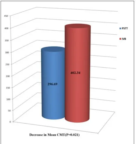
0.003 suggesting that, IVB had better effect on BCVA. These are depicted in Fig.4. With regards to CMT the mean decrease in group I was 296.69 and in group II was 402.34. Group II had more effect in
decreasing the CMT which was confirmed statistically with P value
of 0.021. This is depicted in Fig.5
[image:4.595.48.281.246.493.2]Chart 4. Decrease in Mean CMT: Intergroup variance
Fig. 5. Examples of OCT images in both groups
DISCUSSION
Diabetic macular edema is the main cause for visual impairment in diabetics. The reason for DME is breakdown of BRB. Multiple factors are associated with BRB disruption which includes tight junction leaks, loss of pericytes and endothelial cells, retinal vessel leukostasis, dilatation of retinal vessels and traction of vitreous on
retina[6,7]. Macular edema in diabetes is characterized by retinal
thickening which slowly progresses towards the center of macula thus affecting the vision. Rarely does DME resolves spontaneously with improvement in systemic risk factors, such as glycemic control,
hypertension, hypercholesterolemia or renal status etc. In a study 29%
of untreated eyes developed moderate loss in vision. Spontaneous visual recovery is very rare. Only 5% of cases in a study showed
increase in 3 lines or more, without treatment[9]. The goal of treatment
is to reduce the leak and thus the thickness of the macula. Modified
grid laser was advocated by ETDRS[9].14.5% patients with DME had
improved vision, 60.9% had no change in vision and 24.6% had reduced vision after 3 years of grid treatment, as reported by a
study[10]. Other treatment modalities are thus being evaluated for
DME. Due to its anti-inflammatory, antipermeability and anti-VEGF
properties, Triamcinolone has been used in treatment of DME[11,12].
There is enough evidence to show the efficacy of Transscleral drug
delivery in macular edemas[24]. Bakri and Kaiser evaluated 63 eyes of
50 patients of refractory DME, who had received PSTT of 40 mg. They concluded that the PSTT shows statistically significant
improvement in visual acuity[21]. In a prospective study comparing the
PSTT and intravitreal triamcinolone, the authors did not find much difference between the two groups in the first 3 months, and have concluded that the subtenon approach can be considered a valid
alternative to the intravitreal injection[25]. Beneficial effect of
intravitreal avastin was already established in DME. To our knowledge there is no study which compared these two modalities of treatment. Short term follow up with single injection is a major limiting factor of this study.
Conclusions
Both PSTT and IVB are effective in increasing the visual acuity and decreasing the CMT. IVB has better effect on both these parameters. In selected patients, who cannot afford IVB, PSTT can be tried as a first line of treatment. Prospective randomized studies with long term follow-up are required to study the long-term efficacy and side effects of these treatment modalities.
REFERENCES
1. Yau JW; Rogers SL; Kawasaki R; Lamoureux EL; Kowalski JW;
Bek T; Chen SJ; Dekker JM; Fletcher A; Grauslund J; Haffner S;
Hamman RF; Ikram MK; Kayama T; Klein BE; Klein R;
Krishnaiah S; Mayurasakorn K; O'Hare JP; Orchard TJ; Porta M; Rema M; Roy MS; Sharma T; Shaw J; Taylor H; Tielsch JM; Varma R; Wang JJ; Wang N; West S; Xu L; Yasuda M; Zhang X; Mitchell P; Wong TY. Global prevalence and major risk factors
of diabetic retinopathy. Diabetes Care. 2012; 35(3):556-64.
2. Raman R, Rani PK, Reddi Rachepalle S, Gnanamoorthy P,
Uthra S, Kumaramanickavel G, Sharma T. Prevalence of diabetic retinopathy in India: Sankara Nethralaya Diabetic Retinopathy Epidemiology and Molecular Genetics Study report 2.
Ophthalmology. 2009 Feb;116(2):311-8.
3. Diep TM; Tsui I. Risk factors associated with diabetic macular
edema. Diabetes Res Clin Pract. 2013; 100(3):298-305.
4. Klein R, Knudtson MD, Lee KE, Gangron R, Barbara EK,
Klein.The Wisconsin Epidemiologic Study of Diabetic
Retinopathy XXIII: the twenty-five-year incidence of macular
edema in persons with type 1 diabetes. Ophthalmology
2009;116(3): 497-503.
5. Wenick AS; Bressler NM Diabetic macular edema: current and
emerging therapies. Middle East Afr J Ophthalmol. 2012;
6. Bandello F; Battaglia Parodi M; Lanzetta P; Loewenstein A;
Massin P; Menchini F; Veritti D. Diabetic macular edema. Dev
Ophthalmol. 2010; 47:73-110.
7. Bhagat N; Grigorian RA; Tutela A; Zarbin MA. Diabetic macular
edema: pathogenesis and treatment. Surv Ophthalmol. 2009;
54(1):1-32.
8. Kim BY, Smith SD, Kaiser PK. Optical coherence tomographic
patterns of diabetic macular edema. Am J Ophthalmol. 2006 Sep;
142(3):405-12.
9. Early Treatment Diabetic Retinopathy Study Research Group.
Photocoagulation for diabetic macular edema. Early trearment
diabetic retinopathy study report number 1. Arch Ophthalmol.
1985; 103(12); 1796-1806.
10.Fraser-Bell S; Kaines A; Hykin PG Update on treatments for
diabetic macular edema.CurrOpinOphthalmol.2008; 19(3):185-9.
11.Zhang Y; Ma J; Meng N; Li H; Qu Y.Comparison of intravitreal
triamcinolone acetonide with intravitreal bevacizumab for
treatment of diabetic macular edema: a meta-analysis. Curr Eye
Res. 2013; 38(5):578-87.
12.Qi HP; Bi S; Wei SQ; Cui H; Zhao JB. Intravitreal versus
subtenon triamcinolone acetonide injection for diabetic macular
edema: a systematic review and meta-analysis. Curr Eye Res.
2012; 37(12):1136-47.
13.Konstantopoulos A, Williams CP, Newsom RS, Luff AJ. Ocular
morbidity associated with intravitreal trimcinolone acetonide. Eye
(Lond). 2007; 21(3): 317-320.
14.Veritti D; Di Giulio A; Sarao V; Lanzetta P. Drug safety
evaluation of intravitreal triamcinolone acetonide. Expert Opin
Drug Saf. 2012; 11(2):331-40.
15.Gupta N; Mansoor S; Sharma A; Sapkal A; Sheth J;
Falatoonzadeh P; Kuppermann B; Kenney M.Diabetic retinopathy
and VEGF. Open Ophthalmol J. 2013; 7:4-10.
16.Funatsu H, Noma H, Mimura T, Eguchi S, Hori S. Association of
vitreous inflammatory factors with diabetic macular edema.
Ophthalmology 2009; 116(1):73-9.
17.J.F.Arevalo, J.G. Sancez, L.Wu et al., “Primary intravitreal
bevacizumab for diffuse diabetic macular edema: the Pan-American colloaborative retinal study group at 24 months”,
Ophthalmology 2009;116(8), 1488-1497.
18. P. Massin, F Bandello, JG Garweg et al., “Safety and efficacy of
ranibizumab in diabetic macular edema (RESOLVE study): a 12-mont, randomized, controlled, double-masked, multicenter phase
II study,” Diabetes Care 2010;33(11), 2399-2405.
19.Pacella E; La Torre G; Impallara D; Malarska K; Turchetti P;
Brillante C; Smaldone G; De Paolis G; Muscella R; Pacella F. Efficacy and safety of the intravitreal treatment of Diabetic
Macular Edema with Pegaptanib: a 12-month follow-up. Clin Ter.
2013; 164(2):121-6.
20.Shima C; Sakaguchi H; Gomi F; Kamei M; Ikuno Y; Oshima Y;
Sawa M; Tsujikawa M; Kusaka S; Tano Y. Complications in
patients after intravitreal injection of bevacizumab. Acta
Ophthalmol. 2008; 86(4):372-6.
21. Bakri SJ, Kaiser PK. Posterior subtenon triamcinolone acetonide
for refractory diabetic macular edema. Am J Ophthalmol
2005;139(2):290–294.
22.Rodrigues EB, Meyer CH, Grumann A Jr, Shiroma H, Aguni JS,
Farah ME. Tunneled scleral incision to prevent vitreal reflux after
intravitreal injection. Am J Ophthalmol 2007; 143(6):1035-1037.
23.Nozik RA. Periocular injection of steroids. Trans Am Acad
Ophthalmol Otolaryngol 1972;76(3):695-705.
24.Geroski DH, Edelhauser HF: Transscleral drug delivery for
posterior segment disease. Adv Drug Deliv Rev 2001;
31;52(1):37-48.
25.Cellini M; Pazzaglia A; Zamparini E; Leonetti P; Campos EC.
Intravitreal vs subtenon triamcinolone acetonide for the treatment
of diabetic cystoid macular edema. BMC Ophthalmol. 2008; 8:5.