Abstract
Thomas III, Melvin Edward. The Role of Phosphorylation in Allosteric Modulation of Caspase-3. (Under the direction of Dr. Robert Rose).
Caspases are cysteine-dependentaspartate-specific proteases that responsible for control cellular homeostasis. Caspases control homeostasis by carrying out programmed cell death via apoptosis. Caspase-3 is one of the main executioner caspases that is activated and commits the cell to death. Caspase-3 cleavage of substrates like ROCK 1, acinus, nuclear lamin proteins, CAD, and many others leads to the morphological features of apoptosis: cell membrane blebbing, nuclear condensation, and DNA fragmentation. Because the caspase family of proteases are enzymatically efficient, their expression and activation are under tight cellular regulation. Caspase-3 exist as a stable inactive dimer and its activation requires upstream caspase cleavage. This activation mechanism is controlled by FLIP and XIAP binding and inhibition of initiator caspases, heat shock protein 27 binding to the prodomain of caspase-3 and phosphorylation of the intersubunit linker of caspase-3 all of which block caspase-3 activation. Once activated caspase-3’s activity is regulated by active site inhibitors such as p35 from the baculovirus and XIAP, as well as allosteric inhibitors such as phosphorylation and S-Nitrosylation.
These regulatory events control the phenotype of the cell. When caspase-3 is activated during
non-apoptotic phenotypes, it is hypothesized that the total caspase activity is below the non-apoptotic
threshold. Subthreshold levels of caspase-3 activity are required for the regulation of cell proliferation
of many different cell types such as forebrain cells, lymphoid cells, and B-cells. Regulated caspase-3
activity is also required for differentiation of erythroblasts, keratinocytes, neuronal cells, and many
important area of cancer research. Dysregulation of caspase activity can lead to diseases such as
cancer and Alzheimer’s.
Here we study how caspase-3 activity is regulated by phosphorylation at an allosteric site. Caspase-3
is phosphorylated at Ser150 and Thr152, but the biophysical and structural characterization hasn’t
been defined. We show that this region of the enzyme, C-terminal loop of helix-3 (H3CL), is an apart
of an interaction network that connects this site to the active site of both protomers. Phosphorylation
of Ser150 destabilizes the enzyme, making it more sensitive to cellular pH changes and decreases
enzymatic efficiency for whole protein substrates. Thr152 phosphorylation is an enzymatic
kill-switch, which disrupts a highly conserved hydrophobic pocket between H3Cl and the loop bundle of
the active site of the second protomer. These data for the first time demonstrate how the caspase
family of enzymes are regulated by phosphorylation. This allosteric regulation potentially allows the
cell to bring the total caspase activity in the cell below the apoptotic threshold, which promotes
developmental phenotypes such as erythroid differentiation that require caspase-3 activity at
The Role of Phosphorylation in Allosteric Modulation of Caspase-3
by
Melvin Edward Thomas III
A dissertation submitted to the Graduate Faculty of North Carolina State University
in partial fulfillment of the requirements for the degree of
Doctor of Philosophy Biochemistry
Raleigh, North Carolina 2017
APPROVED BY:
Dr. Robert Rose Dr. Michael B. Goshe
Committee Chair
ii Dedication
iii Biography
Melvin Thomas was born in Philadelphia Pennsylvania in 1989. In 1994 his family moved to Raleigh North Carolina where he grew up. Melvin was always the type of child to question the reasoning of everything he was told or learned. His love for science started very early when he received a microscope set as a Christmas gift, in which he would go outside to collect specimens every day. Melvin went to Eastern University for undergraduate school where he obtained his BS in biochemistry. He then was accepted into NC State for graduate school where he seeks to become the first generation scientist of his family. Melvin worked in Dr. Clay Clark’s lab studying caspase regulation and allostery in NC until the lab was
iv Acknowledgments
I would like to thank Dr. Clark for accepting me into his lab and all of his mentorship and training over the past 6 years. His guidance and understanding of biochemistry has equipped me with the tools I needed to become an effective biochemist. I would also like to thank my committee, Dr. Rose, Dr. Goshe, Dr. Flora Meilleur, and Dr. Hamilton. I greatly appreciate the academic help and scientific guidance throughout my graduate student career, and supportive critiques at every committee meeting. I would like to thank Dr. Swartz for you many contributions from training me in the field of crystallography to helping me fix expensive broken lab equipment. I would like to thank Dr. Lawton for being my first biochemistry mentor, your example of being a Christian scientist is something I will always look up to.
I would like to thank my family for supporting me throughout my life. My Mom, who gave me encouragement every day. My Father, who gave me wisdom and pushed me to only accept the best. To my loving wife, who decided to marry me at one of the busiest phases of my life without complaint and loves me even at my worse. To my son, for being my
v Table of Contents
LIST OF TABLES ………..…..……..ix
LIST OF FIGURES ………...……..ix
CHAPTER 1: CASPASE ACTIVATION AND FUNCTION ……….1
A. Introduction...……….1
B. Programmed Cell death- Apoptosis ………..1
a. Apoptosis Activation Pathways ………2
b. Morphological changes and Apoptotic Cleanup ………...4
C. Caspase Cascade, Activation, and Structure ……….6
a. Caspase Cascade ………...7
b. Activation and Structure ………...………8
D. Conclusions ………...9
E. References ………...11
CHAPTER 2: REGULATION OF CASPASE ACTIVITY ………...16
A. Introduction...………...16
B. Zymogen maturation and Expression ...………16
C. Active Site Inhibitors ………..19
D. Allosteric Regulation ………..21
E. Posttranslational Modifications ………..25
F. Conclusions ……….33
vi
CHAPTER 3: NON-APOPTOTIC FUNCTION OF CASPASES ……….41
A. Proliferation ………41
a. Initiator Caspases in Proliferation ………...41
b. Executioner Caspases in Proliferation ………42
c. Compensatory Cell Proliferation ………42
B. Differentiation ……….44
a. Caspase-1 Differentiation ………...45
b. Caspase-2 Differentiation ………...46
c. Caspase-3 Differentiation ………...47
d. Caspase-6 Differentiation ………...50
e. Caspase-7 Differentiation ………...50
f. Caspase-8 Differentiation ………...50
g. Caspase-9 Differentiation ………...51
h. Caspase-14 Differentiation ……….52
i. Conclusions ……….52
j. References ………...53
CHAPTER 4: MODIFICATIONS TO A COMMON PHOSPHORYLATION NETWORK PROVIDE INDIVIDUALIZED CONTROL IN CASPASES ………. .59
A. Abstract ………...……60
B. Introduction ………...61
C. Methods ………...…64
D. Results ………...69
vii
F. References ………...………..……101
CHAPTER 5: LENGTHENING THE INTERSUBUNIT LINKER OF PROCASPASE 3 LEADS TO CONSTITUTIVE ACTIVATION ………107
A. Abstract …....……….108
B. Introduction ………...109
C. Materials and Methods ………..113
a. Cloning, Protein Expression, and Purification, Western Blots ……….113
b. Enzyme Activity Assays ………...114
c. Identification of Cleavage Sites by Mass Spectrometry ……….……..114
d. Generation of Structural Models and Molecular Dynamics Simulations ….116 D. Results ………...116
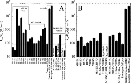
a. A Longer IL Leads to Increased Enzyme Activity in the Zymogen ……….116
b. Disrupting the Hydrophobic Cluster Has Little Effect on the Activity of Procaspase 3 ………..117
c. IL Mutations Result in Minor Populations of Alternately Cleaved Inactive Protein ………...118
d. Molecular Dynamics Simulations Show Increased Flexibility in the IL of Insertion Mutants ………..123
E. Discussion ………..………...126
F. Acknowledgments ………..……….………..132
G. Abbreviations ………..…………..132
viii
APPENDIX: CASPASE PREPARATION AND CHARACTERIZATION ………...147
A. Caspase-3/Mutant Protein Purification ………...147
B. Crystal Trials ………...151
C. Activity Assays ………152
D. Half-Life ………..154
E. In Gel Trypsin Digest and Peptide Mapping Analysis by Mass-Spec …………....156
F. P32 ATP incorporation (Phosphorylation of caspase-3 by p38-MAPKα) …………158
G. Cell Culture ………..………...161
H. Apoptosis Assays in K562 cells ………..164
I. Differentiation assays in K562 cells……….………164
J. siRNA/Plasmid DNA Transfections ………...165
K. Flow cytometry ………..…...166
L. Cell lysis and Western Blots ………...……172
ix List of Tables
Chapter 4
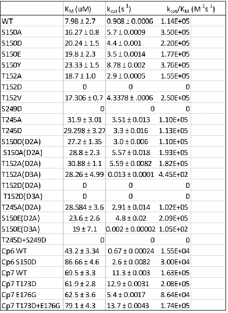
Table 1. Catalytic Parameters………91
Supplamental Table 1. H3CL positional frequencies………...92
Supplamental Table 2. Crystallographic parameters………93
Chapter 5
Table 1. Alternate Cleavage Sites ………...…139
Supplamental Table 1. Primers and templates ………...141 Supplamental Table 2. Activity of procaspase 3 variants ……….…….142
Appendix
Table 1. AFC standards ………...152
Table 2. Substrate Concentration Gradient ……….153
x List of Figures
Chapter 1
Figure 1. Caspase Activation Cascade ………...10
Figure 2. Caspase Domain Organization ………11
Chapter 4 Figure 1. Caspase-3 structure and regulation model………..87
Figure 2. Phylogenetic analysis of H3CL………...88
Figure 3. Electrostatic interactions of H3CL………..89
Figure 4. Biophysical analysis of H3Cl variants and pH………90
Supplamental Figure 1. ΔRMSF of MD-Simulations………...94
Supplamental Figure 2. ΔRMSF of MD-Simulations………...95
Supplamental Figure 3. ΔRMSF of MD-Simulations………...96
Supplamental Figure 4. ΔRMSF of MD-Simulations………...97
Supplamental Figure 5. Structural superimposition of loop 4 and helix-5………98
Supplamental Figure 6. AEW at 280nm vs pH gradient………99
Supplamental Figure 7. FPLC Chromatograms………...100
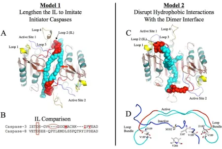
Chapter 5 Figure 1. Proposed models for procaspase 3 activation ………...133
Figure 2. Enzyme activity assays ……….134
Figure 3. SDS-PAGE and Western analyses ………135
Figure 4. Enzyme inhibition and active site titrations ………..136
xi
Figure 6. Distance calculations from molecular dynamics simulations ………...138
Supplamental Figure 1. Distance calculations from MD Simulations ………...…….140
Appendix Figure 1. His-bind Gel ……….……….149
Figure 2. Q-sepharose Gel ………...……….150
Figure 3. Final Concentration Gel ………...………….151
Figure 4. Michaelis Menten Kinetics Plot ………...….154
Figure 5. Half-life Plot ……….156
Figure 6. Kinase Assay Gel ……….……….160
Figure 7. Kinase Assay Autoradiogram ………..….161
Figure 8. Time Dependent γ-32P-ATP Incorporation ………..…….161
Figure 9. Doublet Analysis Plot ………..…….167
Figure 10. FSC vs SSC Plot ………..….168
Figure 11. Count vs Fluorophore Plot ………..………..…168
Figure 12. Gating Statistics ………168
Figure 13. Experimental Group Plots TX Efficiency ………....169
Figure 14. Experimental Group Plots Unstained Control ………..170
Figure 15. Experimental Group Plots Single Stain Control ………...…………171
Figure 16. Experimental Group Plots AnnexinV-PI Assay ………...172
1 Chapter 1: Caspase Activation and Function
Introduction
In order for multicellular organisms to survive, they must maintain an organized system to produce healthy tissue, while equally removing damaged or dying cells. The process of killing cells, or programmed cell death, is a complex system that requires many molecular pathways to converge leading to cell death, which maintains cellular homeostasis. Apoptosis is the most energetically efficient and safest cell death pathway. Apoptosis is activated either by external or internal signals. Both of these signals lead to the activation of caspases, a cysteinyl aspartate-specific protease family responsible for controlling the fate of the cell. These enzymes have hundreds of substrates both nuclear and cytosolic. These tightly regulated enzymes are ubiquitously expressed and found in every species that utilizes programmed cell death via apoptosis. Because of the phenotypic importance of these enzymes, they are an essential protein to study due to their implications in cell death related diseases such as cancer.
A. Programmed Cell death- Apoptosis
2
cellular components are degraded through the formation of the autophagosome and autolysosomes (autophagy) (Fuchs and Steller 2015; Tsujimoto and Shimizu 2005).
Autophagy has been shown to be induced by starvation in Drosophilaand yeast, in order to promote survival of the organism by recycling the degraded molecular components,
(Tsujimoto and Shimizu 2005; Denton et al. 2009; Lum et al. 2005; Klionsky 2000). Autophagic cell death can also be induced by intracellular damage signals (Klionsky 2000; Lum et al. 2005). Regulated necrosis, known as necroptosis, is another cell death mechanism organisms use to control bacterial and viral infections, and can also be induced by reactive oxygen species (Fuchs and Steller 2015; Cho et al. 2009; Galluzzi et al. 2008). The
physiological hallmarks of necroptosis include cell and organelle swelling, which leads to plasma membrane rupturing and loss of intracellular components (Feoktistova and Leverkus 2015; Kaczmarek, Vandenabeele, and Krysko 2013). This pathway has been shown to be an important network between PCD and the innate immune system (Feoktistova and Leverkus 2015; Fuchs and Steller 2015). The most well-studied pathway of PCD, and the main topic of the chapter, is apoptosis. Programmed cell death via apoptosis is a complex biological
process that evolved over 600 million years ago and some of the apoptotic domains can be found in the earliest eukaryotic ancestors (Zmasek and Godzik 2013). The process of apoptosis from start to finish can be broadly broken down into 3 major phases; Activation, morphological changes, and removal of dead cells and these are described below.
Apoptosis Activation Pathways – Apoptosis is an energy-dependent program that has a
3
the cell, there is a superfamily of death receptors called the tumor necrosis factor (TNF) receptors (Locksley, Killeen, and Lenardo 2001). The extrinsic pathway is externally activated by death receptor ligand binding of this superfamily such as FasL/FasR and TNFα/TNFR1 (ligand/receptor)(Cheng et al. 1994). The activation of these cysteine-rich
death receptors induces oligomerization and structural reorganization that leads to their activation (Locksley, Killeen, and Lenardo 2001). After activation, the death receptors form complexes with adaptor proteins like Fas-associated death domain protein (FADD) (Lawton 2016). The formation of this complex is known as the death-inducing signaling complex (DISC) (Kim, Choi, and Joe 2000). This complex recruits and activates caspase-8 and -10, which triggers the activation of the caspase cascade (discussed below) and commitment to cell death (Kim, Choi, and Joe 2000). The intrinsic pathway is activated in response to an internal signal and doesn’t require the death receptors (Tait and Green 2010). These death
inducing signals include DNA damage, hypoxia, viral infections, and many others (Elmore 2007). These signals cause the deregulation of the BCL-2 family of proteins. This family has three classes that regulate each other and thus apoptosis. The first class is the anti-apoptotic members BCL-2 and BCL-XL, which inhibits the second class pro-apoptotic members BAX and BAK, and third class, BH3-only proteins which inhibit the antiapoptotic members (Youle and Strasser 2008; Hockenbery et al. 1990). The internal death signals activate the BH3-only proteins, and through the sequestering of the antiapoptotic members, activate BAX and BAK (Youle and Strasser 2008). Though the mechanism is disputed, activated BAX and BAK go on to bind to the mitochondrial membrane and cause mitochondrial outer membrane
4
Smac/DIABLO and HtrA2/Omi induce apoptosis by inhibiting X-linked inhibitor of apoptosis (XIAP), a protein that can bind to the active site of caspase-3 and -7 (Tait and Green 2010). When cytochrome-c is released into the cytoplasm it forms an ATP dependent, multimeric complex with apoptosis protease-activating factor 1 (APAF-1) to form the apoptosome (Riedl and Salvesen 2007; Chandra et al. 2006). The apoptosome then can recruit and activate caspase-9, which activates the caspase cascade (discussed below)and commitment to cell death(Lawton 2016; Joza et al. 2001).
While each of these pathways can commit the cell to death individually, there can be crosstalk between the extrinsic and intrinsic pathways that lead to an enhanced apoptotic signal (Lawton 2016). This crosstalk is mediated through the extrinsic activation of caspase-8 which cleaves the BH3-only protein BID to form the active tBID (Korsmeyer et al. 2000). tBID then both inhibits the anti-apoptosis function of BCL-2 and is translocated the
mitochondria where it promotes intrinsic apoptosis via outer membrane permeabilization and cytochrome-c release (Youle and Strasser 2008; Korsmeyer et al. 2000).
Morphological changes and Apoptotic Cleanup – Once the cell is committed to death via
apoptosis, the downstream signals elicit very specific morphologies based upon the inducer to cell death (Häcker 2000). There are three common morphological features of apoptosis after commitment to cell death: cell membrane blebbing, nuclear condensation, and DNA fragmentation (Häcker 2000). Membrane blebbing is the protrusions of the cell membrane in an actin/myosin dependent manner (Charras 2008). The activation of caspases leads to the activation of Rho effector protein ROCK 1, which is a kinase that phosphorylates and
5
6
dying cells release four known signals; fractalkine, caspase-dependent
lysophosphatidylcholine (LPC), sphingosine-1-phosphate (SIP), ATP, and UTP, all which bind to specific receptors on the surface of the phagocyte cell (CX3CR1, G2A, SIP-R1/5, and P2Y2, respectively) (Hochreiter-hufford and Ravichandran 2013). The release of these signals creates a chemical gradient that the phagocytes can follow like a trail of breadcrumbs directly to the apoptotic cell (Peter et al. 2008). Once the phagocytes have successfully migrated to the dying cell it needs to be able to recognize the specific cell without engulfing healthy cells. In order to specifically target the dying cell, the process of apoptotic signaling causes the release of specific cell surface markers such as phosphatidylserine (PS), the most common and studied apoptotic signal, which flipped from the inner leaflet of the cell to the outer leaflet (Lauber et al. 2004). Though under-studied, other apoptotic surface markers include oxidized low-density lipoprotein particles (oxLDL), thrombospondin1-(TSP-1) binding sites, cell surface glycosylation, and mannose binding lectin (MBL)(Ravichandran 2011; Lauber et al. 2004). These markers are bound by phagocyte receptors such as TIM-4, BAI1, CD68, CD36, CD14, integrins, and many others and all lead to the uptake and engulfment of the apoptotic cell (Hochreiter-hufford and Ravichandran 2013; Maderna and Godson 2003; Lauber et al. 2004).
Morphological changes to the apoptotic cell efficiently and evolutionarily lead to a rapid removal of dying cells. Because the process of apoptosis is tightly regulated, removal of dead cells allows for the protection of the surrounding normal cells, and thus survival of the host organism (Platt, Da Silva, and Gordon 1998; Häcker 2000).
7
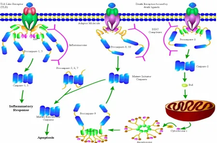
Caspase Cascade - Caspases are cysteinyl aspartate-specific proteases that are required for
programmed cell death. In humans, there are 13 caspases that are either activated in response to inflammation, apoptosis, or an “apoptosis-like” process (Clark 2016). The inflammatory
8
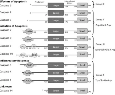
Activation and Structure - All caspases have a similar structural organization. Each has a
short or long N-terminal region call the prodomain that can contain a death effector domain (DED) or a caspase recruitment domain (CARD), and a protease domain that contain a large subunit and a small subunit that are separated by an intersubunit linker (IL) (Figure 2) (Clark 2016). This structural arrangement of caspases is known as the caspase-hemoglobinase fold (Aravind and Koonin 2002). All caspases exist as a stable inactive form (zymogen) called procaspase until cleaved and activated (Salvesen 2002). Caspases with long prodomains that contain either a DED (caspase-8 and -10) or a CARD (caspase-1,-2, -4, -5, and -9) exist as inactive monomers, while short prodomain caspases (caspase-3, -6, and-7) exist as stable inactive dimers (MacKenzie and Clark 2012; Clark 2016; Bose and Clark 2001). As
9
rearrangement of the active site loops (L1-L4 and L2’) to form the active site (Chai et al. 2001; Thomsen, Koerber, and Wells 2013).
For each of the mature caspases, rearrangement of their respective active site loops yields an active site with a substrate binding pocket that can be put into 3 groups based on the
recognition of the tetrapeptide sequence (P4-P1); group 1 (caspase-1, -4, -5, -14 which recognize (W/L)EHD), group 2 (caspase-2, -3, -7 which recognize DXXD, and group 3 (caspase-6, -8, -9, and -10 which recognize (L/V)EXD) (Figure 2) (Talanian et al. 1997; Pop and Salvesen 2009). All caspases are required to have an aspartate at the P1 position and have evolved an S1 pocket for it, but the remaining positions are what allows for each caspase to have a diverse group of substrates (Seaman et al. 2016; Fischer, Jänicke, and Schulze-Osthoff 2003; McStay, Salvesen, and Green 2008). One caveat to this mechanism is due to the structural similarities that exist in the substrate binding pocket amongst the entire caspase family of protein. This leads to redundancies in substrate specificity, such as
caspase-3 having the ability to cleave the substrates of every other caspase with varying processivity (Poreba et al. 2013). This makes designing drugs that have specificity for the active site of individual caspases extremely difficult.
Conclusions
10
macroautophagy, and necrosis). Apoptosis, however, is the safest and most energy efficient mechanism the organism has to control cell fate. The 2 major pathways of apoptosis that lead to cell death (extrinsic and intrinsic pathway) both converge to activate the caspase family of proteins, whose activity is required for nearly all forms of apoptosis. The crosstalk that exists between these pathways also provides another level of feedback regulation. Discussed in chapter 2, the regulation of caspases are mandatory in the cell’s commitment to apoptosis,
thus demonstrating why the study of caspase structure and function is important for the advancement of human health.
11
Figure 2:Caspase Domain Organization (MacKenzie and Clark 2012).
References
Aravind, L., and Eugene V. Koonin. 2002. “Classification of the Caspase-Hemoglobinase Fold: Detection of New Families and Implications for the Origin of the Eukaryotic Separins.”
Proteins: Structure, Function and Genetics 46 (4): 355–67. doi:10.1002/prot.10060.
Barros, Lf, T Kanaseki, R Sabirov, S Morishima, J Castro, Cx Bittner, E Maeno, Y Ando-Akatsuka, and Y Okada. 2003. “Apoptotic and Necrotic Blebs in Epithelial Cells Display Similar Neck Diameters but Different Kinase Dependency.” Cell Death and Differentiation 10: 687–97. doi:10.1038/.
Bose, Kakoli, and A Clay Clark. 2001. “Dimeric Procaspase-3 Unfolds via a Four-State Equilibrium Process.” Biochemistry 40 (47): 14236–42. doi:10.1021/bi0110387.
12
Chandra, Dhyan, Shawn B Bratton, Maria D Person, Yanan Tian, Angel G Martin, Mary Ayres, Howard O Fearnhead, Varsha Gandhi, and Dean G Tang. 2006. “Intracellular Nucleotides Act as Critical Prosurvival Factors by Binding to Cytochrome C and Inhibiting Apoptosome.” Cell
125 (7): 1333–46. doi:10.1016/j.cell.2006.05.026.
Charras, G. T. 2008. “A Short History of Blebbing.” Journal of Microscopy 231 (3): 466–78. doi:10.1111/j.1365-2818.2008.02059.x.
Cheng, J, T Zhou, C Liu, JP Shapiro, MJ Brauer, MC Kiefer, PJ Barr, and JD Mountz. 1994.
“Protection from Fas-Mediated Apoptosis by a Soluble Form of the Fas Molecule.” Science 263 (5154): 1759–62. doi:10.1126/science.7510905.
Chipuk, Jerry E, Tudor Moldoveanu, Fabien Llambi, Melissa J Parsons, and Douglas R Green. 2010. “The BCL-2 Family Reunion.” Molecular Cell. doi:10.1016/j.molcel.2010.01.025.
Cho, YoungSik, Sreerupa Challa, David Moquin, Ryan Genga, Tathagat Dutta Ray, Melissa Guildford, and Francis Ka-Ming Chan. 2009. “Phosphorylation-Driven Assembly of the RIP1-RIP3 Complex Regulates Programmed Necrosis and Virus-Induced Inflammation.” Cell 137 (6): 1112–23. doi:10.1016/j.cell.2009.05.037.
Clark, A Clay. 2016. “Caspase Allostery and Conformational Selection.” Chemical Reviews. doi:10.1021/acs.chemrev.5b00540.
Cohen, J. John, Richard C. Duke, Valerie A. Fadok, and Karen S. Sellins. 1992. “Apoptosis and Programmed Cell Death in Immunity.” Annual Review of Immunology 10 (1). Annual Reviews 4139 El Camino Way, P.O. Box 10139, Palo Alto, CA 94303-0139, USA : 267–93.
doi:10.1146/annurev.iy.10.040192.001411.
Coleman, Mathew L, Erik A Sahai, Margaret Yeo, Marta Bosch, Ann Dewar, and Michael F Olson. 2001. “Membrane Blebbing during Apoptosis Results from Caspase-Mediated Activation of ROCK I.” NATURE CELL BIOLOGY 3. http://cellbio.nature.com.
Denton, Donna, Bhupendra Shravage, Rachel Simin, Kathryn Mills, Deborah L Berry, Eric H Baehrecke, and Sharad Kumar. 2009. “Report Autophagy, Not Apoptosis, Is Essential for Midgut Cell Death in Drosophila.” CURBIO 19: 1741–46. doi:10.1016/j.cub.2009.08.042.
Elmore, Susan. 2007. “Apoptosis: A Review of Programmed Cell Death.” Toxicologic Pathology 35: 495–516. doi:10.1080/01926230701320337.
Feeney, Brett, Cristina Pop, Paul Swartz, Carla Mattos, and A Clay Clark. 2006. “Role of Loop Bundle Hydrogen Bonds in the Maturation and Activity of (Pro)caspase-3.” Biochemistry 45 (44). American Chemical Society: 13249–63. doi:10.1021/bi0611964.
Feoktistova, Maria, and Martin Leverkus. 2015. “Programmed Necrosis and Necroptosis Signalling.”
FEBS Journal 282 (1): 19–31. doi:10.1111/febs.13120.
Fischer, U, R U Jänicke, and K Schulze-Osthoff. 2003. “Many Cuts to Ruin: A Comprehensive Update of Caspase Substrates.” Cell Death and Differentiation 10 (1): 76–100.
doi:10.1038/sj.cdd.4401160.
Fuchs, Yaron, and Hermann Steller. 2015. “Live to Die Another Way: Modes of Programmed Cell Death and the Signals Emanating from Dying Cells.” Nature Publishing Group 16.
doi:10.1038/nrm3999.
13
(7). Elsevier: 1161–63. doi:10.1016/j.cell.2008.12.004.
Häcker, Georg. 2000. “The Morphology of Apoptosis.” Cell and Tissue Research 301 (1): 5–17. doi:10.1007/s004410000193.
Hochreiter-hufford, Amelia, and Kodi S Ravichandran. 2013. “Clearing the Dead : Apoptotic Cell Sensing ,” 1–20. doi:10.1101/cshperspect.a008748.
Hockenbery, David ;, Gabriel ; Nunez, Curt ; Milliman, Robert D Schreiber, and Stanley J Korsmeyer. 1990. “Bcl-2 Is an Inner Mitochondrial Membrane Protein That Blocks Programmed Cell Death.” Nature 348 (6299).
Joza, Nicholas, Santos A Susin, Eric Daugas, and Et Al. 2001. “Essential Role of the Mitochondrial Apoptosis-Inducing Factor in Programmed Cell Death.” Nature 410 (6828): 549–54.
doi:10.1038/35069004.
Kaczmarek, Agnieszka, Peter Vandenabeele, and Dmitri V Krysko. 2013. “Necroptosis: The Release of Damage-Associated Molecular Patterns and Its Physiological Relevance.”
doi:10.1016/j.immuni.2013.02.003.
Kass, George E N, John E Eriksson, Marianne Weis, Sten Orrenius, and Sek C Chow. 1996. “Chromatin Condensation during Apoptosis Requires ATP.” Biochem. J 318: 749–52. http://www.biochemj.org/content/ppbiochemj/318/3/749.full.pdf.
Kim, Jin Woo, E J Choi, and Cheol O Joe. 2000. “Activation of Death-Inducing Signaling Complex (DISC) by pro-Apoptotic C-Terminal Fragment of RIP.” Oncogene 19 (39): 4491–99.
doi:10.1038/sj.onc.1203796.
Klionsky, Daniel J. 2000. “Autophagy as a Regulated Pathway of Cellular Degradation.” Science 290 (5497): 1717–21. doi:10.1126/science.290.5497.1717.
Korsmeyer, S J, M C Wei, M Saito, S Weiler, K J Oh, and P H Schlesinger. 2000. “Pro-Apoptotic Cascade Activates BID, Which Oligomerizes BAK or BAX into Pores That Result in the Release of Cytochrome C.” Cell Death and Differentiation 7 (12): 1166–73.
doi:10.1038/sj.cdd.4400783.
Lamkanfi, Mohamed, and Daniel Jiménez Fernández. 2015. “Inflammatory Caspases: Key Regulators of Inflammation and Cell Death.” Biol. Chem 396 (3): 193–203. doi:10.1515/hsz-2014-0253.
Lauber, Kirsten, Sibylle G. Blumenthal, Michaela Waibel, and Sebastian Wesselborg. 2004. “Clearance of Apoptotic Cells: Getting Rid of the Corpses.” Molecular Cell 14 (3): 277–87. doi:10.1016/S1097-2765(04)00237-0.
Lawton, Jennifer S. 2016. “A Fate Worse than Death.” Journal of Thoracic and Cardiovascular Surgery 152 (1). Nature Publishing Group: 97–98. doi:10.1016/j.jtcvs.2016.03.007.
Locksley, Richard M, Nigel Killeen, and Michael J Lenardo. 2001. “The TNF and TNF Receptor Review Superfamilies: Integrating Mammalian Biology The Receptors and Ligands in This Superfamily Have Unique Structural Attributes That Couple Them Directly to Signaling Pathways for Cell Proliferation, Survival, and Differentiation. Thus, They Have Assumed Prominent Roles in the Generation of Tissues and Transient Microen.” Cell 104: 487–501.
14
MacKenzie, Sarah H, and A Clay Clark. 2012. “Death by Caspase Dimerization.” Advances in Experimental Medicine and Biology 747 (January): 55–73. doi:10.1007/978-1-4614-3229-6_4.
Maderna, Paola, and Catherine Godson. 2003. “Phagocytosis of Apoptotic Cells and the Resolution of Inflammation.” Biochimica et Biophysica Acta - Molecular Basis of Disease.
doi:10.1016/j.bbadis.2003.09.004.
Martinon, Fabio, and Jü Rg Tschopp. 2004. “Review Inflammatory Caspases: Linking an Intracellular Innate Immune System to Autoinflammatory Diseases.” Cell 117: 561–74.
http://www.cell.com/cell/pdf/S0092-8674(04)00490-8.pdf.
McIlwain, David R, Thorsten Berger, and Tak W Mak. 2013. “Caspase Functions in Cell Death and Disease.” Cold Spring Harbor Perspectives in Biology 5 (4). Cold Spring Harbor Laboratory Press: a008656. doi:10.1101/cshperspect.a008656.
Mcluskey, Karen, and Jeremy C Mottram. 2015. “Comparative Structural Analysis of the Caspase Family with Other Clan CD Cysteine Peptidases.” Biochem. J 466: 219–32.
doi:10.1042/BJ20141324.
McStay, G P, G S Salvesen, and D R Green. 2008. “Overlapping Cleavage Motif Selectivity of Caspases: Implications for Analysis of Apoptotic Pathways.” Cell Death and Differentiation 15 (2): 322–31. doi:10.1038/sj.cdd.4402260.
Mills, Jason C, Nicole L Stone, Joseph Erhardt, and Randall N Pittman. 1998. “Apoptotic Membrane Blebbing Is Regulated by Myosin Light Chain Phosphorylation.” The Journal of Cell Biology
140 (3): 627–36. http://www.jcb.org.
Muzio, Marta, Brent R Stockwell, Henning R Stennicke, Guy S Salvesen, and Vishva M Dixit. 2017. “An Induced Proximity Model for Caspase-8 Activation*.” Accessed June 10.
http://www.jbc.org/content/273/5/2926.full.pdf.
Nagata, Shigekazu. 2000. “Apoptotic DNA Fragmentation.” Experimental Cell Research 256 (1): 12– 18. doi:10.1006/excr.2000.4834.
Peter, Christoph, Michaela Waibel, Caius G. Radu, Li V. Yang, Owen N. Witte, Klaus Schulze-Osthoff, Sebastian Wesselborg, and Kirsten Lauber. 2008. “Migration to Apoptotic ‘find-Me’ signals Is Mediated via the Phagocyte Receptor G2A.” Journal of Biological Chemistry 283 (9): 5296–5305. doi:10.1074/jbc.M706586200.
Platt, Nick, Rosangela P. Da Silva, and Siamon Gordon. 1998. “Recognizing Death: The
Phagocytosis of Apoptotic Cells.” Trends in Cell Biology 8 (9): 365–72. doi:10.1016/S0962-8924(98)01329-4.
Pop, Cristina, and Guy S Salvesen. 2009. “Human Caspases: Activation, Specificity, and Regulation.” The Journal of Biological Chemistry 284 (33): 21777–81.
doi:10.1074/jbc.R800084200.
Pop, Cristina, John Timmer, Sabina Sperandio, and Guy S. Salvesen. 2006. “The Apoptosome Activates Caspase-9 by Dimerization.” Molecular Cell 22 (2): 269–75.
doi:10.1016/j.molcel.2006.03.009.
Poreba, Marcin, Aleksandra Strózyk, Guy S Salvesen, and Marcin Drag. 2013. “Caspase Substrates and Inhibitors.” Cold Spring Harbor Perspectives in Biology 5 (8). Cold Spring Harbor Laboratory Press: a008680. doi:10.1101/cshperspect.a008680.
15
Signaling Pathways.” Immunity 35 (4). Elsevier Inc.: 445–55. doi:10.1016/j.immuni.2011.09.004.
Riedl, Stefan J, and Guy S Salvesen. 2007. “The Apoptosome: Signalling Platform of Cell Death.”
Nature Reviews. Molecular Cell Biology 8 (5): 405–13. doi:10.1038/nrm2153.
Sahara, S, M Aoto, Y Eguchi, N Imamoto, Y Yoneda, and Y Tsujimoto. 1999. “Acinus Is a Caspase-3-Activated Protein Required for Apoptotic Chromatin Condensation.” Nature 401 (6749): 168– 73. doi:10.1038/43678.
Salvesen, Guy S. 2002. “Caspases and Apoptosis.” Essays in Biochemistry 38: 9–19. doi:10.1042/bse0380009.
Seaman, J E, O Julien, P S Lee, T J Rettenmaier, N D Thomsen, and J A Wells. 2016. “Cacidases: Caspases Can Cleave after Aspartate, Glutamate and Phosphoserine Residues.” Cell Death and Differentiation 23 (10). Nature Publishing Group: 1717–26. doi:10.1038/cdd.2016.62.
Slee, Elizabeth A, Colin Adrain, and Seamus J Martin. 2001. “Executioner Caspase-3, -6, and -7 Perform Distinct, Non-Redundant Roles during the Demolition Phase of Apoptosis.” Journal of Biological Chemistry 276 (10): 7320–26. doi:10.1074/jbc.M008363200.
Tait, Stephen W. G., and Douglas R. Green. 2010. “Mitochondria and Cell Death: Outer Membrane Permeabilization and beyond.” Nature Reviews Molecular Cell Biology 11 (9). Nature
Publishing Group: 621–32. doi:10.1038/nrm2952.
Talanian, Robert V, Christopher Quinlan, Simone Trautz, Maria C Hackett, John A Mankovich, David Banach, Tariq Ghayur, Kenneth D Brady, and Winnie W Wong. 1997. “Substrate
Specificities of Caspase Family Proteases.” Journal of Biological Chemistry 272 (15): 9677–82. doi:10.1074/jbc.272.15.9677.
Thomsen, Nathan D, James T Koerber, and James A Wells. 2013. “Structural Snapshots Reveal Distinct Mechanisms of Procaspase-3 and -7 Activation.” Proceedings of the National Academy of Sciences 110 (21): 8477–82. doi:10.1073/pnas.1306759110.
Toné, Shigenobu, Kenji Sugimoto, Kazue Tanda, Taiji Suda, Kenzo Uehira, Hiroaki Kanouchi, Kumiko Samejima, Yohsuke Minatogawa, and William C Earnshaw. 2007. “Three Distinct Stages of Apoptotic Nuclear Condensation Revealed by Time-Lapse Imaging, Biochemical and Electron Microscopy Analysis of Cell-Free Apoptosis.” Experimental Cell Research 313 (16): 3635–44. doi:10.1016/j.yexcr.2007.06.018.
Tsujimoto, Y, and S Shimizu. 2005. “Another Way to Die: Autophagic Programmed Cell Death.”
Cell Death and Differentiation 12: 1528–34. doi:10.1038/sj.cdd.4401777.
Witkowski, Witold A, and Jeanne A Hardy. 2009. “L2??? Loop Is Critical for Caspase-7 Active Site Formation.” Protein Science 18 (7): 1459–68. doi:10.1002/pro.151.
Youle, Richard J., and Andreas Strasser. 2008. “The BCL-2 Protein Family: Opposing Activities That Mediate Cell Death.” Nature Reviews Molecular Cell Biology 9 (1). Nature Publishing Group: 47–59. doi:10.1038/nrm2308.
ZHANG, Jian Hua, and Ming XU. 2000. “DNA Fragmentation in Apoptosis.” Cell Research 10 (3): 205–11. doi:10.1038/sj.cr.7290049.
16 Chapter 2:Regulation of Caspase Activity
Introduction
Caspases cleave hundreds of different substrates in the cell and have evolved to be very efficient
enzymes. Their main enzymatic purposes are to carry out programmed cell death via apoptosis.
Because the balance of cells being created and destroyed is required for survival of multicellular
organisms, caspases are a family of proteases that are tightly regulated. Caspases are regulated at
every point in the cellular mechanisms a cell uses to make a protein. The first regulatory point of
regulation is gene expression. Because caspases are expressed as inactive proteins enzyme activation
is another regulatory mechanism. After activation caspase functions are regulated by active site and
allosteric inhibition as well as proteasomal degradation. Each of these regulatory checkpoints gives
the organism precise control over the phenotype of the cell and dictates whether a population of cells
lives or dies. Studying these regulatory checkpoints of caspases is important to the field of cancer
biology, as the disruption of caspase regulation often leads to cancer and other neurodegenerative
diseases.
A. Zymogen maturation and Expression
Caspase regulation starts with how well the cell expresses the enzyme itself. While the majority of
caspase regulation takes place after expression, studies have shown that in certain diseases, caspase
expression levels are altered (Yang et al. 2007). Caspase-3, the executioner protein, has been shown
to downregulated in various forms of cancer (Devarajan et al. 2002; Yang et al. 2007). In breast
cancer, caspase-3 expression has been shown to be reduced in about 75% of tumors (Yang et al.
2007). MCF-7 cells, a breast cancer cell line, lacks the caspase-3 gene and is much more resistant to
chemotherapies than cells transfected with the active enzyme (Yang et al. 2001). Caspase-1 and
17
compared to healthy prostate tissue (Winter et al. 2001). In highly malignant neuroblastoma cells of
children, caspase-8, a key initiator caspase in the extrinsic pathway, is downregulated due to
hypermethylation of its promoter. This makes these cells very resistant therapeutics that activate
apoptosis (Hopkins-Donaldson et al. 2000). The same mechanism of hypermethylation in the CpG
islands of caspase-8’s promoter is also seen in small cell lung cancer, leading to a loss in caspase-8
expression and poor prognosis (Hopkins-Donaldson et al. 2003). This loss of expression at the DNA
or mRNA level is a common occurrence in many forms of cancer but has been best characterized in
breast cancer lines. These data reveal that caspase-2, -8, and -10 also are downregulated in many
types of breast cancers (Yang et al. 2007). Knowledge of how caspases are transcriptionally and
epigenetically regulated in cancers will provide novel tools to help the advancement of cancer
therapeutics.
Another mechanism of caspase regulation just downstream of gene expression is the cells ability to
control caspase maturation. As mentioned in Chapter 1 all caspases exist as a stable inactive
18
(Fas-associated death-domain-like IL-1beta-converting enzyme)-inhibitory proteins (FLIPs) (Tschopp et al. 1998). This family of proteins contains death-inducing domains which is the same domain initiator caspase-8 and -10 use to be recruited to the DISC (Micheau et al. 2002). Because of this domain, the cell can overexpress this protein to quench the DISC of all death receptors which promotes survival by preventing caspase-8 and -10 from proximity induced dimerization at the DISC (Krueger et al. 2001). Though there are conflicting reports, certain variants of FLIPs contain caspase-like domains that can dimerize to caspase-8 at the DISC, leading to an inactive caspase-8/FLIP dimer (Krueger et al. 2001). Caspase-9’s, the other initiator caspase, zymogen maturation also has evolved an inhibitory mechanism with X-linkedinhibitor of apoptosis (XIAP). The BIR3 domain of XIAP is able to form a heterodimer with caspase-9 which blocks homodimerization with a second caspase-9 monomer (Shi 2004).
Because of the loss of the L2’, which would be donated by the second caspase-9 monomer during
homodimerization, the caspase-9/XIAP dimer is catalytically inactive (Shiozaki et al. 2003). During
the mitotic (M) phase of the cell cycle, suppression of apoptosis in mediated through zymogen
maturation inhibition of caspase-2, -8, and -9. The cells do this by using cdk1/cyclin B to
phosphorylate caspase-8 at Ser387, caspase-2 at Ser340, and caspase-9 at the highly conserved
Thr125 (Andersen et al. 2009; Allan & Clarke 2007; Matthess et al. 2010).
Because caspase-3 is the most important executioner caspase, there are two known
19
from processing the zymogen to its mature form (Voss et al. 2007). The second mechanism that evolved to block zymogen maturation is through a posttranslational modification in the IL of caspase-3. Protein kinase CK2 was shown to phosphorylation caspase-3 at Thr174 and Ser176, which is in the intersubunit linker (Duncan et al. 2011). These modifications prevent initiator caspases from being able to bind to the IL and cleave Asp175 leading to zymogen maturation (Duncan et al. 2011).
Protein expression is the first “line of defense” in the regulation of caspases. While caspases are normally basely expressed in nearly all normal cells, the expression of caspases is abrogated in cancer cells (Devarajan et al. 2002; Yang et al. 2007). With gene-editing tools at the forefront of modern medicine, understanding gene expression patterns of the caspase cascade will
play a significant role in gene therapy. In healthy cells, caspase zymogens are expressed well and
waiting for the activation signals that lead to their maturation. This is the second step in the “line of
defense” to regulate caspase activity. Understanding how the overexpression of antiapoptotic proteins
such as Hsp27 and protein kinase CK2 has provided a deeper knowledge of the networks that interact
to regulate cell fate (Voss et al. 2007; Duncan et al. 2011). These data can provide novel therapeutic
targets to diseases that lead to overexpression of these targets.
B. Active Site Inhibitors
After caspases have been processed into their mature active form there are still several levels of
regulation that can reduce or inhibit caspases activity to promote survival. One of these evolutionarily
conserved regulatory mechanisms is competitive inhibition of the substrate binding pocket. There are
two host sources that have evolved caspase active site inhibitors; mammals and viruses (Callus &
Vaux 2007). The Cowpox virus was the first virus that was discovered to house a gene that can inhibit
caspase-1 (Ekert et al. 1999). This protein is called Cytokine Response Modifier A (CrmA) and it has
20
and allows for survival of the infected cells (Miura et al. 1995). Another viral protein with as nearly
identical inhibitory mechanism is p35 from the baculovirus, which can inhibit caspase-1, -3, -6, -7, -8,
and -10 (Ekert et al. 1999). The first mammalian caspase inhibitor discovered was the from the
inhibitor of apoptosis (IAP) family of proteins called X-linked IAP (XIAP) (Eckelman et al. 2006).
XIAP has 3 baculoviral IAP repeat (BIR) domains and a RING domain and has a unique 2-step
mechanism to inhibit caspase-3 and -7 (Silke & Meier 2013). When caspase-3 and -7 zymogens are
processed to their mature form, the formation of the small subunit contains an IAP-binding motif
(IBM) that is able to bind to the BIR2 domain of XIAP (Deveraux et al. 1997). High-resolution NMR
structures show that XIAP residues 140-156 are able to reach and bind to the opening of the active
site, which blocks substrates from docking to the caspase (Sun et al. 1999).
The next class of active site inhibitors are synthetic inhibitors. Since the discovery of caspases’ role in
cell death, therapeutic modulation of their activity has been under study for a variety of disease states.
The range of substrate specificity range from broad, like the cysteine protease inhibitor
iodoacetamide, to specific (Ekert et al. 1999). One of the most widely used methods to make synthetic
inhibitors that are specific to each caspase is to develop peptides that contain each caspase’s substrate
recognition sequence (McStay & Green 2014). As mentioned in the previous chapter, the caspase
substrate binding pocket has 3 groups with a specific recognition of a tetrapeptide sequence (P4-P1); group 1 (caspase-1, -4, -5, -14 which recognize (W/L)EHD), group 2 (caspase-2, -3, -7 which recognizes DXXD, and group 3 (caspase-6, -8, -9, and -10 which recognize
(L/V)EXD) (Talanian et al. 1997; Pop & Salvesen 2009). The majority of these peptides have a “warhead” on the P1 position, which is where the catalytic cysteine-histidine dyad would
21
permanently inhibiting the enzyme (Thornberry et al. 1994; Powers et al. 2002). Reversible inhibitors use an aldehyde (DEVD-CHO) warhead (Ekert et al. 1999).
Active site inhibition of caspases is an efficient mechanism to reduce the amount of active caspase in the cell to promote survival. The cell has evolved some very efficient mechanism to keep caspases in check and man-made therapeutics have come a very long way. Because the structure of the active site in each caspase are nearly identical and all require an aspartate at the P1 position, there are no known active site inhibitors of caspases that can target one caspase within the same substrate specificity group without having anaffinity for the others (Garcia-Calvo et al. 1998). This structural similarity has made finding therapeutics very difficult for the caspase family.
C. Allosteric Regulation
Allosteric regulation of proteins has been studied extensively over the last century. The term allostery
is defined as thesecondary site of a protein, different from the substrate binding pocket, that a
regulatory ligand can bind to affect the activity on the protein (Changeux 2011). Originally, two
models of allostery existed, the sequential model of Koshland, Nemethy, and Filmer (KNF), and the
concerted model of Monod, Wyman, and Changeux (MWC). Both models state that allosteric protein
has two conformational states, a high and low affinity state, which are selected upon ligand binding.
They differ in that the KNF model proposes that an allosteric proteins’ conformational change is due
to a ligand concentration dependent induced-fit, where ligand binding alters the binding site for a
higher affinity, while the MWC model proposes that allosteric proteins exist in an equilibrium of high
and low states in which ligand binding affects the equilibrium by favoring one state (MONOD et al.
1965; Koshland et al. 1966). One of the latest models of protein allostery is the ensemble allosteric
model (EAM), which takes a novel structure-based allostery with the general allosteric models of
22
enzyme as well as the equilibrium of states in the enzyme pre and post ligand binding (Hilser et al.
2012). This conformational selection model of allostery has biological importance, in that the
thermodynamic landscape on an allosteric protein has high energy states (unstable) as well as low
energy states (stable). For a two-state enzyme, while the low energy state, A, are mostly populated,
when in equilibrium the low abundant high energy state, B’, population can be selected upon specific
ligand binding to shift the population to this new more stable ground state, B (Clark 2016). In terms
of allosteric inhibition, an allosteric inhibitor with specificity for a low abundance, high energy,
inactive conformation of an enzyme within the conformational ensemble which is mostly populated
by active states, can stabilize and shift the ensemble to the inactive conformation given a large
abundance of ligand (Clark 2016). The study of theallosteric modulation of enzymes have presented
novel approaches to both study protein regulation and therapeutically modulate protein function.
Caspase regulation has been studied extensively and have been determined to be allosteric enzymes.
Allosteric regulation of caspases have both evolved naturally in biological organisms as well as have
been chemically engineered to develop novel therapeutics. There are two mechanism the cell has
evolved to allosterically control caspases; protein-protein interactions, and phosphorylation. The
protein-protein allosteric mechanism involves the previously mentioned XIAP. As stated above, the
IAP-binding motif within the small subunit of caspase-3 and -7 that is exposed during maturation and
is able to bind to the BIR2 domain of XIAP is an allosteric binding event. This is anallostericbinding
event, through a yet to be determined mechanism, leads to a shift in the ensemble of caspase-3 and -7
from active to inactive, even without to binding of XIAP binding to the active site (Fuentes-Prior &
Salvesen 2004).
It is well known that the caspase cascade and the kinome are intricately intertwined and that caspases
are regulated by phosphorylation. Some of the mechanisms have been mentioned above; zymogen
maturation, but the most understudied mechanism is how phosphorylation allosterically regulates
23
lead to an allosteric change in caspase-3 and -8 is mediated by p38-MAPK (mitogen-activated protein
kinase) (Kurokawa & Kornbluth 2009b; Alvarado-Kristensson et al. 2004). p38-MAPK
phosphorylates caspase-3 and -8 at the base of helix-3, and has been shown to promote survival in
human neutrophils, while the removal of this modification by protein phosphatase 2A (PP2A) leads to
caspase dependent apoptosis (Alvarado-Kristensson et al. 2004; Alvarado-Kristensson & Andersson
2005). Though the allosteric mechanism that causes a loss of enzymatic function at this site is
unknown, it has been proposed that phosphorylation at Ser150 causes the destabilization of both
helix-2 and helix-3 which propagates o the active site (Cade et al. 2014). This serine in the loop at the
C-terminus of caspases is highly conserved in all human caspases, with the exception of caspase-10
and -14. Another type of posttranslational modifications that allosterically affect caspase activity is
the binding of heavy metals. Though this type of modification isn’t a covalent attachment to the
enzyme, it has been shown that zinc can bind and inhibit caspase-6 (Velázquez-Delgado & Hardy
2012b). Zinc binds to an exosite that is made up of 3 amino acids (Lys36, Glu244, and His287) and a
water molecule. This binding event causes a conformational change in the enzyme that traps it in an
inactive conformation that contains an extended helix-3 which disrupts the active site
(Velázquez-Delgado & Hardy 2012b). Other caspases, such as caspase-9, can bind zinc to inhibit the enzyme, but
this is due to active site binding, while caspase-6 contains a novel allosteric site distal from the active
site (Huber & Hardy 2012).
Another important allosteric site that is common in all caspases is in the dimer interface. While there
isn’t a mechanism that has evolved to target this common site, there are a few synthetic molecules
that have been developed to target this site. The first two molecules discovered that can target this
allosteric site is FICA (5-Fluoro-1H-indole-2- carboxylic acid (2-mercapto-ethyl)-amide) and DICA
(2-(2,4-Dichloro- phenoxy)-N-(2-mercapto-ethyl)-acetamide) (Hardy et al. 2004). In caspase-7, when
these molecules bind to the allosteric cavity in the dimer interface, they cause three major allosteric
24
the catalytic dyad, flipping of Try223 and Arg187 which excludes substrate binding by steric clashes,
and disordering of the active site loops (Hardy et al. 2004). These allosteric inhibitors essentially trap
the active enzyme in a conformation that is structurally similar to the zymogen. Another dimer
interface allosteric inhibitor with a similar mechanism is compound 34 (Scheer et al. 2006). This
molecule binds to a cysteine in the dimer interface of caspase-1. This binding event traps Arg286 in
an inactive conformation that prevents the catalytic cysteine from being in its proper orientation for
activity (Scheer et al. 2006). Though not in the dimer interface, there is a small peptide that was
discovered using phage-display to allosterically inhibit caspase-6 function (Stanger et al. 2012). This
small peptide, Pep419, binds to His126, which is distal to the active site and induces tetramerization
of the enzyme trapping it in an inactive conformation using a noncompetitive mechanism. It was also
shown that this inhibitor can inhibit caspase-6 in neuronal cells, which makes it a potentially good
therapeutic for neurodegenerative diseases such as Alzheimer’s (Stanger et al. 2012).
There are also small molecule activators of caspases, which elicit their function allosterically. One of
the first allosteric activators of caspases discovered was procaspase-activating compound (PAC-1)
(Putt et al. 2006). Though the mechanism is under debate, this compound was discovered using a drug
screen against procaspase-3 and was determined to cause an enhanced autoactivation of the enzyme,
which slowed tumor growth and induced apoptosis in cancer cell lines (Porter 2006). Another
allosteric activator that was discovered using a high throughput drug screen was compound 1541
(Wolan et al. 2009). This compound also bound to the zymogen form of both caspase-3 and -6 but not
caspase-7, and lead to their enhanced activation and induction of apoptosis in many different cancer
cell lines (Wolan et al. 2009). Targeting the procaspase enzyme for activation without proteolytic
activation is an important and novel therapeutic approach because the zymogen form of caspases are
poorly inhibited by endogenous XIAP which is overexpressed in many cancers (MacKenzie et al.
25
Understanding how the cell evolved to allosterically regulate caspases is a very young field. Further
studies have a high potential to discover novel therapeutics that have enhanced specificity for
individual caspases with very little crossover.
D. Posttranslational Modifications
Posttranslational modifications are defined as the covalent attachment a molecule to the amino acid of
a translated protein (Prabakaran et al. 2012). This biologically evolved tool allows the cells to give
any particular protein multiple functions, essentially enhancing the functional space of the genome. It
is widely known that caspases are highly regulated at many steps of their enzymatic life-span. One of
the most efficient ways to regulate caspase function is via posttranslational modifications. Though
many of the structural characterization of these modifications are unknown, there are many known
posttranslational modifications of caspases and how each elicits a specific biological response.
Though some of these have been described above, here we will discuss the various posttranslational
modifications of caspases, and how they affect the function of the enzyme.
One of the most well-known posttranslational modifications of caspases is phosphorylation.
Phosphorylation is the covalent attachment of a phosphate group to any serine, threonine, or, tyrosine.
Caspases have been shown to be phosphorylated at all three of these amino acids by serine/threonine
kinases as well as tyrosine kinases, and these modifications are removed by protein phosphatases
(Kurokawa & Kornbluth 2009b). Phosphorylation of caspases either enhance the activity of the
enzyme, or has an inhibitory effect, but the majority of the literature points to phosphorylation mainly
as an inhibitory mechanism. This is likely due to the fact that caspases have already evolved to be a
very catalytically efficient enzyme. One of the mechanisms that have been shown to activate caspases
is the phosphorylation of caspase-2 (Shi et al. 2009). DNA-PK phosphorylates caspase-2 in the
prodomain at Ser122 which leads to its enhanced binding to the death domain of p53-induced protein
with a death domain (PIDD) which leads to its enhanced activation. This mechanism was shown to be
26
(Shi et al. 2009). Caspase-1, a key enzyme in the inflammatory response, is phosphorylated by PAK-1
at Ser376. This modification, through an unknown mechanism, leads to caspase-1’s activation, and
this activation mechanism is halted in Helicobacter pylori infection (Basak et al. 2005). Another
example of caspase activation by phosphorylation is activation of caspase-9 (Raina et al. 2005).
Caspase-9 is phosphorylated by the tyrosine kinase c-Abl at Tyr153 in response to apoptosis caused
by genotoxic stress. This modification leads to the cleavage and autoactivation of caspase-9, which
later goes on to activate caspase-3 (Raina et al. 2005). While the majority of caspase activity
enhancement by phosphorylation seems to only effect initiator caspases, there is evidence that
phosphorylation can also enhance executioner caspases as well. Although the site of phosphorylation
hasn’t been determined, caspase-3 is phosphorylated by protein kinase Cδ (PKCδ) during
spontaneous apoptosis in monocytes (Voss et al. 2005). This phosphorylation event was shown to
lead to an enhancement of caspase-3’s enzymatic efficiency both in vitro and in monocyte cells (Voss
et al. 2005). The remainder of the known caspase phosphorylation events led to the inactivation of the
enzyme.
The following are a list of every known caspase phosphorylation event and function with some
redundancies from above:
Caspase-1 is phosphorylated by PAK-1 at Ser376 whichleads to its activation, and this activation
leads to interleukin-1β release during a Helicobacter pylori infection (Basak et al. 2005).
Caspase-2 has been shown to be phosphorylated by CaMKII, Cdk1/B1, and PKCK2 (Zamaraev et al.
2017; Kurokawa & Kornbluth 2009b). Calcium/calmodulin-dependent protein kinase II (CaMKII)
was first shown to phosphorylate caspase-2 in the prodomain in Xenopus at Ser164 (human caspase-2
numbering). This phosphorylation event prevents caspase-2 from being able to bind to adapter protein
complexes such asRIP-associated ICH-1 homologous protein with a death domain (RAIDD) and
PIDD, therefore blocking the activation of caspase-2 (Nutt et al. 2005). This inhibitory mechanism is
27
phosphorylated by Cdk1/B1 in the intersubunit linker, which prevents zymogen maturation by
blocking cleavage in the IL (Andersen et al. 2009). In a similar mechanism to CaMKII, caspase-2 is
also phosphorylated by protein kinase CK2 (PKCK2) at Ser157 in the prodomain, which promotes
survival by blocking caspase-2 from binding PIDD, inhibiting TRAIL-dependent apoptosis in cancer
cells (Shin et al. 2005).
Caspase-3 has been shown to be phosphorylated by p38-MAPK, PKCK2, and PKCδ (Duncan et al.
2011; Voss et al. 2005; Alvarado-Kristensson et al. 2004). As mentioned above, caspase-3 was first
shown to be phosphorylated by p38-MAPK at Ser150 in human neutrophils to reduce apoptosis and
promote survival (Alvarado-Kristensson et al. 2004). Zymogen maturation of caspase-3 is inhibited
via phosphorylation of Thr174 and Ser176 by PKCK2, which protect Asp175 cleavage by caspase-8
and -9 (Duncan et al. 2011). Although the specific site(s) are of debate, caspase-3 has been shown to
be phosphorylated by PKCδ. In human monocytes, caspase-3 is phosphorylated by PKCδ which,
through an unknown mechanism, increases caspase-3 activation and leads to an increase in apoptosis
(Voss et al. 2005).
Caspase-6 was first shown to be phosphorylated by Ark5 kinase at Ser257 (Suzuki et al. 2004).
Phosphorylation of Ser257 causes a steric clash with L2’ of the active site loops (Wang et al. 2010).
This steric clash between helix-5 and the loop bundle causes the catalytic dyad to shift away from its
active orientation, therefore inhibiting the enzyme (Velázquez-Delgado & Hardy 2012a).
Caspase-7 is phosphorylated by p21-activated protein kinase 2 (PAK2) at Ser30, Thr176, and Ser239.
Phosphorylation at the prodomain of caspase-7 (Ser30) was shown to inhibit the enzyme by blocking
initiator caspases from binding to cleave and activate the enzyme. While Thr176 phosphorylation
alone has no inhibitory function, phosphorylation of Ser239 inhibits the enzyme by blocking the
substrate from binding to the active site (Eron et al. 2017). PAK2 inhibition of caspase-7 is amplified
28
Caspase-8 is phosphorylated by Src, Fyn, Lyn, Plk1, Cdk1/B1, ERK, RSK2, and p38-MAPK. The Src
family of tyrosine kinases (Src, Fyn, and Lyn) phosphorylate caspase-8 at Tyr397 and Tyr465 (Jia et
al. 2008). Phosphorylation at these residues inhibits zymogen maturation by blocking the cleavage
residues Asp384 and Asp374, which traps caspase-8 on the DISC leading to its degradation (Cursi et
al. 2006; Powley et al. 2016). This mechanism reduces apoptosis in cancer cells such as colon cancer
and is a survival mechanism in human neutrophils. This phosphorylation event also has been shown
to recruit caspase-8 to the lamella in migrating cells, and blocking this modification abolishes
localization and decreases migration in neuroblastoma cells (Barbero et al. 2008). A similar inhibitory
mechanism is mediated through caspase-8 phosphorylation by Cdk1/B1. This kinase phosphorylates
the zymogen form of caspase-8 at Ser387, which blocks zymogen maturation, allowing for survival of
breast tissue and lymphocytes and contributes to tumorigenesis (Matthess et al. 2010). In cells
undergoing mitosis, after Cdk1/B1 phosphorylation, polo-like kinase 1 (Plk1) can phosphorylate
caspase-8 at Ser305 to further prevent zymogen maturation (Matthess et al. 2014). Caspase-8 is also
phosphorylated by extracellular signal-regulated kinases 1 and 2 (ERK1/2) at Ser387, which inhibits
zymogen maturation by block dimerization and autocleavage activation (Li et al. 2015). This
mechanism places caspase-8 as a critical target in the growth factor receptor pathway in cancer cells
since ERK1/2 are both over-activated in many cancer types (Mandal et al. 2014). In a similar
mechanism as caspase-3, caspase-8 is phosphorylated by p38-MAPK at the highly conserved Ser347
(Alvarado-Kristensson et al. 2004; Alvarado-Kristensson & Andersson 2005). Through an unknown
allosteric mechanism, this modification leads to inhibition of the enzyme and survival of human
neutrophils. One of the last known kinases that phosphorylate caspase-8 is ribosomal S6 kinase 2
(RSK2). RSK2 phosphorylates caspase-8 at Thr263, which destabilizes the enzyme (Peng et al.
2011). This modification blocks Fas-induced apoptosis by increasing the rate that caspase-8 is
29
Caspase-9 is phosphorylated by Akt, PKA, ERK1/2, p38-MAPK, DYRK1A, CDK1/B1, PKC, cAbl,
and CK2. The first kinase discovered to phosphorylated caspase-9 was Akt (Cardone 1998). Akt
phosphorylated caspase-9 at Ser196 in humans, but not in dog or mouse. This modification has been
predicted to inhibit the enzyme by blocking dimerization, which promotes survival in primary
effusion lymphoma (PEL), an aggressive form of non-Hodgkin’s B-cell lymphoma(Watanabe et al.
2014). Protein kinase A (PKA) has been shown to phosphorylate caspase-9 at Ser99, Ser183, and
Ser195 (Martin et al. 2005). While these modificationsdoesn't directly inhibit the enzyme, they are
predicted to block the complete formation of the apoptosome alongside Apaf-1 phosphorylation,
therefore blocking intrinsic apoptosis (Allan & Clarke 2009). Hyperosmotic stress activates protein
kinase C isoform zeta (PKCζ), which suppresses intrinsic apoptosis by phosphorylating caspase-9 at
Ser144 (Brady et al. 2005). Though the structural mechanism is unknown, it is likely that
phosphorylation of this site inhibits zymogen maturation. Murine caspase-9 has been shown to be
phosphorylated by casein kinase 2 (CK2) at Ser348 (Ser310 in humans) (McDonnell et al. 2008). This
modification blocks caspase-8 from cleaving and activating caspase-9 but does not block
autoprocessing. There is anindication that CK2 can phosphorylate human caspase-9 at Ser302,
Ser307, and Ser310 but the biophysical and phenotypic mechanism has yet to be studied (Li et al.
2017). One interesting phosphorylation event of caspase-9 is the phosphorylation of Tyr153 by c-Abl
tyrosine kinase (Raina et al. 2005). This modification takes place during DNA-damage leads to the
enhancement of caspase-9 processing, which amplifies the apoptotic response. Lastly, one of the most
well studied sites of phosphorylation of caspase-9 is Thr125 which is in the prodomain. Depending on
the cell and cell phenotype this site is phosphorylated by ERK1/2, p38-MAPK, DYRK1A, and
CDK1/B1, all leading to the inhibition of activation by blocking caspase-9 recruitment to the
apoptosome (Zamaraev et al. 2017; Kurokawa & Kornbluth 2009a). In response to growth factors or
mitogenic stimuli, ERK1/2 binds to the prodomain of caspase-9 at Arg10 in order to phosphorylate
Thr125 during G1/S of the cell cycle (Martin et al. 2008). p38-MAPK has been shown to
30
2009). During retinal development, DYRK1A phosphorylates Thr125 to fine-tune apoptosis as
dysregulation of this process can lead to neurological diseases such as Down’s syndrome (Laguna et
al. 2008). DYRK1A phosphorylation of caspase-9 also leads to its nuclear localization, which affects
thetotal amount of caspase available for activation (Seifert et al. 2008). During mitosis, apoptosis is
kept in check by CDK1/B1 phosphorylation of caspase-9, which decreases the cells sensitivity to
antimitotic drugs (Allan & Clarke 2007).
There are no know phosphorylation events for caspase-4, -5, -10, -11, -12, and -14.
There are other posttranslational modifications that the cell uses to regulate the activity of caspases,
such as ubiquitination, SUMOylation, S-Nitrosylation, S-Glutathionylation, and metal binding.
Ubiquitin is a 76-amino acid protein that is covalently attached to Lysine residues of the target
substrate (Swatek & Komander 2016). This modifier can then be further modified by other ubiquitin
molecules at 8 different places as well as phosphorylation, and acetylation, all of which code for a
very diverse set of biological functions such as proteasomal degradation or subcellular localization
(Komander & Rape 2012). Ubiquitination of caspases have been implicated in a variety of diseases
and have been shown to both enhance and downregulate the activity of caspases. The majority of the
E3 ligases that ubiquitinate caspases have been studied in the regulation of caspase-8. HECTD3
(homologous to E6-APCOOH terminus domain-containing E3s) is an E3 ligase that was discovered
to be overexpressed in many breast cancers and was shown to induce Lys63-linked polyubiquitination
of caspase-8 at Lys215 which is in the DED domain (Li et al. 2013). This modification blocks
caspase-8 from being recruited to the DISC for activation, thus hindering the progression of apoptosis
and survival of cancer. Another down-regulator of caspase-8 activity is tumor necrosis factor
receptor-associated factor 2 (TRAF2) E3 ligase. TRAF2 ubiquitinatesthe active form of caspase-8 at
Lys224, Lys229, and Lys231 in the large subunit using K48-linked polyubiquitination, which leads to
its proteasomal degradation causing a decrease in the apoptotic response (Gonzalvez et al. 2012).
31
activated due to a local increase in the concentration of caspase-8 monomers (proximity induced
dimerization) (MacKenzie & Clark 2012; Clark 2016; Bose & Clark 2001; Kim et al. 2000;
Riedl & Salvesen 2007; Chandra et al. 2006). This activation has also been shown to require ubiquitination of caspase-8. Dimerization of the death receptors and recruitment of caspase-8 to the DISC leads to cullin3 (CUL3)-based E3 ligase/RING box protein RBX1K63-linked ubiquitination of caspase-8 at Lys461(Jin et al. 2009). This modification causes the ubiquitin-binding protein p62 to bind to caspase-8 leading to its aggregation and full activation of caspase-8, thus amplifying apoptosis. Outside of the normal extrinsic apoptosis, caspase-8 is also involved in ER stress induced apoptosis and this is mediated through it ubiquitination. During ER stress induced apoptosis, tripartite motif 13 (TRIM13) E3 ligase induces the K-63 linked poly-ubiquitination of caspase-8 which stabilizes the enzyme (Tomar et al. 2013). This leads to caspase-8 translocation to the autophagolysosome and activation to commit the cell to death during ER stress.
There are a few studies that have revealed that executioner caspases are also regulated by ubiquitination. SAG/ROC1 (sensitive to apoptosis gene/regulator of cullin-1)of the Skp1– cullin–F-box protein (SCF) E3 ubiquitin (Ub) ligase complex bind to the prodomain of the caspase-3 zymogen to induce ubiquitination of caspase-3 (Tan et al. 2006). This modification reduces the cellular half-life of caspase-3 leading to its 26S proteasome–mediated