Journal of Blood Medicine
O r i g i n A L r e s e A r c H open access to scientific and medical research Open Access Full Text Article
Dove
press
A randomized study comparing chemotherapy
followed by g-csF alone or in combination with
gM-csF for mobilization of peripheral blood stem
cells in patients with non-Hodgkin’s lymphomas
chitra Hosing1
Mark F Munsell2
James M reuben3
Uday Popat1
Bang-ning Lee3
Hui gao3
Martin Körbling1
elizabeth J shpall1
Partow Kebriaei1
Amin Alousi1
Marcos De Lima1
John McMannis1
Muzaffar Qazilbash1
Paolo Anderlini1
sergio giralt1
richard e champlin1
issa Khouri1
1stem cell Transplantation and
cellular Therapy, 2Biostatistics, 3Hematopathology research, The
University of Texas M.D. Anderson cancer center, Houston, TX, UsA
correspondence: chitra Hosing Department of stem cell Transplantation and cellular Therapy, Unit 423, The University of Texas M.D. Anderson cancer center, 1515 Holcombe Blvd., Houston, TX 77030, UsA
Tel +1 713 792 8750 Fax +1 713 794 4902
email [email protected]
Objective: Granulocyte colony-stimulating factor (G-CSF) and granulocyte-macrophage colony
stimulating factor (GM-CSF) are the two most commonly used cytokines for mobilization of stem cells in patients undergoing high dose chemotherapy with stem cell support. Both cytokines increase the peripheral blood white blood cell count and the stem cell count but there are other differences in the stem cell products mobilized by G-CSF versus those mobilized with GM-CSF. Generally higher numbers of dendritic cells are mobilized with GM-CSF than by G-CSF. The primary objective of this randomized study was to evaluate the safety and efficacy of chemotherapy plus G-CSF versus chemotherapy plus G-CSF and GM-CSF in patients with B-cell non-Hodgkin’s lymphoma (NHL) who were undergoing chemo-mobilization. Secondary objectives were to determine the expression of various dendritic cell subsets in the two groups and to determine the incidence of disease progression or relapse at 12 months.
Methods: We prospectively evaluated 84 patients with relapsed NHL who were candidates
for high dose therapy (HDT). All patients underwent chemo-mobilization using ifosfamide, etoposide, and rituximab. All patients were randomized in an adaptive manner to receive either G-CSF or G-CSF plus GM-CSF (G+GM) starting 24 hours after completion of chemotherapy and continuing until completion of apheresis. The stem cell yield/kg, the number of apheresis procedures needed in the two groups, and the toxicity were recorded. We also enumerated den-dritic cell subsets, myeloid DCs (mDC) and plasmacytoid DCs (pDC), in apheresis products and in peripheral blood (PB) samples collected pre-chemotherapy. The data were expressed as a percentage of peripheral blood mononuclear cells.
Results: A total of 84 patients were treated. Forty-three patients received G-CSF and 41 received G+GM. Both regimens were well tolerated. The median CD34+ cell dose collected was similar in the two groups. A total of 54 (G-CSF N = 25 and G+GM N = 29) paired samples from baseline and post-apheresis were available for analysis of dendritic cell subsets. There was no significant differ-ence in the percentages of mDC subsets between baseline and post-apheresis collected with G-CSF or G+GM mobilization. However, there was a significant increase in the percentage of pDC subsets in the G-CSF alone when compared to the G+GM arm (P = 0.002). Furthermore, the ratio of mDC and pDC was significantly lower after mobilization with G-CSF versus G+GM (P = 0.029).
Conclusion: Addition of GM-CSF to G-CSF to the mobilization regimen resulted in lower
percentages of pDC in the apheresis products when compared to those with G-CSF alone. This shifts the mDC/pDC ratio in the apheresis grafts in favor of mDC in the combination arm. However, these differences did not seem to impact the clinical outcomes in the two groups. (ClinicalTrials.gov Identifier: NCT00499343).
Keywords: lymphoma, filgrastim, sargramostim, stem cell mobilization
Journal of Blood Medicine downloaded from https://www.dovepress.com/ by 118.70.13.36 on 24-Aug-2020
For personal use only.
Number of times this article has been viewed
This article was published in the following Dove Press journal: Journal of Blood Medicine
Introduction
Peripheral blood stem cells (PBSC) are the preferred source of stem cells for autologous transplantation because of the technical advantage and the shorter time to engraftment. Mobilization of CD34+ cells into the peripheral blood can be achieved by the administration of filgrastim (G-CSF or Neupogen®), sargramostim (GM-CSF or Leukine®), or
both, either alone or in combination with chemotherapy. Both cytokines differ somewhat in the number and com-position of PBSCs and effector cells mobilized to the peripheral blood. Previous studies have shown a correlation between clinical outcome and the graft composition.1–4 As
a single agent, G-CSF mobilizes more CD34+ cells than does GM-CSF. Studies using concurrent or sequential mobilization with G+GM suggest that the combination of the two growth factors is superior to either GM-CSF or G-CSF alone in mobilizing CD34+ cells. There are other differences in the stem cell product mobilized by G-CSF versus those mobilized with GM-CSF. Generally higher numbers of dendritic cells are mobilized with GM-CSF than by G-CSF.5,6 Dendritic cells (DCs) are important
anti-gen presenting cells that are necessary for priming naïve T cells. They play an important role in the development of anti-tumor responses. In humans two types of DCs have been described: mDC and pDC.
It is unclear if mobilization of higher numbers of dendritic cells has any clinical advantage for patients undergoing high-dose chemotherapy and autologous PBSC transplantation.
The purpose of this randomized study was to evaluate the safety and efficacy of chemotherapy plus G-CSF versus chemotherapy plus G+GM in patients with B-cell NHL who were undergoing chemo-mobilization. Secondary objectives were to determine the degree of expression of various dendritic cell subsets in the two groups and determine the incidence of disease progression or relapse at 12 months in the two groups.
Patients and methods
Patients up to 70 years of age with relapsed or primary refractory CD20-positive NHL who were to undergo autologous stem cell collection for SCT at The University of Texas MD Anderson Cancer Center were eligible for this prospective study. All patients were required to have adequate hematological, renal, and hepatic function and a Zubrod performance status of 3. Patients with active infections or lymphoma in the central nervous system, prior pelvic
radiation, more than three prior chemotherapy regimens, and more than six cycles of fludarabine-based chemotherapy were excluded. Patients had to be at least three weeks from their last chemotherapy. All patients provided written informed consent. The protocol was reviewed and approved by the Institutional Review Board of The University of Texas MD Anderson Cancer Center, and the study was reviewed annually by The University of Texas MD Anderson Data Safety Monitoring Board (DSMB). The data for this analysis were collected from the institution’s Department of Stem Cell Transplantation database and from patients’ medical records.
chemotherapy
Patients received ifosfamide, 10 g/m2, given by continuous
intravenous infusion over 72 hours on days 1–3, etoposide, 150 mg/m2, given intravenously every 12 hours for a total
of six doses on days 1–3, and rituximab intravenously at 375 mg/m2 on day 1 and at 1000 mg/m2 on day 8. Patients
randomized to the G-CSF arm started G-CSF on day +6 at 6 µg/kg (rounded off to the nearest vial) every 12 hours until completion of apheresis. Patients randomized to the G+GM arm started G-CSF 6 µg/kg (rounded off to the nearest vial) every 12 hours on day +6 along with GM-CSF at 250 µg/m2
daily (rounded off to the nearest vial) till the completion of apheresis.
Peripheral blood stem cell collection
Upon recovery of counts PBSCs were collected using stan-dard apheresis procedures. Apheresis was started when the peripheral blood CD34 counts reached 15 × 104/mL andcontinued till the target CD34+ cell dose of 4 × 10⁶/kg
was reached. All patients underwent leukapheresis using the COBE Spectra cell separator (COBE BCT, Inc., Lakewood, CO). Three times the estimated blood volume was processed during each collection. Anticoagulant citrate dextrose solution was used as an anticoagulant. Calcium was given by continuous infusion through the return line. The total nucleated cell count and the CD34+ cell concentration was measured immediately after completion of apheresis. Patients who failed to reach a peripheral blood CD34 count of 15 × 104/mL or failed to collect a CD34+ cell dose of 2 × 10⁶/kg after four apheresis procedures were classified
as mobilization failures.
graft composition
Dendritic cell subsets, mDC and pDC, were enumerated by immunophenotyping. DCs were identified by their lack of
Journal of Blood Medicine downloaded from https://www.dovepress.com/ by 118.70.13.36 on 24-Aug-2020
leukocyte lineage-specific markers, and expression of HLA DR, CD11c (mDC) and CD123 (pDC). The expression of these molecules in the apheresis product was compared with that of PB samples collected pre-chemotherapy as baseline. Additionally, the percentage of leukocytes expressing these differentiation antigens was compared for patients in the G-CSF arm with those in the GM-CSF arm by the Mann–Whitney U test; differences were considered significant for P 0.05.
Statistical methods
This was a randomized trial to compare chemotherapy followed by G-CSF or G+GM for stem cell mobilization. The primary objective of the study was to determine the efficacy of in vivo purging achieved by rituximab in the two groups and to determine the number of apheresis pro-cedures, total stem cell yield/kg patient body weight, and the toxicity profile in the two groups. Secondary objectives were to determine the expression of various dendritic cell subsets in the two groups and determine the incidence of disease progression or relapse at 12 months in the two groups.
Treatment success was defined as successful purging of stem cell product of monoclonal B-cells and the ability to collect 4 × 10⁶ CD34+ cells/kg in four, or less, apheresis
procedures. As part of the pretreatment evaluation patients provided bone marrow (BM)/PB for the measurement of monoclonal B-cells by flow cytometry. We assumed that if the BM/PB was not involved with monoclonal B-cells then the collected product did not have to be re-tested. Initially, patients were randomized fairly to the two treatment arms. After 40 patients had been randomized, adaptive randomiza-tion was employed to unbalance the randomizarandomiza-tion in favor of the treatment arm having the higher observed success rate. We assumed a beta (1.8, 0.2) prior distribution for the treatment success rate in each treatment arm. This prior dis-tribution has a mean of 0.90 and a standard deviation of 0.17. Beginning with the 41st and successive patients we found the probability that treatment arm G-CSF had a higher success rate than treatment arm G+GM, updating the distributions for the treatment success rates with the stem cell yield and the flow cytometry data that had accumulated during the trial to that point. We then randomized patients to treatment arm G-CSF with this probability. We planned to enroll a maximum of 100 patients, and we planned to stop the study as soon as there was a 0.90 probability that one treatment arm had a higher success rate than the other treatment arm.
Results
Between February 2004 and August 2007, 84 consecutive patients with relapsed or primary refractory B-cell NHL were enrolled in the study. Forty-three patients were randomized to G-CSF and 41 patients were randomized to G+GM. In October 2007 the DSMB recommended the study be closed to new patient accrual, since the predictive probability that either treatment arm would be found to have a higher suc-cess rate than the other with probability more than 0.90 was less than 0.02.
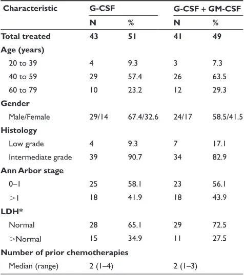
Patient demographics are summarized in Table 1. There were no statistically significant differences between the two treatment groups with regard to demographic or clinical characteristics.
Apheresis data
All the apheresis products were negative for monoclonal B cells by flow cytometric analysis. Thirty-nine of 43 patients (90.7% with 90% CI: 82.7–96.6%) in the G-CSF arm and 35 of 41 patients (85.4% with 90% CI: 76.0–93.3%) in the G+GM arm successfully collected 4 × 10⁶ CD34+
cells/kg body weight. The probability that the G-CSF arm
Table 1 Baseline patient characteristics
Characteristic G-CSF G-CSF + GM-CSF
N % N %
Total treated 43 51 41 49
Age (years)
20 to 39 4 9.3 3 7.3
40 to 59 29 57.4 26 63.5
60 to 79 10 23.2 12 29.3
Gender
Male/Female 29/14 67.4/32.6 24/17 58.5/41.5
Histology
Low grade 4 9.3 7 17.1
intermediate grade 39 90.7 34 82.9
Ann Arbor stage
0–1 25 58.1 23 56.1
1 18 41.9 18 43.9
LDH*
normal 28 65.1 29 72.5
normal 15 34.9 11 27.5
Number of prior chemotherapies
Median (range) 2 (1–4) 2 (1–3)
Notes: *One patient on the g-csF + gM-csF treatment arm has no data on LDH.
Abbreviations: g-csF, granulocyte colony-stimulating factor; gM-csF, granulocyte-macrophage colony-stimulating factor; LDH, lactate dehydrogenase.
Journal of Blood Medicine downloaded from https://www.dovepress.com/ by 118.70.13.36 on 24-Aug-2020
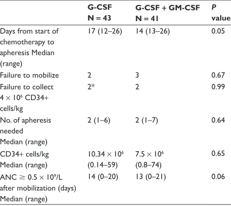
has a higher success rate than the G+GM arm is 0.778. The median CD34+ cell dose collected was 10.34 × 10⁶/kg (range,
0.14–59) in G-CSF arm and 7.5 × 10⁶/kg (range, 0.79–73.7)
in the G+GM arm (P = 0.65). Median numbers of apheresis procedures required were two in both arms (P = 0.64). Collection started a median of 17 days (range, 12–26) from the start of chemotherapy in the G-CSF arm and 14 days (range, 13–26) in the G+GM arm (P = 0.06). These results are summarized in Table 2.
count recovery
The median time to absolute neutrophil count (ANC) of 0.5 × 10⁹/L was 14 days (range, 0–20) in the G-CSF arm
and 13 days (range, 0–21) in the G+GM arm (P = 0.05) as shown in Table 2.
Toxicity data
No unexpected toxicities were observed. Commonly experienced toxicities included bone pain and myalgias secondary to cytokine administration, cytopenias related to chemotherapy, and febrile neutropenia. No one expe-rienced grade 4 or 5 toxicity. The most common grade 3 toxicities that were observed are summarized in Table 3a (CTCAE v 3.0).7 The number of patients who
experi-enced a grade 3 adverse event was similar in the G-CSF and G+GM group (11 versus 13 patients, respectively; P = 0.3) (Table 3b).
Overall and disease-free survival
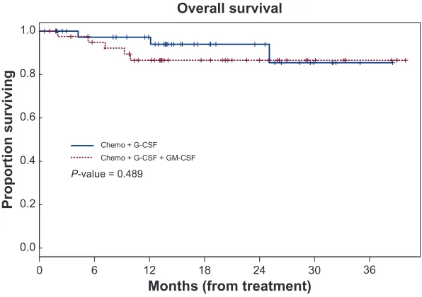
After a median follow up time of 14.5 months (range, 0.6–38.5) in the G-CSF arm and 14.0 months (range, 1.1–39.9) in the G+GM arm, the 3-year overall survival (OS) was 85% (95% CI: 69.5%–100%) and 86.6% (95% CI: 76.2%–98.3%), respectively (P = 0.489). The 3-year disease-free survival (DFS) was 75.8% (95% CI: 57.9%–99.4%) in the G-CSF arm and 77.1% (95% CI: 65.0%–91.5%) in the G+GM arm as shown in Figure 1.
The OS for patients with intermediate grade NHL was 83.7% (95% CI: 72.2%–97.0%) at 3 years. None of the patients with low grade NHL died. The DFS for patients with intermediate grade NHL was 72.8% (95% CI: 60%–88.4%) at 3 years. None of the patients with low grade NHL had recurrent disease.
graft composition
A total of 54 patients (25 G-CSF and 29 G+GM) provided paired baseline and post-apheresis samples DC subset
Table 2 Apheresis data
G-CSF
N = 43 G-CSF + GM-CSF N = 41 P value
Days from start of chemotherapy to apheresis Median (range)
17 (12–26) 14 (13–26) 0.05
Failure to mobilize 2 3 0.67
Failure to collect 4 × 10⁶ cD34+
cells/kg
2* 2 0.99
no. of apheresis needed Median (range)
2 (1–6) 2 (1–7) 0.64
cD34+ cells/kg Median (range)
10.34 × 10⁶
(0.14–59)
7.5 × 10⁶
(0.8–74)
0.65
Anc 0.5 × 10⁹/L
after mobilization (days) Median (range)
14 (0–20) 13 (0–21) 0.06
Notes: *One patient was noncompliant with therapy.
Abbreviations: Anc, absolute neutrophil count; g-csF, granulocyte colony-stimulating factor; gM-csF, granulocyte-macrophage colony-colony-stimulating factor.
Table 3a Toxicity data: grade 3 adverse events (cTcAe v 3.0)
Adverse event G-CSF G-CSF/GM-CSF Total
Bone pain 1 0 1
cytokine release syndrome
0 2 2
Drug fever 0 1 1
esophagitis 1 0 1
Febrile neutropenia 6 7 13
Fever/infection without neutropenia
1 1 2
Muscle weakness (generalized)
0 1 1
nausea/vomiting 2 0 2
Thrombocytopenia 1 0 1
Anemia 2 1 3
syncope 0 1 1
Total 14 14 28
Abbreviations: g-csF, granulocyte colony-stimulating factor; gM-csF, granulocyte-macrophage colony-stimulating factor.
Table 3b number of patients by maximum grade of adverse
event
Maximum rade G-CSF G + GM Total
3 11 13 24
2 10 15 25
1 19 12 31
Total 40 40 80
Notes: There appear to be no statistically significant differences between treatment groups with respect to the number of patients with maximum grade adverse event. Fisher’s exact test P-value = 0.2704.
Journal of Blood Medicine downloaded from https://www.dovepress.com/ by 118.70.13.36 on 24-Aug-2020
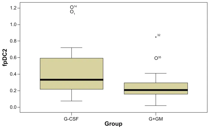
analysis. There was no significant difference between the percentages of mDC subsets in the apheresis products in the two arms. However, there was a significant increase in the percentage of pDC subsets in the apheresis product in the G-CSF arm when compared to G+GM (P = 0.002); consequently, there was a significant decrease in the ratio of mDC and pDC (mDC/pDC) with G-CSF when compared to G+GM (P = 0.029) (Table 4 and Figure 2).
Discussion
Although G-CSF and GM-CSF are widely used for stem cell mobilization in normal donors and in patients with hematological malignancies who are to undergo an autologous stem cell transplant, there is still controversy regarding the role of the two cytokines in mobilizing DC cells and their subsets. The clinical impact, of either of the two mobilization
strategies in patients undergoing autologous PBSC transplan-tation is also unclear. Our study shows that both regimens are well tolerated. The most common grade 3 toxicity was febrile neutropenia. The stem cell yield, number of apheresis pro-cedures required to reach a target cell dose of 4 × 10⁶/kg and
the overall and disease free survival were similar in the two groups. Previous studies have shown that GM-CSF when used alone or in combination with G-CSF mobilizes more mature (CD14-/CD80+ or CD86- cells) and precursor dendritic cells when compared to G-CSF alone.5,6,8–12 What is unclear,
how-ever, is whether there is polarization of these dendritic cells to myeloid dendritic cells or plasmacytoid dendritic cells and its clinical significance. Several groups have reported polar-ization to pDC cells in normal donors who received G-CSF for stem cell mobilization.13–15 In contrast Shaughnessy et al
did not observe any polarization of DCs by either G-CSF or GM-CSF in normal donors, however donors receiving G-CSF mobilized more mDC and pDC cells.16 Gazitt et al did not find
a major difference in the ratio of mDC/pDC cells in the PB of patients with NHL mobilized with cyclophosphamide plus G-CSF compared with patients mobilized with either cyclo-phosphamide plus CSF or cyclocyclo-phosphamide plus GM-CSF followed by G-GM-CSF.12 Others have reported polarization
to pDC with G-CSF17–18 or no polarization of DC subsets.19
Sanchez et al11 found that in patients with acute myelogenous
leukemia who were mobilized with G-CSF alone, there was polarization towards the mDC subset.
Our data show that there was no difference in the total numbers of DC cells mobilized in the two groups; how-ever, we found a polarization towards the pDC subset in
Table 4 changes in the percentages of mDc, pDc and the mDc/
pDc pre and post mobilization with g-csF or g+gM
Median
(mean ± SEM) G-CSF N = 25 G+GM N = 29
P value
%mDC
Pre 55.6 (49.9 ± 3.53) 56.9 (57.2 ± 2.78) 0.125 Post 17.6 (22.5 ± 2.76) 17.8 (19.3 ± 2.11) 0.633
%pDC
Pre 18.9 (21.6 ± 2.83) 22.5 (22.1 ± 1.84) 0.549 Post 7.6 (9.25 ± 1.32) 4.8 (4.82 ± 0.57) 0.002
mDC/pDCmDC
Pre 2.67 (4.80 ± 1.47) 2.68 (3.53 ± 0.52) 0.924 Post 2.52 (3.72 ± 0.80) 4.02 (7.21 ± 2.30) 0.029
Abbreviations: Dc, dentric cells; g-csF, granulocyte colony-stimulating factor; gM, granulocyte-macrophage; seM, standard error of mean.
Months (from treatment) Overall survival
Proportion survivin
g
0.0
0 6 12 18 24 30 36
0.2 0.4 0.6 0.8 1.0
Chemo + G-CSF Chemo + G-CSF + GM-CSF
P-value = 0.489
Figure 1 Kaplan–Meier overall survival.
Abbreviations: g-csF, granulocyte colony-stimulating factor; gM-csF, granulocyte-macrophage colony-stimulating factor.
Journal of Blood Medicine downloaded from https://www.dovepress.com/ by 118.70.13.36 on 24-Aug-2020
patients mobilized with G-CSF alone. We did not observe a survival difference in the two arms of the study at least with the short follow-up time of 14 months. On the contrary Gazitt et al12 reported that there was a survival
advantage for patients mobilized with GM-CSF contain-ing regimens. Similarly Dean et al also found that higher numbers of mDC cells and total DCs were significantly associated with improved survival.1 These differences may
be explained by the different patient populations studied and whether the evaluations were done on the PB or in apheresis products.
Conclusion
Mobilization of peripeheral blood progenitor cells with a combination of G-CSF plus GM-CSF results in the mobilization of higher numbers of pDC cells. However, this did not seem to have any clinical advantage.
Acknowledgments
This study was partially supported by a research grant from Bayer Health Corporation. The authors report no conflicts of interest in this work.
References
1. Dean R, Masci P, Pohlman B, et al. Dendritic cells in autologous hematopoietic stem cell transplantation for diffuse large B-cell lymphoma: graft content and post transplant recovery predict survival.
Bone Marrow Transplant. 2005;36:1049–1052.
2. Porrata LF, Gastineau DA, Padley D, Bundy K, Markovic SN. Re-infused autologous graft natural killer cells correlates with absolute lymphocyte count recovery after autologous stem cell transplantation.
Leuk Lymphoma. 2003;44:997–1000.
3. Porrata LF, Gertz MA, Geyer SM, et al. The dose of infused lymphocytes in the autograft directly correlates with clinical outcome after autologous peripheral blood hematopoietic stem cell transplantation in multiple myeloma. Leukemia. 2004;18:1085–1092.
4. Siena S, Schiavo R, Pedrazzoli P, Carlo-Stella C. Therapeutic relevance of CD34 cell dose in blood cell transplantation for cancer therapy.
J Clin Oncol. 2000;18:1360–1377.
5. Gazitt Y. Comparison between granulocyte colony-stimulating factor and granulocyte-macrophage colony-stimulating factor in the mobilization of peripheral blood stem cells. Curr Opin Hematol. 2002;9:190–198.
6. Gazitt Y, Shaughnessy P, Devore P. Mobilization of dendritic cells and NK cells in non-Hodgkin’s lymphoma patients mobilized with different growth factors. J Hematother Stem Cell Res. 2001;10:177–186. 7. Trotti A, Colevas AD, Setser A, et al. CTCAE v3.0: development
of a comprehensive grading system for the adverse effects of cancer treatment. Semin Radiat Oncol. 2003;13:176–181.
8. Gazitt Y. Recent developments in the regulation of peripheral blood stem cell mobilization and engraftment by cytokines, chemokines, and adhesion molecules. J Hematother Stem Cell Res. 2001;10: 229–236.
9. Bernhard H, Disis ML, Heimfeld S, Hand S, Gralow JR, Cheever MA. Generation of immunostimulatory dendritic cells from human CD34+
hematopoietic progenitor cells of the bone marrow and peripheral blood.
Cancer Res. 1995;55:1099–1104.
10. Avigan D, Wu Z, Gong J, et al. Selective in vivo mobilization with granulocyte macrophage colony-stimulating factor (GM-CSF)/ granulocyte-CSF as compared to G-CSF alone of dendritic cell progenitors from peripheral blood progenitor cells in patients with advanced breast cancer undergoing autologous transplantation. Clin
Cancer Res. 1999;5:2735–2741.
11. Sanchez J, Herrera C, Torres A, Roman-Gomez J, Alvarez MA. Chemotherapy plus G-CSF mobilized peripheral blood stem cell harvests from acute myeloid leukemia patients contain large amounts of polyclonal myeloid linnegCD11cpos dendritic precursor cells.
Br J Haematol. 2004;124:636–644.
12. Gazitt Y, Akay C, Thomas C 3rd. No polarization of type 1 or type 2 precursor dendritic cells in peripheral blood stem cell collections of non-hodgkin’s lymphoma patients mobilized with cyclophosphamide plus G-CSF, GM-CSF, or GM-CSF followed by G-CSF. Stem Cells
Dev. 2006;15:269–277.
1.2
O1 O14
O35 1.0
0.8
0.6
0.4
0.2
0.0
G-CSF
Group
fpDC2
G+GM *32
Figure 2 Percentage fold change in pDc subsets compared to baseline. Notes: Fold% Dc2, P= 0.002.
Abbreviations: Dc, dentric cells; g-csF, granulocyte colony-stimulating factor; g+gM, granulocyte + granulocyte-macrophage.
Journal of Blood Medicine downloaded from https://www.dovepress.com/ by 118.70.13.36 on 24-Aug-2020
Journal of Blood Medicine
Publish your work in this journal
Submit your manuscript here: http://www.dovepress.com/Journal-of-blood-medicine-journal
The Journal of Blood Medicine is an international, peer-reviewed, open access, online journal publishing laboratory, experimental and clinical aspects of all topics pertaining to blood based medicine including but not limited to: Transfusion Medicine; Blood collection, Donor issues, Transmittable diseases, and Blood banking logistics; Immunohematology; Artificial and alternative
blood based therapeutics; Hematology; Biotechnology/nanotechnology of blood related medicine; Legal aspects of blood medicine; Historical perspec-tives. The manuscript management system is completely online and includes a very quick and fair peer-review system. Visit http://www.dovepress.com/ testimonials.php to read real quotes from published authors.
Dove
press
13. Arpinati M, Green CL, Heimfeld S, Heuser JE, Anasetti C. Granulocyte-colony stimulating factor mobilizes T helper 2-inducing dendritic cells.
Blood. 2000;95:2484–2490.
14. Hock BD, Haring LF, Ebbett AM, Patton WN, McKenzie JL. Differential effects of G-CSF mobilisation on dendritic cell subsets in normal allogeneic donors and patients undergoing autologous transplantation. Bone Marrow Transplant. 2002;30:733–740. 15. Klangsinsirikul P, Russell NH. Peripheral blood stem cell harvests
from G-CSF-stimulated donors contain a skewed Th2 CD4 phenotype and a predominance of type 2 dendritic cells. Exp Hematol. 2002;30: 495–501.
16. Shaughnessy PJ, Bachier C, Lemaistre CF, Akay C, Pollock BH, Gazitt Y. Granulocyte colony-stimulating factor mobilizes more den-dritic cell subsets than granulocyte-macrophage colony-stimulating factor with no polarization of dendritic cell subsets in normal donors.
Stem Cells. 2006;24:1789–1797.
17. Lonial S, Hicks M, Rosenthal H, et al. A randomized trial comparing the combination of granulocyte-macrophage colony-stimulating factor plus granulocyte colony-stimulating factor versus granulocyte colony-stimulating factor for mobilization of dendritic cell subsets in hematopoietic progenitor cell products. Biol Blood Marrow Transplant. 2004;10:848–857.
18. Morse MA, Vredenburgh JJ, Lyerly HK. A comparative study of the generation of dendritic cells from mobilized peripheral blood progenitor cells of patients undergoing high-dose chemotherapy. J Hematother
Stem Cell Res. 1999;8:577–584.
19. Ferrari S, Rovati B, Porta C, et al. Lack of dendritic cell mobilization into the peripheral blood of cancer patients following standard- or high-dose chemotherapy plus granulocyte-colony stimulating factor.
Cancer Immunol Immunother. 2003;52:359–366.
Journal of Blood Medicine downloaded from https://www.dovepress.com/ by 118.70.13.36 on 24-Aug-2020