Optical Biosensors based on Silicon-On-Insulator
Ring Resonator: A Review
Patrick Steglich1,2,∗, Marcel Hülsemann3, Birgit Dietzel2and Andreas Mai1,2
1 IHP – Leibniz-Institut für innovative Mikroelektronik, Im Technologiepark 25, 15236 Frankfurt (Oder),
Germany
2 Technische Hochschule Wildau, Hochschulring 1, 15745 Wildau, Germany
3 First Sensor AG, Peter-Behrens-Straße 15, 12459 Berlin, Germany
* Correspondence: Patrick Steglich (steglich@ihp-microelectronics.com)
Abstract: Recent developments in optical biosensors based on integrated photonic devices are reviewed with a special emphasis on silicon-on-insulator ring resonator. The review is mainly devoted to the following aspects: (1) Principles of sensing mechanism, (2) sensor design, (3) biofunctionalization procedures for specific molecule detection and (4) measurement set-ups and advances in chip-integration. The inherent challenges of implementing photonics-based biosensors to meet specific requirements of applications in medicine, food analysis, and environmental monitoring are discussed.
Keywords: Biosensors; Biophotonics; Integrated optical sensors; Aptamers; Biomaterials; Optical sensor; Silicon photonics; Ring resonators; Lab-on-a-chip.
1. Introduction
Silicon-based photonic biosensors integrated into a semiconductor chip technology can lead to major advances in point-of-care applications, food diagnostics, and environmental monitoring through the rapid and precise analysis of various substances. In recent years, there has been an increasing interest in sensors based on photonic integrated circuits (PIC) because they give rise to cost effective, scalable and reliable on-chip biosensors for a broad market.
The silicon-on-insulator (SOI)-technology is the most attractive technology for PICs from commercial point of view since it provides a scalable platform for mass production and the opportunity for monolithic integration of electronic and photonic devices, which is known as electronic photonic integrated circuits (EPIC) [1]. This allows the integration of sensors, detectors and read-out electronics in a single chip.
Once the photonic chip is fabricated, the silicon surface of the sensor can be coated with a covalently attached sensing layer. This layer determines the specific detection and, hence, the application. This step, however, is independent from the fabrication of the chip, making the SOI-technology attractive for both, science and industry. A further advantage of SOI-based biosensors is the possibility to realize sensor arrays. This allows for the detection of several substances in parallel (multiplexing) [2].
The photonic biosensor can be realized by utilizing interferometric or resonant structures. The former one is usually based on a Mach-Zehnder interferometer [3] configuration and the later most often on a ring resonator [4]. Ring resonators, however, have the advantage of a high sensitivity and small footprint, which allows for a dense integration.
Possible fields of application for SOI-based ring resonators are for example, but not limited to, the detection of antibiotics in milk, monitoring of pesticides and hormones in water, point-of-care devices for the diagnosis of cancer, infections, cardiovascular diseases, and other pathological states. Table 1 gives an overview of competitive biosensing techniques. The main advantage of SOI-based ring resonators is their small size and fast readout as well as the possibility for low cost, portable devices for point-of-care applications.
In this work, we focus on SOI-based ring resonators and provide in the first part an overview of the working principle and sensing mechanism. The detection limits and integration challenges are critically discussed. In the second part, we review recent advances on surface functionalization and report on the detection of various biomarkers. Finally, we discuss typical experimental set-ups and recent developments regarding integration approaches.
Table 1.Comparison of different biosensing techniques.
pros cons Ref.
enzyme-linked
immunosorbent assay (ELISA)
high selectivity, portable expensive, time consuming [5]
electrochemical sensor high selectivity, fast expensive, sample
pre-treatment and
pre-concentration required
[6]
bilayer lipid membranes (BLM)
high selectivity, low cost, fast, portable, small
detection limit [7]
high performance liquid chromatography (HPLC)
high selectivity expensive, time consuming, non-portable, sample pre-treatment required
[8,9]
micro-electrode immunoassay
high selectivity, low cost non-portable, time consuming
[10]
Field immunoassay high selectivity, low cost, fast, easy to use
non-portable [11]
surface plasmon
resonance spectroscopy (SPR)
high selectivity, fast expensive, non-portable [12,13]
optical ring resonators high selectivity, fast, low cost, portable, multiplexing
sample pre-concentration required
this work
2. Photonic devices and sensing mechanisms
This section provides an introduction to photonic biosensors based on ring resonators and to the underlying sensing mechanism. Further, we discuss recent advances in device design and operation principles.
2.1. Operation principle
This work is focusing on photonic sensors based on optical ring resonators in a SOI-technology. A ring resonator is composed of a silicon-based waveguide on top of a buried oxide substrate. In general, the light of a tunable laser or a superlumineszenzdiode is coupled to the waveguide via grating coupler or by butt coupling. The light is then partly coupled to the ring resonator if the resonance condition is fulfilled leading to resonance peaks at the output spectrum, as illustrated in Figure 1(a). At the output, the light is coupled to a photodetector or an optical spectrum analyzer depending on the light source. Current advances in heterogeneous as well as monolithically integration give rise to implement laser [14] and photodiodes [15] on the same chip together with the photonic sensors.
Figure 1. (a) Schematic representation of a silicon-on-insulator ring resonator. According to the resonance condition, only selected wavelengths can propagate in the ring and distinct resonance peaks appear in the output spectrum. (b) Molecular binding takes place if a sample of the analyte gets in touch with the adsorbed layer on top of the silicon waveguide leading to a resonance wavelength shift ∆λ.
However, the general sensing mechanism underlying their operation is evanescent field sensing. If the evanescent field is altered due to an immobilization of analytes on the silicon waveguide, the resonance condition of the ring resonator is changed leading to a resonance wavelength shift. In this way, antibodies that only attach to their corresponding antigens are detected with high specificity by detecting either the resonance peak shift∆λor the intensity change∆I. Ones the analyte-antibody binding took place; the residuals can be removed by drying or flushing to enhance the specific measurement. The wavelength shift can be calculated from resonator metrics, that is [17]
∆λ= λres
ng ∆nclad, (1)
wherengis the group index. It can be determined by
ng=ne f f−λ δne f f
δλ . (2)
Assuming a small resonance wavelength shift and, hence, a flat dispersion of the effective refractive index, the group index can be calculated by
ng≈ne f f = λ20 FSR
1
Lring, (3)
whereLringis the ring circumference.
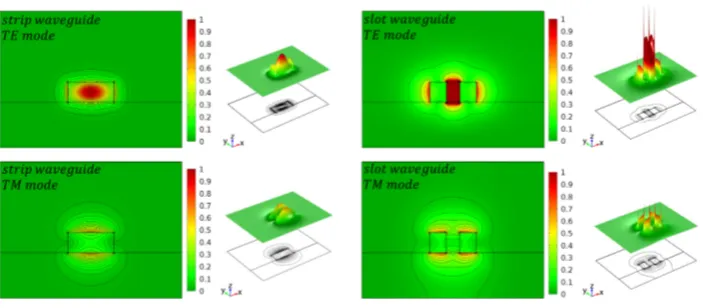
Figure 2.Typical silicon-on-insulator waveguide geometries for optical biosensing.
Figure 3.Simulation of the normalized E-field intensity for the first TE- and TM-mode for a strip and slot waveguide. Reproduced from Ref. [18] (CC BY 4.0).
appropriated waveguide type for a specific application can be found in Refs. [21,22]. The waveguide sensitivity is given by
Swg = ∆ne f f
∆nclad
, (4)
where ne f f represents the effective refractive index of the waveguide and nclad is the cladding refractive index. Such a definition is useful for waveguide optimization through simulation studies. However, the ring resonator sensitivity depends not only on the waveguide geometry and, therefore, a second definition defining the ring resonator sensitivity is given by
Srr= ∆λ
∆ne f f
. (5)
Here,∆λis a small shift of the resonance peak. Taken both definitions into account, we get the overall photonic device sensitivity defined by
S=SwgSrr= ∆ne f f
∆nclad
∆λ
∆ne f f
= ∆λ
∆nclad
. (6)
by the measurement set-up. For example, an optical spectrum analyzer has a typical wavelength resolution of 20 pm. This measurement resolution (MR) can be also expressed with the system noise variance by
MR=∆λmin=3σλ. (7)
This leads to theLODgiven by
LOD= ∆λmin
S . (8)
To get a metric which is independently from the measurement set-up, an intrinsic LOD(iLOD) is necessary [23]. It can be obtained by setting the measurement resolutionMR as full width at half maximum (FW HM) of the resonance peak, which leads to
iLOD= FW HM
S =
λ0
QS, (9)
whereλ0denotes the resonance wavelength andQthe optical quality factor, which is determined by
Q= λ0
FW HM. (10)
Finally, we provide a strategy to improve the waveguide geometry by design. Towards this, we consider the most important characteristics of integrated photonic biosensors, which can be devided into five categories [4]:
1. Increasing the waveguide sensitivity Swg increases the light-analyte-interaction. In fact, this determines the wavelength shift∆λand has a strong impact on the overall sensitivity.
2. Enhancing the ring resonator sensitivity Srr, which determines the wavelength shift ∆λ depending on the refractive index change ∆ne f f. This can be achieved by increasing the light-matter interaction using slot waveguide structures.
3. A smallFW HM, i.e. a high Q-factor, impacts the sensitivity of ring resonator sensors since the impact of noise on the determination of the resonance wavelength will be reduced [24,25]. A higher Q-factor leads to a lower attenuation in the ring and minimizes the smallest detectable wavelength shift∆λand consequently the detection limit.
4. A small footprint is directly related to the detection time and reduces the area consumption and therefore device costs significantly. Further, this allows a high integration density, which is of special interest for multiplexing.
5. Compatibility with a semiconductor production platform, which gives the ability for an industrial production flow. The compatibility with an electronic-photonic integrated circuit (EPIC) allows for a monolithic integration.
As mentioned before, each waveguide-type has advantages and disadvantages and therefore, a design trade-off regarding sensitivity and optical losses is necessary. Recently, a hybrid-waveguide ring resonator was proposed to combine a strip waveguide with a slot waveguide in such a way that the figure of meritFOM=Srr/FW HMis maximized [4]. Figure4shows a schematic representation of this SOI ring resonator, which consists of both a strip waveguide and a slot waveguide. This type of ring resonator has been demonstrated to have an improved figure of merit compared to more common strip or slot waveguide-based ring resonators, as it is summarized in Table2. A comparative study on the sensor performance of slot and strip waveguide ring resonators is given in Ref. [26]. Here, glucose level monitoring in blood samples in the range 10 to 200 mg/dL using minimal invasive technique is simulated. Additionally, a six times higherSrr of the slot waveguide ring resonator is estimated using the Finite Element Method (FEM).
Figure 4. Schematics of different ring resonator concepts: (a) Common silicon strip-waveguide ring resonator. (b) Fully slotted ring resonator with a strip-waveguide as bus waveguide. (c) Hybrid-waveguide ring resonator consisting of a slot- and strip waveguide. The strip-to-slot optical mode transition is achieved by a slow-varying waveguide taper. ( c2018 IEEE. Reprinted, with permission, from Ref. [4])
another regime that allows to reduce the detection limit. This method were several times adopted and highly sensitive biosensors were demonstrated that exceed the sensitivity of more common single-ring sensors [29–31].
Table 2.Comparison of different ring resonators based on SOI-technology. ( c2018 IEEE. Reprinted, with permission, from Ref. [4])
slot-waveguide strip-waveguide hybrid-waveguide
footprint [µm2] 130 100 2,720
Srr[nm/RIU] 298 70 106.29
Q 330 20,000 18,500
FOM 63 903 1,337
Ref. [32] [24] [4]
It is also notable that advances on planar silicon ring resonators with innovative guiding structures have been theoretically investigated recently. Such resonator structures show ring resonator sensitivities of up to 120 nm/RIU and high Q-factors of 105 [33], which could result in a record highFOMof about 7, 742. More recently, polarization independent slot-waveguide structures were theoretically demonstrated to double the waveguide sensitivity [34]. More recently, in 2019, sub-wavelength grating (SWG) waveguides have been demonstrated to exhibit a bulk sensitivity up to 579.5 nm/RIU and a surface sensitivity of 1900 pm/nm [35]. These results show the potential for integrated high sensitive optical biosensors in a SOI technology and give prospective to further improvements.
3. Functionalization procedures and applications
In this section we give a basic introduction of to label-free functionalization procedures and a short overview of recent advances in the bio-functionalization of photonic sensors based on SOI-technology.
inflammation, for example, can support the decision of the correct medicine to provide primary medical care inside and outside of doctors’ offices and hospitals as well as to monitor therapeutic interventions.
For experimental development antibody-antigen model systems like anti bovine serum albumin (antiBSA)-bovine serum albumin (BSA) [36] are typically used for proof of concepts. In a standard procedure (e.g. ELISA), the high specificity and affinity biotin-streptavidin biotin binding is widely used as linker. Therefore, this system is also used as model system for proof of concepts and to validate SOI ring resonators [24,37,38]. In general, the biospecific interaction is following the key-lock principle allowing for the selection of one specific particle of one million particles.
Over the past 10 years, several researchers have successfully demonstrated functionalized SOI ring resonators for the detection of acute inflammations, viral diseases and cancer by biomarkers such as proteins [2,24,37], interleukins [39], nucleic acids [40,41], and viruses [42]. A brief overview is given in table3.
Table 3.Examples of application and selection of biomolecules that have been detected by integrated photonic biosensors based on SOI ring resonators.
Application Analyte/Biomarker Receptor/Target Detection limit Ref.
Acute inflammation C-reactive protein (CRP) Anti-CRP 6.5 pM [39,43] Acute inflammation Interleukin 2,4,5 Anti-CRP 6−100 pM [43,44]
HIV Human immunoglobulin (Hu-IgG) Anti-Hu-lgG 1 ng [45]
Hepatitis Human serum albumin Anti-Albumin 3.4 pg [24]
Meningitis tmRNA DNA 0.524 nM [41]
Prostate cancer Prostate specific antigen (PSA) Anti-PSA 0.4 nM [2,46]
Liver cancer α-fetoprotein (AFP) Anti-AFP 100 pM [2]
Bowel cancer Carcinoembrionic antigen (CEA) Anti-CEA 10 pM [47] Bladder cancer Tumor necrosis factor (TNF) Antibody 100 pM [44] Model system Green fluorescent protein (GFP) Antibody 0.1 mg/ml [46]
Model system Streptavidin Biotin 60−150 fM [24,37,38]
Food monitoring Bean pod mottle virus Antibody 1.43 pM [42]
For this purpose, the silicon surface of the ring resonator has to be functionalized with the corresponding bioactive receptors. The coupling of these bioactive receptors to the silicon surface can be covalently or adsorptivly immobilized.
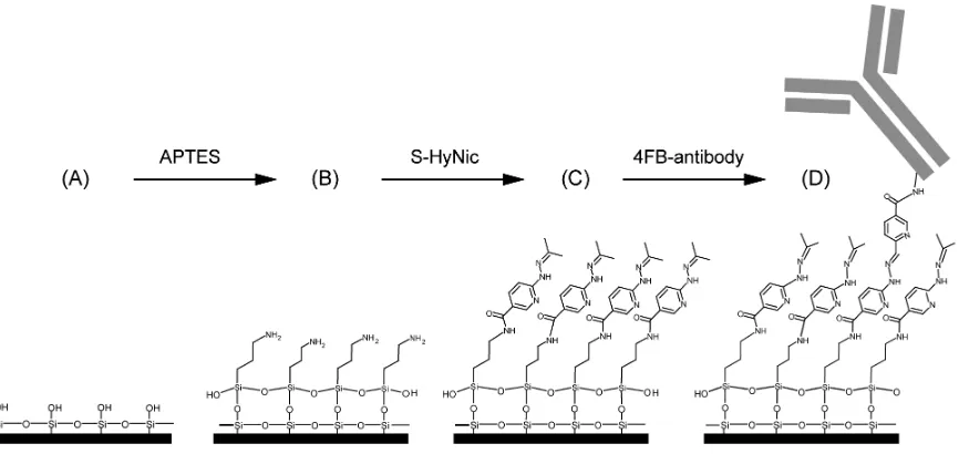
Covalent immobilization gives a tight binding of the organic receptors on the inorganic silicon surface. As a rule, up to four reaction steps (A-D) are required for this, as shown in Figure5by means of an example from Ref. [47]:
1. (A) Surface activation
The surface activation carried out by cleaning with Piranha solution or hydrogen peroxide-ammonium hydroxide solution and an argon plasma to generate hydroxyl groups for the following functionalization step.
2. (B) Functionalization
As coupling, agents are often bifunctional organosilane (in Figure 5, for example, (3-Aminopropyl)triethoxysilane (APTES)) of the general formula R3-Si-(CH2)n-X with hydrolysable groups R (OCH3, CH2CH3, Cl, F, SH) used. The choice of functional groups X (NH2, epoxy, SH, C=C) depending on the desired specification. The condensation of these with the surface hydroxyl groups results in the formation of siloxane bonds (Si-O-Si). Such coupling leads to monolayers that is covalent bonded on the silicon surface and therefore among the most stable.
3. (C) Linker
(BS3), but may also carry two different functional groups, e.g. N-γ-maleimidobutyryl oxysuccinimide ester (GMBS) is an amine-to-sulfhydryl crosslinker that contains NHS esters and maleimide reactive groups at opposite ends of a short spacer arm. In the example use the succinimidyl-6-hydrazino-nicotinamide (S-HyNic), a heterobifunctional crosslinker reacts with the amino-modified surfaces.
4. (D) Immobilisation of receptor
Immobilization of biomolecules as receptore such as antibodies first requires the modification of these. Thus, biotinylation can introduce functionality into the biomolecule that selectively addressed for coupling. In the lower example, a 4-formylbenzamide (4FB)-modified antibody to form a stable covalent hydrazone linkage at the 6-hydrazinonicotinamide (HyNic) moieties of the linker.
Figure 5.Representative example for surface functionalization. (A) Silicon surface of an unmodified SOI ring resonator. (B) In order to generate an amino-terminated surface, APTES is reacting with the surface siloxane groups. (C) Afterwards, S-HyNic is reacting with primary amines to build a HyNic-displaying surface. (D) Finally, the addition of 4FB-modified antibodies leads to a hydrazone bond formation between the 4FB moieties on the antibodies and the HyNic moieties on the surface. Reprinted (adapted) with permission from Ref. [47]. Copyright (2018) American Chemical Society.
On the one hand, this allows a stable binding but, on the other hand, it requires a sophisticated synthetization, but the application justifies the effort.
Adsorptive immobilization via ionic or van der Waals interactions is easier to use, and allows for fast measurements without specific reagents and is applied despite the disadvantages how low sensitivity due to an incorrect orientation, show in Figure6(a). Using a protein layer, Caroselli and co-workers improved the alignment of antibody receptors [36]. However, adsorption is the weakest compound because it can be resolved by varying pH, temperature or ionic strength changes. Another problem is the possible inactivation due to the change in the 3D-structure of the biomolecule after adsorption on the sensor surface. For this reason, the covalent immobilization is preferred for a highly sensitive and selective measurement.
Figure 6. (a) The antibody receptors are usually randomly-oriented on the silicon surface when they are directly immobilized using physical adsorption. (b) Using a protein A layer leads to properly-oriented antibody receptors. Reproduced from Ref. [36] (CC BY 4.0).
One great advantage of integrated photonic biosensor is the ability for multiplexing making this technology attractive for diagnostics and interaction screening [49]. For example, Luchansky et al. have demonstrated a fast multiplexing system using 32-element arrays of ring resonators to quantify several species with excellent time-to-result [44,50]. In particular, they have detected the cytokines interleukin-2 (IL-2), interleukin-4 (IL-4), interleukin-5 (IL-5), and tumor necrosis factor alpha (TNFα) in parallel with high accuracy in serum-containing cell media.
Recent developments in antifouling coatings have led to a further reduction of nonspecific protein binding to the sensor surface. For example, Jäger et al. [51] have examined methylated dendritic polyglycerol (dPG(OMe)) as a protective layer. In this case, fibrinogen was used to test the antifouling properties. A reduction of 87% in the binding of fibrinogen to the silicon surface was demonstrated by using a SOI rib waveguide-based ring resonator.
4. System integration
In this section we discuss different measurement set-ups used in laboratories and review current advances to integrate them into a SOI platform.
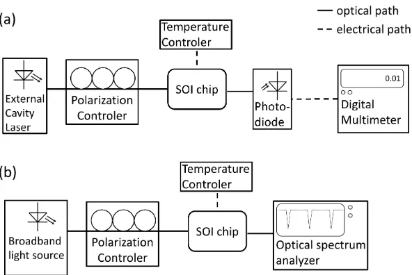
The most common set-up is shown in Figure 7. It comprises either a tunable laser source in combination with a photodiode (Figure7(a)) or a broadband light source with a optical spectrum analyzer (Figure7(b)). The polarization of the light is typically adjusted by a 3-paddle polarization controller. To avoid temperature fluctuations, the sample holder is heated just above room temperature. The main disadvantage of this measurement is the light coupling since it requires a precise adjustment of optical fibers.
To avoid fiber coupling, current research in SOI technology is focusing on the integration of light sources and photodiodes. While Ge-photodiodes have been successfully integrated in a SOI platform [15,52–54], the integration of laser sources is still challenging [55]. Current approaches employ wafer-to-wafer [56,57] or die-to-wafer [14,58,59] bonding.
Figure 7. Schematic of typical measurement set-ups in laboratories. (a) The light source consists of an external cavity laser, which can tune its wavelength (tunable laser). In this case, a photodiode can be used as detector. (b) If a broad band light source (e.g. superluminescent diode) is employed, an optical spectrum analyzer is needed on the detector side.
Current integration issues are related to chip packaging. Since the integration of light sources, photodetectors and readout require conductive interconnects and occupy many space, a backend of line (BEOL) until five metal levels are necessary for a monolithic integration. This, on the other hand, requires a relatively deep etching procedure through the BEOL in order to release the sensing area (ring resonator). This leads to a high aspect ratio and makes surface functionalization and the implementation of micro-fluids challenging. Therefore, current integration approaches prefer a hybrid integration; i.e. the integration light source and detector unit on a separate chip. The disadvantage of this approach is the sophisticated optical interconnection between each chip such as, for example, photonic wire bonding realized by direct-write two-photon lithography [63–65]. Therefore, the system integration still requires further developments and is in the focus of current research.
5. Conclusion
Biosensors based on SOI ring resonators are reviewed and discussed. The theoretical background in terms of waveguide and resonator sensitivity as well as detection limits is provided and current developments in ring resonator geometries are reviewed. An overview of functionalized SOI ring resonators and their applications is provided. Finally, experimental set-ups for the optical characterization are described and current integration approaches are reviewed.
Acknowledgments: This work is funded by European Regional Development Fund (10.13039/501100008530). We acknowledge support by the German Research Foundation and the Open Access Publication Funds of the TH Wildau.
Author Contributions:This article was jointly written by and proof-read by all authors. All authors contributed in various degrees to the review.
Conflicts of Interest:The authors declare no conflict of interest.
References
Figure 8.Schematic of ring resonator array. Each ring is separately addressed in the electronic regime to individually measure the transmission. The sinusoidal input signal is divided in certain time slots. Adopted from Ref. [62].
electronic-photonic integrated circuits. Electron Devices Meeting (IEDM), 2015 IEEE International. IEEE, 2015, pp. 15–6.
2. Washburn, A.L.; Luchansky, M.S.; Bowman, A.L.; Bailey, R.C. Quantitative, Label-Free Detection of Five Protein Biomarkers Using Multiplexed Arrays of Silicon Photonic Microring Resonators. Analytical Chemistry2010,82, 69–72,[https://doi.org/10.1021/ac902451b]. PMID: 20000326.
3. Densmore, A.; Xu, D.X.; Janz, S.; Waldron, P.; Mischki, T.; Lopinski, G.; Delâge, A.; Lapointe, J.; Cheben, P.; Lamontagne, B.; Schmid, J.H. Spiral-path high-sensitivity silicon photonic wire molecular sensor with temperature-independent response.Opt. Lett.2008,33, 596–598.
4. Steglich, P.; Villringer, C.; Pulwer, S.; Heinrich, F.; Bauer, J.; Dietzel, B.; Mai, C.; Mai, A.; Casalboni, M.; Schrader, S. Hybrid-Waveguide Ring Resonator for Biochemical Sensing. IEEE Sensors Journal2017, 17, 4781–4790.
5. Wang, H.; Zhou, X.; Liu, Y.; Yang, H.; Guo, Q. Determination of aflatoxin M1 in milk by triple quadrupole liquid chromatography-tandem mass spectrometry. Food Additives & Contaminants: Part A 2010,27, 1261–1265,[https://doi.org/10.1080/19440049.2010.487501].
6. Paniel, N.; Radoi, A.; Marty, J.L. Development of an electrochemical biosensor for the detection of aflatoxin M1 in milk. Sensors2010,10, 9439–9448.
7. Tothill, I. Biosensors and nanomaterials and their application for mycotoxin determination. World Mycotoxin Journal2011,4, 361–374.
8. Markaki, P.; Melissari, E. Occurrence of aflatoxin M1 in commercial pasteurized milk determined with ELISA and HPLC. Food Additives & Contaminants 1997, 14, 451–456, [https://doi.org/10.1080/02652039709374551]. PMID: 9328529.
9. Behfar, A.; Khorasgani, Z.N.; Alemzadeh, Z.; Goudarzi, M.; Ebrahimi, R.; Tarhani, N. Determination of Aflatoxin M1 levels in produced pasteurized milk in Ahvaz City by using HPLC. Jundishapur journal of natural pharmaceutical products2012,7, 80.
10. Parker, C.O.; Lanyon, Y.H.; Manning, M.; Arrigan, D.W.; Tothill, I.E. Electrochemical immunochip sensor for aflatoxin M1 detection. Analytical Chemistry2009,81, 5291–5298.
11. Sibanda, L.; Saeger, S.D.; Peteghem, C.V. Development of a portable field immunoassay for the detection of aflatoxin M1 in milk. International Journal of Food Microbiology1999,48, 203 – 209.
12. Ince, R.; Narayanaswamy, R. Analysis of the performance of interferometry, surface plasmon resonance and luminescence as biosensors and chemosensors.Analytica chimica acta2006,569, 1–20.
14. Zhang, J.; Haq, B.; O’Callaghan, J.; Gocalinska, A.; Pelucchi, E.; Trindade, A.J.; Corbett, B.; Morthier, G.; Roelkens, G. Transfer-printing-based integration of a III-V-on-silicon distributed feedback laser. Opt. Express2018,26, 8821–8830.
15. Lischke, S.; Knoll, D.; Mai, C.; Zimmermann, L.; Peczek, A.; Kroh, M.; Trusch, A.; Krune, E.; Voigt, K.; Mai, A. High bandwidth, high responsivity waveguide-coupled germanium p-i-n photodiode. Opt. Express 2015,23, 27213–27220.
16. Shang, J.; Cheng, F.; Dubey, M.; Kaplan, J.M.; Rawal, M.; Jiang, X.; Newburg, D.S.; Sullivan, P.A.; Andrade, R.B.; Ratner, D.M. An Organophosphonate Strategy for Functionalizing Silicon Photonic Biosensors. Langmuir2012,28, 3338–3344,[https://doi.org/10.1021/la2043153]. PMID: 22220731.
17. Taniguchi, T.; Hirowatari, A.; Ikeda, T.; Fukuyama, M.; Amemiya, Y.; Kuroda, A.; Yokoyama, S. Detection of antibody-antigen reaction by silicon nitride slot-ring biosensors using protein G.Optics Communications 2016,365, 16 – 23.
18. Steglich, P. Silicon-on-Insulator Slot Waveguides: Theory and Applications in Electro-Optics and Optical Sensing. InEmerging Waveguide Technology; You, K.Y., Ed.; IntechOpen: Rijeka, 2018; chapter 10.
19. Steglich, P.; Villringer, C.; Dümecke, S.; Michel, Y.P.; Casalboni, M.; Schrader, S. Silicon-on-insulator slot-waveguide design trade-offs. 2015 International Conference on Photonics, Optics and Laser Technology (PHOTOPTICS), 2015, Vol. 2, pp. 47–52.
20. Steglich, P.; Villringer, C.; Pulwer, S.; Casalboni, M.; Schrader, S. Design Optimization of Silicon-on-Insulator Slot-Waveguides for Electro-optical Modulators and Biosensors. Photoptics 2015; Ribeiro, P.; Raposo, M., Eds.; Springer International Publishing: Cham, 2016; pp. 173–187.
21. Milvich, J.; Kohler, D.; Freude, W.; Koos, C. Surface sensing with integrated optical waveguides: a design guideline.Opt. Express2018,26, 19885–19906.
22. Luan, E.; Shoman, H.; Ratner, D.; Cheung, K.; Chrostowski, L. Silicon Photonic Biosensors Using Label-Free Detection. Sensors2018,18, 3519.
23. Chrostowski, L.; Grist, S.; Flueckiger, J.; Shi, W.; Wang, X.; Ouellet, E.; Yun, H.; Webb, M.; Nie, B.; Liang, Z.; others. Silicon photonic resonator sensors and devices. Laser Resonators, Microresonators, and Beam Control XIV. International Society for Optics and Photonics, 2012, Vol. 8236, p. 823620.
24. Vos, K.D.; Bartolozzi, I.; Schacht, E.; Bienstman, P.; Baets, R. Silicon-on-Insulator microring resonator for sensitive and label-free biosensing. Opt. Express2007,15, 7610–7615.
25. White, I.; Zhu, H.; Suter, J.; Hanumegowda, N.M.; Oveys, H.; Zourob, M.; Fan, X. Refractometric Sensors for Lab-on-a-Chip Based on Optical Ring Resonators.Sensors Journal, IEEE2007,7, 28–35.
26. Singh, R.R.; Kumari, S.; Gautam, A.; Priye, V. Glucose Sensing Using Slot Waveguide-Based SOI Ring Resonator. IEEE Journal of Selected Topics in Quantum Electronics2019,25, 1–8.
27. Dai, D. Highly sensitive digital optical sensor based on cascaded high-Q ring-resonators. Opt. Express 2009,17, 23817–23822.
28. Claes, T.; Bogaerts, W.; Bienstman, P. Experimental characterization of a silicon photonic biosensor consisting of two cascaded ring resonators based on the Vernier-effect and introduction of a curve fitting method for an improved detection limit. Opt. Express2010,18, 22747–22761.
29. Hoste, J.W.; Soetaert, P.; Bienstman, P. Improving the detection limit of conformational analysis by utilizing a dual polarization Vernier cascade. Opt. Express2016,24, 67–81.
30. Jiang, X.; Ye, J.; Zou, J.; Li, M.; He, J.J. Cascaded silicon-on-insulator double-ring sensors operating in high-sensitivity transverse-magnetic mode. Opt. Lett.2013,38, 1349–1351.
31. Liu, Y.; Li, Y.; Li, M.; He, J.J. High-sensitivity and wide-range optical sensor based on three cascaded ring resonators. Opt. Express2017,25, 972–978.
32. Claes, T.; Molera, J.; De Vos, K.; Schachtb, E.; Baets, R.; Bienstman, P. Label-Free Biosensing With a Slot-Waveguide-Based Ring Resonator in Silicon on Insulator.Photonics Journal, IEEE2009,1, 197–204. 33. Ciminelli, C.; Dell’Olio, F.; Conteduca, D.; Campanella, C.; Armenise, M. High performance SOI
microring resonator for biochemical sensing. Optics & Laser Technology2014,59, 60 – 67.
35. Luan, E.; Yun, H.; Laplatine, L.; Dattner, Y.; Ratner, D.M.; Cheung, K.C.; Chrostowski, L. Enhanced Sensitivity of Subwavelength Multibox Waveguide Microring Resonator Label-Free Biosensors. IEEE Journal of Selected Topics in Quantum Electronics2019,25, 1–11.
36. Caroselli, R.; García Castelló, J.; Escorihuela, J.; Bañuls, M.J.; Maquieira, A.; García-Rupérez, J. Experimental Study of the Oriented Immobilization of Antibodies on Photonic Sensing Structures by Using Protein A as an Intermediate Layer. Sensors2018,18.
37. Iqbal, M.; Gleeson, M.A.; Spaugh, B.; Tybor, F.; Gunn, W.G.; Hochberg, M.; Baehr-Jones, T.; Bailey, R.C.; Gunn, L.C. Label-Free Biosensor Arrays Based on Silicon Ring Resonators and High-Speed Optical Scanning Instrumentation. IEEE Journal of Selected Topics in Quantum Electronics2010,16, 654–661. 38. Xu, D.X.; Densmore, A.; Delâge, A.; Waldron, P.; McKinnon, R.; Janz, S.; Lapointe, J.; Lopinski, G.;
Mischki, T.; Post, E.; Cheben, P.; Schmid, J.H. Folded cavity SOI microring sensors for high sensitivity and real time measurement of biomolecular binding.Opt. Express2008,16, 15137–15148.
39. Luchansky, M.S.; Washburn, A.L.; McClellan, M.S.; Bailey, R.C. Sensitive on-chip detection of a protein biomarker in human serum and plasma over an extended dynamic range using silicon photonic microring resonators and sub-micron beads. Lab Chip2011,11, 2042–2044.
40. Qavi, A.J.; Kindt, J.T.; Gleeson, M.A.; Bailey, R.C. Anti-DNA:RNA Antibodies and Silicon Photonic Microring Resonators: Increased Sensitivity for Multiplexed microRNA Detection. Analytical Chemistry 2011,83, 5949–5956,[https://doi.org/10.1021/ac201340s]. PMID: 21711056.
41. Scheler, O.; Kindt, J.T.; Qavi, A.J.; Kaplinski, L.; Glynn, B.; Barry, T.; Kurg, A.; Bailey, R.C. Label-free, multiplexed detection of bacterial tmRNA using silicon photonic microring resonators. Biosensors and Bioelectronics2012,36, 56 – 61.
42. McClellan, M.S.; Domier, L.L.; Bailey, R.C. Label-free virus detection using silicon photonic microring resonators. Biosensors and Bioelectronics2012,31, 388 – 392.
43. Luchansky, M.S.; Bailey, R.C. Silicon photonic microring resonators for quantitative cytokine detection and T-cell secretion analysis.Analytical chemistry2010,82, 1975–1981.
44. Luchansky, M.S.; Bailey, R.C. Rapid, Multiparameter Profiling of Cellular Secretion Using Silicon Photonic Microring Resonator Arrays. Journal of the American Chemical Society2011, 133, 20500–20506, [https://doi.org/10.1021/ja2087618]. PMID: 22040005.
45. Vos, K.D.; Girones, J.; Claes, T.; Koninck, Y.D.; Popelka, S.; Schacht, E.; Baets, R.; Bienstman, P. Multiplexed Antibody Detection With an Array of Silicon-on-Insulator Microring Resonators. IEEE Photonics Journal 2009,1, 225–235.
46. Fukuyama, M.; Nishida, M.; Abe, Y.; Amemiya, Y.; Ikeda, T.; Kuroda, A.; Yokoyama, S. Detection of Antigen–Antibody Reaction Using Si Ring Optical Resonators Functionalized with an Immobilized Antibody-Binding Protein.Japanese Journal of Applied Physics2011,50, 04DL07.
47. Washburn, A.L.; Gunn, L.C.; Bailey, R.C. Label-Free Quantitation of a Cancer Biomarker in Complex Media Using Silicon Photonic Microring Resonators. Analytical Chemistry 2009, 81, 9499–9506, [https://doi.org/10.1021/ac902006p]. PMID: 19848413.
48. Luan, E.; Shoman, H.; Ratner, D.M.; Cheung, K.C.; Chrostowski, L. Silicon Photonic Biosensors Using Label-Free Detection. Sensors2018,18.
49. Kindt, J.T.; Bailey, R.C. Biomolecular analysis with microring resonators: applications in multiplexed diagnostics and interaction screening. Current Opinion in Chemical Biology2013,17, 818 – 826. In vivo chemistry•Analytical techniques.
50. Shia, W.W.; Bailey, R.C. Single domain antibodies for the detection of ricin using silicon photonic microring resonator arrays. Analytical chemistry2012,85, 805–810.
51. Jäger, M.; Becherer, T.; Bruns, J.; Haag, R.; Petermann, K. Antifouling coatings on SOI microring resonators for bio sensing applications.Sensors and Actuators B: Chemical2016,223, 400 – 405.
52. Li, G.; Luo, Y.; Zheng, X.; Masini, G.; Mekis, A.; Sahni, S.; Thacker, H.; Yao, J.; Shubin, I.; Raj, K.; Cunningham, J.E.; Krishnamoorthy, A.V. Improving CMOS-compatible Germanium photodetectors.Opt. Express2012,20, 26345–26350.
54. Chen, H.; Verheyen, P.; Heyn, P.D.; Lepage, G.; Coster, J.D.; Balakrishnan, S.; Absil, P.; Yao, W.; Shen, L.; Roelkens, G.; Campenhout, J.V. −1 V bias 67 GHz bandwidth Si-contacted germanium waveguide p-i-n photodetector for optical links at 56 Gbps and beyond.Opt. Express2016,24, 4622–4631.
55. Bo, R.; Yan, H.; Yanan, L. Research progress of III–V laser bonding to Si. Journal of Semiconductors2016, 37, 124001.
56. Duprez, H.; Descos, A.; Ferrotti, T.; Sciancalepore, C.; Jany, C.; Hassan, K.; Seassal, C.; Menezo, S.; Bakir, B.B. 1310 nm hybrid InP/InGaAsP on silicon distributed feedback laser with high side-mode suppression ratio. Optics express2015,23, 8489–8497.
57. Szelag, B.; Hassan, K.; Adelmini, L.; Ghegin, E.; Rodriguez, P.; Bensalem, S.; Nemouchi, F.; Bria, T.; Brihoum, M.; Brianceau, P.; others. Hybrid III-V/Si DFB laser integration on a 220 mm fully CMOS-compatible silionn photonlcsplotform. 2017 IEEE International Electron Devices Meeting (IEDM). IEEE, 2017, pp. 24–1.
58. Juvert, J.; Cassese, T.; Uvin, S.; de Groote, A.; Snyder, B.; Bogaerts, L.; Jamieson, G.; Campenhout, J.V.; Roelkens, G.; Thourhout, D.V. Integration of etched facet, electrically pumped, C-band Fabry-Pérot lasers on a silicon photonic integrated circuit by transfer printing. Opt. Express 2018,26, 21443–21454.
59. Uvin, S.; Kumari, S.; Groote, A.D.; Verstuyft, S.; Lepage, G.; Verheyen, P.; Campenhout, J.V.; Morthier, G.; Thourhout, D.V.; Roelkens, G. 1.3 μm InAs/GaAs quantum dot DFB laser integrated on a Si waveguide circuit by means of adhesive die-to-wafer bonding. Opt. Express2018,26, 18302–18309. 60. Jäger, M.; Bruns, J.; Ehrentreich-Förster, E.; Petermann, K. Arrays of Individually Addressable SOI Micro
Ring Resonators for Bio Sensing. Advanced Photonics 2013. Optical Society of America, 2013, p. ST4B.3. 61. Jäger, M.; Volkmann, D.; Bruns, J.; Petermann, K. Multiplexed Single Wavelength Bio Sensor for Low Cost
Applications. Advanced Photonics 2015. Optical Society of America, 2015, p. SeT1C.4.
62. Moock, P.; Kasper, L.; Jäger, M.; Stolarek, D.; Richter, H.; Bruns, J.; Petermann, K. TDM-controlled ring resonator arrays for fast, fixed-wavelength optical biosensing.Opt. Express2018,26, 22356–22365. 63. Billah, M.R.; Blaicher, M.; Hoose, T.; Dietrich, P.I.; Marin-Palomo, P.; Lindenmann, N.; Nesic, A.;
Hofmann, A.; Troppenz, U.; Moehrle, M.; Randel, S.; Freude, W.; Koos, C. Hybrid integration of silicon photonics circuits and InP lasers by photonic wire bonding. Optica2018,5, 876–883.
64. Lindenmann, N.; Balthasar, G.; Hillerkuss, D.; Schmogrow, R.; Jordan, M.; Leuthold, J.; Freude, W.; Koos, C. Photonic wire bonding: a novel concept for chip-scale interconnects. Opt. Express 2012, 20, 17667–17677.