Western University Western University
Scholarship@Western
Scholarship@Western
Electronic Thesis and Dissertation Repository
12-12-2014 12:00 AM
Prostate Cancer Microparticles as a Next Generation Screening
Prostate Cancer Microparticles as a Next Generation Screening
Tool for Prostate Cancer
Tool for Prostate Cancer
Khurram M. Siddiqui
The University of Western Ontario Supervisor
Dr. Joseph L. Chin
The University of Western Ontario Joint Supervisor Dr. Nicholas Power
The University of Western Ontario Graduate Program in Surgery
A thesis submitted in partial fulfillment of the requirements for the degree in Master of Science © Khurram M. Siddiqui 2014
Follow this and additional works at: https://ir.lib.uwo.ca/etd
Part of the Analytical, Diagnostic and Therapeutic Techniques and Equipment Commons, Male Urogenital Diseases Commons, Nanotechnology Commons, Neoplasms Commons, and the Translational Medical Research Commons
Recommended Citation Recommended Citation
Siddiqui, Khurram M., "Prostate Cancer Microparticles as a Next Generation Screening Tool for Prostate Cancer" (2014). Electronic Thesis and Dissertation Repository. 2641.
https://ir.lib.uwo.ca/etd/2641
This Dissertation/Thesis is brought to you for free and open access by Scholarship@Western. It has been accepted for inclusion in Electronic Thesis and Dissertation Repository by an authorized administrator of
THE PROSTATE CANCER MICROPARTICLES AS A NEXT GENERATION SCREENING TOOL FOR PROSTATE CANCER»
(Thesis format: Integrated Article)
by
Khurram Siddiqui
Graduate Program in Surgery
A thesis submitted in partial fulfillment of the requirements for the degree of
Master of Science in Surgery
The School of Graduate and Postdoctoral Studies The University of Western Ontario
London, Ontario, Canada
ii
Abstract
Currently available screening tests for prostate cancer (PCa) are neither very sensitive nor specific. Microparticles (MP) are submicron tumor cell fragments released by PCa cells into the circulation and offer a possible means of sampling the tumor. We evaluated the utility of a MP blood test using nanoscale flow cytometry to distinguish patients with PCa from patients with benign prostatic hyperplasia (BPH). We used monoclonal antibodies against prostate specific membrane antigen, gastrin releasing peptide receptor and ghrelin peptide ligand.
We found higher but statistically insignificant, PSMA and Ghrelin dual positive MP counts in the BPH group. Our results show that although MP can be enumerated, better more exclusive surface antigens and different antibodies are required to test the utility of MP for this to be used as a "Next Generation Screening Tool" for PCa. In addition, comparison with a more suitable control group would improve accuracy of the experimental test.
Keywords
iii
Co-Authorship Statement
Chapter 2: Pilot Study: Enumeration of Prostate Cancer Microparticles as a Tool to
Identify Prostate Cancer
The experiment presented in this chapter was performed in the laboratory of Drs. Ann Chambers and Hon Leong. In this experiment samples were obtained from the patients of Dr. Nicholas Power and Ontario Cancer Institute of Research. The concept of this study was designed by Dr. Leong and funded through his grant (Prostate Cancer Canada). Dr. Leong and his laboratory technician, Colleen Biggs invented the microparticle assay and performed the initial optimization of this test. Both of them taught the technique and helped in analyzing the samples for this pilot study. Dr. Michele Billia also helped in analyzing the samples. Drs. Joseph Chin and Nicholas Power provided clinical insights and helped in writing of abstract and presentations of this pilot study in various conferences.
Chapter 3: Prostate Cancer Microparticles as a Next Generation Screening Tool for
Prostate Cancer
iv
Acknowledgments
I would like to express my special appreciation for all who contributed to the completion of the experiments and this writing of the thesis. It could not have been accomplished without the guidance and contributions of many great people. Firstly, I would like to thank the members of my graduate committee, especially Dr. Nicholas Power, who has been a tremendous mentor for me. I would like to thank him for his perseverance and encouragement during my research. His advice on both research as well as on my career have been priceless. I would also like to thank Prof. Joseph Chin for his guidance and advice; he always took time out of his busy scheduled whenever I need his advice. This thesis could never have been possible without Dr. Hon Leong who has been the main architect of this project. His help was instrumental from conception to the completion of this project. I will like to acknowledge the support of Dr. Michele Bilia and Sohrab Ali for helping me in conducting this experiment. I would also like to thank, Dr. Leong’s lab laboratory technicians, Colleen Biggs, whose help was critical during the initial optimization of the assay. This whole venture would have not been possible without the support of Dr. Jonathan Izawa, who provided the opportunity to join this wonderful research program.
v
Table of Contents
Abstract ... ii
Co-Authorship Statement... iii
Acknowledgments... iv
Table of Contents ...v
List of Tables ... viii
List of Figures ...x
Appendices... xii
Chapter 1 ... 1
1 Introduction ... 1
1.1 General Overview ... 1
1.2 Prostate Cancer ... 3
1.3 Prostate Specific Antigen (PSA)... 15
1.4 Trans-rectal ultrasound (TRUS) guided Biopsy ... 17
1.5 Microparticles ... 19
1.6 Instruments... 25
1.7 Prostate Cancer Surface Receptors ... 29
1.7.1 Prostate specific Membrane Antigen (PSMA) ... 29
1.7.2 Ghrelin ... 31
1.7.3 Gastrin Releasing Peptide Receptor (GRPR) ... 32
1.8 Bibliography... 34
Chapter 2 ... 46
2 Pilot Study: Enumeration of Prostate Cancer Microparticles as a Tool to Identify Prostate Cancer... 46
vi
2.2 Material and Methods ... 47
2.3 Results ... 48
2.4 Discussion ... 53
2.5 Conclusion ... 54
2.6 Limitations ... 55
2.7 Bibliography... 56
Chapter 3 ... 58
3 Prostate Cancer Microparticles as a Next Generation Screening Tool for Prostate Cancer... 58
3.1 Introduction ... 58
3.2 Objective ... 59
3.3 Materials and Methods... 59
3.4 Statistical Analysis ... 63
3.5 Results ... 65
3.6 Discussion ... 81
3.7 Conclusions ... 84
3.8 Bibliography... 85
4 Chapter 4 ... 87
4.1 General Discussion ... 87
4.1 Conclusion ... 92
4.2 Future Directions... 93
4.3 Bibliography... 94
vii
viii
List of Tables
Table 1. Incidence and mortality rate of prostate cancer in different ethnic groups (Adopted from Wein AJ, Kavoussi LR, Novick AC, Partin AW & Peters CA, eds. Campbell-Walsh
Urology, 10th Edition. Philadelphia: Saunders, 2010. ...6
Table 2. Modified Gleason’s histological grading system Adopted from Wein AJ, Kavoussi LR, Novick AC, Partin AW & Peters CA, eds. Campbell-Walsh Urology, 10th Edition. Philadelphia: Saunders, 2010. ...11
Table 3. Tumor staging (TNM) according AJCC 7th edition...13
Table 4. Distribution of Pathologic T stage in Group 1 ...66
Table 5. Distribution of Gleason Score in Group 1 ...67
Table 6. Microparticle Events Expressing Various Biomarkers (Prostate Specific Membrane Antigen/PSMA, Gastrin-Releasing Peptide Receptor/GRPR, Ghrelin peptide ligand/Ghrelin) in Plasmas from Patients with Prostate Cancer (PCa, Group 1). N=249. The Mean, Median and Standard Error were calculated using SPSS version 21.0. ...69
Table 7. Microparticles Events Expressing Various Biomarkers (Prostate Specific Membrane Antigen/PSMA, Gastrin-Releasing Peptide Receptor/GRPR, Ghrelin peptide ligand/Ghrelin) in Plasmas from Patients with Benign Prostatic Hyperplasia (BPH, Group 2). N=156. The Mean, Median and Standard Error were calculated using SPSS version 21.0. ...70
Table 8. Concentration of MPs Expressing Various Biomarkers in Prostate Cancer Patient Plasmas (Group 1) and BPH Patient Plasmas (Group 2). One-way ANOVA (Ordinary) test was used for comparison using SPSS version 21.0. ...72
ix
Table 10. Comparison of Concentration of MPs Expressing Various Biomarkers in Prostate Cancer Patient Plasmas (Group 1) and BPH Patient Plasmas (Group 2) for Patients with between PSA 4 to 10 ng/mL. One-way ANOVA (Ordinary) test was used for comparison. 77
x
List of Figures
Figure 1. Anatomical location of the prostate gland. The gland is shown in purple color lying at the base of the urinary bladder and just anterior to the rectum. Adopted from Wein AJ, Kavoussi LR, Novick AC, Partin AW & Peters CA, eds. Campbell-Walsh Urology, 10th Edition. Philadelphia: Saunders, 2010. ...8
Figure 2. Zonal anatomy of prostate showing arrangement of zones of prostate. Adopted from Wein AJ, Kavoussi LR, Novick AC, Partin AW & Peters CA, eds. Campbell-Walsh Urology, 10th Edition. Philadelphia: Saunders, 2010. ...9
Figure 3 Atomic force microscopy images depicting the structure of individual prostate specific membrane antigen (PSMA) positive microparticles ...22
Figure 4. Apogee A50 Nanoscale Flow cytometer ...27
Figure 5. Fundamental framework of a flow cytometer with florescence channels and the laser detectors...28
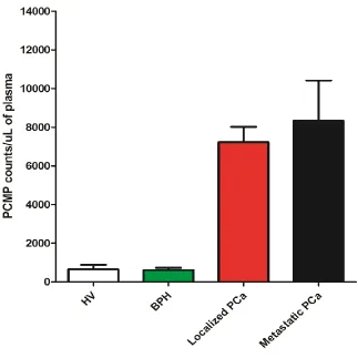
Figure 6. Comparison of Number of Prostate Cancer Microparticles (PCMP) Expressing PSMA+ Ghrelin, Dual Positive Events in Plasmas from Four Groups, Including Healthy Volunteers (HV) and Benign Prostatic Hypertrophy (BPH), Representing A Population with No Cancer and Other Two Groups; Localized Prostate Cancer and Metastatic Prostate Cancer, Representing A Population with Cancer.. ...49
Figure 7. Comparison of Number of Prostate Cancer Microparticles (PCMP) Expressing PSMA+ Ghrelin, Dual Positive Events in Plasmas from Prostate Cancer Patients with
Gleason Score 3+3, 3+4, 4+3, 4+4 and 5+4. ...50
Figure 8. Comparison of Number of Prostate Cancer Microparticles (PCMP) Expressing PSMA+ Ghrelin, Dual Positive Events in Plasmas from Prostate Cancer Patients with Tumor Stages; T2a, T2b, T2c and T3. ...51
xi
Volunteers (HV) and Benign Prostatic Hypertrophy (BPH), Representing A Population with No Cancer and Other Two Groups; Localized Prostate Cancer and Metastatic Prostate Cancer, Representing A Population with Cancer.. The Green Dashed Line Distinguishes Patients with PCa from patients with No Cancer...52
Figure 10. Histogram from Apogee A-50 nanoscale flow cytometer obtained from a patient plasma from group 1 representing patients with prostate cancer The top panel (A) represents all microparticle events analyzed according to size (long angle vs. small angle light scatter, Y vs. X axis respectively). The bottom panel (B) represents those same events but analyzed for PSMA + Ghrelin, dual positive MP, On Y axis is Ghrelin-Cy5 and on X axis is PSMA-PE. ...64
Figure 11. Mean Concentration of MPs Expressing Various Biomarkers; PSMA, PSMA + GRPR, PSMA + Ghrelin and PSMA + GRPR + Ghrelin in Prostate Cancer Patient Plasmas (Blue, N=249) and BPH Patient Plasmas (Red, N=156). One-way ANOVA (Ordinary) test was used to compare the means of the two groups. ...71
Figure 12. Mean Concentration of MPs Expressing Various Combinations of Biomarkers in Prostate Cancer Patient Plasmas (N=72) Stratified into Three PSA Groups (PSA < 4 ng/mL, 4-10 ng/mL and >10 ng/mL)...73
Figure 13. Mean Concentration of MPs Expressing Various Combinations of Biomarkers in BPH Patient Plasmas (N=143) stratified in Three PSA Groups (PSA < 4 ng/mL, 4-10 ng/mL and >10 ng/mL)...74
Figure 14.Comparison of Mean Concentration of MPs Expressing Various Combinations of Biomarkers in Prostate Cancer Patient Plasmas (Blue, N=74) and BPH Patient Plasmas (Red, N=143) stratified in Three PSA Groups (PSA < 4 ng/mL, 4-10 ng/mL and >10 ng/mL). One-way ANOVA (Ordinary) test was used to compare the means of the two groups. ...79
xii
Appendices
1
Chapter 1
1
Introduction
1.1
General Overview
Prostate Cancer (PCa) is leading the list of all newly diagnosed visceral cancers in men, and each year it is responsible for approximately 9.3% of all cancer related deaths (Jemal
et al. 2010). Currently, detection of PCa relies on a blood test known as Prostate Specific Antigen (PSA), digital rectal examination (DRE), and transrectal ultrasound guided biopsy (TRUS) of prostate. PCa is among the few solid organ malignancies which have a surrogate tumor marker to detect the disease and monitor its course. Although PSA is a highly sensitive and specific marker in the post treatment setting, especially post radical prostatectomy, it has a very low sensitivity and specificity as a screening tool. In 2011, the U.S. Preventive Services Task Force (USPSTF) drafted a recommendation against the routine use of PSA screening at any age and in October 2012 the USPSTF website posted that “Prostate cancer is a serious health problem that affects thousands of men and their
families. But before getting a PSA test, all men deserve to know what the science tells us
about PSA screening: there is a very small potential benefit and significant potential
harms. We encourage clinicians to consider this evidence and not screen their patients
with a PSA test unless the individual being screened understands what is known about
PSA screening and makes the personal decision that even a small possibility of benefit
2
A large prospective study from Europe Organization for Research and Treatment of Cancer (EORTC) looking at the role of PSA based screening program, ERSPC trial (European Randomised Study of Screening for Prostate Cancer) in reduction of mortality reported that a very large number of men need to be screened (1410 men) in order to save one life (Schroder et al. 2009). The Prostate, Lung, Colorectal and Ovarian Cancer (PLCO) Screening Trial, also investigating the usefulness of PSA as a screening tool, showed that the incidence of death per 10,000 person-years was 2.0 (50 deaths) in the PSA based screening group and 1.7 (44 deaths) in the control group (Andriole et al.
3
the majorities of men with suspicious PSA levels actually do not have PCa and are unnecessarily exposed to the risks of biopsy.
To meet the challenge of developing a screening test superior to PSA, we propose a blood test based on prostate cancer microparticles (PCMP). Microparticles (MP) are submicron (< 1µm) particles released from cells during their growth, malignant transformation or apoptosis (Rak, 2013). MP are released in the circulation and express surface receptors inherited from their cells of origin (Andreu et al. 2014). We enumerated PCMP using a combination of monoclonal antibodies (mAb) namely prostate specific membrane antigen, gastrin releasing peptide receptor and ghrelin peptide ligand which have been shown to bind to the extracellular portion of the receptors inherited by the MP derived from the PCa cells. We used these PCMP counts to distinguish between plasmas from patients with localized PCa and benign prostatic hyperplasia (BPH). The MP based test has the potential of functioning as a “fluid biopsy” which can continually sample the primary tumor to gain insight regarding the biology of these tissues.
1.2
Prostate Cancer
4
diagnosed and approximately 11 men dying of PCa every day (www.cancer.ca). These statistics place prostate cancer as one of the leading cancers affecting males in North America. Prostate cancer typically has a long course, thus making it a major consumer of the health care budget. Overall, the lifetime risk of developing PCa is about 16.7%; therefore one in six men will be diagnosed with prostate cancer in their life time. The incidence of harboring a focus of clinically insignificant PCa is even higher as autopsy studies performed on prostate glands obtained from men dying of all causes has shown that 20% of men aged 50 to 60 years and 50% of men, aged 70 to 80 years, have histologic evidence of carcinoma prostate (Carter et al. 1990). This disparity between clinically significant cancer and incidental or indolent cancer has led to numerous efforts to risk stratify this disease. None are perfect and study in this area is the major focus of prostate cancer research. Naturally, this uncertainty provokes a genuine anxiety among men at the risk of developing prostate cancer (Kotwal et al. 2012).
5
among immigrants, moving from an area of low incidence to higher incidence locations have reported significant increase in incidence of PCa in the immigrants compared to the natives in their country of origin. The increase in the incidence of PCa is more if immigration happened earlier in life of an individual. The higher incidence is also seen in the second generation of immigrants originating from areas of lower incidence.. This signifies the influence of environmental factors in development of cancers (Shimizu et al.
6
7
A high dietary intake of red meat, animal and polyunsaturated fats and milk appear to increase the risk of prostate cancer whereas, fruit and vegetables and polyphenols may be of preventive value for development of PCa. A review of dietary factors influencing the risk of PCa does not suggest any conclusive evidence to elucidate the role of the above agents in the development or prevention of PCa (Mandare et al. 2014). Anecdotal experiences and small nonrandomized studies have long promoted Selenium, Lycopene and Vitamin E as dietary supplements to prevent the development of PCa. To provide conclusive answer to this query a large prospective, multicenter study was designed. This Selenium, lycopene and Vitamin E Cancer Prevention Trial (SELECT) found no preventive effects of any of these substances in the development of prostate cancer (Klein
et al. 2011). On the contrary, it provided some evidence that Vitamin E in high dose actually increased the risk of development of PCa.
8
9
10
McNeal, in his original description of the zonal anatomy of prostate gland, described four basic anatomic zones (McNeal, 1980). This description was further modified later and Figure 2 shows the current understanding of the zonal anatomy of the prostate gland
(Campbell-Walsh Urology, tenth edition, 2010).
11
12
Prostatic carcinoma can metastasize through lymphatic or hematogenous dissemination. Bone metastases are the most common site of hematogenous spread. Lymphatic metastases occur frequently to the obturator lymph nodes (Campbell-Walsh Urology, tenth edition, 2010).
Prostate cancer is staged according to the guidelines of the 7th edition of American Joint Committee on Cancer (AJCC). This staging system is commonly referred to as TNM system. T stage refers to the volume of disease or the local extend of tumor, N reflects the lymph node status and M categorize the metastasis, if present. Clinical TNM stage based on local examination and imaging results is commonly referred to as cTNM. The pathological TNM or pTNM, however, is the final stage assigned after histological evaluation of the surgically removed prostate gland, along with regional lymph nodes. Table 3 illustrates the details of TNM staging system (Campbell-Walsh Urology, tenth
13
Tx Primary Tumor could not be assessed
T0 No evidence of Tumor
T1 Tumor not palpable
T2a Tumor palpable in less than half of one lobe
T2b Tumor palpable in more than half of one lobe
T2c Tumor palpable in both lobes
Nx Nodal metastasis cannot be assessed
N0 No nodal metastasis
N1 Nodal metastasis in single node less than 2 cm
N2 Nodal metastasis in single/multiple nodes less than 5 cm
N3 Nodal metastasis in multiple nodes more than 5 cm
Mx Distant metastasis cannot be assessed
M0 No distant metastasis
M1a Involvement of non regional lymph nodes
M1b Involvement of bones
M1c Involvement of other sites
14
15
1.3
Prostate Specific Antigen (PSA)
PSA is a glycoprotein enzyme from the kallikrein-related peptidase family and is produced by prostatic epithelial cells. It is also known as gamma-seminoprotein or kallikrein-3 (KLK3). The physiological role is in the liquefaction of semen. It helps in dissolving the coagulum and allows the sperm to swim freely for insemination.
16
with prostate disease and is not specific for PCa. The area under the curve (AUC) of the receiver operating characteristic (ROC) curve is between 0.56 and 0.70 for the ability of PSA to identify patients with cancer, where a score of 1.0 is perfect discrimination and 0.5 is a coin toss (Brawer et al. 1999). Common non-cancerous causes of elevated PSA levels are prostatic infection, trauma, and BPH. Studies have also shown that using the classic cutoff of 4mg/ml will result in missing significant number of prostate cancer cases (Schroder et al. 2008). If the cutoff of < 4mg/ml is considered as a negative test, a large study found 15.2% men in this group were actually later diagnosed to have prostate cancer, and according to AUA risk classification, 14.9% of these patients exhibited high risk disease (Thompson et al. 2004).
17
surveillance. A higher PSAD is found to be positively associated with the risk of progression and need for active treatment (Welty et al. 2014).
Age related PSA is another method to improve diagnostic efficiency. These age-specific reference ranges are designed to enhance the predictive value of PSA as a more discriminating tumor marker for detecting clinically significant cancers in older men (increasing specificity) and to find more potentially curable cancers in younger men (increasing sensitivity) (Oesterling et al. 1993).
PSA in circulation exists in both free form and complex form, bound with alpha 1-antichymotrypsin (ACT). Complex PSA is raised in PCa (Leinonen et al. 1993). A free to total PSA ratio of < 15 to 20% is generally considered to increase the risk of cancer (Oesterling et al. 1995). However, there is no exact watershed level. PSA velocity, the rate at which the PSA rises, is also used to improve the predictive value, where if the PSA increases by >0.75mg/ml per year it is considered to be an indication for prostate biopsy (Carter et al. 1993).
Extensive research in finding ways to promote the utility of PSA as a screening tool has failed to develop a perfect model capable of confidently selecting men suitable for aggressive and life-saving treatment.
1.4
Trans-rectal ultrasound (TRUS) guided Biopsy
18
biopsies. Specific templates are used to systematically sample the prostate gland. Additional samples may be obtained from sonographic or clinically suspicious areas.
The chances of finding a focus of prostate cancer on TRUS biopsy increases with the number of cores taken but this may also increase the risk for complications which include hematuria, rectal bleeding and urinary tract infection. In North America, more than 1 million prostate biopsy procedures are performed each year, with approximately 25% being positive for cancer and approximately 75% negative for cancer (Thompson et al.
19
needed for screening PCa. In theory, every additional 5% increase in accuracy rates for such a prostate screening test would eliminate approximately 165,000 unnecessary biopsies and 6,930 hospitalizations each year in North America. This would potentially result in significant health care savings and improve the quality of life of our patients.
To meet the challenge of a screening test superior to PSA, we propose a prostate microparticle-based “fluid biopsy” which continually samples the prostate and its primary tumor. The microparticles may contain biological information from the parent cells and may help to gain insight regarding the biology of these tissues without the need of obtaining a tissue sample.
1.5
Microparticles
20
labeled as “cell dust” or “cell garbage”, but advancements in imaging technology has enabled us to understand them better and this has opened up new avenues of research (Leong et al. 2011).
Living cells, through outward blebbing of the plasma membrane, generate MP from the region of membrane lipid rafts. They exhibit high levels of exposed phosphatidylserine (PS), integrins and metalloproteinases (Rak, 2013).
21
MP can be visualized using electron microscopy for morphological characterization. Gold-labeled immune electron microscopy was first used to assess the urinary MP (Mitchell et al. 2009). With the microparticles fluorescently labeled, MP can also be visualized indirectly using confocal microscopy (CM). However, MP are too small for direct visualization with standard CM. The lipophilic fluorescent dyes conjugated with antibodies against the antigens expressed on the plasma membranes of the MP can help detect them with relative ease. Dr. Leong, at our laboratory has validated the presence of MP analyzed using flow cytometry (Leong et al. 2011) and by atomic force microscopy (Leong et al. 2010). Figure 3 shows atomic force microscopy images of prostate cancer microparticles.
The exact role of these MP is still elusive but there is great enthusiasm in the scientific community to explore the role of these structures in the vesicular transportation system both for intracellular and extracellular communication. In 2013, James Rothman, Randy Schekman and Thomas Sudhof were presented with the Nobel Prize for their work on the role of micro vesicles for intracellular transportation. Similarly, plasma membrane derived microparticles may also have a role in the extracellular transportation/communication (Camussi et al. 2010).
22
category are the Exosomes that have a size up to 500nm (Pan, 1983) and have an established role in cellular communication.
.
23
However, the mechanism of release of exosomes is very different as they are first internalized and then reprocessed for release (Duijvesz et al. 2011). Ectosomes are plasma cell derived structures but are mainly released as a result of apoptosis (Diamant et al. 2004). To avoid use of confusing terminologies we will refer to microvesicles, exosomes, oncosomes, secretory vesicles and ectosomes as ‘Microparticle (MP)’ in our discussion. There is growing consensus that MP is the best-suited terminology to describe these biologically diverse structures.
The exact physiological importance of these structures in the development of cancer is not known. MP are enriched with specific antigens (Clayton et al. 2009). Elegant experiments in immunology have shown that MP affects the immune system by expressing and processing antigens (Raposo et al. 1996). As MP retains the surface characteristics of parent cells they may have a role in antigen presentation and immunomodulation. They may play a role in either promotion or prevention of metastasis. Assuming this is the mechanism of their release into the circulation, this feature may be exploited to develop diagnostic markers, which can function as a “fluid biopsy” of the entire gland.
24
Fluorescence-activated cell sorting (FACS) technology i.e. flow cytometer is capable of enumerating MP but identification of organ specific MP, which are relatively tiny compared to a cell, is difficult using a regular flow cytometry system due to the limitations of laser detectors. Other visualization techniques such as electron microscopy (EM) and confocal microscopy (CM) described previously are good for morphological characterization but cannot perform quantitative assays in a high throughput manner. A combination of enzyme-linked immunosorbent assay (ELISA) for exosome quantification has been used (Logozzi et al. 2009). In this experiment they used two different transmembrane proteins that are present on all exosomes. They postulated that by using one general transmembrane protein or so called ‘capture protein’, it is possible to identify exosomes and by using ‘tissue- or cancer-specific’ transmembrane protein, the number of
exosomes derived from a specific tissue can be measured.
25
1.6
Instruments
We used a specialized instrument that analyzes cell fragments in a high-throughput, multi-parametric manner. This nanoscale flow cytometer is manufactured by Apogee Flow systems Incorporation, Hertfordshire, UK. The “Apogee A-50 micro nanoscale flow cytometer” shown in Figure 4, is specifically designed to analyze and enumerate
cancer microparticles. This machine has three lasers installed; the Laser wavelengths are 375nm, 405nm, 488nm and 635nm. The multiple light scattering and fluorescence detectors help increase the detection limit to <100nm and increase the resolution to <10nm. Figure 5 shows the basic design of the machine, the florescence channels and the laser with exposure to the column of cells. This machine is equipped with Peripheral Component Interconnect Express (PCIe), high-speed computer software used for data acquisition. It employs the latest Altera™ technology for data acquisition at speed of up to 100k events per second. Conventional flow cytometers rely on fluorescent probes to measure biological particles smaller than 500nm, this may produce dim signals and data may be inconsistent. The A50-Micro's light scatter performance allows small particles to be better discriminated.
26
27
28
29
1.7
Prostate Cancer Surface Receptors
Cells originating from a specific organ typically manifest some receptors exclusive to that organ. This usually helps in classifying the cell to the organ of origin. Histological interpretation of specimens from metastatic sites with unknown primary lesion have long relied on using monoclonal antibodies against cell specific receptors to identifying the origin of these metastatic lesions. Recently, they have attracted attention and are increasingly used to develop therapeutic and diagnostic agents for treatment of cancers (Deckert et al. 2009). These antibodies are conjugated with florescence dyes and used in imaging modalities. For example, the Prostascint scan uses PSMA to look for metastatic lesions from PCa (Rosenthal et al. 2001). Ghrelin has been used as a PET imaging agent to identify foci of carcinomas in the prostate gland (Fowkes, 2014). We hypothesize that these antibodies may be used to identify microparticles, as it is postulated that a MP retains the surface receptors from the parent cells. With the use of appropriate antibodies it may be possible to establish the lineage of the MP. Monoclonal antibodies (mAbs) are highly specific and adaptable for targeting cells. We used two antibodies against two surface receptors expressed on PCa cells namely, prostate specific membrane antigen (PMSA) and gastrin releasing peptide receptor (GRPR). We also used a peptide-ligand called Ghrelin. The antibodies and the peptide-ligand were conjugated with fluorescent agents and used to enumerate the MP using flow cytometry.
1.7.1
Prostate specific Membrane Antigen (PSMA)
30
protein consists of a small intracellular domain of 19 amino acids, a transmembrane domain of 24 amino acids, and a large extracellular domain of 707 amino acids. The extracellular portion consists of a binding motif, including two zinc ions. The presence of PSMA is not actually unique to the prostate (Sacha et al. 2007). Its structure is almost identical to folate hydrolase and it is confirmed to be present in four sites in the body: prostate (secretory acinar epithelium), kidney (proximal tubules), nervous system glia (astrocytes and schwann cells), and the small bowel (Mhawech et al 2007). The physiological significance of PSMA in not completely understood in the prostate gland but it may be linked to the presence of folates in the seminal fluid. As intracellular folate is more abundant in rapidly dividing cells it is postulated that it may be more expressed in the higher grade prostate cancers.
PSMA was first identified in the prostate using IgG1 monoclonal antibody called 7E11-C5.3. This antibody was developed using the prostate cancer cell line known as LNCaP and was also used in development of Prostascint scan (Horoszewicz et al. 1987). This scan was approved by U.S. Food and Drug Administration (FDA) to be used as 111 In-labeled form (ProstaScint, Cytogen, Philadelphia, PA) but gained limited success (Rosenthal et al 2001). The main flaw in this antibody was that it had affinity for the intracellular portion and hence was not useful to identify living cells. Wolf et al developed three other mAbs (3/A12, 3/E7, 3/F11), which show a strong and specific extracellular binding to PSMA (Wolf et al. 2010). Wolf also demonstrated that 3/E7 was compatible with flow cytometric analysis and showed high affinity to human prostate tissue. This antibody could be obtained with >95% purity from the hybridoma.
31
prostatic intraepithelial neoplasia (PIN) and prostate cancer (Bostwick et al. 1998). Similar association of PSMA expression in high grade prostate cancer has been shown by other investigators (Wright et al. 1995). This data support potential clinical use of PSMA in the diagnosis of PCa.
1.7.2
Ghrelin
32
tissue, we were able to demonstrate that this novel fluorescein-ghrelin probe was able to distinguish between benign and cancerous cell lines and therefore has potential to be explored as a marker for diagnosis of PCa (Lu et al. 2012). In our pilot study, we have shown the usefulness of Ghrelin as our cancer-specific biomarker to define the population of prostate cancer microparticles in plasma. In the pilot study, we enumerated PSMA+ and GHSR+ microparticles in plasmas from three patient cohorts and found higher counts of PSMA+ GHSR+ dual positive MP in the metastatic and localized prostate cancer patient cohorts compared to the low counts observed in healthy volunteers. Based on this experience we elected to investigate Ghrelin as a biomarker in this study.
1.7.3
Gastrin Releasing Peptide Receptor (GRPR)
33
34
1.8
Bibliography
Ananias HJ, van den Heuvel MC, Helfrich W & de Jong IJ. (2009). Expression of the gastrin-releasing peptide receptor, the prostate stem cell antigen and the prostate-specific membrane antigen in lymph node and bone metastases of prostate cancer. Prostate 69, 1101–1108.
Andreu Z & Yáñez-Mó M. (2014). Tetraspanins in extracellular vesicle formation and function. Front Immunol 5, 442.
Andriole GL, Crawford ED, Grubb RL 3rd, Buys SS, Chia D, Church TR, Fouad MN, Gelmann EP, Kvale PA, Reding DJ, Weissfeld JL, Yokochi LA, O'Brien B, Clapp JD, Rathmell JM, Riley TL, Hayes RB, Kramer BS, Izmirlian G, Miller AB, Pinsky PF, Prorok PC, Gohagan JK & Berg CD; PLCO Project Team. (2009). Mortality results from a randomized prostate-cancer screening trial. N Engl J Med 360, 1310-9.
Beer M, Montani M, Gerhardt J, Wild PJ, Hany TF, Hermanns T, Müntener M & Kristiansen G. (2012). Profiling gastrin-releasing peptide receptor in prostate tissues: clinical implications and molecular correlates. Prostate. 72, 318-25.
35
Bologna M, Festuccia C, Muzi P, Biordi L & Ciomei M. (1989). Bombesin stimulates growth of human prostatic cancer cells in vitro. Cancer 63, 1714-1720.
Bostwick DG, Pacelli A, Blute M, Roche P & Murphy GP. (1998). Prostate specific membrane antigen expression in prostatic intraepithelial neoplasia and adenocarcinoma:a study of 184 cases. Cancer 82, 2256–61.
Brawer MK. (1999) Prostate-specific antigen: current status. CA Cancer J Clin 49, 264-81.
Camussi G, Deregibus MC, Bruno S, Cantaluppi V & Biancone L. (2010). Exosomes/microvesicles as a mechanism of cell-to-cell communication. Kidney Int 8, 838–48.
Carter HB & Pearson JD. (1993). PSA velocity for the diagnosis of early prostate cancer. A new concept. Urol Clin North Am 20, 665-670.
Carter HB, Piantadosi S & Isaacs JT. (1990). Clinical evidence for and implications of the multistep development of prostate cancer. J Urol 143, 742–746.
36
Catalona WJ, Smith DS, Ratliff TL, Dodds KM, Coplen DE, Yuan JJ, Petros JA & Andriole GL. (1991). Measurement of prostate-specific antigen in serum as a screening test for prostate cancer. N Engl J Med 324, 1156-61.
Chun FK, Epstein JI, Ficarra V, Freedland SJ, Montironi R, Montorsi F, Shariat SF, Schröder FH & Scattoni V. (2010). Optimizing performance and interpretation of prostate biopsy: a critical analysis of the literature. Eur Urol 58, 851-64.
Clayton A & Mason MD. (2009). Exosomes in tumour immunity. Curr Oncol 16,46–49.
Crawford ED. (2003). Epidemiology of prostate cancer. Urology 22, 3-12.
Deckert PM. (2009) Current constructs and targets in clinical development for antibody-based cancer therapy. Curr Drug Targets 10, 158–175.
DeSantis CE, Lin CC, Mariotto AB, Siegel RL, Stein KD, Kramer JL, Alteri R, Robbins AS & Jemal A. (2014). Cancer treatment and survivorship statistics, 2014. CA Cancer J Clin. 64, 252-71.
Diamant M, Tushuizen ME, Sturk A & Nieuwland R. (2004). Cellular microparticles: new players in the field of vascular disease?. Eur J Clin Invest 34, 392–401.
Diederick Duijvesz , Theo Luider , Chris H. Bangma & Guido Jenster. (2011) Exosomes as Biomarker Treasure Chests for Prostate Cancer Eur Urol 5 9, 8 2 3 – 8 3 1.
37
Erspamer V. (1988). Discovery, isolation, and characterization of bombesin-like peptides. Ann N Y Acad Sci 547, 3-9.
Fowkes & Milan M. "Peptidomimetic GHS-R1a Agonists as PET Imaging Agents for Prostate Cancer" (2014). University of Western Ontario - Electronic Thesis and Dissertation Repository. Paper 1972.
Giacchetti S, Gauville C, De Cremoux P, Bertin L, Berthon P, Abita JP, Cuttitta F & Calvo F. (1990). Characterization, in some human breast cancer cell lines, of gastrin-releasing peptide-like receptors which are absent in normal breast epithelial cells. Int J Cancer; 46: 293-298.
Gleason DF & Mellinger GT. (1974). Prediction of prognosis for prostatic adenocarcinoma by combined histological grading and clinical staging. J Urol 111, 58-64.
Horoszewicz JS, Kawinski E & Murphy GP. (1987). Monoclonal antibodies to a new antigenic marker in epithelial prostatic cells and serum of prostatic cancer patients. AnticancerRes 7, 927–35.
38
receptor in pituitary and hypothalamus that functions in growth hormone release. Science. 273, 974-977.
http://www.cancer.ca/en/cancer-information/cancer-type/prostate/statistics/region.
http://www.uspreventiveservicestaskforce.org/prostatecancerscreening. htm.
Israeli RS, Powell CT, Fair WR & Heston WD. (1993) Molecular cloning of a complementary DNA encoding a prostate-specific membrane antigen. Cancer Res 53,227–30.
Jeffery PL, Herington AC & Chopin LK. (2002). Expression and action of the growth hormone releasing peptide ghrelin and its receptor in prostate cancer cell lines. J Endocrinol 172, 7–11.
Jemal A, Siegel R, Xu J & Ward E. (2010). CA Cancer J Clin 60, 277-300.
Jensen RT, Battey JF, Spindel ER & Benya RV. (2008). International Union of Pharmacology. LXVIII. Mammalian bombesin receptors: Nomenclature, distribution, pharmacology, signaling, and functions in normal and disease states. Pharmacol Rev 60, 1–42.
39
Kojima M, Hosoda H, Date Y, Nakazato M, Matsuo H & Kangawa K. (1999). Ghrelin is a growth-hormone-releasing acylated peptide from stomach. Nature. 402, 656-660.
Kotwal AA, Schumm P, Mohile SG & Dale W. (2012). The influence of stress, depression, and anxiety on PSA screening rates in a nationally representative sample. Med Care. 50, 1037-44.
Leong HS, Podor TJ, Manocha B & Lewis JD. (2011). Validation of flow cytometric detection of platelet microparticles and liposomes by atomic force microscopy. J Thromb Haemost 9, 2466-76.
Leong HS, Steinmetz NF, Ablack A, Destito G, Zijlstra A, Stuhlmann H, Manchester M & Lewis JD (2010). Intravital imaging of embryonic and tumor neovasculature using viral nanoparticles. Nat Protoc 5,1406-1417.
Leinonen J1, Lövgren T, Vornanen T & Stenman UH. (1993). Double-label time-resolved immunofluorometric assay of prostate-specific antigen and of its complex with alpha 1-antichymotrypsin. Clin Chem 39, 2098-103.
Loeb S, Carter HB, Berndt SI, Ricker W & Schaeffer EM. (2011). Complications after prostate biopsy: data from SEER-Medicare. J Urol 186, 1830-1834.
40
Lu C, McFarland MS, Nesbitt RL, Williams AK, Chan S, Gomez-Lemus J, Autran-Gomez AM, Al-Zahrani A, Chin JL, Izawa JI, Luyt LG, Lewis JD. Ghrelin receptor as a novel imaging target for prostatic neoplasms. Prostate. 2012 Jun 1;72(8):825-33.
Mandair D, Rossi RE, Pericleous M, Whyand T & Caplin ME. (2014). Prostate cancer and the influence of dietary factors and supplements: a systematic review. Nutr Metab 6, 30.
Martinez CH, Williams AK, Chin JL, Stitt L& Izawa JI. (2013). Perineural invasion and TRUS findings are complementary in predicting prostate cancer biology. Can J Urol 20, 6696-701.
McNeal JE. (1980). Anatomy of the prostate: an historical survey of divergent views. Prostate 1, 3-13.
Mhawech-Fauceglia P, Zhang S, Terracciano L, Sauter G, Chadhuri A, Herrmann FR, & Penetrante R.. (2007). Prostate- specific membrane antigen (PSMA) protein expression in normal and neoplastic tissues and its sensitivity and specificity in prostate adenocarcinoma: an immunohistochemical study using mutiple tumour tissue microarray technique. Histopathology 50, 472–83.
41
Mitchell PJ, Welton J, Staffurth J, Court J, Mason MD, Tabi Z & Clayton A. (2009). Can urinary exosomes act as treatment response markers in prostate cancer? J Transl Med 7, 4.
Nadji M, Tabei SZ, Castro A, Chu TM, Murphy GP, Wang MC & Morales AR. (1981). Prostatic-specific antigen: an immunohistologic marker for prostatic neoplasms. Cancer 48, 1229-32.
Nakajima T, Yasuhara T, Erspamer V, Erspamer GF, Negri L & Endean R. (1980). Physalaemin- and bombesin-like peptides in the skin of the Australian leptodactylid frog Uperoleia rugosa. Chem Pharm Bull 28, 689-95.
Nilsson J, Skog J, Nordstrand A, Baranov V, Mincheva-Nilsson L, Breakefield XO & Widmark A. (2009). Prostate cancer-derived urine exosomes: a novel approach to biomarkers for prostate cancer. Br J Cancer 100, 1603–7.
Oesterling JE, Jacobsen SJ, Chute CG, Guess HA, Girman CJ, Panser LA & Lieber MM. (1993). Serum prostate-specific antigen in a community-based population of healthy men. Establishment of age-specific reference ranges. JAMA. 270, 860-4.
Oesterling JE, Jacobsen SJ, Klee GG, Pettersson K, Piironen T, Abrahamsson PA, Stenman UH, Dowell B, Lövgren T & Lilja H. (1995). Free, complexed and total serum prostate specific antigen: the establishment of appropriate reference ranges for their concentrations and ratios. J Urol. 154, 1090-1095.
42
Rak J. (2013). Extracellular vesicles - biomarkers and effectors of the cellular interactome in cancer. Front Pharmacol 4, 21.
Raposo G, Nijman HW, Stoorvogel W, Liejendekker R, Harding CV, Melief CJ, & Geuze HJ. (1996). B lymphocytes secrete antigen-presenting vesicles. J Exp Med 183,1161–72.
Rosenthal SA, Haseman MK & Polascik TJ. (2001). Utility of capromab pendetide (ProstaScint) imaging in the management of prostate cancer. Tech Urol 7, 27–37.
Rosita D, Dewit MA & Luyt LG. (2009). Fluorine and rhenium substituted ghrelin analogues as potential imaging probes for the growth hormone secretagogue receptor. J Med Chem 52, 2196-2203.
Sacha P, Zamecnik J, Barinka C, Hlouchová K, Vícha A, Mlcochová P, Hilgert I, Eckschlager T, Konvalinka J. (2007). Expression of glutamate carboxypeptidase II in human brain.Neuroscience 144, 1361–72.
Schröder FH, Carter HB, Wolters T, van den Bergh RC, Gosselaar C, Bangma CH, & Roobol MJ. Early detection of prostate cancer in 2007. Part 1: PSA and PSA kinetics. Eur Urol 53, 468-77.
43
Schröder FH, Hugosson J, Roobol MJ, Tammela TL, Zappa M, Nelen V, Kwiatkowski M, Lujan M, Määttänen L, Lilja H, Denis LJ, Recker F, Paez A, Bangma CH, Carlsson S, Puliti D, Villers A, Rebillard X, Hakama M, Stenman UH, Kujala P, Taari K, Aus G, Huber A, van der Kwast TH, van Schaik RH, de Koning HJ, Moss SM, & Auvinen A. (2014). Screening and prostate cancer mortality: results of the European Randomised Study of Screening for Prostate Cancer (ERSPC) at 13 years of follow-up. Lancet, Aug 6.
Scosyrev E, Wu G, Mohile S & Messing EM. (2012). Prostate-specific antigen screening for prostate cancer and the risk of overt metastatic disease at presentation : Analysis of trends over time. Cancer 118, 5768-76.
Siddiqui KM, Biggs C, Billia M, Mazzola CR, Izawa J, PowerN, Chin J & Leong HS.. (2014). Enumeration of Prostate Cancer Microparticles as a Tool to Identify Prostate Cancer. CUAJ8, 5-6
Siegel R, Ma J, Zou Z, Jemal A. (2014). CA Cancer J Clin 64, 9-29.
Stamey TA, Yang N, Hay AR, McNeal JE, Freiha FS & Redwine. (1987). Prostate-specific antigen as a serum marker for adenocarcinoma of the prostate. N Engl J Med. 317, 909-16.
Shimizu H, Ross RK, Bernstein L, Yatani R, Henderson BE & Mack TM. (1991). Cancers of the prostate and breast among Japanese and white immigrants in Los Angeles County. Br J Cancer 63, 963-6.
44
Prevalence of prostate cancer among men with a prostate-specific antigen level < or =4.0 ng per milliliter. N Engl J Med 350, 2239-46.
Trams EG, Lauter CJ, Salem N Jr & Heine U. (1981). Exfoliation of membrane ecto-enzymes in the form of micro-vesicles. Biochim Biophys Acta 645, 63-70.
Wang MC, Valenzuela LA, Murphy GP & Chu TM. (1979). Purification of a human prostate specific antigen.Invest Urol 17, 159-63.
Wein AJ, Kavoussi LR, Novick AC, Partin AW & Peters CA, eds. Campbell-Walsh Urology, 10th Edition. Philadelphia: Saunders, 2010.
Welch HG, Fisher ES, Gottlieb DJ & Barry MJ. (2007). Detection of prostate cancer via biopsy in the Medicare-SEER population during the PSA era. J Natl Cancer Inst. 99, 1395.
Welty CJ, Cowan JE, Nguyen H, Shinohara K, Perez N, Greene KL, Chan JM, Meng MV, Simko JP, Cooperberg MR & Carroll PR. (2014). Extended Follow-Up and Risk Factors for Disease Reclassification from a Large Active Surveillance Cohort for Localized Prostate Cancer. J Urol. Sep 24.
Wolf P. (1967). The nature and significance of platelet products in human plasma. Br J Haematol 13, 269-288.
45
PSMA Epitopes Are Promising Diagnostic and Therapeutic Tools for Prostate Cancer. The Prostate 70, 562-569.
46
Chapter 2
2
Pilot Study: Enumeration of Prostate Cancer Microparticles
as a Tool to Identify Prostate Cancer
2.1 Introduction
47
2.2
Material and Methods
We used A-50 Apogee, nanoscale flow cytometer which is specifically designed to analyze and enumerate cancer microparticles, to study prostate cancer microparticles present in four cohorts of patients:
a. Healthy volunteers (HV) (n=24); Included young men and women aged <35 with no known cancers.
b. Benign Prostatic Hypertrophy (BPH) (n=10); Included men who had a normal digital rectal examination and had a transurethral resection of prostate with a pathologic diagnosis of BPH.
c. Localized Prostate Cancer (n=112): Samples obtained from Ontario Institute of Cancer (OICR) from patients undergoing radical prostatectomy for localized prostate cancer.
d. Metastatic Prostate Cancer (n=23): Included samples from patients with metastatic castrate resistant prostate cancer.
We used a monoclonal antibody specific to the extracellular portion of PSMA (PSMA-RPE mAb) and ghrelin peptide, a growth hormone secretagogue receptor (GHSR) ligand (Ghrelin-FITC ligand) to identify and enumerate dual positive prostate cancer
microparticles (PSMA + Ghrelin dual positive).
48
Before the experiment, we thawed PPP in the tube and transferred 20 uL to a sterile 1.5 mL eppendorf tube. This was designated as tube A. Using a 20 uL pipetman, another 20 uL of PPP was transferred to another sterile 1.5 mL eppendorf tube and labeled as tube B. 2uL each of IgG-FITC antibody and IgG-RPE antibody was then added to tube A. 2uL of Ghrelin-FITC ligand and 2uL of PSMA-RPE antibody was also added to tube B. Both the tubes A and B were mixed and immediately incubate in the dark for 30 min. 600uL of sheath fluid (1X PBS, pH 7.4) was then added to each tube and samples were vortexed. Samples were then analyzed using flow cytometry. Samples were analyzed in triplicates and the average calculated.
2.3
Results
We analyzed 169 plasmas and found significantly higher counts (p<0.01, ANOVA, bon ferroni test) of PSMA + Ghrelin, dual positive prostate cancer microparticle (PCMP) in patients with prostate cancer as compared to BPH and healthy volunteers. The numbers of PCMP in each group are shown in Figure 6.
However we did not find any significant correlation between the number of PCMP and Gleason score (one-way ANOVA test). The number of PCMP in each category of Gleason score are shown in Figure 7.
49
50
51
52
53
PCa. With this cutoff, the blood test is 89% accurate in identifying patients with PCa (localized PCa cohort) with 20% of patients being mistakenly identified as having PCa.
2.4
Discussion
Prostate cancer (PCa) is a leading cause of cancer-related death of men in the western world (Siegel et al. 2014). Majority of men are diagnosed with PCa based on a raised prostate specific antigen (PSA). Since its introduction, PSA has generated intense debate as an effective screening tool for PCa (Catalona et al. 1991). The recent recommendations by United States Preventive Task Force (USPSTF) has concluded that PSA based screening produces unacceptably high rates of false positive results and causes more harm than benefit.
The National Cancer Institute defines a biomarker as “a biological molecule found in the blood, other body fluids, or tissues that is a sign of a normal or abnormal process or of a condition or disease”. An ideal biomarker for PCa should not only be able to screen with high sensitivity and specificity but also predict the course of disease and help select high risk individuals for aggressive treatment. The ideal tumor marker should be economical, reproducible, non-invasive, not time consuming to perform and easily accessible for majority of population (Velonas et al. 2013).
54
thus if reliably identified, they can be exploited as biomarker treasure chests for prostate cancer (Dujivesz D et al. 2011). The main function of MP is proposed to be cell-to-cell communication but they are likely to play a significant role in oncogenesis (Yang et al. 2011). Exosomes (very similar to MP) have also been shown to promote metastasis by avoiding detection by immune system (Yang et al. 2007).
We developed a MP based blood test to identify patients with PCa using a combination of monoclonal antibody for prostate specific membrane antigen (PSMA) and ghrelin peptide, a ligand for growth hormone scretagogue receptor (GHSR). PSMA has been shown to be highly expressed on prostate cells and differential expression has been documented on high grade prostate cancer compared to BPH and normal prostate gland (Wright et al. 1995). Similarly, ghrelin has been shown to bind more strongly with PCa in both in vitro and ex vivo experiments (Lu et al. 2012).
Our results showed that PSMA + Ghrelin, dual positive MP are more abundant in PCa patient plasmas compared to plasma samples from healthy volunteers and patients with BPH. Compared with other established makers like PSA and prostate cancer antigen (PCA) 3 score, which have a diagnostic accuracy of 56% to 72% (Crawford et al. 2012). This blood test is able to identify PCa with a diagnostic accuracy of 89% in only a minute volumes of patient blood in a high-throughput and multi-parametric manner.
2.5
Conclusion
55
2.6
Limitations
56
2.7
Bibliography
Catalona WJ, Smith DS, Ratliff TL, Dodds KM, Coplen DE, Yuan JJ, Petros JA & Andriole GL. (1991). Measurement of prostate-specific antigen in serum as a screening test for prostate cancer. N Engl J Med 324, 1156-61.
Duijvesz D, Luider T, Bangma CH & Jenster G. 2011. Exosomes as biomarker treasure chests for prostate cancer. Eur. Urol 59, 823–831.
Gyorgy B, Szabo TG, Pasztoi M, Pal Z, Misjak P, Aradi B, Laszlo V, Pallinger E, Pap E, & Kittel, A. 2011. Membrane vesicles, current state-of-the-art: Emerging role of extracellular vesicles. Cell Mol. Life Sci 68, 2667–2688
Lu C, McFarland MS, Nesbitt RL, Williams AK, Chan S, Gomez-Lemus J, Autran-Gomez AM, Al-Zahrani A, Chin JL, Izawa JI, Luyt LG, Lewis JD. Ghrelin receptor as a novel imaging target for prostatic neoplasms. Prostate. 2012 Jun 1;72(8):825-33.
Rak J. (2013). Extracellular vesicles - biomarkers and effectors of the cellular interactome in cancer. Front Pharmacol 4, 21.
Schröder FH & ERSPC Investigators. 2014. Screening and prostate cancer mortality: results of the European Randomised Study of Screening for Prostate Cancer (ERSPC) at 13 years of follow-up. Lancet 384. 2027-2035.
Velonas VM1, Woo HH, Remedios CG & Assinder SJ. 2013. Current status of biomarkers for prostate cancer. Int J Mol Sci. 14, 11034-11060.
Wright GLJr, Haley C, Beckett ML & Schellhammer PF. (1995).Expression of prostate-specific membrane antigen in normal, benign ,and malignant prostate tissues. UrolOncol 1,18–28.
57
58
Chapter 3
3
Prostate Cancer Microparticles as a Next Generation
Screening Tool for Prostate Cancer
3.1
Introduction
Prostate cancer (PCa) is the leading cancer among adult males and is one of the few solid organ malignancies, which can be diagnosed and monitored using a tumor marker i.e. prostate specific antigen (PSA). However PSA is not a true tumor marker and is also produced by normal prostate gland and may be raised in a number of benign conditions. Large studies have shown that >50% of men with raised PSA do not have PCa and are unnecessarily subjected to transrectal ultrasound (TRUS) guided systematic biopsy of prostate gland (Martinez et al. 2013). In 2012, the U.S. Preventive Services Task Force (USPSTF) recommended against the routine use of PSA screening at any age. Clearly, PSA does not fulfill any currently acceptable standard of a screening test and a new screening test is needed. To meet this challenge, we propose a prostate cancer microparticle based test.
59
3.2
Objective
The objectives of this study were;
1. To differentiate patients with prostate cancer from those with BPH or ‘no cancer’.
2. To determine the difference in PCMP levels in the two groups namely,
a. Men with localized PCa
b. Men with biopsy proven BPH and no evidence of PCa.
3.3
Materials and Methods
After obtaining the necessary ethical approval was obtained from Western University (Appendix 1), we analysed 405 plasma samples. 249 PCa plasma samples were obtained from Ontario Institute of Cancer Research (OICR) and 147 samples of patients with prostate biopsy proven BPH were obtained from Princess Margaret Hospital GU Tissue Bank (PMH). We also recruited 9 patients with BPH at London Heath Sciences (LHSC), these patients had a TRUS biopsy to rule out PCa and also underwent transurethral resection with histologically confirmed BPH.
Collection of Samples
60
(Purple top tubes). The vacutainer was centrifuged at 2000 RCF for 20 min. This resulted in the blood separating into two distinct layers, a red layer (erythrocytes) and a yellow upper layer (platelet poor plasma-PPP). Using a plastic disposable Pasteur pipet, the PPP was transferred into tube for storage at -80 degree Celsius.
Samples from London Health Sciences: Nine samples were obtained from patients undergoing transurethral resection of prostate (TURP). These patients were previously worked up for lower urinary tract symptoms and presence of PCa was ruled out by transrectal ultrasound guided biopsy. The final pathology report from the TURP specimen was consistent with histological diagnosis of BPH. Prior to surgery 10-15 ml of blood was drawn in a purple top tube and centrifuged at 2000 RCF for 20 min. This resulted in the blood separating into two distinct layers, a red layer (erythrocytes) and a yellow upper layer (platelet poor plasma-PPP). Using a plastic disposable Pasteur pipet, the PPP was transferred into tube for storage at -80 degree Celsius.
Randomization and blinding
All the samples were thawed and aliquoted again. The samples were randomized and numbered to blind the observer during analysis of samples. These randomized and relabelled samples were stored at -80 degree Celsius until they were used during the experiment. The master list was kept separately in the office of Dr. Hon Leong, PhD.
Preparation of samples
61
antibody’. Isotype antibodies were used as control for each sample as the florescence-conjugated antibodies known to bind with trace amounts of nonspecific proteins and produce auto florescence. The use of isotype antibodies helped us in identifying this background noise. The numbers of events in the isotype sample were then subtracted from the counts observed in the sample, labeled as ‘positive antibody’. This methodology
helped in eliminating the effect of this non specific binding. The positive antibody tube was used to prepare a mixture of antibodies namely PSMA-PE antibody (3/E7), Ghrelin peptide, GRPR antibody. Similarly we prepared a mixture of isotype controls for using mouse IgG-RPE antibody for PSMA, Ghrelin/LCE antibody for ghrelin and rabbit IgG antibody for GRPR for the tube labeled as isotype antibody. The concentration of PSMA-PE antibody (3/E7) used was 408.42 µg/ml, the concentration of Ghrelin peptide used was 62.5mM and the concentration GRPR used was 0.5 µg/ml. The concentrations of isotype antibodies were matched.
To simplify the process of conjugation of the antibodies we prepared two cocktails. The tube labeled positive contained; 24µL of GRPR, 24 µL of 2° antibody, 12 µL each of PSMA mAb and Ghrelin peptide. The total volume of this mixture was 72 µL. Similarly we used 24 µL of rabbit IgG antibody for GRPR, 24 µL of 2° antibody and 12 µL each of mouse IgG-RPE antibody for PSMA, Ghrelin/LCE antibody for Ghrelin. The total volume of this mixture was also 72 µL. This volume was enough to conduct the experiment on a batch of 10 samples at one time.
62
according to previous randomization. We added 6 µL from tube labeled ‘positive antibody’ to the tube labeled ‘+ve’ xxx and 6 µL from tube labeled ‘isotype antibody’ to the tube labeled ‘-ve’xxx.
Both the tubes were mixed well and immediately incubate in the dark for 30 min. After incubation we added 600uL of sheath fluid (1X PBS, pH 7.4) using 1000 uL pipetman to each tube and vortexed.
Analysis of samples on Flow cytometer
63
We then analyzed our study samples and used the same settings (identified during optimization) throughout the study. The numbers of events per microliter were recorded for each sample. The isotype controls were used to calculate the background activity of each sample. We subtracted the number of events in the isotype from the sample to calculate the ‘compensated’ number of microparticles events for each specimen.
3.4
Statistical Analysis
64 PSMA-PE
Small Angle
Light Scatter (size)
Ghrelin-Cy5 Long Angle
Light Scatter (size)
65
3.5
Results
Four hundred and five randomized samples were analyzed. The patients were divided in two groups. Group 1 representing 249 patients with prostate cancer and Group 2 included 156 patients with BPH.
In Group 1 (PCa), 65% patients were aged between 40-64 years (range 49-79 years). The mean PSA was 13.88 + 62.22 ng/mL (range 1.5 ng/mL to 541 ng/mL) and the median PSA was 7 ng/ml. The pathologic T stage of the tumors (Group 1) is shown in Table 4 and the distribution of Gleason Score is shown in Table 5. In Group 2 (BPH), the mean age of patients was 64.5 + 6.19 years (range 49-79 years). All patients in Group 2 had biopsy proven BPH, the median size of the prostate gland was 67.5 grams calculated on trans-rectal ultrasound. The mean PSA was 10.07 + 5.82ng/mL and the median PSA was 8.7 ng/ml.
66
pT Stage (n=164) Number of Patients (%)
pT2a 38 (23.1)
pT2b 16 (9.7)
pT2c 39 (23.7)
pT3a 46 (28)
pT3b 23 (14)
pTx 2 (1.2)