Lung Cancer Detection Using Machine
Learning Techniques
K.Mohanambal1, Y.Nirosha2, E.Oliviya Roshini3, S.Punitha4, M.Shamini5
Assistant Professor, Dept. of IT, Valliammai Engineering College, Chennai, Tamil Nadu, India1
UG Student, Dept. of IT, Valliammai Engineering College, Chennai, Tamil Nadu, India2
UG Student, Dept. of IT, Valliammai Engineering College, Chennai, Tamil Nadu, India3
UG Student, Dept. of IT, Valliammai Engineering College, Chennai, Tamil Nadu, India4
UG Student, Dept. of IT, Valliammai Engineering College, Chennai, Tamil Nadu, India5
ABSTRACT: Early detection of lung cancer cells can help in a sharp decrease in the lung cancer mortality rate hence it is an aggressive disease which carrying a dismal prognosis with a 5-year survival rate at 18%.Several computer-aided diagnosis systems have been developed to help reduce lung cancer mortality rates.Thus structural co-occurrence matrix (SCM)-based approach is used to extract the feature and to classify nodules into malignant or benign nodules and also into their malignancy level. The computed tomography(CT) scanfrom the lung image database consortium and image database resource initiative datasets provide knowledge concerning nodule positions and their malignancy levels is been deployed here as a model. Support vector machine is been used as a classifier which is (i) to classify the nodule images into malignant or benign nodules and (ii) to classify the lung nodules into malignancy levels (1 to 5).These experimental results reveal that the SCM successfully extracted features of the nodules from the images and, therefore may be considered as a promising tool to support medical specialist to make a more precise diagnosis concerning the malignancy of lung nodules.
KEYWORDS: Structural Co-occurrence Matrix(SCM),Malignant nodule, Benign nodule.
I.INTRODUCTION
(Lung Cancer Data Consortium) [3], Image-based techniques for analyzing lesions are normally performed with detection, segmentation hand-crafted feature engineering and category labelling Zinovev et al. adopted a belief decision tree approach to predict nodule semantic attributes. Related studies on the Lung Image Database Consortium and Image Database Resource Initiative (LIDC-IDRI) dataset and at simultaneous the CADx system provides a second opinion to help in decision-making [6].However, all these methods rely on nodule segmentation as a prerequisite. Notably, automatic nodule segmentation may influence classification results since methods such as region growing and level set typically depend on initialization [5].Working on these segmented regions may yield inaccurate features that lead to erroneous outputs [5]. The applications of automatic tumour segmentation are broad, including measuring treatment response, planning of radiation treatment, and to facilitate extraction of robust features for high-throughput radiomics by the MRRN [8]. Hence, good results obtained with the proposed method would be a potential to improve medical diagnoses and assist in making more accurate and efficient decisions for these two major health problems.
II. RELATED WORKS
In [1][2]``Automatic detection of lung nodules in computed tomography images and cancer statistics: Training and validation of algorithms using public research databases,'' Lung cancer is manifests itself as a non-calcified pulmonary nodules that can be detected reading lung Computed Tomography (CT) images and Computer Aided Detection (CAD) and it is composed of two CAD sub-procedures is presented: CADi which is to analyse the internal parenchymal nodules and CADJP is to identify the nodules attached to the pleura surface.These CAD algorithm will perform the – lung segmentation;
– candidate detection; – candidate classification.
clinical point of view. The Time to Treat program, which is designed for patients with clinical or radiographic suspicion of lung cancer is introduced.We can decompose such a complex network into a collection of serial processes. In [8] Multiple Resolution Residually Connected Feature Streams For Automatic Lung Tumor Segmentation From CT Images”, we developed two multiple resolution residually connected network (MRRN) formulations called incremental-MRRN and dense-MRRN. An algorithm was instructed by using the 377 tumors from the TCIA dataset and validated on the MSKCC and tested on LIDC datasets. The residual stream input consists of feature maps from one of the preceding higher resolution residual streams. Feature integration from multiple resolutions obviates the need for additional un-pooling following RCUBs as used in conventional encoder-decoder like the Unet. CT scan have been used in lung cancer detection, risk assessment, and clinical management. In particular, the increasing quantity of CT image assays has created a unique address for data-driven analysis to capture underlying cancer characteristics at a macroscopic level, allowing identification of prognostic imaging biomarkers [10].
III.PROPOSED SYSTEM
The image processing techniques are mostly used for prediction of lung cancer and also for early detection and treatment to intercept the lung cancer. To prognosis the lung cancer various features are extracted from the images therefore, pattern recognition based approaches are useful to predict the lung cancer. In the proposed system, we use Python software for analysis. In image processing procedures, the methods involved are image pre-processing, segmentation and feature extraction techniques have been discussed in detail. We are line up to get the more accurate results by using various enhancement and segmentation techniques. The structural co-occurrence matrix (SCM) proficiency was appeal to pluck out features from images of nodules and categorize them into malignant or benign nodules and their malignancy levels. The CT test from the lung image database consortium and image database resource induce datasets provide information referring to nodule positions and their malignancy levels. The classification stage utilise Support Vector Machine algorithm (SVM) and applied them to two tasks:
(i) to classify the nodule images into malignant or benign nodules and
(ii) to classify the lung cells into malignancy levels (1 to 5).
Fig.1 Architecture of the system IV.METHODOLOGY
The lung cancer detection system using image processing is used to classify the present of lung cancer in a CT- images. In this study, we use Python software for analysis. In image processing procedures, methods such as image pre-processing, segmentation and feature extraction have been discussed in detail. We are line up to get the more accurate results by using various enhancement and segmentation techniques. The structural co-occurrence matrix (SCM) proficiency was appeal to pluck out features from images of nodules and categorize them into malignant or benign nodules and their malignancy levels.The classification stage utilize support vector machine algorithm and applied them to two tasks: (i) to classify the nodule images into malignant or benign nodules and (ii) to classify the lung nodules into malignancy levels (1 to 5). These experimental results reveal that the SCM successfully extracted features of the nodules from the images and, therefore may be considered as a promising tool to support medical expert to make a more sparse detecting concerning the malignancy of lung nodules. Thus, computer-aided diagnosis (CAD) systems have arisen to overcome such situations. CAD systems are categorized into two groups[9], to wit, the
(i) detection systems (CADe) and,
CADx systems perform an automatic diagnosis based on features extracted from the system input images [9].The automatic classification of the nodules into malignant or benign using CT images supports the medical specialist when assessing nodules and at the same time the CADx system provides a second opinion to help in decision-making.
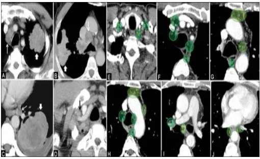
Fig 2.Lung cancer nodules. V.FUTURE ENHANCEMENT
The lung cancer detection system using the machine learning technique is much efficient and gives the betterment result to the radiologist and assist them. This enhances with the additional features for upgrading in the future. On this image processing system to support the radiologist to detect the malignancy nodules and the level of malignancy of the lung.
VI.RESULT AND CONCLUSION
Here we processed the CT scan images to differentiate the benign and the malignant nodule and its level of the growth of the cancer cells by the machine learning system and detecting the growth of the cancerous cell in the initial stage can be made in our project.Here it presented an approach to differentiate the pulmonary nodules into malignant and benign nodules to assist the radiologist and for the future enhancement. Further loads ought be directed at improving the SVM for classifying malignancy levels of nodules through experiments with various alternatives.
REFERENCES
[1].N.Camarlinghi, ``Automatic detection of lung nodules in computed tomography images: Training and validation of algorithms using public research databases,'' Eur. Phys. J. Plus, vol. 128, no. 9, p. 110,Sep. 2013.
[2]. R. L. Siegel, K. D. Miller, and A. Jemal, ``Cancer statistics, 2016,'' CA,Cancer J. Clin., vol. 66, no. 1, pp. 730, Jan. 2016
[3].D. Kumar, A. Wong, and D. A. Clausi, ``Lung nodule classification using deep features in CT images,'' in Proc. 12th Conf. Comput. Robot Vis. (CRV), Jun. 2015, pp. 133138.
[4]. P. P. RebouçasFilho, E. D. S. Rebouças, L. B. Marinho, R. M. Sarmento,J. M. R. Tavares, and V. H. C. de Albuquerque, ``Analysis of human tissue densities: A new approach to extract features from medical images,''PatternRecognit. Lett., vol. 94, pp. 211218, Jul. 2017.
[5]. W. Shen et al., ``Multi-crop convolutional neural networks for lung nodule malignancy suspiciousness classication,'' Pattern Recognit., vol. 61, pp. 663673, Jan. 2017.
[6]. FangfangHan ,Guopeng Zhang, Huafeng Wang, Bowen Song, Hongbing Lu, Dazhe Zhao, Hong Zhao and Zhengrong Liang “A Texture Feature Analysis for Diagnosis of Pulmonary Nodules Using LIDC-IDRI Database” 2013.
[7]. Hyo Kyung Lee,Student Member, IEEE,FengJu,Member, IEEE, Raymond U. Osarogiagbon, Nicholas Faris, Xinhua Yu, FedoriaRugless, Shan Jiang, and JingshanLi,Fellow,” A System-Theoretic Method for Modeling, Analysis, and Improvement of Lung Cancer Diagnosis-to-Surgery Process”,2017.
[9]. I. R. S. Valente, P. C. Cortez, E. C. Neto, J. M. Soares, V. H.C.de Albuquerque, and J. M. R. Tavares,``Automati3D pulmonary nodule detection in CT images: A survey,'' Comput. Methods Programs Biomed., vol. 124, pp. 91107, Feb. 2016.
[10]. P. Lambin, E. Rios-Velazquez, R. Leijenaar, S. Carvalho, R.G. van Stiphout, P. Granton, C.M. Zegers, R. Gillies, R. Boellard, A. Dekker, et al., Radiomics: extracting more information from medical images using advanced feature analysis, Eur. J. Cancer 48 (4) (2012) 441–446.
[11]. M.M. Wahidi, J.A. Govert, R.K. Goudar, M.K. Gould, D.C. Mc Crory, Evidence for the treatment of patients with pulmonary nodules: when is it lung cancer?:Accp evidence-based clinical practice guidelines, CHEST J 132 (3_suppl) (2007) 94S–107S.