International Journal of COPD
Dove
press
O r I g I n a l r e s e a r C h
open access to scientific and medical research
Open access Full Text article
Prevalence of comorbidities according to
predominant phenotype and severity of chronic
obstructive pulmonary disease
gianna Camiciottoli1,2
Francesca Bigazzi1
Chiara Magni1
Viola Bonti1
stefano Diciotti3
Maurizio Bartolucci4
Mario Mascalchi5
Massimo Pistolesi1
1section of respiratory Medicine,
Department of Clinical and
experimental Medicine, 2Department
of Clinical and experimental Biomedical sciences, University of Florence, Florence, 3Department
of electrical, electronic, and Information engineering “guglielmo Marconi,” University of Bologna, Cesena, 4Department of Diagnostic
Imaging, Careggi University hospital,
5radiodiagnostic section, Department
of Clinical and experimental Biomedical sciences, University of Florence, Florence, Italy
Background: In addition to lung involvement, several other diseases and syndromes coexist in patients with chronic obstructive pulmonary disease (COPD). Our purpose was to investigate the prevalence of idiopathic arterial hypertension (IAH), ischemic heart disease, heart failure, peripheral vascular disease (PVD), diabetes, osteoporosis, and anxious depressive syndrome in a clinical setting of COPD outpatients whose phenotypes (predominant airway disease and predominant emphysema) and severity (mild and severe diseases) were determined by clinical and functional parameters.
Methods: A total of 412 outpatients with COPD were assigned either a predominant airway disease or a predominant emphysema phenotype of mild or severe degree according to predic-tive models based on pulmonary functions (forced expiratory volume in 1 second/vital capacity; total lung capacity %; functional residual capacity %; and diffusing capacity of lung for carbon monoxide %) and sputum characteristics. Comorbidities were assessed by objective medical records.
Results: Eighty-four percent of patients suffered from at least one comorbidity and 75% from at least one cardiovascular comorbidity, with IAH and PVD being the most prevalent ones (62% and 28%, respectively). IAH prevailed significantly in predominant airway disease, osteoporosis prevailed significantly in predominant emphysema, and ischemic heart disease and PVD prevailed in mild COPD. All cardiovascular comorbidities prevailed significantly in predominant airway phenotype of COPD and mild COPD severity.
Conclusion: Specific comorbidities prevail in different phenotypes of COPD; this fact may be relevant to identify patients at risk for specific, phenotype-related comorbidities. The highest prevalence of comorbidities in patients with mild disease indicates that these patients should be investigated for coexisting diseases or syndromes even in the less severe, pauci-symptomatic stages of COPD. The simple method employed to phenotype and score COPD allows these results to be translated easily into daily clinical practice.
Keywords: COPD phenotypes, COPD severity, comorbidities
Introduction
Chronic obstructive pulmonary disease (COPD) is a heterogeneous condition diagnosed by not completely reversible airflow obstruction detected during spirometry. Beyond this unifying functional definition, it is known that the lung may react to cigarettes smoking and environmental exposure with conductive airways remodeling or paren-chymal destruction. The prevalence of airways or parenparen-chymal changes determines the prevalent phenotype of COPD. The extent and the concomitance of airways and parenchymal changes determine the severity of the disease. In the last few years, several
Correspondence: gianna Camiciottoli Pulmonary Pathophysiology Unit, Department of Clinical and experimental Medicine, University of Florence, largo Brambilla 3, Florence 50134, Italy Tel +39 55 347 6733664 email gianna.camiciottoli@unifi.it
Journal name: International Journal of COPD Article Designation: Original Research Year: 2016
Volume: 11
Running head verso: Camiciottoli et al
Running head recto: Comorbidities according to phenotypes and severity of COPD DOI: http://dx.doi.org/10.2147/COPD.S111724
International Journal of Chronic Obstructive Pulmonary Disease downloaded from https://www.dovepress.com/ by 118.70.13.36 on 22-Aug-2020
For personal use only.
Number of times this article has been viewed
This article was published in the following Dove Press journal: International Journal of COPD
Dovepress Camiciottoli et al
methods to identify the predominant phenotype and the sever-ity of COPD in clinical practice have been proposed.1–5
In addition to lung involvement, several “comorbidities” may coexist in patients with COPD such as cardiovascular disease, metabolic dysfunctions, and anxious–depressive disorders as the most commonly reported. A number of poten-tial biological mechanisms linking COPD and comorbidities have been proposed: the proinflammatory milieu,6–8 increased
oxidative stress,9 increased arterial stiffness,10,11 catabolic
state,12 and sedentary lifestyle.13 As a matter of fact, patients
with COPD are often “multi-diseased patients,” presenting different clinical pictures, with a poorer quality of life and outcomes,14 and a high disease-related burden.15,16 They also
need a complex diagnostic and therapeutic approach.17
The prevalence of the various comorbidities in COPD varies widely according to the methods used to define phe-notype and severity of the disease.18–22 The purpose of this
study is to investigate the prevalence of idiopathic arterial hypertension (IAH), ischemic heart disease (IHD), heart failure (HF), peripheral vascular disease (PVD), diabetes (D), osteoporosis (O), and anxious depressive syndrome (ADS) in a clinical setting of COPD outpatients whose predominant phenotype and severity were determined by a standardized method, derived from computerized tomography (CT),2
allowing patients to be classified based on simple clinical and pulmonary function parameters.
Materials and methods
Patients
A total of 412 outpatients with COPD (299 males), in stable clinical condition were consecutively enrolled and completed the study. The ethical committee of the University Hospital of Florence approved the study and an informed consent was signed by each patient.
Patients’ evaluation consisted of a thorough clinical history and physical examination. Severity of dyspnea was assessed by the modified Medical Research Council (mMRC) dyspnea scale. Static and dynamic lung volumes and single breath diffusing capacity (DLCO) were measured (V6200 Autobox Body Plethysmograph; Sensor Medics, Yorba Linda, CA, USA) according to American Thoracic Society/European Respiratory Society guidelines and expressed as percentage of the predicted values (%).23Each patient was assigned a
predominant airway or predominant emphysematous pheno-type and a mild or severe category of disease according to the results of a multivariate analysis of 14 continuous variables (age, years; body mass index [BMI]; pack/years; forced vital capacity [FVC]% predicted; forced expiratory volume
in 1 second [FEV1] % predicted; FEV1/vital capacity [VC] and FEV1/FVC ratios, total lung capacity [TLC] % predicted, residual volume [RV] % predicted, RV/TLC ratio, func-tional residual capacity [FRC] % predicted; VC% predicted; inspiratory capacity [IC] % predicted; DLCO % predicted plus three categorical variables (mMRC dyspnea scale, cough, and sputum, scored as follows: dyspnea [0= none; 1= slight; 2=
moderate; 3= severe; 4= very severe]; cough [0= absent; 1=
occasional; 2= chronic]; and sputum [0= absent/occasional; 1= chronic non-purulent; 2= chronic purulent]), which have been validated by quantitative CT.2 The variables entered
to classify COPD patients according to the predominant mechanism of airflow limitation were DLCO%, TLC%, and sputum purulence. These variables were linearly combined to determine the relative predominance of an airway or emphysema phenotype according to the following computa-tion. Sputum purulence was assigned a categorical value (0, absent; 1, present).
Phenotype 0.324 0.018 DL % 0.58 (sputum purulence) 0.011
CO
= − × −
× + ×TTLC%
Patients were classified as being affected by predominant emphysema or by predominant airway disease when the result of the above-mentioned equation was either positive or negative, respectively.2
FEV1/VC ratio, FRC%, and sputum purulence were entered to classify the severity of the disease. These variables were linearly combined to compute the COPD severity score according to the following equation:
Severity 0.575 0.013 FEV /VC 0.013 (sputum purulence) 0.0
1
=− − × +
× + 113 FRC%×
Patients were classified as being affected by severe or mild disease when the result of the computation was positive or negative, respectively.2
Comorbidities
The following comorbidities were chosen to investigate car-diovascular, metabolic, and behavioral domains in patients with COPD. We ascertained the presence of IAH, IHD, HF, PVD, D, O, and ADS by means of an interview, outcomes of functional diagnostic procedures, and current medical treatment. The presence of each comorbidity was confirmed by the investigators through a detailed review of available medical records and medications or therapy specific to any disease. Self-reported diagnoses were not considered.
International Journal of Chronic Obstructive Pulmonary Disease downloaded from https://www.dovepress.com/ by 118.70.13.36 on 22-Aug-2020
Dovepress Comorbidities according to phenotypes and severity of COPD
statistical analysis
Data were presented as mean ± standard deviation for continuous variables, and as counts and proportions for nominal and ordinal variables. The Student’s t-test was used to compare the mean of continuous variables among different subsets of patients. The Fisher’s exact test was used to compare the prevalence of categorical variables among different subsets of patients. The significance level was set at P,0.05.
Results
Anthropometric, smoke exposure, and functional data of the recruited patients and classification according to the prevailing phenotype and severity of the disease are reported in Table 1. Mild and severe COPD patients were matched for age. No difference in smoke exposure was found across the subgroups. Compared with patients with predominant airway disease, those with predominant emphysema were
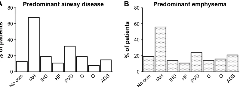
younger, with a lower BMI and a more impaired lung function involving all the considered functional variables. Table 2 shows the number of comorbidities in the whole set and according to prevailing phenotype and disease severity. No comorbidities were found in 16% of patients (65 out of 412). In more details, 29 (13%) predominant airway disease patients and 36 (19%) predominant emphysema patients had no detectable comorbidities. The number of patients with no comorbidities was similar in both the subgroups. The vast majority of patients turned out to be affected by two or three comorbidities (Table 2). Figure 1 reports the overall prevalence of each comorbidity. Figures 2 and 3 display the prevalence of each comorbidity within each of the two phenotypes. Out of 422 patients, 347 (84%) had at least one comorbidity. The most frequent was IAH, which affected 62% of the patients, followed by PVD (28%). Figure 3 shows the prevalence of comorbidities in the two phenotypes. While IAH was significantly more prevalent Table 1 anthropometric and functional data in patients with COPD according to phenotypes and severity of the disease
COPD (n=412)
Predominant airway disease (n=222)
Predominant emphysema (n=190)
P-value Mild COPD
(n=284)
Severe COPD (n=128)
P-value
Males/females 299/113 174/48 125/65 195/89 104/24
age 70±9 71±8 69±9 #0.05 70±9 69±8 ns
p/y 46±27 45±29 48±25 ns 45±28 50±24 ns
BMI 26±5 28±5 25±4 #0.0001 27±5 25±4 #0.0001
FeV1 (%) 69±25 76±23 61±26 #0.0001 79±22 48±19 #0.0001
FeV1/VC (%) 52±14 57±11 46±14 #0.0001 58±11 39±12 #0.0001
FrC (%) 120±31 110±25 132±32 #0.0001 107±21 150±29 #0.0001
TlC (%) 105±17 100±16 110±16 #0.0001 100±15 116±15 #0.0001
DlCO (%) 76±23 89±16 61±16 #0.0001 81±21 66±22 #0.0001
Note: Data are presented as mean ± sD.
Abbreviations: BMI, body mass index (weight/height2); COPD, chronic obstructive pulmonary disease; Dl
CO%, diffusing capacity of lung for carbon monoxide (% of
predicted); FeV1%, forced expiratory volume in 1 second (% of predicted); FeV1/VC%, FeV1–vital capacity ratio; FrC%, functional residual capacity (% of predicted); ns, nonsignificant; p/y, number of daily cigarettes × number of years/20; sD, standard deviation; TlC%, total lung capacity (% of predicted).
Table 2 number of comorbidities in patients with COPD according to phenotypes and severity of the disease Number of
comorbidities*
Predominant airway disease (n=222)
Predominant emphysema (n=190)
χ2
P-value
Mild disease (n=284)
Severe disease (n=128)
χ2
P-value
0 29 (13%) 36 (19%) 39 (13%) 26 (20%)
1 72 (32.4%) 67 (35.3%) 96 (34%) 43 (34%)
2 74 (33.3%) 52 (27.4%) 88 (31%) 38 (29.7%)
3 32 (14.4%) 22 (11.6%) 40 (14%) 14 (11%)
4 11 (5%) 9 (4.7%) 16 (6%) 4 (3%)
5 2 (1%) 3 (1.6%) 2 (0.7%) 3 (2.3%)
6 2 (1%) 1 (0.5%) 3 (1%) 0
0–2 175 155 0.53
ns
223 107 0.28
ns
3–6 47 35 61 21
Notes: *no patient has more than six comorbidities. The Fisher’s exact test was used to compare the prevalence of comorbidities in the different subsets of patients.
Significance was set at P,0.05.
Abbreviations: COPD, chronic obstructive pulmonary disease; ns, nonsignificant.
International Journal of Chronic Obstructive Pulmonary Disease downloaded from https://www.dovepress.com/ by 118.70.13.36 on 22-Aug-2020
Dovepress Camiciottoli et al
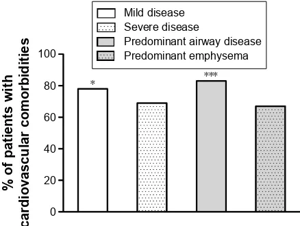
in patients with predominant airway disease, osteoporosis prevailed in those with predominant emphysema. As for COPD severity (Figure 4), we found a significantly higher prevalence of IHD and PVD in patients with mild COPD. Out of 412 patients, 309 (75%) were affected by at least one cardiovascular comorbidity. The prevalence of all considered cardiovascular comorbidities was significantly higher in patients with mild disease and in those with predominant airway phenotype of COPD (Figure 5).
Discussion
The main finding of this study was that specific comorbidi-ties prevailed in the two considered phenotypes of COPD; notably the highest prevalence of comorbidities was found among patients with mild stage of the disease. In particu-lar, IAH was significantly more prevalent in patients with the predominant airway phenotype, whereas osteoporosis seemed to prevail in the predominant emphysema phenotype.
In addition, we found a significantly higher prevalence of IHD and PVD in the subset of patients with mild COPD. The second relevant result was that the cardiovascular comorbidi-ties were found to be significantly more prevalent in patients with the predominant airways phenotype and with a milder expression of the disease.
A growing body of evidence suggests that COPD is associated with comorbidities whose prevalence varies largely among epidemiological studies. In this study, the prevalence of the whole set of considered comorbidity and cardiovascular comorbidities was consistent with those of Anecchino et al,18 which was based upon a different method
for comorbidity detection and classification. Indeed, they found at least one prescription of drugs for a chronic condi-tion other than respiratory disease in a vast majority of their wide cohort of patients with COPD. It is important to under-line that the presence of comorbidities in our study has been investigated by an objective method, with the exclusion of patients with a single drug prescription and/or a self-reported diagnosis, which was not considered probative for the pres-ence of a comorbidity.
In our study, patients with predominant airway disease were slightly older than those with predominant emphy-sema. This result is in contrast with those of Feary et al20
and Sode et al19 who showed the highest risk of comorbidity
in the youngest patients with COPD, suggesting to look for comorbidities at earlier stages of the disease. On the other hand, the present results of a higher prevalence of comorbidities in mild diseases’ severity confirm the hypothesis of individual susceptibility to factors implicated in the genesis of COPD, which could arise and develop up to different degrees of severity, and progress more or less into more severe stages independently of the age of the patients.
RISDWLHQWVZLWK&23'
1RFRP ,$+ ,+' +) 39' $'6
' 2
Figure 1 Prevalence of comorbidities in 412 outpatients with COPD.
Abbreviations: aDs, anxious depressive syndrome; com, comorbidities; COPD,
chronic obstructive pulmonary disease; D, diabetes; hF, heart failure; Iah, idiopathic arterial hypertension; IhD, ischemic heart disease; O, osteoporosis; PVD, peripheral vascular disease.
$
%
RISDWLHQWV
1RFRP ,$+ ,+' +) 39' $'6 ' 2
RISDWLHQWV
1RFRP ,$+ ,+' +) 39' $'6
' 2
3UHGRPLQDQWDLUZD\GLVHDVH 3UHGRPLQDQWHPSK\VHPD
Figure 2 Prevalence of comorbidities in 222 patients with the predominant airway disease (A) phenotype and in 190 patients with the predominant emphysema phenotype (B). Abbreviations: aDs, anxious depressive syndrome; com, comorbidities; D, diabetes; hF, heart failure; Iah, idiopathic arterial hypertension; IhD, ischemic heart disease;
O, osteoporosis; PVD, peripheral vascular disease.
International Journal of Chronic Obstructive Pulmonary Disease downloaded from https://www.dovepress.com/ by 118.70.13.36 on 22-Aug-2020
Dovepress Comorbidities according to phenotypes and severity of COPD
In patients with predominant airway phenotype of COPD, we observed that the most prevailing comorbidity was IAH. This result is consistent with that of Watz et al,24 who detected
IAH in a high proportion (73%) of patients with symptoms of chronic bronchitis without airflow limitation, as well as in those with mild COPD (77%). Interestingly, they found that circulatory markers of systemic inflammation were increased in patients with chronic bronchitis and COPD when metabolic syndrome was detected, irrespective of the degree of lung function impairment. Visceral adipocytes have been sus-pected to play a role by producing interleukin-6, which in turn induces the synthesis of C-reactive protein by hepatocytes. This finding has been confirmed in a study by Poulain et al,25
who demonstrated that systemic inflammation was higher in
obese patients with moderate COPD compared with those with normal weight and severe disease. We confirmed these observations indirectly, as BMI was significantly higher in patients with the predominant airway phenotype compared with those with predominant emphysema phenotype, and in moderate disease compared with severe disease.
Recently, new evidence has emerged suggesting that hypoxia, a frequent finding in patients with COPD, may be an additional cardiovascular risk factor for its role in the atherosclerosis process.26–29 Moreover, patients with COPD
are often affected by a neglected condition such as sleep disorders, resulting in nighttime hypoxia. Obesity strongly contributes along with age and active smoking for sleep disordered breathing, accelerated pulmonary hypertension, obesity–hypoventilation syndrome, and obstructive sleep apnea, irrespective of the degree of airflow obstruction.29
Hypoxia resulting from the above-mentioned conditions can be continuous or intermittent.26 These high
fre-quency cycles of hypoxia and reoxygenation are similar to ischemia–reperfusion injury and result in an increased production of reactive oxygen species and oxidative stress. Cell damage, remodeling of extracellular matrix and blood vessels, endothelial dysfunction, and inactivation of antipro-teases are some of the consequences of oxidative imbalance. Furthermore, in animals models, chronic intermittent hypoxia increases the amount of oxidized lipids and low-density lipoproteins in serum and arterial walls, which have stronger atherogenic effects, such as the formation of plaques in arter-ies which start due to lipid peroxidation. These results have been confirmed and extended in a small cohort of patients with COPD and sustained nocturnal, nonapneic hypoxia,
RISDWLHQWV
1RFRP ,$+ ,+' +) 39' $'6
' 2
3UHGRPLQDQWDLUZD\GLVHDVH 3UHGRPLQDQWHPSK\VHPD
Figure 3 Comparisons of the prevalence of each examined comorbidity in
222 patients with a predominant airway disease phenotype and in 190 patients with a predominant emphysema phenotype.
Note: *P,0.05.
Abbreviations: aDs, anxious depressive syndrome; com, comorbidities; D,
diabetes; hF, heart failure; Iah, idiopathic arterial hypertension; IhD, ischemic heart disease; O, osteoporosis; PVD, peripheral vascular disease.
RISDWLHQWV
1RFRP ,$+ ,+'
+) 39' ' 2 $'6
0LOGGLVHDVH 6HYHUHGLVHDVH
Figure 4 Prevalence of comorbidities in 412 outpatients according to mild or
severe grade of COPD.
Notes: *P,0.05; ***P,0.0001.
Abbreviations: aDs, anxious depressive syndrome; com, comorbidities; COPD,
chronic obstructive pulmonary disease; D, diabetes; hF, heart failure; Iah, idiopathic arterial hypertension; IhD, ischemic heart disease; O, osteoporosis; PVD, peripheral vascular disease.
RISDWLHQWVZLWK
FDUGLRYDVFXODUFRPRUELGLWLHV
0LOGGLVHDVH 6HYHUHGLVHDVH
3UHGRPLQDQWDLUZD\GLVHDVH 3UHGRPLQDQWHPSK\VHPD
Figure 5 Prevalence of cardiovascular comorbidities in 412 outpatients according
to mild or severe grade of COPD and according to the predominant phenotype.
Notes: *P,0.05; ***P,0.0001.
Abbreviation: COPD, chronic obstructive pulmonary disease.
International Journal of Chronic Obstructive Pulmonary Disease downloaded from https://www.dovepress.com/ by 118.70.13.36 on 22-Aug-2020
Dovepress Camiciottoli et al
which showed increased levels of serum lipid peroxidation and decreased paraoxonase activity.28
Patients with COPD and obesity, hypoventilation syn-drome, and obstructive sleep apnea resemble the so-called “blue bloater” phenotype29 and also the predominant airway
phenotype objectively classified in this paper.
Among the extrapulmonary effects of COPD, osteo-porosis has been recognized as a major comorbidity. This association has been explained in terms of age, sex, reduced daily activity, chronic hypoxemia, and systemic corticoster-oids therapy.30 Recently, the presence of osteoporosis also
in patients with milder airflow obstruction and clinically stable conditions has led to linking osteoporosis to a low-grade systemic inflammation sustained by increased plasma levels of C-reactive protein, interleukin-6, and tumor necrosis factor-α.31 Furthermore, COPD exacerbations, associated
with increased systemic inflammation, have been demon-strated to enhance osteoporosis progression.32 The high
prevalence of osteoporosis in the predominant emphysema phenotype in our study supports the view that, beyond a pure catabolic process, a mechanical derangement of lung structure due to hyperinflation and parenchymal disruption might also be relevant in sustaining a widespread chronic inflammation and its consequences. It could not be otherwise explained that a significant improvement of bone mineral density occurred 1 year after lung volume reduction surgery in patients with a pure, severe, CT-detected emphysema phenotype of COPD compared with a control group undergoing rehabilitation.33
The other important finding of this study was that IHD and PVD are significantly more prevalent in the subset of patients with mild COPD. This finding is in keeping with the previ-ous studies19,20 demonstrating that the association between
cardiovascular comorbidities and COPD is a risk factor for poor prognosis, hospitalization, and death of patients with mild COPD.19 Our results showed that the highest burden
of comorbidities is seen mostly in patients with less severe pulmonary disease. These findings were in agreement with findings of Watz et al24 and in contrast with those of Mahboub
et al22 and Dal Negro et al21 who reported an association
between the prevalence of comorbidity and COPD severity measured by a COPD assessment test score .10 or by Global Initiative on Obstructive Lung Disease stages, respectively. Our results were also in contrast with Mannino et al34 and
Johnston et al35 who reported a direct relationship between
severity of airflow limitation and risk for cardiovascular diseases, and for IHD, arrhythmias, stroke, and IAH, each one considered separately. Furthermore, other studies failed to demonstrate any significant association between the
prevalence of cardiovascular comorbidities and the severity of COPD evaluated by the level of airflow obstruction in a small cohort of patients,36 or by the degree of airflow
obstruc-tion, exercise capacity, and BODE index.37
The discrepancies may be related to the method by which the disease severity was scored. Conceivably, disease severity is unlikely to be satisfactorily depicted by the reduction of a single functional parameter (eg, FEV1). Indeed, this has been the sole criterion employed to assess COPD severity in many studies, actually before the publication of the revised Global Initiative on Obstructive Lung Disease guidelines in 2011; here symptoms and clinical events (exacerbations) have been introduced first, along with airway obstruction, to describe a disease that is actually far more complex than airway obstruc-tion alone. Furthermore, this funcobstruc-tional feature also reflects at least two pathological mechanisms, namely reduction of airway caliber (due to inflammation) and reduction of elastic recoil (due to parenchymal destruction). We are confident that the method employed to investigate COPD in this study takes into account both the predominant mechanism of airway obstruction for phenotyping each patient and the total amount of parenchymal and airways involvement to score severity.2
We acknowledge that this study has some limitations: 1) the small cohort of patients recruited; 2) the retrospec-tive collection of data concerning diagnosis and therapy of each examined comorbidity; and 3) the methodology used to report the presence of a comorbidity that is not completely objective because it was not based on repeating or seeking confirmatory tests for each disease, a task that would have been prohibitively expensive.14,16 Therefore, based on this
consideration and in keeping with most of the studies on the subject, we preferred to assess the presence of a comorbidity by means of a combination of clinical history and medical records documentation.
Conclusion
Understanding the association between comorbidities and phenotypes of COPD may be relevant to clarify the relation-ship between different pathophysiological mechanisms and such a disease: we can hypothesize to assign each phenotype of COPD a specific comorbidity panel.
The increased prevalence of comorbidities among patients with mild disease has several relevant practical aspects: first of all, the need of an early diagnosis of COPD, especially in those patients with slight symptoms who turn to general practitioners more often than to pulmonologists. For this purpose, we wish to stress that the results of this study can easily be translated into daily clinical practice due
International Journal of Chronic Obstructive Pulmonary Disease downloaded from https://www.dovepress.com/ by 118.70.13.36 on 22-Aug-2020
Dovepress Comorbidities according to phenotypes and severity of COPD
to the simple method suggested here to phenotype and score the disease. Accordingly, the complementary role of general practitioners and the specialist is highlighted in this study: given the large number of potential candidates to develop COPD, it would be impossible to care for all patients at a specialist level. A need exists to lay bridges and to share a common language that allow both specialists and general practitioners to diagnose and treat COPD and its comor-bidities early and effectively, in order to slow the disease progression and improve the quality of life.38,39
Increasing knowledge on links between COPD and comorbidities could provide innovative treatment strategies and it would assign each patient with COPD targeted thera-pies according to their personalized profile of phenotype, severity, and comorbidity.40
Acknowledgment
Professor Camiciottoli and Dr Diciotti were responsible for the funds allocation of the grant obtained by the Ministry of Health of Italy, Project Code: RF-2010-2321362.
Disclosure
The authors report no conflicts of interest in this work.
References
1. Paoletti M, Camiciottoli G, Meoni E, et al. Explorative data analysis techniques and unsupervised clustering methods to support clinical assessment of chronic obstructive pulmonary disease (COPD) pheno-types. J Biomed Inform. 2009;42(6):1013–1021.
2. Camiciottoli G, Bigazzi F, Paoletti M, Cestelli L, Lavorini F, Pistolesi M. Pulmonary function and sputum characteristics predict computed tomography phenotype and severity of COPD. Eur Respir J. 2013;42(3):626–635.
3. Burgel PR, Paillasseur JL, Roche N. Identification of clinical phenotypes using cluster analyses in COPD patients with multiple comorbidities. Biomed Res Int. 2014;420134.
4. Weatherall M, Travers J, Shirtcliffe PM, et al. Distinct clinical phe-notypes of airways disease defined by cluster analysis. Eur Respir J. 2009;34(4):812–818.
5. Marsh SE, Travers J, Weatherall M, et al. Proportional classifications of COPD phenotypes. Thorax. 2008;63(9):761–767.
6. Agustí A, Edwards LD, Rennard SI, et al. Evaluation of COPD Lon-gitudinally to Identify Predictive Surrogate Endpoints (ECLIPSE) Investigators. Persistent systemic inflammation is associated with poor clinical outcomes in COPD: a novel phenotype. PLoS One. 2012; 7(5):e37483.
7. Agustí AG, Noguera A, Sauleda J, Sala E, Pons J, Busquets X. Sys-temic effects of chronic obstructive pulmonary disease. Eur Respir J. 2003;21(2):347–360.
8. Gan WQ, Man SF, Senthilselvan A, Sin DD. Association between chronic obstructive pulmonary disease and systemic inflammation: a systematic review and a meta-analysis. Thorax. 2004;59(7):574–580. 9. MacNee W. Pathogenesis of chronic obstructive pulmonary disease.
Proc Am Thorac Soc. 2005;2(4):258–266.
10. Sabit R, Bolton CE, Edwards PH, et al. Arterial stiffness and osteopo-rosis in chronic obstructive pulmonary disease. Am J Respir Crit Care Med. 2007;175(12):1259–1265.
11. Maclay JD, McAllister DA, Mills NL, et al. Vascular Dysfunction in chronic obstructive pulmonary disease. Am J Respir Crit Care Med. 2009; 180(6):513–520.
12. Schols AM, Broekhuizen R, Weling-Scheepers CA, Wouters EF. Body composition and mortality in chronic obstructive pulmonary disease. Am J Clin Nutr. 2005;82(1):53–59.
13. Garcia-Aymerich J, Lange P, Benet M, Schnohr P, Antó JM. Regular physical activity modifies smoking-related lung function decline and reduces risk of chronic obstructive pulmonary disease: a population-based cohort study. Am J Respir Crit Care Med. 2007;175(5):458–463. 14. Mannino DM, Higuchi K, Yu TC, et al. Economic burden of COPD in
the presence of comorbidities. Chest. 2015;148(1):138–150. 15. Gershon AS, Mecredy GC, Guan J, Victor JC, Goldstein R, To T.
Quantifying comorbidity in individuals with COPD: a population study. Eur Respir J. 2015;45(1):51–59.
16. Wacker ME, Jörres RA, Schulz H, et al; for COSYCONET-consortium. Direct and indirect costs of COPD and its comorbidities: results from the German COSYCONET study. Respir Med. 2016;111:39–46. 17. Tsiligianni IG, Kosmas E, Van der Molen T, Tzanakis N. Managing
comorbidity in COPD: a difficult task. Curr Drug Targets. 2013;14(2): 158–176.
18. Anecchino C, Rossi E, Fanizza C, De Rosa M, Tognoni G, Romero M; Working Group ARNO project. Prevalence of chronic obstructive pulmonary disease and pattern of comorbidities in a general population. Int J Chron Obstruct Pulmon Dis. 2007;2(4):567–574.
19. Sode BF, Dahl M, Nordestgaard BG. Myocardial infarction and other co-morbidities in patients with chronic obstructive pulmonary disease: a Danish Nationwide Study of 7.4 million individuals. Eur Heart J. 2011; 32(19):2365–2375.
20. Feary JR, Rodrigues LC, Smith CJ, Hubbard RB, Gibson JE. Preva-lence of major comorbidities in subjects with COPD and incidence of myocardial infarction and stroke: a comprehensive analysis using data from primary care. Thorax. 2010;65(11):956–962.
21. Dal Negro RW, Bonadiman L, Turco P. Prevalence of different comor-bidities in COPD patients by gender and GOLD stage. Multidiscip Respir Med. 2015;10(1):24.
22. Mahboub B, Alzaabi A, Iqbal MN, et al. Comorbidities associated with COPD in the Middle East and North Africa region: association with severity and exacerbations. Int J Chron Obstruct Pulmon Dis. 2016;11: 273–280.
23. Pellegrino R, Viegi G, Brusasco V, et al. Interpretative strategies for lung function tests. Eur Respir J. 2005;26(5):948–968.
24. Watz H, Waschki B, Kirsten A, et al. The metabolic syndrome in patients with chronic bronchitis and COPD: frequency and associated consequences for systemic inflammation and physical inactivity. Chest. 2009;136(4):1039–1046.
25. Poulain M, Doucet M, Drapeau V, et al. Metabolic and inflammatory profile in obese patients with chronic obstructive pulmonary disease. Chron Respir Dis. 2008;5(1):35–41.
26. Song D, Fang G, Greenberg H, Liu SF. Chronic intermittent hypoxia exposure-induced atherosclerosis: a brief review. Immunol Res. 2015; 63(1–3):121–130.
27. Bernardo I, Bozinovski S, Vlahos R. Targeting oxidant dependent mechanisms for the treatment of COPD and its comorbidities. Phar-macol Ther. 2015;155:60–79.
28. Okur HK, Pelin Z, Yuksel M, Yosunkaya S. Lipid peroxidation and paraoxonase activity in nocturnal cyclic and sustained intermittent hypoxia. Sleep Breath. 2013;17(1):365–371.
29. McNicholas W, Verbraecken J, Marin JM. Sleep disorders in COPD: the forgotten dimension. Eur Respir Rev. 2013;22(129):365–375. 30. Jebouck A, Boonen S, Decramer M, Janssens W. COPD, bone
metabo-lism and osteoporosis. Chest. 2011;139(3):648–657.
31. Lind B, Feng Y. The association of low bone mineral density with systemic inflammation in clinically stable COPD. Endocrine. 2012; 42(1):190–195.
32. Kiyokawa H, Muro S, Oguma T, et al. Impact of COPD exacerbations on osteoporosis assessed by chest CT scan. COPD. 2012;9(3):235–242.
International Journal of Chronic Obstructive Pulmonary Disease downloaded from https://www.dovepress.com/ by 118.70.13.36 on 22-Aug-2020
International Journal of COPD
Publish your work in this journal
Submit your manuscript here: http://www.dovepress.com/international-journal-of-chronic-obstructive-pulmonary-disease-journal
The International Journal of COPD is an international, peer-reviewed journal of therapeutics and pharmacology focusing on concise rapid reporting of clinical studies and reviews in COPD. Special focus is given to the pathophysiological processes underlying the disease, intervention programs, patient focused education, and self management protocols.
This journal is indexed on PubMed Central, MedLine and CAS. The manuscript management system is completely online and includes a very quick and fair peer-review system, which is all easy to use. Visit http://www.dovepress.com/testimonials.php to read real quotes from published authors.
Dovepress
Dove
press
Camiciottoli et al
33. Mineo TC, Ambrogi V, Mineo D, Fabbri A, Fabbrini E, Massoud R. Bone mineral density improvement after lung volume reduction surgery for severe emphysema. Chest. 2005;127(6):1960–1966.
34. Mannino DM, Thorn D, Swensen A, Holguin F. Prevalence and out-comes of diabetes, hypertension and cardiovascular disease in COPD. Eur Respir J. 2008;32(4):962–969.
35. Johnston AK, Mannino DM, Hagan GW, Davis KJ, Kiri VA. Relation-ship between lung function impairment and incidence or recurrence of cardiovascular events in a middle-aged cohort. Thorax. 2008;63(7): 599–605.
36. Methvin JN, Mannino DM, Casey BR. COPD prevalence in Southeastern Kentucky: the burden of lung disease study. Chest. 2009;135(1): 102–107.
37. Vanfleteren LE, Spruit MA, Groenen M, et al. Clusters of comorbidities based on validated objective measurements and systemic inflammation in patients with chronic obstructive pulmonary disease. Am J Respir Crit Care Med. 2013;187(7):728–735.
38. Celli BR. Recommendation for the early diagnosis of COPD: The AIMAR view. Multidiscip Respir Med. 2015;10(1):6.
39. Welte T, Vogelmeier C, Papi A. COPD: early diagnosis and treatment to slow disease progression. Int J Clin Pract. 2015;69(3):336–349. 40. Brown JP, Martinez CH. Chronic obstructive pulmonary disease
comorbidities. Curr Opin Pulm Med. 2016;22(2):113–118.
International Journal of Chronic Obstructive Pulmonary Disease downloaded from https://www.dovepress.com/ by 118.70.13.36 on 22-Aug-2020