International Journal of General Medicine
Exercise electrocardiogram testing in two
brothers with different outcome – a case study
exercise testing in master cyclists
Christoph Alexander Rüst1 Beat Knechtle1,2
Thomas Rosemann1 1Institute of General Practice and
Health Services Research, University of Zurich, Zurich, Switzerland;
2Gesundheitszentrum St Gallen,
St Gallen, Switzerland
Correspondence: Beat Knechtle Facharzt FMH für Allgemeinmedizin, Gesundheitszentrum St Gallen, Vadianstrasse 26, 9001 St Gallen, Switzerland
Tel +41 071 226 8282 Fax +41 071 226 8272 Email [email protected]
Abstract: The cases of two brothers training and competing as master cyclists and both preparing for a cycling tour are presented. The older brother aged 66 years went first to the primary care physician and presented with an asymptomatic depression in the exercise stress test of the ST segment in V5 and V6 during recovery after complete exhaustion. Coronary angiography revealed a multi vessel coronary artery disease and he underwent bypass surgery.
One year later, he successfully completed his planned cycling tour of ∼600 km in seven stages
and covering ∼12,000 m of total ascent. The younger brother aged 59 years went a few months
later to the primary care physician and also performed asymptomatic exercise stress testing without changes in the ST segments. Unfortunately, 2 months later he suffered a cardiac arrest during his cycling tour and survived following immediate successful cardiopulmonary resuscitation on the road by his cycling colleagues. Immediate invasive coronary arteriography showed a complete stenosis of the trunk of arteria coronaria sinistra (left coronary artery), a 40%–50% stenosis of ramus circumflexus, and a 20% stenosis of arteria coronaria dextra (right coronary artery). The left coronary artery was dilated and he continued cycling 2 months later. In both brothers, familial hypercholesterolemia was the main cardiovascular risk factor for the multi vessel coronary artery disease. A negative exercise electrocardiogram in siblings with an increased risk for coronary artery disease seemed not to exclude an advanced multi vessel coronary artery disease. In master athletes with asymptomatic exercise electrocardiogram but a positive family history, further examinations should be performed in order to detect relevant stenosis in coronary arteries.
Keywords: cyclist, exercise stress test, electrocardiogram, coronary artery, hypercholesterolemia
Introduction
The exercise electrocardiogram (ECG) is widely considered the best available test for screening asymptomatic adults without known cardiovascular disease prior to initiating a vigorous exercise program due to its prognostic value, widespread availability, and low cost.1 Generally, the exercise ECG is used as a non-invasive method to evaluate
coronary artery disease.2 It has been reported that the exercise ECG has value in
diagnosing coronary artery disease and predicting its severity.3 The diagnostic
accuracy of an exercise ECG varies depending on the age, the gender, and the clinical characteristics of the patient.4 We report the case of two brothers without known
cardiovascular disease training and preparing as master cyclists defined as athletes who exceeded the age requisite for success in elite competition; ie, typically 35–40 years5,6
for an event with a different outcome after exercise stress testing.
Dove
press
C A S E R E P O RT open access to scientific and medical research
Open Access Full Text Article
International Journal of General Medicine downloaded from https://www.dovepress.com/ by 118.70.13.36 on 22-Aug-2020
For personal use only.
Number of times this article has been viewed
This article was published in the following Dove Press journal: International Journal of General Medicine
Case presentation
In June 2011, the 66 year old brother A went for a checkup prior to a planned cycling tour from Geneva (Switzerland) over the Alps to Nice (France) at the Mediterranean coast. He was healthy, had no medical problems, and trained frequently during his work life while he regularly competed in master races. During the year, he completed on average ∼12,000 cycling kilometers mainly in road cycling. After his retire-ment, he intensified his training to prepare for the planned cycling tour and went for a routine check before starting the tour. The primary care physician performed an exercise ECG after a physical examination and some basic laboratory measurements. The physical examination of the lean master athlete with 1.76 m body height and 61 kg body mass (body mass index of 19.7 kg/m2) showed no abnormalities. The
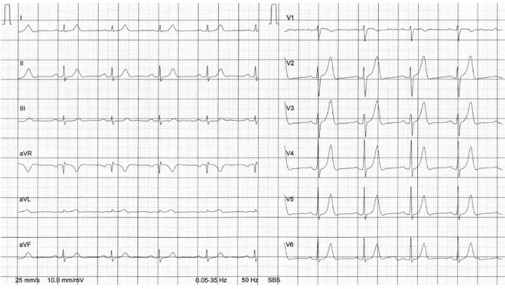
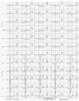
laboratory results are presented in Table 1 and the ECG at rest is shown in Figure 1. In the ECG at rest, high T waves in the precordial leads impress. In the exercise ECG (Figure 2), he achieved 197 Watt at maximum load, equal to 146% of his target. Blood pressure was 120/60 mmHg at rest and achieved a maximum of 180/80 mmHg at maximum exertion. The ST segment decreased 0.9 mm in V5 and 1.0 mm in V6 during the recovery phase (4 minutes) without symptoms. The primary care physician decided to send him for invasive coronary angiography. This examination showed a 50%–70% stenosis of ramus interventricularis anterior (RIVA; left anterior
descending, LAD), a 95% stenosis of arteria coronaria dextra (right coronary artery; RCA) and a 95% stenosis of ramus posterolateralis of the RCA. Due to these multiple stenoses, the cardiologist recommended cancelling the planned cycling tour and sent the patient to coronary artery bypass surgery. The heart surgeon performed a myocardial revascularization with arteria mammaria interna dextra on ramus posterolatera-lis dexter and ramus interventricularis posterior, and arteria mammaria interna sinistra on ramus diagonalis and ramus interventricularis anterior. The patient recovered well during a stationary rehabilitation and completed his planned cycling tour successfully in the summer of 2012.
In March 2012, 59-year old brother B went to the same primary care physician also prior to a planned cycling tour in the Alps. He was also training frequently for mountain bike cycling and had neither cardiac symptoms nor any other medical problems. During the year, he completed on average ∼9,000 cycling kilometers in both road and moun-tain bike cycling. The physical examination of the master athlete with 1.72 m body height and 69 kg body mass (body mass index of 23.3 kg/m2) showed no abnormalities. The
laboratory results are presented in Table 1 and the ECG at rest is shown in Figure 3. In the exercise ECG (Figure 4), he achieved 229 Watt, equal to 151% of his target. Blood pressure was 120/80 mmHg at rest and achieved a maximum of 220/90 mmHg at maximum exertion. The ST segment showed no changes at both maximum exertion and in the recovery phase. Blood pressure returned to 140/80 mmHg by the end of the test. Due to the high cholesterol and the increased thyroid stimulating hormone, the primary care physician decided to start with a lipid-lowering drug (40 mg of simvastatin) and 0.05 mg levothyroxine due to the latent hypothyroidism. The patient prepared for this planned cycling tour in the Alps starting May 17, 2012. The first day in a steep ascent in a mountain pass, he suffered an acute heart attack with cardiac arrest. His colleagues performed a successful cardiopulmonary resuscitation on the road and he was trans-ferred by helicopter to the nearest hospital. The ECG by the rescue team showed ventricular fibrillation. Immediate inva-sive coronary arteriography showed a complete stenosis of arteria coronaria sinistra (LCA), a 40%–50% stenosis of ramus circumflexus, and a 20% stenosis of arteria coronaria dextra (RCA). The LCA was dilated. Due to the fall off the bike, he also suffered a fracture of his first vertebra of the lumbar spine (L1). The ejection fraction of the left ventricle was 55% upon discharge from hospital. He was sent to car-diac rehabilitation where he fully recovered. Two months after the cardiac arrest, he was again training and cycling.
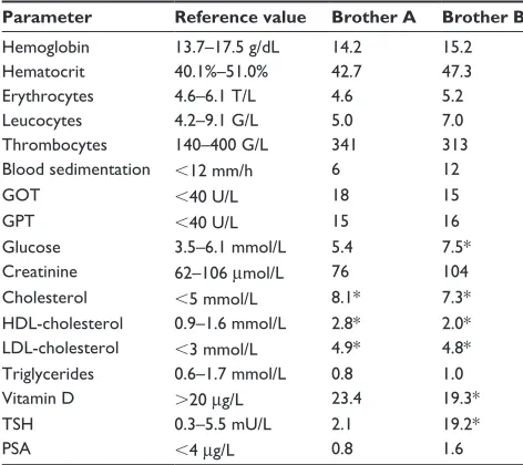
Table 1 Laboratory results of the two brothers at the time of the checkup
Parameter Reference value Brother A Brother B Hemoglobin 13.7–17.5 g/dL 14.2 15.2
Hematocrit 40.1%–51.0% 42.7 47.3
Erythrocytes 4.6–6.1 T/L 4.6 5.2
Leucocytes 4.2–9.1 G/L 5.0 7.0
Thrombocytes 140–400 G/L 341 313
Blood sedimentation ,12 mm/h 6 12
GOT ,40 U/L 18 15
GPT ,40 U/L 15 16
Glucose 3.5–6.1 mmol/L 5.4 7.5*
Creatinine 62–106 μmol/L 76 104
Cholesterol ,5 mmol/L 8.1* 7.3*
HDL-cholesterol 0.9–1.6 mmol/L 2.8* 2.0* LDL-cholesterol ,3 mmol/L 4.9* 4.8* Triglycerides 0.6–1.7 mmol/L 0.8 1.0
Vitamin D .20 μg/L 23.4 19.3*
TSH 0.3–5.5 mU/L 2.1 19.2*
PSA ,4 μg/L 0.8 1.6
Note: *Out of range, blood samples for brother B were not taken in an absolute fasting state.
Abbreviations: GOT, glutamic oxaloacetic transaminase; GPT, glutamic pyruvate transaminase; HDL, high-density lipoprotein; LDL, low-density lipoprotein; TSH, thyroid stimulating hormone; PSA, prostate-specific antigen.
Dovepress
Rüst et al
International Journal of General Medicine downloaded from https://www.dovepress.com/ by 118.70.13.36 on 22-Aug-2020
In the family history of the two brothers, the father died at the age of 70 years due to a heart attack. The mother died at the age of 90 years with no history of coronary heart disease. In autumn 2012, the third brother of the two brothers described herein was sent for an exercise ECG in order to assess the risk of coronary artery disease. This brother also achieved more than the target in the exercise stress test and the exercise ECG revealed no ST segment depression. Due to hypercholesterolemia, he was treated since 2008 with 40 mg of atorvastatin. He agreed to invasive coronary arteriography due to his high risk of heart attack. This examination showed advanced atherosclerosis in all of his coronary arteries.
Discussion
In brother A, the ST segment depression appeared in the recovery phase of the exercise ECG, but not during exercise. Lanza et al investigated the diagnostic and prognostic value of an ST segment depression limited to the recovery phase of an exercise stress test and compared it to that of an ST segment depression during exercise.7 They found that the
diagnostic and prognostic power of the ST segment depres-sion limited to the recovery phase of an exercise ECG was largely similar to that of the ST segment depression induced during effort. Also, Rashid et al reported that in patients undergoing exercise ECG, ST segment depression occurring only in the recovery phase of an exercise ECG had diagnostic value largely comparable with that of ST segment depression induced during the exercise phase of the exercise ECG.8 The
post-exercise ST segment depression without ST segment depression during exercise ECG may be associated with
an abnormal response to imbalances in the cardiovascular sympathetic system in the post-exercise period rather than with myocardial ischemia.9
Both brothers suffered from a multi vessel coronary artery disease. However, only in brother A did the exercise ECG reveal an ST segment depression in the recovery phase. Sun et al found that patients with multi vessel disease had a higher positive rate in exercise ECG compared to those with single vessel disease.10 Wu et al compared the results
of exercise ECG and coronary angiography in 461 patients.11
They showed that positive results of exercise ECG with downslope or horizontal depression of the ST segment more than 0.1 mV during or after exercise may predict coronary artery disease, the sensitivity of which was 74.5% (322 cases) and the specificity as 84.2% (139 cases). With the use of ST depression more than 0.2 mV as the super-positive standard, the sensitivity and specificity of positive exercise ECG to predict triple vessel disease and left main coronary disease was 61.3% and 79.1%, respectively. The duration of the ST segment depression had a positive correlation with the number of diseased coronary arteries. The patients with multi vessel disease and left main coronary artery disease had a higher positive rate of exercise ECG testing compared to patients with single or double vessel disease. It was concluded that exercise ECG testing had some value in diagnosing coronary artery disease and predicting its severity. Some parameters in exercise ECG were also helpful for predicting the severity of coronary artery disease.
Pigozzi et al evaluated the prevalence and clinical significance of ST segment depression during an exercise
Figure 1 ECGat rest from brother A.
Abbreviations: aVF,augmented unipolar limb lead in which the positive electrode is on the left leg; aVL, augmented unipolar limb lead in which the positive electrode is on the left arm; aVR, augmented unipolar limb lead in which the positive electrode is on the right arm; ECG, electrocardiogram; SBS, SCHILLER Baseline-Stabilizer.
Dovepress Electrocardiogram in master cyclists
International Journal of General Medicine downloaded from https://www.dovepress.com/ by 118.70.13.36 on 22-Aug-2020
ECG in 113 asymptomatic apparently healthy elderly male athletes.12 A significant ST segment depression at peak
exercise was detected in one athlete at the first evaluation. A further case was seen during the follow up period in a pre-viously “negative” athlete. Both athletes were asymptomatic and single photon emission computed tomography (SPECT) and/or stress echocardiography (ECHO) were negative for myocardial ischemia. They concluded that athletes with exercise induced ST segment depression with no associated symptoms and/or complex ventricular arrhythmias and no adverse findings at second level cardiologic testing should be
considered free from coronary disease and safe to continue athletic training.
For master athletes, Freeman et al recommend screening exercise ECG in asymptomatic men with diabetes and asymptomatic men over the age of 45 years with two or more cardiovascular risk factors prior to initiating a vigorous exercise program.1 In the present case, it was pure luck that
the coronary artery disease was discovered. The primary care physician performed the exercise stress test in the older brother only to show the wife that the master athlete would be able to complete the planned bike ride.
Figure 2 ExerciseECG from brother A.
Notes: Firstrow = at rest; second row = last step before maximum exertion; third row = maximum exertion; fourth row = after 2 minutes of recovery; fifth row = after 4 minutes of recovery.
Abbreviations: aVF,augmented unipolar limb lead in which the positive electrode is on the left leg; aVL, augmented unipolar limb lead in which the positive electrode is on the left arm; aVR, augmented unipolar limb lead in which the positive electrode is on the right arm; ECG, electrocardiogram.
Dovepress
Rüst et al
International Journal of General Medicine downloaded from https://www.dovepress.com/ by 118.70.13.36 on 22-Aug-2020
Figure 3 ECGat rest from brother B.
Abbreviations: aVF, augmented unipolar limb lead in which the positive electrode is on the left leg; aVL, augmented unipolar limb lead in which the positive electrode is on the left arm; aVR, augmented unipolar limb lead in which the positive electrode is on the right arm; ECG, electrocardiogram; SBS, SCHILLER Baseline-Stabilizer.
Figure 4 ExerciseECG from brother B.
Notes: Firstrow = at rest; second row = last step before maximum exertion; third row = maximum exertion; fourth row = after 2 minutes of recovery; fifth row = after 4 minutes of recovery.
Abbreviations: aVF,augmented unipolar limb lead in which the positive electrode is on the left leg; aVL, augmented unipolar limb lead in which the positive electrode is on the left arm; aVR, augmented unipolar limb lead in which the positive electrode is on the right arm; ECG, electrocardiogram.
Dovepress Electrocardiogram in master cyclists
International Journal of General Medicine downloaded from https://www.dovepress.com/ by 118.70.13.36 on 22-Aug-2020
In these two brothers, the father died at the age of 70 years due to a myocardial infarction. Leander et al showed that family history of coronary heart disease is not only a strong risk factor for myocardial infarction in both sexes, but that its effect was synergistic with other cardiovascular risk factors, as well.13 Siblings of patients with coronary artery disease
would benefit from investigating and treating cardiovascular risk factors. Hengstenberg et al analyzed the risk profile as well as the current utilization of preventive strategies in asymptomatic siblings of patients with myocardial infarction.14 Most asymptomatic individuals with positive
fraternal family history had more than one modifiable risk factor. Rissanen investigated the occurrence of different clinical manifestations of coronary heart disease among the parents and siblings of 309 men with coronary heart disease, including 103 men with fatal and 100 with nonfatal myocar-dial infarction, and 106 men with angina pectoris and among the relatives of 106 reference men.15 Coronary heart disease
was equally common among relatives of all subjects. It was four times as common in case brothers and twice as common in case sisters as in the respective reference siblings. Cardiac deaths were most common in the siblings of men with fatal myocardial infarction, and uncomplicated angina in the men with angina pectoris. The familial resemblance in the clinical manifestations of coronary heart disease suggested familial influence in the mechanisms determining the clinical expres-sion of the disease.
Most of the strong familial component in coronary heart disease of early onset thus appears to be mediated by familial hyperlipidemia and hypertension.16 The risk of premature
coronary heart disease in persons at highest risk could be largely eliminated if information about family history were used to identify such persons at an early stage and if they were treated properly for their correctable risk factors.16 Both
brothers showed hypercholesterolemia upon their first visit to the primary care physician. The high cholesterol values as a high risk for heart diseases can explain the result of both brothers, instead of the positive family history. The younger brother also had a subclinical hypothyroidism with increased thyroid stimulating hormone levels. High thyroid stimulat-ing hormone levels are associated with a non-favorable lipid profile17 and with an increased risk for all-cause and
cardiovascular mortality in adults.18 Most of the heart attacks,
however, happen not with non-significant stenosis but with mild to moderated disease with plaque rupture, causing ST-elevation myocardial infarction.19 Overall, the patient would
have needed better risk factor management rather than a further cardiac evaluation as he was asymptomatic.
Apart from hypercholesterolemia, diabetes mellitus type II is also an important risk factor for coronary artery disease.20,21 Brother B had elevated blood glucose upon the
check-up. However, the blood samples were not drawn in an absolute fasting state and subsequent determination of blood glucose showed normal values. Further, lipoprotein(a) is an established risk marker of coronary artery disease, which is independent from other risk factors.22,23 Unfortunately, this
marker was not determined in these two brothers.
In brother A, impressive T waves were recorded in the precordial leads prior to the exercise ECG. A potential reason for high T waves in precordial leads could be hyperkalaemia. However, the electrolytes potassium (3.9 mmol/L), sodium (138 mmol/L), and creatinine (82 μmol/L) were in the normal range during the hospital stay for the coronary angiography.
A negative exercise ECG in siblings with an increased risk for coronary artery disease seemed not to exclude advanced multi vessel coronary artery disease. In master athletes with asymptomatic exercise ECG but a positive fam-ily history, further examinations should be performed in order to detect relevant stenosis in coronary arteries. Technological advances in recent decades have produced several methods of non-invasive cardiac imaging for the diagnosis of coronary artery disease, such as stress ECHO, myocardial perfusion radionuclide imaging, cardiac computed tomography, and cardiac magnetic resonance imaging. There is always a bal-ance of cost and benefit for further examinations. The choice of an appropriate technique depends on the availability of the method, the patient’s clinical characteristics, and other factors, such as costs, risks, radiation, and potential contrast agent toxicity.24,25 Stress ECHO and myocardial perfusion
scintigraphy are widely available and most extensively validated. Stress ECHO is a non-invasive technology that images the heart using ultrasound and is a useful imaging modality for the diagnosis of coronary artery disease in suspected patients.26 Myocardial perfusion scintigraphy is a
widely used nuclear, non-invasive image acquisition tech-nique for investigating ischemic heart disease.27
Disclosure
The authors report no conflicts of interest in this work.
References
1. Freeman J, Froelicher V, Ashley E. The ageing athlete: screening prior to vigorous exertion in asymptomatic adults without known cardiovascular disease. Br J Sports Med. 2009;43(9):696–701.
2. Sharma K, Kohli P, Gulati M. An update on exercise stress testing. Curr Probl Cardiol. 2012;37(5):177–202.
Dovepress
Rüst et al
International Journal of General Medicine downloaded from https://www.dovepress.com/ by 118.70.13.36 on 22-Aug-2020
International Journal of General Medicine
Publish your work in this journal
Submit your manuscript here: http://www.dovepress.com/international-journal-of-general-medicine-journal The International Journal of General Medicine is an international,
peer-reviewed open-access journal that focuses on general and internal medicine, pathogenesis, epidemiology, diagnosis, monitoring and treat-ment protocols. The journal is characterized by the rapid reporting of reviews, original research and clinical studies across all disease areas.
A key focus is the elucidation of disease processes and management protocols resulting in improved outcomes for the patient.The manu-script management system is completely online and includes a very quick and fair peer-review system. Visit http://www.dovepress.com/ testimonials.php to read real quotes from published authors. 3. Ovrehus KA, Jensen JK, Mickley HF, et al. Comparison of usefulness
of exercise testing versus coronary computed tomographic angiography for evaluation of patients suspected of having coronary artery disease.
Am J Cardiol. 2010;105(6):773–779.
4. Banerjee A, Newman DR, Van den Bruel A, Heneghan C. Diagnostic accuracy of exercise stress testing for coronary artery disease: a system-atic review and meta-analysis of prospective studies. Int J Clin Pract. 2012;66(5):477–492.
5. Ransdell LB, Vener J, Huberty J. Master athletes: an analysis of running, swimming and cycling performance by age and gender. J Exerc Sci Fit. 2009;7:61–73.
6. Reaburn P, Dascombe B. Endurance performance in masters athletes.
Eur Rev Aging Phys Act. 2008;5:31–42.
7. Lanza GA, Mustilli M, Sestito A, Infusino F, Sgueglia GA, Crea F. Diagnostic and prognostic value of ST segment depression limited to the recovery phase of exercise stress test. Heart. 2004;90(12): 1417–1421.
8. Rashid MA, Mallick NH, Alam SN, Noeman A, Ehsan A, Hussain A. Usefulness of ST segment depression limited to the recovery phase of exercise stress test. J Coll Physicians Surg Pak. 2009;19(1):3–6. 9. Shinmura K, Suganuma Y, Hasegawa H, et al. Clinical significance
and mechanism of ST-segment changes during the post-exercise recov-ery period in male patients with coronary artrecov-ery disease. J Cardiol. 1996;27(5):231–239. Japanese.
10. Sun JL, Han R, Guo JH, Li XY, Ma XL, Wang CY. The diagnostic value of treadmill exercise test parameters for coronary artery disease. Cell Biochem Biophys. 2013;65(1):69–76.
11. Wu L, Zhang J, Shao G. A comparative study on coronary angiogra-phy and exercise ECG testing. Zhonghua Nei Ke Za Zhi. 1996;35(2): 107–109. Chinese.
12. Pigozzi F, Spataro A, Alabiso A, et al. Role of exercise stress test in master athletes. Br J Sports Med. 2005;39(8):527–531.
13. Leander K, Hallqvist J, Reuterwall C, Ahlbom A, de Faire U. Family history of coronary heart disease, a strong risk factor for myocardial infarction interacting with other cardiovascular risk factors: results from the Stockholm Heart Epidemiology Program (SHEEP). Epidemiology. 2001;12(2):215–221.
14. Hengstenberg C, Holmer SR, Mayer B, et al. Siblings of myocardial infarction patients are overlooked in primary prevention of cardiovascular disease. Eur Heart J. 2001;22(11):926–933.
15. Rissanen AM. Familial occurrence of coronary heart disease according to clinical manifestation. Acta Med Scand. 1985;218(4):355–363. 16. Rissanen AM. Familial occurrence of coronary heart disease: effect of
age at diagnosis. Am J Cardiol. 1979;44(1):60–66.
17. Duntas LH, Brenta G. The effect of thyroid disorders on lipid levels and metabolism. Med Clin North Am. 2012;96(2):269–281.
18. Tseng FY, Lin WY, Lin CC, et al. Subclinical hypothyroidism is associated with increased risk for all-cause and cardiovascular mortality in adults. J Am Coll Cardiol. 2012;60(8):730–737.
19. Anderson JL, Adams CD, Antman EM, et al; 2011 Writing Group Members; ACCF/AHA Task Force Members. 2011 ACCF/AHA Focused Update Incorporated Into the ACC/AHA 2007 Guidelines for the Management of Patients With Unstable Angina/Non-ST-Elevation Myocardial Infarction: a report of the American College of Cardiology Foundation/American Heart Association Task Force on Practice Guidelines. Circulation. 2011;123(18):e426–e579.
20. Sheikh-Ali M, Raheja P, Borja-Hart N. Medical management and strategies to prevent coronary artery disease in patients with type 2 diabetes mellitus. Postgrad Med. 2013;125(1):17–33.
21. Malik S, Billimek J, Greenfield S, Sorkin DH, Ngo-Metzger Q, Kaplan SH. Patient complexity and risk factor control among multimorbid patients with type 2 diabetes: results from the R2D2C2 study. Med Care. 2013;51(2):180–185.
22. Jaeger BR, Richter Y, Nagel D, et al. Longitudinal cohort study on the effectiveness of lipid apheresis treatment to reduce high lipoprotein(a) levels and prevent major adverse coronary events. Nat Clin Pract Cardiovasc Med. 2009;6(3):229–239.
23. Jaeger BR, Labarrere CA. Genetic variants in Lp(a) lipoprotein and coronary disease. N Engl J Med. 2010;362(12):1146–1147; author reply 1147.
24. Jaarsma C, Leiner T, Bekkers SC, et al. Diagnostic performance of noninvasive myocardial perfusion imaging using single-photon emission computed tomography, cardiac magnetic resonance, and positron emission tomography imaging for the detection of obstructive coronary artery disease: a meta-analysis. J Am Coll Cardiol. 2012;59(19):1719–1728.
25. Dowsley T, Al-Mallah M, Ananthasubramaniam K, Dwivedi G, McArdle B, Chow BJ. The role of noninvasive imaging in coronary artery disease detection, prognosis, and clinical decision making. Can J Cardiol. 2013;29(3):285–296.
26. Health Quality Ontario. Stress echocardiography for the diagnosis of coronary artery disease: an evidence-based analysis. Ont Health Technol Assess Ser. 2010;10(9):1–61.
27. Health Quality Ontario. Single photon emission computed tomography for the diagnosis of coronary artery disease: an evidence-based analysis.
Ont Health Technol Assess Ser. 2010;10(8):1–64.
Dovepress
Dove
press
Electrocardiogram in master cyclists
International Journal of General Medicine downloaded from https://www.dovepress.com/ by 118.70.13.36 on 22-Aug-2020