© IDOSI Publications, 2009
Corresponding Author: Dr. Hesham A. El Enshasy, Chemical Engineering Pilot Plant (CEPP), Faculty of Chemical and Natural Resources Engineering, University Technology Malaysia (UTM), 81310 Skudai, Johor, Malaysia
Serum Concentration Effects on the Kinetics and Metabolism of HeLa-S3 Cell Growth
and Cell Adaptability for Successful Proliferation in Serum Free Medium
1,2Hesham A. EL-Ensahsy,3Ahmed Abdeen, 3Sherif Abdeen, 4Elsayed A. Elsayed, 5Maha EL Demellawy, 2Abdallah A. EL Shereef
1CEPP, Faculty of Chemical and Natural Resources Engineering,
University Technology Malaysia, UTM, Skudai, Johor, Malaysia2 Department of Bioprocess Development, GEBRI,
Mubarak City for Scientific Research, New Burg Al Arab, Alexandria, Egypt
3Department of Zoology, Faculty of Science, Mansoura University, Mansoura, Egypt 4Department of Natural and Microbial Products, National Research Centre, Dokki, Cairo, Egypt
5Department of Medical Biotechnology, GEBRI, Mubarak City for Scientific Research,
New Burg Al Arab, Alexandria, Egypt
Abstract:The effect of serum content of culture medium on the kinetics of cell growth and metabolism has been studied in HeLa S3 culture. Increasing the serum concentration from 5 to 15% in Ham’s F12 mediu m increased the total number of living cells by about 30%. Further increase in serum concentration resulted in significant reduction in viable cell number. Meanwhile, increasing serum concentration in culture medium from 5 to 20% shows positive effect on cell viability and increased it from 38 to 74% in 144 h cultivated cultures. On the other hand, both of glucose consumption and lactate production rates were increased by the increase of serum concentration in the range between 5 and 20%. Based on these data, cells grown in Ham’s F12 medium of 10% serum were used for adaptation to serum free medium (SFM). Successive adaptation of cells to complete SFM was done by cultivating cells in different passages with the increased fraction of SFM in the cultivation medium. The ratios between SM (Ham’s F12 supplemented with 10%) to SFM (SMIF-6) were (100:0; 75:25; 50:50; 25:75; 10:90; 0:100). For each medium, cells were cultivated in 2-3 short passages for only 72 h to keep the cells in all cultures in their mid-exponential phase. Based on this cultivation strategy, HeLa S3 cells were completely adapted to serum free medium in only 880 hours. Neither growth kinetics nor physiological characterization (based on the data of glucose consumption rate and lactate production rate) were changed during the cell adaptation to SFM.
Key words: Hela cells • cell adaptation • serum free medium • cell growth kinetics INTRODUCTION
HeLa cells were firstly isolated from cervix adenocarcinoma tissue in early 1950s and fully adapted forin vitro cultivation in cell culture laboratories. As cancer cell line, HeLa cells can divide into unlimited number in a culture plate as long as basic cell requirements are provided. This type of cells is also robust for unoptimized cultivation conditions compared to other cell lines. Nowadays, HeLa cells are one of the few model cell lines which are routinely used in cell culture laboratories worldwide for drug quality control and throughout screening as well as acting as good host cells for the expression of different recombinant proteins and vaccines [1-3]. HeLa cells are also widely
by 5-10% serum [9]. The concentration of serum added to the cultivation medium is based on the type of media, type of serum and the specific application. The supplementation of culture media with undefined components, such as serum, has many disadvantages. The use of serum in culture media in research and R&D laboratories is associated with several problems such as: limited availability of good fetal bovine serum suppliers, batch to batch variation causing inconsistency in both cell growth pattern and products formation and the risk of viral, mycoplasmal and prions contamination, interfering with the effect of hormones or growth factors upon studying their interaction with cells. Beside all these drawbacks for serum application in cell culture research, other disadvantages are also considered by biotechnological industries for large scale cultivations. These include: the cost of serum can account for 70-85% of the overall cost of the medium, based on its complex structure and high rich protein content which will hinder product isolation and interferes in many separation and purification steps. Furthermore, the recent threat to human health caused by the undefined agents of bovine spongiform encephalopathy (BSE) will cause certain limit of using bovine serum in the production process of health products such as vaccines and therapeutic proteins [10]. Therefore, there are growing demands to develop better serum free medium for both research and industrial communities. Nowadays, different cell lines were adapted to grow in serum free media (SFM) for use in research or large scale production. These media were used successfully for the cultivation of many non-tumor cells such as: human mucosal epithelial cells [11]; human endothelial cells [12]; BHK cells [13]; vero cells [14]; CHO cells [15] and hybridoma cells [16, 17]. Beside these non cancer cell lines, different tumor cells such as human prostatic carcinoma [18]; lung carcinoma [19] and human bladder carcinoma [20] were successfully cultivated in SFM. The aim of the first part of this work was to study the relation between the serum concentration in culture medium and the kinetics of HeLa cell growth and metabolism (based on glucose consumption and lactate production). Followed by this part, studies on cell adaptation for growth in complete serum free medium were successfully done using short term successive adaptation strategy. The kinetics of cell growth and cellular metabolism during cell adaptation were studied in detail.
MATERIAL AND METHODS
Cell line: HeLa-S3 (ACC161) cells, derived from a human cervix adenocarcinoma [21], were obtained from the German Resource Center for Biological
material (DSMZ, Braunschweig, Germany). Cells grew in the form of adherent cells growing monolayer (Anchrogenous dependent).
Cultivation media: Two types of media were used in this study. The serum rich medium was composed of Ham’s F12 medium (Cambrex Bioscience, Verviers, Belgium) supplemented by 10% (v/v) Foetal Bovine Serum, FBS (Sigma, USA). The serum free medium used in this study was Scharfenberg’s modification 6 (SMIF-6) protein -free medium. This medium was developed by Drs. Klaus Scharfenberg and Roland Wagner (GBF, Braunschweig, Germany) and purchased from Biochrom, Berlin, Germany. This medium is characterized by its high glucose concentration of 4.00 g L-1 and well-balanced medium constituents.
Medium preparation: All media in this study were delivered in powder form. For basal medium Ham’s F12 medium as well as Serum Free medium (SMIF-6), all medium components were dissolved in high-purity grade water Milli-Q-water (Millipore, Eschborn, Germany). (TPP-Europe, Switzerland). Before medium sterilization, pH was adjusted to 7 by 1N HCl or 1N NaOH using pH meter (model 215, Denver Instrument company, Aviada, Colorado, USA). The medium osmolarity was adjusted to 340 mOsmol Kg-1 for both cultures using digital osmometer (advanced micro-osmometer Model 3300, Norwood, Massachusetts, USA). The medium was then filtered in microbiological filter system 0.22 µm. A sterility test was performed for 3 days at 37°C. Medium contamination was detected depending on the color change of phenol red, which gives a yellow-orange color through the pH change to acidic value as function of microbial growth. A sample was also examined microscopically after passing the sterility test. Medium was then stored at 4°C until being used.
For serum rich medium, 10% (v/v) FBS (Sigma-Aldrich Chemical, St. Louis, USA) was added to the Ham’s F12 medium before inoculation. The fetal bovine serum, FBS, was preserved at -20°C until being used. It was thawed overnight at room temperature and then was incubated for 30 min. at 56°C in water bath in order to deactivate the complement-system cytolytic proteins. The serum was aliquot in sterile 50 mL falcon tubes and stored at -20°C.
medium or HBSS buffer and then incubated with 2 mL Trypsin/EDTA buffered solution at 37°C for 2 min. Cell scrapers were used to help in detaching the cells and to decrease the exposure time to Trypsin enzyme. After that time, 5 mL of serum enriched medium was added to stop trypsin effect and to prevent cell death. Detached cells were collected and washed twice with HBSS and centrifuged at 1000 rpm for 5 min. (Eppendrof centrifuge 5804, Eppendrof-Netheler-Hinz.GmbH, Hamburg, Germany). The supernatant was discarded and the harvested cells were used to inoculate T-flask containing fresh medium. The inoculum size in all experiments was 1×10⁵ cells mL-1. The inoculated flasks were incubated in CO2-Incubator at 37°C, 5% (v/v) CO2 and 90% relative humidity (New Brunswick Scientific Co., Inc., USA).
Analysis
Cell harvest and sample preparation: Cells were harvested from T-flasks by the addition of 2 mL of 0.25% Trypsin-EDTA and incubating for 1-2 min. at 37°C. The further step of vigorous shaking or tapping was necessary to detach cells from the surface and then cells were collected by cell scraper. One mL sample was used immediately for cell counting and determination of cell viability. The rest of the sample was centrifuged in 2 mL Eppendorff microtube 5 min. at 5000 rpm. The supernatant was frozen immediately at -20°C for further analysis.
Cell enumeration: Cell number was determined by inverted microscope Olympus 1 × 70 equipped by digital camera and image analysis system. Additionally, cell concentration was determined by direct counting of cell number under Heamo cytometer slide (Improved Neubauer Type, Germany) under phase contrast microscope.
Estimation of viable cell concentration: Viable cell concentration was determined using Trypan blue exclusion method (Roche Pharmaceuticals, Mannheim, Germany). The difference between total and dead cells will give the concentration of the viable cells.
Determination of glucose and lactate concentration:
The cell culture supernatant sample in 2 mL Eppendroff was centrifuged for 5 min. at 5000 rpm. A small fraction was used for the determination of glucose and lactate concentrations. Glucose and lactate concentrations were determined using Biochemistry Analyzer, YSI 2700 SELECT (Yellow Springs Instruments, Yellow Springs, Ohio, USA).
RESULTS AND DISCUSSION
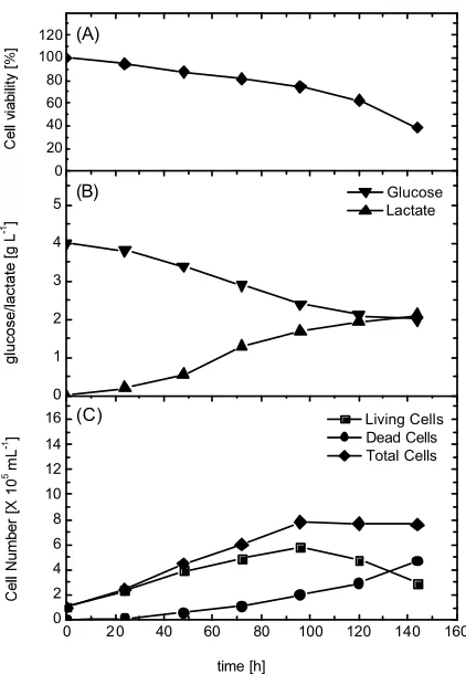
Effect of serum concentration on cell growth, metabolism and viability of HeLa cells: This experiment was designed to have good understanding of the role of serum, as one of the main medium constituents of Ham F12 medium, on cell growth and metabolism before cell adaptation to serum free medium. Therefore, four different experiments were conducted using media of different serum concentrations ranging from 5 up to 20% FBS. Inoculum size/origin (passage number) and cultivation conditions were the same for all experiments. The cell concentrations, cell viability and glucose/lactate concentrations were monitored. As shown in Fig. 1-4 and summarized in Table 1, cells grew exponentially in all cultures and the maximal number of living cells was obtained after 96 h cultivation regardless of serum concentration. On the other hand, living cell concentration increased by the increase of serum concentration in medium from 5 to 15%. Further increase resulted in significant reduction in the number of living cells. On the other hand, the maximal total cell
0 20 40 60 80 100 120 140 160
0 2 4 6 8 10 12 14 16 0 1 2 3 4 5 0 20 40 60 80 100 120
(C)
time [h]
Living Cells Dead Cells Total Cells
(B) Glucose
Lactate
(A)
Table 1: Kinetics of cell growth and cell metabolism during cell growt h in different serum concentration supplemented media Serum concentration in medium (%)
---Parameters 5 10 15 20
Maximal No. of living cells [×105 mL-1] 5.800 7.200 7.500 6.800
Maximal No. of dead Cells [×105 mL-1] 4.700 5.700 3.100 2.000
Maximal No. of total Cells [×105 mL-1] 7.800 10.500 9.900 8.000
Cell viability [%]* 38.200 43.000 59.000 74.000
-Qglucose [g L-1 h-1] 0.018 0.025 0.028 0.029
Qlactate [g L-1 h-1] 0.018 0.031 0.035 0.039
*, this value was calculated after 144 h cultivations
0 20 40 60 80 100 120 140 160
0 2 4 6 8 10 12 14 16 0 1 2 3 4 5 0 20 40 60 80 100 120
(C)
time [h]
Living Cells Dead Cells Total Cells
(B) Glucose
Lactate
(A)
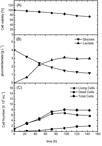
Fig. 2: Time profile of cell growth, glucose consumption and lactate production during HeLa S3 cells cultivation in Ham’s F12 medium supplemented with 10% FBS
number of 10.5×10⁵ mL-1 was obtained in 10% serum supplemented culture after 120 h. As shown in Fig. 2, after 96 h the rate of cell death increased significantly in this culture and the fraction of dead cells in maximal cell growth observed after 120 h accounts for about 40%.
Direct relation between medium serum content and cell viability was also observed in all cultures. For data taken after 144 h, the cell viability in culture increased from 38 to 74% by the increase of serum concentration
0 20 40 60 80 100 120 140 160
0 2 4 6 8 10 12 14 16 0 1 2 3 4 5 0 20 40 60 80 100 120
(C)
time [h]
Living Cells Dead Cells Total Cells
(B) Glucose
Lactate
(A)
Fig. 3: Time profile of cell growth, glucose consumption and lactate production during HeLa S3 cells cultivation in Ham’s F12 medium supplemented with 15% FBS
0 20 40 60 80 100 120 140 160 0
2 4 6 8 10 12 14 16 0 1 2 3 4 5 0 20 40 60 80 100 120
(C)
time [h]
Living Cells Dead Cells Total Cells
(B) Glucose
Lactate
(A)
Fig. 4: Time profile of cell growth, glucose consumption and lactate production during HeLa S3 cells cultivation in Ham’s F12 medium supplemented with 20% FBS
kinetics and MAb production by hybridoma cells was studied by Van der Pol et al. [25]. They reported that the cell death rate was higher at lower serum concentrations, whereas no decrease in MAb was observed when reducing the serum concentration from 10 to only 2.5%.
In all cultures studied, glucose was not the limiting substrate for HeLa cell growth where it was added to the culture medium in relatively high concentration (4.0 g L-1). As shown in Table 1, the rate of lactate production is directly related to the rate of glucose consumption. On the other hand, both of glucose consumption rate and lactate production rate increased gradually by the increase of serum concentrations in the cultivation medium under the range of applied concentrations. The relation between the serum concentration in cultivation medium and glucose production and lactate production rates is highly dependent on the cell line applied. In has been observed in other study that the increase of serum concentration from 2.5 to 10% decreased the glucose consumption and lactate production rates by human promyelocytic leukemia cells (HL60) by 10 and 15%, respectively[26].
On contrary, the other study of Ozturk and Palsson found that increasing serum concentration from 0.625 up to 20% had no effect on glucose consumption nor lactate production rates in hybridoma cells [27]. The conversion of glucose to lactate, obtained by dividing the rate of lactate formation over the rate of glucose consumption [Qlactate / Qglucose], showed that this value increased by 13.5% by increasing the serum concentration from 5 to 20% in the cultivation medium.
Adaptation of cell cultures to a serum-free medium:
0 200 400 600 800 1000 1200 0
2 4 6 8 10 0 2 4 6 8 10 70 80 90 100
time [h]
SFM 10% SM
90% SFM 25% SM
75% SFM 50% SM
50% SFM 75% SM
25% SFM SM
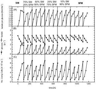
Fig. 5: Cell growth and viability during HeLa S3 cells adaptation to serum free medium in T-flask cultures
0 200 400 600 800 1000 1200
0 2 4 6 8 10 0 1 2 3 4 5 0.0 0.2 0.4 0.6 0.8 1.0 1.2 1.4 1.6 1.8 2.0
(C)
time [h]
(B)
SFM
10% SM 90% SFM 25% SM
75% SFM 50% SM
50% SFM 75% SM
25% SFM
SM
(A)
0 2 4 6 8 10 12 14 0
2 4 6 8 10 12 14 16 18 20 22 24
glucose consumption [mMol L-1]
SM 25 SM : 75 SFM 50 SM : 50 SFM 25 SM : 75 SFM 10 SM : 90 SFM SFM
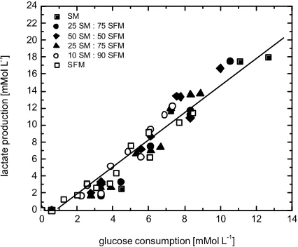
Fig. 7: Metabolic yield: the re lation between glucose consumption and lactate production in different cultures during cell adaptation to serum free medium (data of different experiments of this study)
adaptation of cells to SFM. As shown in Fig. 7, the relation between glucose consumption and lactate production can be represented as straight line for all points obtained from different cultivations at different ages. However, this relation between the concentrations of glucose consumed to the concentrations of lactate produced can be eas ily represented by the simple linear equation as follows:
Lactate produced [Mol] = -1.0325 + 1.5546 × Glucose consumed [Mol]
On the other hand, the average value of yield coefficient for lactate production per glucose consumed [Ylactate/gluc.(cons)] for all cultivations was about 1.26±0.36 (Mol Mol-1). The yield of lactate production from glucose ranged between 1.0 and 2.0 mole [Mol Mol-1] for most cell lines. This is usually based on the cultivation conditions such as aeration and medium osmotic stress[29].
Under anaerobic growth conditions, the yield of lactate production from glucose is (2:1) based on the stoichiometry of anaerobic metabolism. To eliminate both of these factors in our study, all cultivations were carried out under the same static cultivation in T-flask and medium osmolarity was adjusted to 340 mMol Kg-1 during media preparation. The obtained average yield [Ylactate/gluc.(cons)] of 1.26±0.36 (Mol Mol-1) in our study may be due to the cultivation of cells in static culture. But the stability of this value, in all cultures confirms that there was no change in cell physiological activity during cell adaptation.
REFERENCES
1. Kretzmer, G., 2002. Industrial processes with animal cells. Appl. Microbiol. Biotechnol., 59: 135-142.
2. Bleckwenn, N.A., W. Bentley and J. Shiloach, 2003. Exploring vaccinia virus as a tool for large-scale recombinant protein expression. Biotechnol. Prog., 19: 130-136.
3. Hendrick, V., D. Ribeiro de Sousa, A.R. dos Santos Pedregal, C. Bassens, P. Rigaux, K. Sato, K. Kotarsky and J. Werenne, 2006. Expression of recombinant protein in CHO and HeLa cells and its follow-up using EGF reporter gene, In: Ijima, S. and K.I. Nishijma, (Eds.). Animal Cell Technology: Basic and Applied Aspects, Springer, Netherlands, pp: 55-59.
4. Kronenberger, P., D. Schober, E. Prchla, O. Ofori-Anyinam, R. Vrijsen, B. Rombaut, D. Blaas, R. Fuchs and A. Boeyé, 1998. Uptake of poliovirus into the endosomal system of HeLa cells. Arch. Virol., 143: 1417-1424.
5. Morizono, K. and S. Harada, 1998. Human immunodeficiency virus type 1 (HIV-1) infection and transcytosis activity of a HIV-1 susceptible clone from HeLa cell. Microbiol. Immunol., 42: 313-320.
6. Kong, L.B., L.B. Ye, L. Ye, K.A. Timani, Y. Zheng, Q.J. Liao, B.Z. Li and B. Gao, 2006. Establishment of stable HeLa cell lines expressing enzymatically active hepatitis C virus RNA polymerase. Arch. Virol., 151: 361-367.
7. Witherell, G., 2000. In vitro Translation Using HeLa Extract,. In: Bonifacino J.S., M. Dasso, J.B. Harford, J. Lippincott-Schwartz and K.M. Yamada (Eds.). Current Protocols in Cell Biology. John Wiley and Sons, Inc, New York, pp: 1509-10510. 8. Masters, J.R., 2002. HeLa cells 50 years on: the
good, the bad and the ugly. Nature Rev. Cancer., 2: 315-319.
9. Jun, S.C., G.M. Lee, S.H. Chang and H. Kim, 1996. Effect of serum type and concentration on the expression of β-galactosidase in recombinant vaccinia virus infected HeLa S3 cells. Cytotechnol., 19: 153-159.
10. Burgener, A. and M. Butler, 2006. Medium development, In: Ozturk, S.S. and W.S. Hu (Eds.). Cell Culture Technology for Pharmaceutical and Cell-Based Therapies, Taylor & Francis Group, New York, London, pp: 41-79.
12. Labitzke, R. and P.A. Friedl, 2001. A serum-free medium supporting growth of human umbilical cord vein endothelial cells in long-term cultivation. Cytotechnol., 35: 87-92.
13. Perrin, P., S. Madhusudana, C. Gontier-Jallet, S. Petres, N. Tordo and O.W. Merten, 1995. An experimental rabies vaccine production with a new BHK-21 suspension cell culture process: use of serum-free medium and perfusion-reactor system. Vaccine., 13: 1244-1250.
14. Butler, M., A. Burgener, M. Patrick, M. Berry, D. Moffatt, N. Huzel, N. Barnabe and K. Coombs, 2000. Application of a serum-free medium for the growth of vero cells and the production of reovirus. Biotechnol. Prog., 16: 854-858.
15. Haldankar, R., J.J. Kopchick and D. Ridgway, 1999. Stable production of a human growth hormone antagonist from CHO cells adapted to serum-free suspension culture. Biotechnol. Prog., 15: 336-346.
16. Chen, Z., Y. Ke and Y.A. Chen, 1993. A serum-free medium for hybridoma cell culture. Cytotechnol., 11: 169-174.
17. Voigt, A. and F. Zind, 1999. Hybridoma cell growth and anti-neuroblastoma monoclonal antibody production in spinner flasks using a protein-free medium with microcarriers. J. Biotechnol., 68: 213-226.
18. Hedlund, T.E. and G.J. Miller, 1994. A serum-free defined medium capable of supporting growth of four established human prostatic carcinoma cell lines. Prostate, 24: 221-228.
19. Brower, M., D.N. Crney, H.K. Pie, A.F. Gazdar and J.D. Minna, 1986. Growth of cell lines and clinical specimens of human non-small cell lung cancer in a serum-free defined medium. Cancer Res., 46: 798-806.
20. Ruck, A., E. Jakobson, S. Bjorkman and S. Paulie, 1994. Adaptation of human bladder carcinoma cell lines to serum-free growth. Evidence for autocrine growth, stimulation. Anticancer Res., 14: 55-60. 21. Puck, T.T., P.I. Marcus and S.J. Cieciura, 1956.
Clonal growth of mammalian cellsin vitro: Growth characteristics of colonies from single HeLa cells with and without A Feeder layer. J. Exp. Med., 103: 273-284.
22. Kunas, K.T. and E.T. Papoutsakis, 1990. The protective effect of serum against hydrodynamic damage of hybridoma cells in agitated and surface-aerated bioreactor. J. Biotechnol., 15: 57-70.
23. Kadohama, T., K. Nishimura, Y. Hoshino, T. Sasajima and B.E. Sumpio, 2007. Effects of different types of fluid shear stress on endothelial cell proliferation and survival. J. Cell. Physiol., 212: 244-251.
24. Zanghi, J.A., F. Martin and J.E. Bailey, 1999. Serum protects protein-free competent Chinese hamster ovary cells against apoptosis induced by nutrient deprivation in batch culture. Biotechnol. Bioeng., 64: 108-119.
25. Van der Pol, L., G. Zijistra, M. Thalen and J. Tramper, 1990. Effect of serum concentration on production of monoclonal antibodies and on shear sensitivity of a hybridoma. Bioprocess Biosys. Eng., 5: 241-245.
26. McDowell, C.L. and E.T. Papoutsakis, 1998. Seru m increases the CD13 receptor expression, reduces the transduction of fluid-mechanical forces and alters the metabolism of HL60 cells cultured in agitated bioreactors. Biotechnol. Bioeng., 60: 259-268.
27. Ozturk, S.S. and B.O. Palsson, 1991. Examination of serum and bovine serum albumin as shear protective agents in agitated cultures of hybridoma cells. J. Biotechnol., 18: 13-28.
28. Sinacore, M.S., T.S. Charlebois, S. Harrison, S. Brennan, T. Richards, M. Hamilton, S. Scott, S. Brodeur, P. Oakes, M. Leonard, M. Switzer, A. Anagnostopoulos, B. Foster, A. Harris, M. Jankowski, M. Bond, S. Martin and S.R. Adamson, 1996. CHO DUKX cell lineages preadapted to growth in serum-free suspension culture enable rapid development of cell culture processes for the manufacture of recombinant proteins. Biotechnol. Bioeng., 52: 518-528.