ABSTRACT
THERIOT, CASEY MICHELLE. The Structure and Function of Thermostable Prolidases from Pyrococcus sp. (Under the direction of Dr. Amy M. Grunden).
Prolidase is a metallopeptidase that is ubiquitous in nature and has been isolated from mammals, bacteria and archaea. Prolidase specifically hydrolyzes dipeptides with a prolyl residue in the carboxy terminus (NH2-X-/-Pro-COOH). Current applications using prolidase to detoxify OP nerve agents include its incorporation into enzyme-based cocktails, fire-fighting foams and as biosensors for OP compound detection. Prolidases are also employed in the cheese ripening process to improve cheese taste and texture. In humans, prolidase deficiency (PD) is a rare autosomal recessive disorder that affects the connective tissue. Symptoms of PD include skin lesions, mental retardation and recurrent respiratory infections. Enzyme replacement therapies are currently being studied in an effort to optimize enzyme delivery and stability for this application. Previously, prolidase has been linked to collagen metabolism and more recently is being associated with melanoma. Increased prolidase activity in melanoma cell lines has lead investigators to create cancer prodrugs targeting this enzyme. Thus, there are many biotechnological applications using recombinant and native forms of prolidase.
cocktail are limited by long-term enzyme stability and reactivity over a broad range of temperatures. To obtain a better enzyme for organophosphorus nerve agent decontamination and to investigate the structural factors that may influence protein thermostability and thermoactivity, randomly mutated P. furiosus prolidases were prepared by using both XL1-red-based mutagenesis and error prone PCR. An Escherichia coli strain JD1 ( DE3) (auxotrophic for proline [ proA] and with deletions in pepQ and pepP dipeptidases with specificity for proline-containing dipeptides) was constructed for screening mutant P. furiosus prolidase expression plasmids. By using this positive selection, Pyrococcus prolidase mutants with improved activity at low temperatures were isolated. These mutants were further characterized to obtain better understanding of substrate catalysis with proline-dipeptides and OP nerve agents over a range of temperatures, and the relationship of these features with thermoactivity and thermostability is discussed.
The Structure and Function of Thermostable Prolidases from Pyrococcus sp.
by
Casey Michelle Theriot
A dissertation submitted to the Graduate Faculty of North Carolina State University
in partial fulfillment of the requirements for the degree of
Doctor of Philosophy
Microbiology
Raleigh, North Carolina 2010
APPROVED BY:
_______________________________ ______________________________
Dr. Jonathan Olson Dr. Robert Kelly
BIOGRAPHY
ACKNOWLEDGMENTS
I would first like to thank my family and friends for all of their support through the last 5 years of graduate school. It has been a long journey. I especially would like to thank my father and mother, Donald and Penny Theriot, for always being there when I was stressed out and for always believing in me, always telling me that I could do anything. To my many friends in Atlanta (Michelle, Amy and Steph), around the country and the world for their many kind words and conversations over the years. To my best friend and co-worker Michael Taveirne for always understanding and being there for me when I needed it most.
A special thanks to Dr. Amy Grunden for allowing me to come into her lab and giving me a strong foundation in microbiology research that I will be able to build on in the future. To all the lab members in the Grunden lab that were there from the beginning, past and present, Dr. Sherry Tove, Dr. Xuelian Du, Dr. Alice Lee, Dr. Mikyoung Ji, Dr. Drew Devine, Dr. Jimmy Gosse, Oscar Tirado-Acevado, and our newest member Rushyannah Killens. The Grunden lab has always been my home away from home and I thank all the members for making it a special time in my life. Thanks to all the graduate students in the Department of Microbiology for enduring the long journey together. I have made lifelong friends. I would like to also thank my committee members, Dr. Jon Olson, Dr. Bob Kelly and Dr. Jim Brown, for supporting and leading me in the right direction over the years.
TABLE OF CONTENTS
LIST OF TABLES………....viii
LIST OF FIGURES………...x
CHAPTER 1. Literature Review………1
Abstract………..2
1.1Introduction………..3
1.2Prolidase………...6
1.2.1 Mechanism of Substrate Specificity and Catalysis……….…..6
1.2.2 Proposed Reaction Mechanism……….9
1.2.3 Structure-function Information Provided by the Solved P. furiosus Prolidase Structure……….….10
1.2.4 Molecular and Catalytic Properties of Recombinant Prolidases……….…13
1.3Applications of Prolidases……….15
1.3.1 Detoxification of Organophosphorus Compounds….………15
1.3.2 Uses in the Food Industry………...21
1.3.3 Impact on Human Health………23
1.3.3a Prolidase Deficiency……….……23
1.3.3b Collagen Catabolism……….26
Acknowledgments………....32
References Cited………..33
CHAPTER 2. Improving the Catalytic Activity of Hyperthermophilic Pyrococcus Prolidases for Detoxification of OP Nerve Agents over a Broad Range of Temperatures……….64
Abstract………65
2.1 Introduction………66
2.2 Materials and Methods………...70
2.3 Results………77
2.4 Discussion………..87
Acknowledgments………....93
References Cited………..94
CHAPTER 3. Characterization of Two Proline Dipeptidases (Prolidases) from the Hyperthermophilic Archaeon Pyrococcus horikoshii……….…110
Abstract………..111
3.1 Introduction………..112
3.2 Materials and Methods……….116
3.3 Results………..123
APPENDIX. Screening Pyrococcus horikoshii Prolidase (Ph1prol) Mutants for
Increased Activity over a Broad Range of Temperature with OP Nerve Agents….….155
Abstract………..156
A.1 Introduction……….157
A.2 Materials and Methods………158
A.3 Results and Discussion………162
LIST OF TABLES
CHAPTER 1. Literature Review
Table 1-1 Pita-bread enzymes and their substrates………..54 Table 1-2 Specific activity of purified OPAAs/prolidases when DFP, other G-type
nerve agents and the proline dipeptides, Leu-Pro and Ala-Pro, are used as substrates………55 Table 1-3 Effects of Alteromonas sp. strain JD6.5 enzyme in the presence or absence of
various biodegradable and water-soluble wetting agents, degreasers,
or foams………..56
CHAPTER 2. Improving the Catalytic Activity of Hyperthermophilic Pyrococcus
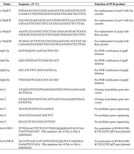
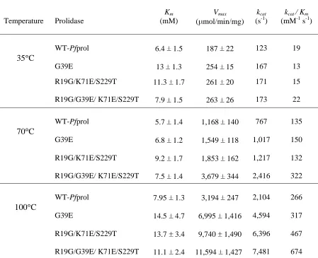
Prolidases for Detoxification of OP Nerve Agents over a Broad Range of Temperatures Table 2-1 PCR primers used in this study………..100 Table 2-2 Kinetic parameters of wild type and mutated prolidases from Pyrococcus
furiosus………....………...101
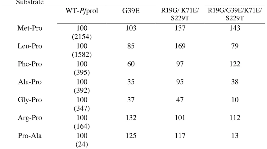
Table 2-3 Pot-life reactivity of recombinant wild type and mutant P. furiosus prolidases at 70°C………...102 Table 2-4 Substrate specificity of recombinant wild type and mutant P. furiosus
CHAPTER 3. Characterization of Two Proline Dipeptidases (Prolidases) from the Hyperthermophilic Archaeon Pyrococcus horikoshii
Table 3-1 Substrate specificity of recombinant P. furiosus and P. horikoshii prolidases
with different dipeptides………142
Table 3-2 Kinetic parameters of prolidases from P. furiosus and P. horikoshii………143
Table 3-3 Effect of metal ions on Ph1prol activity………144
Table 3-4 Effect of metal ions on Phprol activity………..144
Table 3-1S Metal content of purified prolidases from P. horikoshii………149
Table 3-2S Metal reconstitution assay under anaerobic conditions……….150
APPENDIX. Screening Pyrococcus horikoshii Prolidase (Ph1prol) Mutants for Increased Activity over a Broad Range of Temperature with OP Nerve Agents Table A-1 Relative activity of recombinant Pyrococcus prolidases with OP nerve agent DFP………168
LIST OF FIGURES
CHAPTER 1. Literature Review
Figure 1-1 Structure of basic R-group amino acid and cyclic proline……….…..57
Figure 1-2 Proline-specific peptidase cleavage map……….…58
Figure 1-3 Proposed reaction mechanism for P. furiosus prolidase………..59
Figure 1-4 The structure of a monomer and dimer of P. furiosus prolidase……….60
Figure 1-5 Dinuclear metal center active site of P. furiosus prolidase………..61
Figure 1-6 Alignment of P. furiosus prolidases with other prolidases………...62
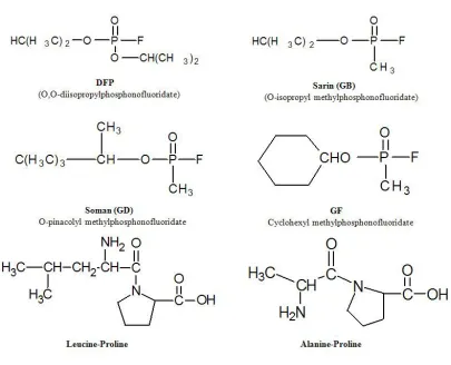
Figure 1-7 Chemical structures of G-type nerve agents and proline dipeptides…………63
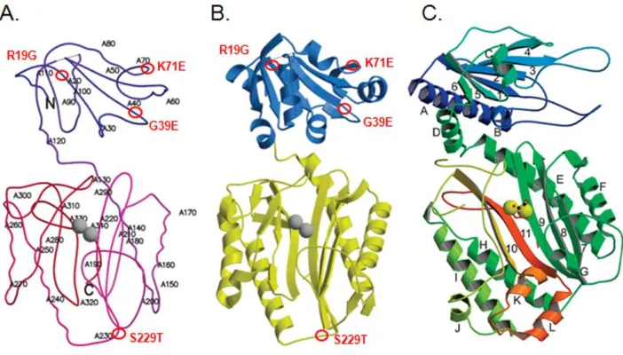
CHAPTER 2. Improving the Catalytic Activity of Hyperthermophilic Pyrococcus Prolidases for Detoxification of OP Nerve Agents over a Broad Range of Temperatures Figure 2-1 Mapping of mutations in the monomer structure of P. furiosus prolidase…105 Figure 2-2 Effects of temperature on the activities of wild type Pfprol, G39E-, R19G/K71E/S229T-, R19G/G39E/K71E/S229T- prolidases………106
Figure 2-3 Effects of pH on the activities of wild type and mutant prolidases………...107
p-CHAPTER 3. Characterization of Two Proline Dipeptidases (Prolidases) from the Hyperthermophilic Archaeon Pyrococcus horikoshii
Figure 3-1 Alignment of P. horikoshii prolidases with other prolidases……….145 Figure 3-2 Effects of pH on the activites of P. horikoshii prolidases………..146 Figure 3-3 Effects of different metals on the activities of P. horikoshii prolidases……147 Figure 3-4 Effects of cobalt and manganese concentrations on the activities of P.
horikoshii prolidases ……….148 Figure 3-1S Purification scheme and SDS-PAGE of P. horikoshii prolidases…..………151 Figure 3-2S Effects of temperature on the activities of P. horikoshii prolidases………..153 Figure 3-3S Effects of different metals on the activities of P. horikoshii prolidases under
anaerobic conditions………..154
APPENDIX. Screening Pyrococcus horikoshii Prolidase (Ph1prol) Mutants for Increased Activity over a Broad Range of Temperature with OP Nerve Agents Figure A-1 Effects of temperature on the activities of Pyrococcus prolidases with
CHAPTER 1 Literature Review
Biotechnological Applications of Recombinant Prolidases
Casey M. Theriot, Sherry R. Tove, Amy M. Grunden
Department of Microbiology, North Carolina State University, Raleigh, NC 27695
Citation: Theriot, C.M., S.R. Tove, A.M. Grunden. (2009) Biotechnological Applications of Recombinant Microbial Prolidases. In Advances in Applied Microbiology
Abstract
1.1 Introduction
and a penultimate proline in both short and long peptides (NH2-X-/-Pro--X-); proline iminopeptidase, that cleaves the N-terminal proline residue from any length polypeptide (Pro-/-X-); proline specific C-terminal exopeptidase, which releases an amino acid from the C terminus of a peptide with a penultimate proline (-X--Pro-/X-COOH); and prolidase, (NH2-X-/-Pro-COOH) (Ghosh et al., 1998) (Fig. 1-2). Prolidase is a proline specific peptidase that can hydrolyze dipeptides with proline at the C-terminus and a non-polar amino acid at the N-terminus (X-Pro). Some prolidases have also shown the ability to hydrolyze substrates with proline in the N-terminus as well as the C-terminus.
acetylcholinesterase (AChE), which leads to a buildup of acetylcholine in the body and can result in hypersecretion, convulsions, respiratory problems, coma and finally death. Previously, enzymes that catalyze the hydrolysis of OPs from the species Alteromonas were known as OPAAs or organophosphorus acid anhydrolases. OPAAs were shown to be capable of cleaving the P-F, P-O, P-CN and P-S bonds of the nerve agents, sarin and soman (Cheng et al., 1998). However, OPAA has been reclassified as a prolidase because it is able to efficiently hydrolyze specific X-Pro dipeptides, which is characteristic of prolidases. OP compounds appear tomimic X-Pro substrates in shape, size and surface charges (Cheng and DeFrank, 2000). Based on the activity that prolidases have against some OP agents, prolidases are being studied for use as biodecontaminants for detoxification of OP nerve agents in the field.
Prolidase is also important to the food and dairy industry because it can be used in ripening processes to reduce bitterness of cheese. The reduction in bitterness is due to the release of proline when prolidase is added in the cheese ripening process (Bockelmann, 1995). It can also be a critical enzyme for degrading proline-containing peptides generated in fermentation processes, which is important for creating desired flavor and texture attributes for fermented foods (Sullivan and Jago, 1972; Yang and Tanaka, 2008).
abnormalities result. While an increase in prolidase activity and a decrease in collagen in breast cancer tissue may cause increased cancer risk (Cechowska-Pasko et al., 2006). The use of prolidase as a potential biomarker for melanoma is currently being considered, as is its use as a potential therapeutic (Lupi et al., 2006; Mittal et al., 2007; Mittal et al., 2005).
Although there have been a number of important biotechnological applications that prolidases have been identified as being potentially suitable for, there are currently limitations preventing the wide-spread use of prolidases in all of these applications. However, by using appropriate bioengineering techniques, candidate prolidases can be tailored to each specific application. This review will provide an update on experimentally defined properties of a number of native and recombinant prolidases, as well as a discussion of current and future applications of prolidase enzymes.
1.2 Prolidase
1.2.1 Mechanism of Substrate Specificity and Catalysis
Streptomyces griseus aminopeptidase (SgAP) (Gilboa et al., 2001), human methionine aminopeptidase-2 (HsMetAP) (Liu et al., 1998), P. furiosus methionine aminopeptidase-1 (PfMetAP) (Tahirov et al., 1998), and carboxypeptidase G2 from Pseudomonas sp. strain
RS-16 (Rowsell et al., 1997). These enzymes share a dinuclear metal center bridged by a water molecule or hydroxide ion. The metal cluster is essential for the activation of catalysis. It functions to activate a nucleophile for the reaction, as well as participating in substrate binding and stabilizing the transition state (Maher et al., 2004). Some enzymes require two metals in the active site to activate catalysis and others only need one (Maher et al., 2004).
Based upon structural homologies of these enzymes, prolidases can be further categorized into a smaller class of metalloenzymes known as the “pita-bread enzymes”. Other enzymes within this class include methionine aminopeptidase (MetAP) (Roderick and Matthews, 1993), aminopeptidase P (APP) (Taylor, 1993) and creatinase (Coll et al., 1990), each of which have slightly different substrate specificity (Table 1-1), but the same conserved metal binding pocket suggesting they might have a conserved catalytic mechanism (Lowther and Matthews, 2002). These enzymes all contain a characteristic pita bread fold that encompasses a highly conserved metal center and substrate-binding pocket that is located in the enzyme‟s C-terminal domain. The substrate specificity of individual prolidases is dependent on the nature of the metal occupying the metal centers (Table 1-1).
numbering) (Bazan et al., 1994; Chang et al., 1992; Tsunasawa et al., 1997). P. furiosus prolidase (Pfprol) has a similar metal binding center to MetAP and APP. Pfprol also has similar N-terminal domains to those seen for APPro and creatinase, whereas E. coli MetAP lacks this N-terminal domain. MetAP is active as a monomer, while Pfprol and creatinase are dimers, and APPro functions as a tetramer.
MetAP is specific for dipeptides with N-terminal methionine in the P1 position
and a small uncharged residue such as Gly, Ala, Ser, Thr, Pro, Val or Cys in the P1 position
(Graham et al., 2006; Hirel et al., 1989). Prolidases are specific for dipeptides with proline in the trans configuration in the P1 position and non-polar residues in the P1 position (Ghosh
et al., 1998; Grunden et al., 2001; King et al., 1986; Lin and Brandts, 1979), whereas APPro has affinity for substrates with a hydrophobic or basic residue in the P1 position and a trans
Pro at P1 (Lowther and Matthews, 2002). The reaction centers of APP and prolidase require
the occupancy of two divalent ions such as Co2+ and Mn2+ in order to catalyze their reactions.
Although, two cations must be bound in the metal sites, the relative binding affinity of the metals differs, with there being one tightly bound metal atom and one more loosely bound (Lowther and Matthews, 2002).
E. coli MetAP has been shown to be maximally active when bound with 2 atoms of Co2+ per monomer under aerobic assay conditions or when bound with one Fe2+ atom per
APP and prolidase demonstrate the highest activities when bound with Co2+ and Mn2+,
whereas MetAP‟s highest activities are observed when it is loaded with Fe2+
(Lowther and Matthews, 2002). Although initially E. coli and P. furiosus MetAPs were described as requiring the occupancy of two metals for activity, more recent studies have indicated that they actually function more efficiently as Fe-containing monometallic hydrolases under anaerobic assay conditions (Copik et al., 2005; Cosper et al., 2001; D'Souza V and Holz, 1999). A study by Du et al. demonstrated that P. furiosus prolidase also showed the highest activity when assayed anaerobically with Fe2+, and the second highest activity when Co2+
was bound to the enzyme under aerobic assay conditions (Du et al., 2005). This suggests that both Pfprol and E. coli and P. furiosus MetAPs could preferentially use an iron mononuclear metal center in vivo under anaerobic conditions but switch to the use of a dimetal Co2+ reaction center under iron limitation or aerobic/oxidizing conditions.
1.2.2 Proposed Reaction Mechanism
and is thought to activate the nucleophile (ON) and facilitate proton transfer to glutamate
residue 313 (E-313), (2) the carboxy anion of the resultant tetrahedral intermediate, originating from the oxygen of the scissile bond (OC) is stabilized by the expanded
coordination sphere of Co1 and interactions with histidine-192 (H-192) and histidine-291 (H- 291), (3) resolution of the intermediate to products returns the coordination of Co1 to five, while the metal bridging and H-291 interactions are maintained, and (4) the active site is fully regenerated upon release of the proline and deprotonation of solvent molecules. Note that the amino acid residues refer to P. furiosus prolidase numbering (Fig. 1-3).
1.2.3 Structure-function Information Provided by the Solved P. furiosus
Prolidase Structure
„pita bread‟ family of proteins. There are a number of hydrogen bonds between the two domains, possibly enhancing stability. Locations where hydrogen bonds are present include the end of small helix domain I (residues 24-32) and -turn domain II (residues 284-294) (Maher et al., 2004). The proposed determinants for substrate specificity are thought to be localized to a region containing amino acid residues 113-123 in Pfprol. These residues link the two domains, and the angle is more acute with respect to the C-terminal domain when overlaid with the APPro domain. The active site of Pfprol is further crowded by the N-terminal domain of the subunits (residues 36B-39B). This suggests that the substrates coming in to the active site will be discriminated primarily based on size, where greater specificity will occur for smaller proline-containing peptides such as dipeptides versus polypeptides (Maher et al., 2004).
When isolated in either a native or recombinant form, Pfprol contains one Co(II) atom per monomer (Ghosh et al., 1998). During the crystallization process, Zn(II) replaced Co(II) in the prolidase active site as a consequence of the crystallization method used (Maher et al., 2004). However, Zn could be replaced with Co in the crystallized prolidase, and it restored enzyme activity.
Although the X-ray crystal structure analysis of Pfprol was able to definitively establish the structure of the enzyme‟s metal center-containing active site, the question as to which of the metal sites (Co1 or Co2) is the tightly-bound and which is the loosely-bound (Kd
0.24 mM) site remained unresolved. Therefore, further studies were undertaken which analyzed key site-directed Pfprol mutants to differentiate the binding affinities between the two Co atoms (Du et al., 2005). To look at different affinities of the metal binding sites, targeted mutations were made in the following locations: Asp209Ala, His284Ala, His284Leu, and Glu327Leu within Pfprol, where the three-letter code preceding the indicated amino acid position indicates the original amino acid and the three-letter code following the number indicates the mutated residue (Du et al., 2005). Results showed that Co1 is the tightly bound metal and Co2 is the looser bound metal (Kd 0.24 mM) (Du et al.,
2005). Similar findings were observed for E. coli MetAP where the Co1 site is the tight binding site, and the dissociation constants, Kd of Co1 and Co2 were estimated to be 0.3
1.2.4 Molecular and Catalytic Properties of Recombinant Prolidases The first prolidase both structurally and biochemically characterized was isolated from the hyperthermophilic archaeon P. furiosus (Ghosh et al., 1998; Grunden et al., 2001; Maher et al., 2004). It is a homodimer, with a molecular mass of 39.4 kDa per subunit, and as purified, it was determined to contain one bound Co2+ per subunit (Ghosh et al., 1998).
With the addition of cobalt, itshows maximum activity at 100 C and pH 7.0 with Met-Pro as the substrate (Ghosh et al., 1998; Grunden et al., 2001). Pfprol has a narrow substrate specificity, only hydrolyzing dipeptides with a proline in the C-terminus and non-polar amino acids (Leu, Met, Val, Phe or Ala) in the N-terminal position (Ghosh et al., 1998). The dipeptidase is maximally active with the addition of the divalent cations Co2+ and Mn2+, and
it cannot be substituted with other divalent cations (Mg2+, Ca2+, Fe2+, Ni2+, Cu2+ or Zn2+)
(Ghosh et al., 1998).
most active with manganese, while Lactobacillus delbrueckii prolidase requires zinc (Morel et al., 1999; Stucky et al., 1995).
Besides the differences in metal requirements, prolidases also demonstrate differences in substrate specificities. Pfprol has the greatest affinity for dipeptides with proline in the C-terminus and cannot cleave dipeptides with proline in the N-terminus. Likewise, L. delbrueckii pepQprolidase can also only cleave X-Pro dipeptides (Stucky et al., 1995). However, L. lactis prolidase can cleave dipeptides with proline in either the C- or N-terminal positions (Cheng et al., 1996). On the other hand, L. casei and guinea pig brain prolidase can cleave substrates without a prolyl residue.
Pyrococcus horikoshii and E. coli MetAP. Percent similarities compared to Pfprol for all listed prolidases are included.
1.3 Applications of Prolidases
1.3.1 Detoxification of Organophosphorus Compounds
Previous forms of disposal have consisted of chemical treatment, open-pit burning, evaporative burial, and deep ocean dumping, and presently, the EPA has approved incineration (Chen et al., 2000). Incineration is an expensive process, and it has raised environmental concerns. As a result, other environmentally friendly technologies are now being considered to degrade the stockpiles, including enzyme-based decontamination systems (Cheng and DeFrank, 2000).
In 1946 Mazur described the first work investigating hydrolysis of DFP (diisopropylfluorophosphate), an analogue of G-type nerve agents, by enzymes found in rabbit and human tissue extracts (Mazur, 1946). Most of these enzymes were first labeled DFPases and sarinases specific to the nerve agents they degraded. In 1992 they were listed in the category of Phosphoric Triester Hydrolases, named by the Nomenclature Committee of the International Union of Biochemistry and Molecular Biology. These enzymes were further separated into two subgroups based on their substrate specificities. The first subgroup is the organophosphate hydrolases (also referred to as paraoxonase and phosphotriesterase), which prefer the substrates paraoxon, and P-esters, which have a P-O bond. The second subgroup is diisopropyl-fluorophosphatases (also including OPAA or organophosphorus acid anhydrolase) which are most active against OP compounds with P-F or P-CN bonds (Cheng and DeFrank, 2000).
et al., 1995). It can also cleave the P-F and P-CN bonds, and the hydrolysis rates are 40-2450 times faster than chemical hydrolysis at temperatures up to 50 C (Munnecke, 1979). OPH is able to degrade a broad list of substrates including organophosphate pesticides (paraoxon and coumaphos) and OP nerve agents (DFP and sarin) (Chen et al., 2000; Cheng and DeFrank, 2000; Dumas et al., 1989; Dumas et al., 1990). Of its substrates, OPH can hydrolyze paraoxon the fastest with a rate of 104 s-1 (Grimsley et al., 1997). OPH mutants have been constructed in order to increase the substrate specificity with nerve agents. The following mutations were made in the metal binding center area of OPH: His257Leu, His257Val and His254Arg, resulting in even higher activity with soman and VX (Lai et al., 1996; Vanhooke et al., 1996).
and Fa-Aroonsawat, 2008) expressing OPH enzymes have been conducted. Both native and recombinant OPH can also be immobilized onto surfaces such as nylon (Caldwell and Raushel, 1991a), porous glass, and silica beads (Caldwell and Raushel, 1991b) as well as added to enzyme reactors, but this method still requires pure OPH enzyme which is very costly (Mulchandani et al., 1999; Mulchandani et al., 1998b). In recombinant E. coli with active OPH on the cell surface, the enzyme was stable and remained 100% active for more than a month (Chen and Mulchandani, 1998). Immobilized cells expressing OPH were used in batch reactors to test against many OP chemicals. It showed 100% hydrolysis of OP pesticides paraoxon and diazinon in less than 3.5 hours (Chen et al., 2000; Cheng and DeFrank, 2000). OPH is being used in medical applications as well. It can be used as an antidote or a therapeutic in preventing OP poisoning (Grimsley et al., 2000). Mice treated with OPH intravenously prevented cholinesterase inhibition when exposed to DFP, sarin or soman (Tuovinen et al., 1994; Tuovinen et al., 1996). When mice were pre-treated with OPH they were able to resist even higher doses of nerve agents.
OPAA from A. haloplanktis and OPAA-2 from Alteromonas sp. JD6.5 are very similar with 81% amino acid sequence identity and 91% similarity (Cheng et al., 1997). Both OPH and OPAA enzymes can hydrolyze many of the same substrates; however, there is no significant sequence homology found between any of the known OPH and OPAA enzymes (Cheng and DeFrank, 2000; Cheng et al., 1996), suggesting they are not the same enzyme.
suggests that Alteromonas OPAAs and prolidases may have evolved from the same ancestral gene (Cheng et al., 1997).
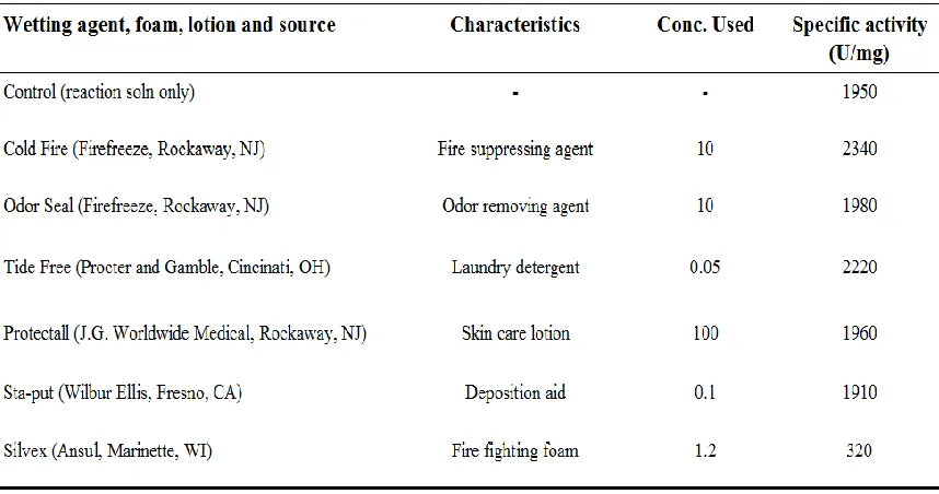
In order to effectively incorporate prolidases into an acceptable decontamination formulation, the enzyme has to be stable over time and not be inhibited by the water-based system employed. Table 1-3 shows the current systems including fire-fighting foams or sprays, degreasers, laundry detergent, and aircraft deicing solutions (Cheng and DeFrank, 2000). Foams appear to be the best delivery option because they have surface-active agents that help with the solubilization of the substrate, and they are able to adhere to vertical surfaces, enabling the enzyme to have significant contact time with substrates over a large surface area.
Currently to detoxify nerve agent exposed environments, a decontamination solution known as DS2 is being used in conjunction with bleach (Cheng et al., 1999). DS2 is environmentally harmful because it is corrosive and contributes additional hazardous waste to the environment. Since the use of current decontamination solution formulations is not a good long-term decontamination strategy, there is a perceived need to optimize an enzyme-based decontamination system. However, limitations of the enzymes that have thus far been examined for use in this process include poor activity at low pH and over a broad temperature range, and instability of the enzymes in the presence of harsh solvents, metals, detergents and or denaturants.
heavily on microbial metabolism. Cheese is made by the coagulation of milk using starter culture bacteria, usually Lactic Acid Bacteria (LAB), and the enzyme rennet. LAB acidifies milk by converting lactose into lactic acid, and the rennet coagulates the mixture. Ripening is driven by the microbial proteolysis process and results in casein protein being broken down into many different peptides and amino acids (Stucky et al., 1995). Some amino acids are known to produce a bitter taste. Hydrophobic peptides ranging from 2-23 residues play a significant role in the bitterness of cheddar cheese (Sullivan and Jago, 1972). Peptides isolated from casein hydrolysate and cheese result in high hydrophobicity and a high number of aromatic amino acids, causing bitterness (Agboola et al., 2004). In the study by Agboola et al., the role that a number of hydrophobic peptides play in determining the bitterness of ovine milk cheese was examined. To reduce the overall bitterness of the cheese, particular peptides need to be removed by proteolysis. Because of their unique structure, most of the remaining dipeptides are Xaa-Pro and or Pro-Xaa type dipeptides. These proline-containing dipeptides have been shown to significantly contribute to bitterness, especially the Xaa-Pro class of dipeptides (Yang and Tanaka, 2008).
(El Soda, 1993). Lactic acid bacteria (LAB) are used in the fermentation of foods, especially dairy products, and they contain many peptidases specific for proline including: proline iminopeptidase (PepI), prolinase (PepR), X-prolyl dipeptidyl aminopeptidase (PepX) and prolidase (PepQ) (Christensen et al., 1999; Sousa et al., 2001). By using prolidase to hydrolyze the bond of Xaa-Pro dipeptides, bitterness in cheese and other fermented foods can be reduced. In a study by Courtin et al., a Lactococcus lactis proteolytic system was investigated for its ability to increase the ripening process of cheese. By adding excess proline specific peptidases from lactobacilli, including prolidases, they were able to increase the total amount of free amino acids 3-fold, in turn speeding up cheese ripening (Courtin et al., 2002). The role of proline specific dipeptides in the flavor of cheese has been explored, but the role that the single amino acid proline plays is still unknown and requires further examination.
1.3.3 Impact on Human Health 1.3.3a Prolidase Deficiency
prolidase gene are linked to this deficiency (Ledoux et al., 1996; Lupi et al., 2006). Key point mutations Arg184Gln (Arg122 in Pfprol), Asp276Asn (Asp209 in Pfprol), Gly278Asp (Gly211 in Pfprol), and Gly448Arg (Gly323 in Pfprol) have been found in patients with this disorder (Maher et al., 2004). These mutations were carefully compared to the Pfprol enzyme to evaluate what impact they have on the enzyme‟s structure and function. The same point mutations in Pfprol result in disruption of function and structure of the enzyme (Maher et al., 2004). Diagnosis of PD in the past has been difficult and has resulted in significant numbers of misdiagnosed cases (Lupi et al., 2006; Viglio et al., 2006). Presently, detection methods include screening for prolidase activity in erythrocytes, leukocytes and skin fibroblast cultures and also screening urine for excess X-Pro imidodipeptides (Viglio et al., 2006). Therapeutic approaches have been explored for PD in the past. Topical treatments with glycine and proline have been used with minimal effectiveness on leg ulcers (Arata et al., 1986; Jemec and Moe, 1996). Oral administration of L-proline was also tried, however, this treatment failed to prevent the ulcerations (Isemura et al., 1979; Ogata et al., 1981; Sheffield et al., 1977). Other methods in preventing PD have included blood transfusions and aphaeresis (Endo et al., 1982; Lupi et al., 2002), the use of corticosteroids (Shrinath et al., 1997; Yasuda et al., 1999), application of growth hormone (Monafo et al., 2000), and an antibiotic topical treatment (Ogata et al., 1981).
transfer resulted in 7.5 times the normal activity of prolidase in the fibroblasts. The main consideration in enzyme replacement therapy is in the type of enzyme delivery system and the stability of the enzyme once it is delivered to the target location. Prolidase enzyme delivery using micro- and nano-particulate systems has been done, and the delivery efficiency of the enzyme into fibroblasts was poor (Colonna et al., 2007; Genta et al., 2001; Lupi et al., 2004). More recently, enzyme replacement studies have been done using liposomes to deliver native prolidase trancellularly into fibroblasts in PD patients (Perugini et al., 2005). Although, the delivery of the enzyme to fibroblasts was efficient, the enzyme was only active for 6 days (Perugini et al., 2005). A study by Colonna, et al., addressed the issue of enzyme stability and efficient enzyme delivery, using PEGylated prolidase loaded in chitosan nanoparticles to restore the normal prolidase activity in PD patient cells (Colonna et al., 2008).
system, showed stability and activity at 37 C for up to 6 days (Lupi et al., 2006). While these findings represent progress in enzyme replacement therapy, clearly further advances in enzyme stability need to occur for human prolidase replacement therapy to be a viable therapeutic option in the treatment of PD.
1.3.3b Collagen Catabolism
Primarily produced in fibroblasts, collagen is the main fibrous structural protein that makes up connective tissue in vertebrates (Bornstein and Sage, 1989). Up to 25% of total
body protein is collagen (Di Lullo et al., 2002). ECM or extracellular matrix is made up of 80% collagen, and connective tissue and the organic part of bone are made of 90-95% collagen (Dixit et al., 1977). Hydroxylproline and proline make up over 25% of collagen amino acids (Dixit et al., 1977). Collagen has also been recognized as a ligand for integrin ( 1-integrin) cell surface receptors, which are important for regulating ion transport, lipid
metabolism, kinase activation and gene expression (Akiyama et al., 1990; Bissell et al., 1982; Carey, 1991; Donjacour and Cunha, 1991; Surazynski et al., 2008). When collagen structure is affected, it can have an impact on cell signaling, metabolism and function, which can lead to tumorigenicity and invasiveness (Surazynski et al., 2008). The secretion of matrix metalloproteinases or MMPs, which break down ECM or collagen, is also an important event in the progression and metastasis of cancer (Cechowska-Pasko et al., 2006).
prolidase facilitated degradation. Prolidase is able to hydrolyze the most abundant substrate Gly-pro from degraded procollagen and collagen (Surazynski et al., 2008). It is suggested that prolidase plays a role in metabolism of collagen and recycling of proline for collagen resynthesis (Jackson et al., 1975; Palka, 1996; Yaron and Naider, 1993). The mechanism to explain this is currently being investigated but at this time is unclear.
The absence of prolidase, or PD, has been associated with slow wound healing, due to an abnormal nitric oxide (NO) signaling pathway. NO is associated with collagen metabolism and matrix degradation because it shows high expression when tissues need repair (Lupi et al., 2008; Surazynski et al., 2005). Overexpression of prolidase has been linked to increased levels of nuclear hypoxia inducible factor (HIF-1 ), which plays an important role in stress responsive gene expression (Jaakkola et al., 2001; Semenza, 2001; Surazynski et al., 2008). Prolidase activity has also been implicated as a factor in other diseases such as osteogenesis imperfecta (Galicka et al., 2005; Galicka et al., 2001), pancreatic diseases (Palka et al., 2002), lung carcinoma planoepitheliale (Karna et al., 2000) and metastasis of breast cancer MCF-7 cells (Miltyk et al., 1999; Palka and Phang, 1998).
In a study by Cechowski-Pasko et al., it was observed that increased prolidase activity in breast cancer tissue correlated with deficiencies in collagen and 1-integrin
a study by Mittal et al, high expression of prolidase activity in melanoma cell lines was also observed (Mittal et al., 2005). Prolidase was selected as a drug target due to its consistently high expression in melanoma cell lines and its high substrate specificity (Mittal et al., 2005). Prodrugs are being used to decrease the toxicity and side effects of chemotherapeutic agents in cancer patients. Currently, using prolidase as a drug target for selective activation of the prodrug, melphalan, for specific drug delivery to the tumor is being evaluated (Mittal et al., 2007; Mittal et al., 2005).
1.4 Advances in and Limitations of the Use of Prolidase for Biotechnological Applications
There are advantages and disadvantages of using certain prolidases in all of the applications previously discussed. The advantages of using Alteromonas recombinant prolidase in biodecontamination foams are due to its high activity against G-type nerve agents, such as soman and sarin. The limitations in using a mesophilic prolidase in the DS2 foam formulation owe to its limited stability under harsh conditions. The formulation includes solvents and other denaturing solutions that reduce the enzyme‟s ability to function and hydrolyze the target nerve agents optimally. The enzyme also requires the addition of a metal for maximum activity.
a 50% loss of activity after incubation for 6 hours at 100 C (Ghosh et al., 1998). The disadvantages in using Pfprol enzyme for application purposes are due to its thermoactivity. At 80 C there is a 50% loss of activity of Pfprol, and there is little activity detected below 50 C (Ghosh et al., 1998). Although the enzyme‟s thermoactivity currently limits its use at low temperatures, this enzyme is of particular interest because of its stability at high temperatures and because it may be able to remain active in a decontamination formulation containing organic solvents and/or other denaturants. Purified Pfprol was tested against DFP, a G-series OP nerve agent, and it exhibited a specific activity of 30 U/mg at 55 C. This is comparable to human and squid prolidases, which have been evaluated and were shown to have specific activities averaging between 10-75 U/mg at 30 C (data provided by Dr. Joseph DeFrank of the U.S. Army Edgewood Research, Development and Engineering Center).
The limitations of using P. furiosus prolidase as a potential biodecontaminant include its low activity at temperatures below 50ºC and its need for cobalt metal for activity. Using the structural information provided from Pfprol, there are bioengineering strategies that could address the enzyme‟s negligible activity at temperatures below 50ºC. The Pfprol gene has been altered using both random and targeted mutation strategies. Random mutagenesis strategies that have been used include: error-prone PCR, hydroxylamine mutagenesis, serial passage of the Pfprol expression plasmid in the E.coli XL1-Red mutator strain, and the Genemorph II mutagenesis method (Stratagene, La Jolla CA) (Theriot et al., 2008). The mutated prolidase genes were transformed into E. coli host strain, JD1(λDE3), which is auxotrophic for proline and does not express E. coli encoded prolidases. This strain was used to select and screen mutants at 30°C. Targeted site-directed mutagenesis was also performed, using the solved crystal structure of P. furiosus prolidase as a model. Mutants were screened to determine if key amino acid changes affected catalytic activity, metal dependency, and substrate specificity. The goal is to generate a prolidase with increased ability to hydrolyze OP nerve agents at lower temperatures (35 C-55 C).
VX analogue, and a decrease in hydrolysis of DFP (P-F bond). Both His257Leu and the double mutant His254Arg/His257Leu demonstrated 11- and 18-fold increased activity for NPPMP, an analogue of soman (P-S bond), respectively (diSioudi et al., 1999; Grimsley et al., 2000). By changing the amino acid residues, hydrogen bonds are disrupted along with electrostatic interactions with side-chains (Vanhooke et al., 1996). It has been suggested that this could add flexibility for larger substrates entering the binding pocket and decrease the affinity for smaller substrates such as DFP (Grimsley et al., 2000).
Genetic engineering of OPH is a good example of how changing one or two amino acids can produce a new enzyme that is altered in its metal binding and substrate specificity properties. Enzyme engineering could be the solution to generating an optimum prolidase suitable for each application. For the detoxification of OPs, a prolidase that shows increased hydrolytic cleavage of OP nerve agents can be made. It would also be useful to consider generating a highly expressed prolidase in lactic acid bacteria for reducing bitterness and enhancing flavor during the cheese making process. For enzyme replacement therapy studies, a more stable recombinant human prolidase is needed for treatment of patients suffering from Prolidase Deficiency.
1.5 Conclusions
as a potential therapeutic for Prolidase Deficiency and currently is being studied for its role in tumorgenesis. Each application that uses prolidase requires an enzyme with particular properties for optimum performance. Genetic engineering of prolidase is being conducted in order to tailor each enzyme for each application. Currently, the only solved crystal structure model of prolidase is from the hyperthermophile P. furiosus, and as such, it is being used as the model for directed mutation studies for the improvement of prolidases for a variety of applications. Furthermore, studies designed to alter the structure of prolidases will not only provide better optimized enzymes but will also provide critical information about metalloenzymes, hyperthermophilic enzymes, and enzyme catalysis that can be applied to other important technologies.
Acknowledgments
References Cited
Adams, M. W., Kletzin, A., (1996). Oxidoreductase-type enzymes and redox proteins involved in fermentative metabolisms of hyperthermophilic Archaea. Adv Protein Chem. 48, 101-80.
Agboola, S., Chen, S., Zhao, J., (2004). Formation of bitter peptides during ripening of ovine milk cheese made with different coagulants. Lait 84. 567-578.
Akiyama, S. K., Nagata, K., Yamada, K. M., (1990). Cell surface receptors for extracellular matrix components. Biochim Biophys Acta. 1031, 91-110.
Anderson, R. S., Durst, H. D., Landis, W. G., (1988). Organofluorophosphate-hydrolyzing activity in an estuarine clam, Rangia cuneata. Comp Biochem Physiol C. 91, 575-8. Arata, J., K. , Hatakenaka, K., Oono, T., (1986). Effect of topical application of glycine and
proline on recalcitrant leg ulcers of prolidase deficiency. Arch Dermatol. 122, 626-627.
Attaway, H., Nelson, J. O., Baya, A. M., Voll, M. J., White, W. E., Grimes, D. J., Colwell, R. R., (1987). Bacterial detoxification of diisopropyl fluorophosphate. Appl Environ Microbiol. 53, 1685-9.
Bazan, J. F., Weaver, L. H., Roderick, S. L., Huber, R., Matthews, B. W., (1994). Sequence and structure comparison suggest that methionine aminopeptidase, prolidase,
Benning, M. M., Kuo, J. M., Raushel, F. M., Holden, H. M., (1994). Three-dimensional structure of phosphotriesterase: an enzyme capable of detoxifying organophosphate nerve agents. Biochemistry. 33, 15001-7.
Benning, M. M., Kuo, J. M., Raushel, F. M., Holden, H. M., (1995). Three-dimensional structure of the binuclear metal center of phosphotriesterase. Biochemistry. 34, 7973-8.
Bissell, M. J., Hall, H. G., Parry, G., (1982). How does the extracellular matrix direct gene expression? J Theor Biol. 99, 31-68.
Bockelmann, W., (1995). The proteolytic system of starter and non-starter bacteria:
Components and their importance for cheese ripening. International Dairy Journal. 5, 977-994.
Bornstein, P., Sage, H., (1989). Regulation of collagen gene expression. Prog Nucleic Acid Res Mol Biol. 37, 67-106.
Bradbury, A. F., Finnie, M. D., Smyth, D. G., (1982). Mechanism of C-terminal amide formation by pituitary enzymes. Nature. 298, 686-8.
Browne, P., O'Cuinn, G., (1983). The purification and characterization of a proline dipeptidase from guinea pig brain. J Biol Chem 258, 6147-54.
Caldwell, S. R., Raushel, F. M., (1991b). Detoxification of organophosphate pesticides using an immobilized phosphotriesterase from Pseudomonas diminuta. Biotechnol Bioeng. 37, 103-9.
Carey, D. J., (1991). Control of growth and differentiation of vascular cells by extracellular matrix proteins. Annu Rev Physiol. 53, 161-77.
Cechowska-Pasko, M., Palka, J., Wojtukiewicz, M. Z., (2006). Enhanced prolidase activity and decreased collagen content in breast cancer tissue. Int J Exp Pathol. 87, 289-96. Chang, Y. H., Teichert, U., Smith, J. A., (1992). Molecular cloning, sequencing, deletion,
and overexpression of a methionine aminopeptidase gene from Saccharomyces cerevisiae. J Biol Chem. 267, 8007-11.
Chen, W., Mulchandani, A., (1998). The use of live biocatalysts for pesticide detoxification. Trends Biotechnol. 16, 71-6.
Chen, W., Richins, R. D., Mulchandani, P., Kaneva, I., Mulchandani, A., (2000). Biodegradation of organophosphorus nerve agents by surface expressed
organophosphorus hydrolase. Enzymes in Action Green Solutions for Chemical Problems. 33, 211-221.
Cheng, T., Liu, L., Wang, B., Wu, J., DeFrank, J. J., Anderson, D. M., Rastogi, V. K., Hamilton, A. B., (1997). Nucleotide sequence of a gene encoding an
Cheng, T. C., DeFrank, J. J., (2000). Hydrolysis of Organophosphorus Compounds by Bacterial Prolidases. Enzymes in Action Green Solutions for Chemical Problems. 33, 243-261.
Cheng, T. C., DeFrank, J. J., Rastogi, V. K., (1999). Alteromonas prolidase for
organophosphorus G-agent decontamination. Chem Biol Interact. 119-120, 455-62. Cheng, T. C., Harvey, S. P., Chen, G. L., (1996). Cloning and expression of a gene encoding
a bacterial enzyme for decontamination of organophosphorus nerve agents and nucleotide sequence of the enzyme. Appl Environ Microbiol. 62, 1636-41.
Cheng, T. C., Harvey, S. P., Stroup, A. N., (1993). Purification and Properties of a Highly Active Organophosphorus Acid Anhydrolase from Alteromonas undina. Appl Environ Microbiol. 59, 3138-3140.
Cheng, T. C., Rastogi, V. K., DeFrank, J. J., Sawiris, G. P., (1998). G-type nerve agent decontamination by Alteromonas prolidase. Ann N Y Acad Sci. 864, 253-8.
Chevrier, B., Schalk, C., D'Orchymont, H., Rondeau, J. M., Moras, D., Tarnus, C., (1994). Crystal structure of Aeromonas proteolytica aminopeptidase: a prototypical member of the co-catalytic zinc enzyme family. Structure. 2, 283-91.
Christensen, J. E., Dudley, E. G., Pederson, J. A., Steele, J. L., (1999). Peptidases and amino acid catabolism in lactic acid bacteria. Antonie van Leeuwenhoek. 76, 217-246. Chungjatupornchai, W., Fa-Aroonsawat, S., (2008). Biodegradation of organophosphate
Coll, M., Knof, S. H., Ohga, Y., Messerschmidt, A., Huber, R., Moellering, H., Russmann, L., Schumacher, G., (1990). Enzymatic mechanism of creatine amidinohydrolase as deduced from crystal structures. J Mol Biol. 214, 597-610.
Colonna, C., Conti, B., Perugini, P., Pavanetto, F., Modena, T., Dorati, R., Genta, I., (2007). Chitosan glutamate nanoparticles for protein delivery: development and effect on prolidase stability. J Microencapsul. 24, 553-64.
Colonna, C., Conti, B., Perugini, P., Pavanetto, F., Modena, T., Dorati, R., Iadarola, P., Genta, I., (2008). Site-directed PEGylation as successful approach to improve the enzyme replacement in the case of prolidase. Int J Pharm. 358, 230-7.
Copik, A. J., Waterson, S., Swierczek, S. I., Bennett, B., Holz, R. C., (2005). Both
nucleophile and substrate bind to the catalytic Fe(II)-center in the type-II methionyl aminopeptidase from Pyrococcus furiosus. Inorg Chem. 44, 1160-2.
Cosper, N. J., D'Souza V, M., Scott, R. A., Holz, R. C., (2001). Structural evidence that the methionyl aminopeptidase from Escherichia coli is a mononuclear metalloprotease. Biochemistry. 40, 13302-9.
Courtin, P., Nardi, M., Wegmann, U., Joutsjoki, V., Ogier, J. C., Gripon, J. C., Palva, A., Henrich, B., Monnet, V., (2002). Accelerating cheese proteolysis by enriching Lactococcus lactis proteolytic system with lactobacilli peptidases. International Dairy Journal 12, 447-454.
D'Souza V, M., Bennett, B., Copik, A. J., Holz, R. C., (2000). Divalent metal binding properties of the methionyl aminopeptidase from Escherichia coli. Biochemistry. 39, 3817-26.
D'Souza V, M., Holz, R. C., (1999). The methionyl aminopeptidase from Escherichia coli can function as an iron(II) enzyme. Biochemistry. 38, 11079-85.
DeFrank, J. J., Beaudry, W. T., Cheng, T. C., Harvey, S. P., Stroup, A. N., Szafraniec, L. L., (1993). Screening of halophilic bacteria and Alteromonas species for
organophosphorus hydrolyzing enzyme activity. Chem Biol Interact. 87, 141-8. DeFrank, J. J., Cheng, T. C., (1991). Purification and properties of an organophosphorus acid
anhydrase from a halophilic bacterial isolate. J Bacteriol. 173, 1938-43. DeFrank, J. J., M., G., Harvey, S., Fry, I. J., Earley, J. P., Lupton, F. S., (2000).
Biodegradation of hydrolyzed chemical warfare agents by bacterial consortia. Enzymes in Action Green Solutions for Chemical Problems. 33, 193-209.
Di Lullo, G. A., Sweeney, S. M., Korkko, J., Ala-Kokko, L., San Antonio, J. D., (2002). Mapping the ligand-binding sites and disease-associated mutations on the most abundant protein in the human, type I collagen. J Biol Chem. 277, 4223-31. diSioudi, B., Grimsley, J. K., Lai, K., Wild, J. R., (1999). Modification of near active site
residues in organophosphorus hydrolase reduces metal stoichiometry and alters substrate specificity. Biochemistry. 38, 2866-72.
Donjacour, A. A., Cunha, G. R., (1991). Stromal regulation of epithelial function. Cancer Treat Res. 53, 335-64.
Du, X., Tove, S., Kast-Hutcheson, K., Grunden, A. M., (2005). Characterization of the dinuclear metal center of Pyrococcus furiosus prolidase by analysis of targeted mutants. FEBS Lett. 579, 6140-6.
Dumas, D. P., Caldwell, S. R., Wild, J. R., Raushel, F. M., (1989). Purification and properties of the phosphotriesterase from Pseudomonas diminuta. J Biol Chem. 264, 19659-65. Dumas, D. P., Durst, H. D., Landis, W. G., Raushel, F. M., Wild, J. R., (1990). Inactivation
of organophosphorus nerve agents by the phosphotriesterase from Pseudomonas diminuta. Arch Biochem Biophys. 277, 155-9.
El Soda, M., (1993). The role of lactic acid bacteria in accelerated cheese ripening FEMS Microbiol Rev 12, 239-252.
Endo, F., Matsuda, I., Ogata, A., Tanaka, S., (1982). Human erythrocyte prolidase and prolidase deficiency. Pediatr Res. 16, 227-31.
Endo, F., Tanoue, A., Nakai, H., Hata, A., Indo, Y., Titani, K., Matsuda, I., (1989). Primary structure and gene localization of human prolidase. J Biol Chem. 264, 4476-81. Fernandez-Espla, M. D., Martin-Hernandez, M. C., Fox, P. F., (1997). Purification and
characterization of a prolidase from Lactobacillus casei subsp. casei IFPL 731. Appl Environ Microbiol. 63, 314-6.
lack of enzyme activity causes necrosis-like cell death in cultured fibroblasts. Hum Genet. 111, 314-22.
Fujii, M., Nagaoka, Y., Imamura, S., Shimizu, T., (1996). Purification and characterization of a prolidase from Aureobacterium esteraromaticum. Biosci Biotechnol Biochem. 60, 1118-22.
Galicka, A., Surazynski, A., Wolczynski, S., Palka, J., Popko, J., Gindzienski, A., (2005). Phenotype variability in a daughter and father with mild osteogenesis imperfecta correlated with collagen and prolidase levels in cultured skin fibroblasts. Ann Clin Biochem. 42, 80-4.
Galicka, A., Wolczynski, S., Anchim, T., Surazynski, A., Lesniewicz, R., Palka, J., (2001). Defects of type I procollagen metabolism correlated with decrease of prolidase activity in a case of lethal osteogenesis imperfecta. Eur J Biochem. 268, 2172-8. Genta, I., Perugini, P., Pavanetto, F., Maculotti, K., Modena, T., Casado, B., Lupi, A.,
Iadarola, P., Conti, B., (2001). Enzyme loaded biodegradable microspheres in vitro ex vivo evaluation. J Control Release. 77, 287-95.
Ghosh, M., Grunden, A. M., Dunn, D. M., Weiss, R., Adams, M. W., (1998).
Characterization of native and recombinant forms of an unusual cobalt-dependent proline dipeptidase (prolidase) from the hyperthermophilic archaeon Pyrococcus furiosus. J Bacteriol. 180, 4781-9.
Graham, S. C., Lilley, P. E., Lee, M., Schaeffer, P. M., Kralicek, A. V., Dixon, N. E., Guss, J. M., (2006). Kinetic and crystallographic analysis of mutant Escherichia coli aminopeptidase P: insights into substrate recognition and the mechanism of catalysis. Biochemistry. 45, 964-75.
Grimsley, J. K., Disioudi, B. D., Holton, T. R., Sacchettini, J. C., Wild, J. R., (2000). Active site modifications of organophosphorus hydrolase for improved detoxification of organophosphorus neurotoxins.
Enzymes in Action Green Solutions for Chemical Problems. 33, 223-242.
Grimsley, J. K., Scholtz, J. M., Pace, C. N., Wild, J. R., (1997). Organophosphorus hydrolase is a remarkably stable enzyme that unfolds through a homodimeric intermediate. Biochemistry. 36, 14366-74.
Grunden, A. M., Ghosh, M., Adams, M. W., (2001). Proline dipeptidase from Pyrococcus furiosus. Methods Enzymol. 330, 433-45.
Hirel, P. H., Schmitter, M. J., Dessen, P., Fayat, G., Blanquet, S., (1989). Extent of N-terminal methionine excision from Escherichia coli proteins is governed by the side-chain length of the penultimate amino acid. Proc Natl Acad Sci U S A. 86, 8247-51. Hoskin, F. C., Roush, A. H., (1982). Hydrolysis of nerve gas by squid-type diisopropyl
phosphorofluoridate hydrolyzing enzyme on agarose resin. Science. 215, 1255-7. Ikeda, K., Tohyama, J., Tsujino, S., Sato, K., Oono, T., Arata, J., Endo, F., Sakuragawa, N.,
Isemura, M., Hanyu, T., Gejyo, F., Nakazawa, R., Igarashi, R., Matsuo, S., Ikeda, K., Sato, Y., (1979). Prolidase deficiency with imidodipeptiduria. A familial case with and without clinical symptoms. Clin Chim Acta. 93, 401-7.
Ishibashi, N., T., K., Chino, M., Fukui, H., Shinoda, I., Kikuchi, E., Okai, H., Fukui, S., (1988). Taste of Proline-containing Peptides. Agric. Biol. Chem. 52, 95-98.
Jaakkola, P., Mole, D. R., Tian, Y. M., Wilson, M. I., Gielbert, J., Gaskell, S. J., Kriegsheim, A., Hebestreit, H. F., Mukherji, M., Schofield, C. J., Maxwell, P. H., Pugh, C. W., Ratcliffe, P. J., (2001). Targeting of HIF- to the von Hippel-Lindau ubiquitylation complex by O2-regulated prolyl hydroxylation. Science. 292, 468-72.
Jackson, S. H., Dennis, A. W., Greenberg, M., (1975). Iminodipeptiduria: a genetic defect in recycling collagen; a method for determining prolidase in erythrocytes. Can Med Assoc J. 113, 759, 762-3.
Jalving, R., Bron, P., Kester, H. C., Visser, J., Schaap, P. J., (2002). Cloning of a prolidase gene from Aspergillus nidulans and characterisation of its product. Mol Genet Genomics. 267, 218-22.
Jemec, G. B., Moe, A. T., (1996). Topical treatment of skin ulcers in prolidase deficiency. Pediatr Dermatol. 13, 58-60.
Karna, E., Surazynski, A., Palka, J., (2000). Collagen metabolism disturbances are
accompanied by an increase in prolidase activity in lung carcinoma planoepitheliale. Int J Exp Pathol. 81, 341-7.
King, G. F., Middlehurst, C. R., Kuchel, P. W., (1986). Direct NMR evidence that prolidase is specific for the trans isomer of imidodipeptide substrates. Biochemistry. 25, 1054-62.
Kokturk, A., Kaya, T. I., Ikizoglu, G., Koca, A., (2002). Prolidase deficiency. Int J Dermatol. 41, 45-8.
Lai, K., Dave, K. I., Wild, J. R., (1994). Bimetallic binding motifs in organophosphorus hydrolase are important for catalysis and structural organization. J Biol Chem. 269, 16579-84.
Lai, K., Grimsley, J. K., Kuhlmann, B. D., Scapozza, L., Harvey, S. P., DeFrank, J. J., Kolalowski, J. E., Wild, J. R., (1996). Rational enzyme design: Computer modeling and site-directed mutagenesis for the modification of catalytic specificity in
organophosphorus hydrolase. Chimia. 50, 430-431.
Lai, K., Stolowich, N. J., Wild, J. R., (1995). Characterization of P-S bond hydrolysis in organophosphorothioate pesticides by organophosphorus hydrolase. Arch Biochem Biophys. 318, 59-64.
Ledoux, P., Scriver, C. R., Hechtman, P., (1996). Expression and molecular analysis of mutations in prolidase deficiency. Am J Hum Genet. 59, 1035-9.
LeJeune, K. E., Wild, J. R., Russell, A. J., (1998). Nerve agents degraded by enzymatic foams. Nature. 395, 27-8.
Lin, L. N., Brandts, J. F., (1979). Evidence suggesting that some proteolytic enzymes may cleave only the trans form of the peptide bond. Biochemistry. 18, 43-7.
Little, J. S., Broomfield, C. A., Fox-Talbot, M. K., Boucher, L. J., MacIver, B., Lenz, D. E., (1989). Partial characterization of an enzyme that hydrolyzes sarin, soman, tabun, and diisopropyl phosphorofluoridate (DFP). Biochem Pharmacol. 38, 23-9.
Liu, S., Widom, J., Kemp, C. W., Crews, C. M., Clardy, J., (1998). Structure of human methionine aminopeptidase-2 complexed with fumagillin. Science. 282, 1324-7. Lowther, W. T., Matthews, B. W., (2000). Structure and function of the methionine
aminopeptidases. Biochim Biophys Acta. 1477, 157-67.
Lowther, W. T., Matthews, B. W., (2002). Metalloaminopeptidases: common functional themes in disparate structural surroundings. Chem Rev. 102, 4581-608.
Lowther, W. T., Zhang, Y., Sampson, P. B., Honek, J. F., Matthews, B. W., (1999). Insights into the mechanism of Escherichia coli methionine aminopeptidase from the
structural analysis of reaction products and phosphorus-based transition-state analogues. Biochemistry. 38, 14810-9.
Lupi, A., Della Torre, S., Campari, E., Tenni, R., Cetta, G., Rossi, A., Forlino, A., (2006). Human recombinant prolidase from eukaryotic and prokaryotic sources. Expression, purification, characterization and long-term stability studies. Febs J. 273, 5466-78. Lupi, A., Perugini, P., Genta, I., Modena, T., Conti, B., Casado, B., Cetta, G., Pavanetto, F.,
Iadarola, P., (2004). Biodegradable microspheres for prolidase delivery to human cultured fibroblasts. J Pharm Pharmacol. 56, 597-603.
Lupi, A., Tenni, R., Rossi, A., Cetta, G., Forlino, A., (2008). Human prolidase and prolidase deficiency: an overview on the characterization of the enzyme involved in proline recycling and on the effects of its mutations. Amino Acids. 35, 739-752.
Maher, M. J., Ghosh, M., Grunden, A. M., Menon, A. L., Adams, M. W., Freeman, H. C., Guss, J. M., (2004). Structure of the prolidase from Pyrococcus furiosus.
Biochemistry. 43, 2771-83.
Mazur, A., (1946). An enzyme in animal tissues capable of hydrolyzing the phosphorus-fluorine bond of alkyl fluorophosphates. J. Biol. Chem. 164, 271-289.
Meng, L., Ruebush, S., D'Souza V, M., Copik, A. J., Tsunasawa, S., Holz, R. C., (2002). Overexpression and divalent metal binding properties of the methionyl
aminopeptidase from Pyrococcus furiosus. Biochemistry. 41, 7199-208.
Mentlein, R., (1988). Proline residues in the maturation and degradation of peptide hormones and neuropeptides. FEBS Lett. 234, 251-6.
Mittal, S., Song, X., Vig, B. S., Amidon, G. L., (2007). Proline prodrug of melphalan targeted to prolidase, a prodrug activating enzyme overexpressed in melanoma. Pharm Res. 24, 1290-8.
Mittal, S., Song, X., Vig, B. S., Landowski, C. P., Kim, I., Hilfinger, J. M., Amidon, G. L., (2005). Prolidase, a potential enzyme target for melanoma: design of proline-containing dipeptide-like prodrugs. Mol Pharm. 2, 37-46.
Monafo, V., Marseglia, G. L., Maghnie, M., Dyne, K. M., Cetta, G., (2000). Transient
beneficial effect of GH replacement therapy and topical GH application on skin ulcers in a boy with prolidase deficiency. Pediatr Dermatol. 17, 227-30.
Morel, F., Frot-Coutaz, J., Aubel, D., Portalier, R., Atlan, D., (1999). Characterization of a prolidase from Lactobacillus delbrueckii subsp. bulgaricus CNRZ 397 with an unusual regulation of biosynthesis. Microbiology. 145 ( Pt 2), 437-46.
Mulbry, W. W., Karns, J. S., Kearney, P. C., Nelson, J. O., McDaniel, C. S., Wild, J. R., (1986). Identification of a plasmid-borne parathion hydrolase gene from
Flavobacterium sp. by southern hybridization with opd from Pseudomonas diminuta. Appl Environ Microbiol. 51, 926-30.
Mulchandani, A., Kaneva, I., Chen, W., (1999). Detoxification of organophosphate nerve agents by immobilized Escherichia coli with surface-expressed organophosphorus hydrolase. Biotechnol Bioeng. 63, 216-23.
Mulchandani, A., Mulchandani, P., Kaneva, I., Chen, W., (1998b). Biosensor for direct determination of organophosphate nerve agents using recombinant Escherichia coli with surface-expressed organophosphorus hydrolase. 1. Potentiometric microbial electrode. Anal Chem. 70, 4140-5.
Munnecke, D. M., (1979). Hydrolysis of organophosphate insecticides by an immobilized-enzyme system. Biotechnol Bioeng. 21, 2247-61.
Myara, I., Cosson, C., Moatti, N., Lemonnier, A., (1994). Human kidney prolidase--purification, preincubation properties and immunological reactivity. Int J Biochem. 26, 207-14.
Niehaus, F., Bertoldo, C., Kahler, M., Antranikian, G., (1999). Extremophiles as a source of novel enzymes for industrial application. Appl Microbiol Biotechnol. 51, 711-29. Ogata, A., Tanaka, S., Tomoda, T., Murayama, E., Endo, F., Kikuchi, I., (1981). Autosomal
recessive prolidase deficiency. Three patients with recalcitrant ulcers. Arch Dermatol. 117, 689-97.
Palka, J., Surazynski, A., Karna, E., Orlowski, K., Puchalski, Z., Pruszynski, K., Laszkiewicz, J., Dzienis, H., (2002). Prolidase activity disregulation in chronic pancreatitis and pancreatic cancer. Hepatogastroenterology. 49, 1699-703.
Palka, J. A., (1996). The role of prolidase as an enzyme participating in the metabolism of collagen. Rocz Akad Med Bialymst. 41, 149-60.
Palka, J. A., Phang, J. M., (1998). Prolidase in human breast cancer MCF-7 cells. Cancer Lett. 127, 63-70.
Park, M. S., Hill, C. M., Li, Y., Hardy, R. K., Khanna, H., Khang, Y. H., Raushel, F. M., (2004). Catalytic properties of the PepQ prolidase from Escherichia coli. Arch Biochem Biophys. 429, 224-30.
Persson, B., Flinta, C., von Heijne, G., Jornvall, H., (1985). Structures of N-terminally acetylated proteins. Eur J Biochem. 152, 523-7.
Perugini, P., Hassan, K., Genta, I., Modena, T., Pavanetto, F., Cetta, G., Zanone, C., Iadarola, P., Asti, A., Conti, B., (2005). Intracellular delivery of liposome-encapsulated
prolidase in cultured fibroblasts from prolidase-deficient patients. J Control Release. 102, 181-90.
Rainina, E. I., Efremenco, E. N., Varfolomeyev, S. D., Simonian, A. L., Wild, J. R., (1996). The development of a new biosensor based on recombinant E. coli for the direct detection of organophosphorus neurotoxins. Biosens Bioelectron. 11, 991-1000. Rao, V. H., Royce, P. M., Steinmann, B., (1993). Normal production, nature, and extent of
Richins, R. D., Kaneva, I., Mulchandani, A., Chen, W., (1997). Biodegradation of
organophosphorus pesticides by surface-expressed organophosphorus hydrolase. Nat Biotechnol. 15, 984-7.
Roderick, S. L., Matthews, B. W., (1993). Structure of the cobalt-dependent methionine aminopeptidase from Escherichia coli: a new type of proteolytic enzyme. Biochemistry. 32, 3907-12.
Rowsell, S., Pauptit, R. A., Tucker, A. D., Melton, R. G., Blow, D. M., Brick, P., (1997). Crystal structure of carboxypeptidase G2, a bacterial enzyme with applications in cancer therapy. Structure. 5, 337-47.
Royce, P. M., Steinmann, B. Eds.), (2002). Prolidase deficiency. Wiley-Liss, New York. Semenza, G. L., (2001). Hypoxia-inducible factor 1: oxygen homeostasis and disease
pathophysiology. Trends Mol Med. 7, 345-50.
Sheffield, L. J., Schlesinger, P., Faull, K., Halpern, B. J., Schier, G. M., Cotton, R. G., Hammond, J., Danks, D. M., (1977). Iminopeptiduria, skin ulcerations, and edema in a boy with prolidase deficiency. J Pediatr. 91, 578-83.
Shimazu, M., Mulchandani, A., Chen, W., (2001). Simultaneous degradation of organophosphorus pesticides and p-nitrophenol by a genetically engineered Moraxella sp. with surface-expressed organophosphorus hydrolase. Biotechnol Bioeng. 76, 318-24.
Sjostrom, H., Noren, O., Josefsson, L., (1973). Purification and specificity of pig intestinal prolidase. Biochim Biophys Acta. 327, 457-70.
Sousa, M. J., Ardo, Y., McSweeney, P. L. H., (2001). Advances in the study of proteolysis during cheese ripening. International Dairy Journal. 11, 327-345.
Strater, N., Lipscomb, W. N., (1995). Transition state analogue L-leucinephosphonic acid bound to bovine lens leucine aminopeptidase: X-ray structure at 1.65 A resolution in a new crystal form. Biochemistry. 34, 9200-10.
Stucky, K., Klein, J. R., Schuller, A., Matern, H., Henrich, B., Plapp, R., (1995). Cloning and DNA sequence analysis of pepQ, a prolidase gene from Lactobacillus delbrueckii subsp. lactis DSM7290 and partial characterization of its product. Mol Gen Genet. 247, 494-500.
Suga, K., Kabashima, T., Ito, K., Tsuru, D., Okamura, H., Kataoka, J., Yoshimoto, T.,
(1995). Prolidase from Xanthomonas maltophilia: purification and characterization of the enzyme. Biosci Biotechnol Biochem. 59, 2087-90.
Sullivan, J. J., Jago, G. R., (1972). The structure of bitter peptides and their formation from caseins. Aust Journal Dairy Technology 27, 98-104.
Surazynski, A., Liu, Y., Miltyk, W., Phang, J. M., (2005). Nitric oxide regulates prolidase activity by serine/threonine phosphorylation. J Cell Biochem. 96, 1086-94.