Article
1
Visualization of Annular Gap Junction Vesicle
2
Processing: The Interplay between Annular Gap
3
Junctions and Mitochondria
4
Cheryl L. Bell 1, Teresa I. Shakespeare 2 and Sandra A. Murray 1,*
5
1 Department of Cell Biology, School of Medicine, University of Pittsburgh, Pittsburgh, PA 15261, USA;
6
[email protected] (C.L.B.); [email protected] (S.A.M.)
7
2 Department of Biology, Savannah State University, Savannah, GA 31404, USA; [email protected] (T.I.S.)
8
* Correspondence: [email protected]; Tel.: +01-412-648-9566
9
10
Abstract: It is becoming clear that in addition to gap junctions, playing a role in cell-cell communication,
11
gap junction proteins, connexins, located in cytoplasmic-compartments may have other important
12
functions. Mitochondrial connexin 43 (Cx43) is increased after ischemic preconditioning and has been
13
suggested to play a protective role in the heart. How Cx43 traffics to the mitochondria and the interactions
14
of mitochondria with other Cx43-containing structures are unknown. In this study, immunocytochemical,
15
super-resolution and transmission electron microscopy were used to detect cytoplasmic Cx43-containing
16
structure and to demonstrate their interactions with other cytoplasmic organelles. The most prominent
17
cytoplasmic Cx43-containing structures, annular gap junctions, were demonstrated to form intimate
18
associations with lysosomes as well as with mitochondria. Surprisingly, the frequency of associations
19
between mitochondria and annular gap junctions was greater than that between lysosomes and annular
20
gap junctions. The benefits of annular gap junction/mitochondrial associations are not known. However,
21
it is tempting to suggest that the contact between annular gap junction vesicles and mitochondria
22
facilitates Cx43 deliver to the mitochondria. Furthermore, it points to the need for investigating trafficking
23
of Cx43 to cytoplasmic compartments and annular gap junction as more than only a vesicle destined for
24
degradation.
25
26
Keywords: Gap Junction; Connexin; Annular Gap Junction Vesicle; Mitochondria; Lysosome
27
28
1. Introduction
29
Gap junction channels play a pivotal role in a vast number of physiological events by providing
30
channels for the intercellular communication of regulatory molecules between cells[1]. Gap junction
31
channels are composed of gap junction proteins, termed connexin[2]. All of the members of the multigene
32
connexin family have a similar topology and the sequences of several connexin gap junction proteins have
33
been determined[3,4]. Connexin 43 (Cx43) gap junction protein, the most ubiquitously expressed
34
connexin, is thought to be synthesized in the endoplasmic reticulum, oligomerized into a hemichannel in
35
the Golgi[5] and then transported to the cell surface in secretory vesicles. On the cell surface,
36
hemichannels from apposing cells align (dock) to form channels[6]. Gap junction channels then aggregate
37
to form gap junction plaques[1]. These gap junction plaques are composed of thousands of channels[6]
38
and plaque size is determined by the number of hemichannels that are delivered to the membrane in
39
secretory vesicles and aggregated on the surface to form these plaques[6]
40
Gap junction plaques are removed from the cell surface by an internalization event[7,8]. The
41
disassembly and removal of gap junction plaques from the plasma membrane involve a clathrin/dynamin
42
dependent unique internalization process[9-12] in which the gap junction plaque that connects two
43
adjacent cells is internalized into the cytoplasm of one of the cells to form an annular gap junction[13-15].
44
These annular gap junctions were first identified with transmission electron microscopy (TEM) and were
45
distinguished from other organelles by the presence of a double-membrane (pentalaminar membrane) and
46
a central lumen[7,13-18]. Annular gap junctions (also called connexosomes) have now been demonstrated
47
with live cell imaging techniques to form from internalization at central regions of the gap junction
48
plaque[19-22] or internalization of the entire gap junction plaque[23].
49
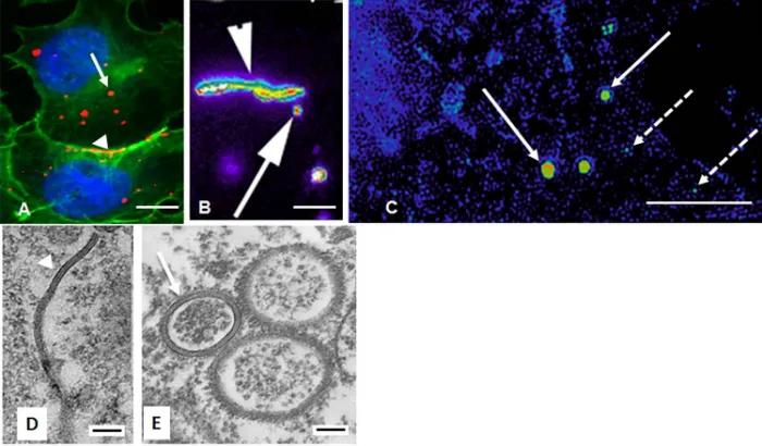
The central dogma has been that the gap junction proteins (connexins) in these annular gap
50
junctions are degraded, and thus the internalization process is only a method of eliminating old
51
connexins[7,14,24]. However, data from our laboratory and that of others has demonstrated that
52
connexins, specifically annular gap junction vesicle connexin 43 (Cx43), recycle back to the cell surface to
53
form functional gap junctions[25,26]. While the aggregation of gap junction channels into plaques and
54
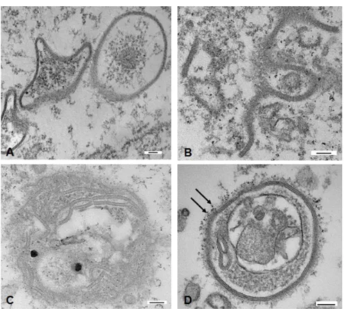
channel gating have been extensively studied[19,27,28], mechanisms involved in the processing of annular
55
gap junction vesicles have only recently gained attention. However, it is clear that, the rates of gap junction
56
plaque disassembly as well as assembly are critical to gap junction function in cell-cell communication and
57
cell-cell adhesion[29,30]. Changes in gap junction location and connexin trafficking and internalization is
58
thought to play several pivotal physiological roles during embryonic development [31,32], mitosis[26],
59
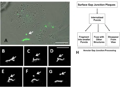
wound healing[33], and cell migration and these changes are thought to facilitate tumor growth and
60
metastasis as well as cardiac muscle changes during ischemia[34-36]. Other than considering secretory
61
vesicles as the mechanism of Cx43 delivery, alternative Cx43 cytoplasmic delivery sources have received
62
little attention. It has been demonstrated however that there is Cx43 on mitochondrial membranes, and this
63
mitochondrial Cx43 was shown to be essential for ischemic preconditioning[34]. Further, N-terminal
64
truncated isoforms of Cx43 have been demonstrated to play a role in mitochondrial movement to the cell
65
periphery and in maintaining the mitochondrial network integrity during oxidative stress in
66
cardiomyocytes and cold stress in adipocytes [37-39]. The method of Cx43 delivery to the mitochondria is
67
not understood. Furthermore, the possible pathophysiological role of Cx43 within cytoplasmic
68
compartments or associations between these compartments is not known. Understanding Cx43 trafficking,
69
fate and the translocation to Cx43-containing organelles is essential to an understanding of a host of normal
70
and pathological events.
71
In this study, we demonstrated cytoplasmic Cx43-containing structures and evaluated their
72
organelle interactions. We found that the most abundant Cx43-containing structures, annular gap junction
73
vesicles, associated more frequently with mitochondria than with lysosomes. These findings are consistent
74
with the possibility that some of the Cx43 present in annular gap junctions may be utilized by mitochondria
75
rather than being only degraded.
76
2. Results
77
2.1. Characterization of Cx43 Containing Gap Junctions Structure Behavior
78
A human adrenal tumor cell line (SW-13) that expresses Cx43 gap junction protein was used to
79
analyze cytoplasmic Cx43-containing structures, particularly that of annular gap junction vesicles, which
80
are the most prominent Cx43-containing organelle in the cytoplasm (Figure 1A). This cell line forms
81
relatively large gap junctions which are spontaneously and frequently internalized to form annular gap
82
junction vesicles, which is particularly advantageous for the study aspects of Cx43 trafficking. With
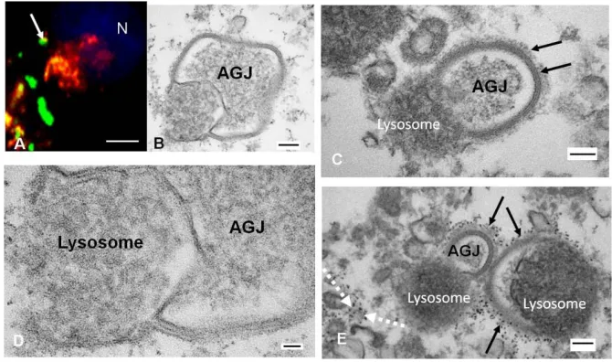
83
surface between contacting cells (Figures 1A-B, and 2). Puncta indicative of annular gap junction vesicles
85
could be easily seen within the cytoplasm (Figures 1 and 2). In addition, secretory vesicles that deliver new
86
Cx43 to the plasma membrane could be detected at the light level of resolution, but they were more easily
87
discerned with super-resolution microscopy (Figure 1C). Secretory and annular gap junction vesicles both
88
appeared as puncta at the light microscopic level of resolution and are distinguished mainly by the
89
differences in their sizes. Annular gap junction vesicles (≥0.5µm) have been demonstrated to be larger
90
than the secretory vesicles (≤150nm)[19,40]. With transmission electron microscopy the pentalaminar
91
membrane of annular gap junction vesicles, which is a typical characteristic of the gap junction plaque
92
membrane, can be used to distinguish this vesicle from other cytoplasmic organelles. (Figure 1D-E).
93
94
95
Figure 1. Localization of gap junction plaques and annular gap junction vesicles. A) Immunocytochemistry
96
of Cx43 (red) and cortical actin (green). Cortical actin (green) staining was used to define the boundaries of
97
the cell and to aid in distinguishing intracellular annular gap junction puncta (red, arrow) from surface gap
98
junction plaques (red, arrowhead) . (B) Pseudo-colored image of Cx43-GFP from a time lapse frame
99
demonstrating gap junction plaques (arrowhead) and annular gap junction vesicles (arrow). (C)
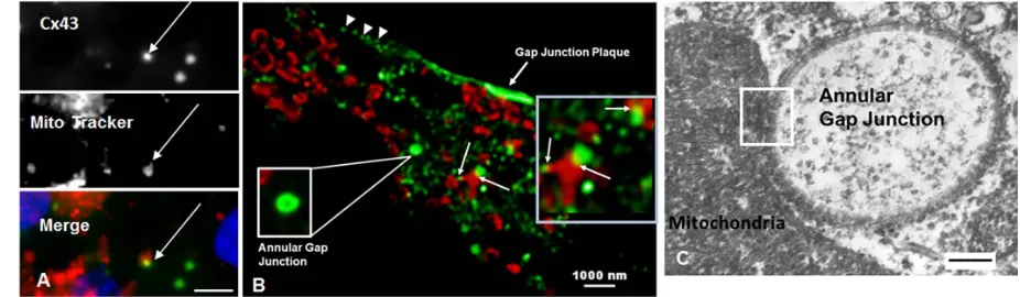
100
Super-resolution microscopy demonstrating annular gap junction (arrow) and secretory vesicles (broken
101
arrow). (D, E) TEM demonstrating the typical pentalaminar membrane of the gap junction plaque
102
(arrowhead in E) and annular gap junction vesicle (arrow in E).. Scale Bars= (A-B):10 um, (C): 5 um, (D-E):
103
In addition to annular gap junctions and secretory vesicles, ultrastructural analysis revealed
105
occasional membrane fragments of various sizes, atypical aggregates of gap junction membrane as well as
106
bizarre looking structures in the cytoplasm which were composed of gap junction membranes (Figure 3).
107
The fragments were identified by the typical gap junction membrane while the more bizarre structures
108
were further confirmed as composed of Cx43 with immuno-electron microscopy (Figure 3D). Given the
109
recent suggestion that Cx43 in annular gap junctions could have fates other than only degradation, we did a
110
detailed analysis of annular gap junction processing and their associations with other organelles.
111
112
113
114
115
Figure 2. Time lapse imaging of cells expressing Cx43-GFP. (A) DIC image of cell population seen in the
116
time lapse images collected at 1 minute intervals in B-G. The gap junction plaque between two cells is
117
evident as well as the cytoplasmic annular gap junction (arrow in A). (B-G) Time lapse montage (1 minute
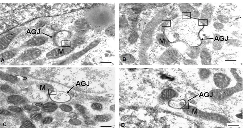
118
intervals) demonstrating the formation and release of the annular gap junction vesicle (arrows) into the
119
cytoplasm of one of the two contacting cells. (H) Flow chart summary of annular gap junction formation
120
122
Figure 3. TEM of cytoplasmic gap junction structures. (A) Two star shaped annular gap junctions seen next
123
to a typical annular gap junction vesicle. (B) Fragmented gap junction membrane. Note the pentalaminar
124
membrane. (C) Aggregates of gap junction membrane arranged in an annular shape. (D) Bizarre annular
125
gap junction with annular structure and other unknown material within the lumen. Immuno-electron
126
microscopic Cx-43 Q-dots (arrows) can be seen labeling the annular gap junction membrane. Scale Bars=
127
(A-D): 100 nm
128
129
130
2.1.1. Live Cell Imaging of Gap Junction Plaque Endoexocytosis
131
Since at the light microscopic level the distinguishing pentalaminar membrane was not visible,
132
positive identification of annular gap junctions in these live cell imaging studies was made by monitoring
133
them, following observations of their internalization from gap junction plaques. Annular gap junction
134
vesicles were observed to form both as a result of internalization of portions of gap junction plaques or
135
entire gap junction plaques. In both cases the annular gap junction vesicle was released into one of two
136
contacting cells (Figure 2). The internalization occurred rapidly if only a portion of the central area of the
137
gap junction plaque was internalized, while internalization of the entire gap junction was a much slower
138
process and resulted in larger annular gap junction vesicle being released into the cytoplasm. As
139
summarized in figure 2H, over the 26-hour monitoring period, 93 annular gap junctions from four different
140
movies were observed to either (a) remain relatively stationary within the cytoplasm, (b) fragment to form
141
smaller puncta, (c) join with cytoplasmic organelles, (d) disappear from view or (e) move toward and
142
appear to associate with the cell surface gap junction plaques.
143
Annular gap junctions that were observed to join with other cytoplasmic organelles, remained
144
with these organelles throughout the monitoring period. These results prompted us to determine the
145
identity of structures that associated with the annular gap junction vesicles.
146
147
Annular gap junctions were observed With immunocytochemical localization techniques, annular
149
gap junction vesicles were found to associate with two different organelles, lysosomes (Figure 4) as
150
expected, and mitochondria (Figures 5,6). The details of these interactions were analyzed.
151
152
2.2.1. Analysis of Lysosomes/ Annular Gap Junction Associations
153
Lysosomes, detected with a lysosomal marker, Lamp 1, were shown to colocalize with annular gap
154
junctions in cells expressing endogenous Cx43 (Figure 4A) as well as transfected to express Cx43-GFP (data
155
not shown). An analysis of cytoplasmic puncta indicated that 8.3 ± 3.6 % of the endogenous Cx43 was
156
colocalized with lysosomes. A higher percentage of the Cx43-GFP was found colocalized with the lysosome
157
marker. We believe the higher localization with Cx43-GFP was due to increased aggregation of
158
non-specific staining within the cytoplasm of transfected cells.
159
160
161
162
Figure 4. Localization of annular gap junction vesicles and Lysosomes (A) Immunocytochemical
163
localization of Cx43 (green) and lysosomes detected with LAMP1 (red). The colocalization (yellow) of an
164
annular gap junction vesicle and lysosome can be seen (arrow). (B-E) The intimate contact between annular
165
gap junction (AGJ) and lysosomes is shown. In B and D the outer membrane of the annular gap junction can
166
be seen to be continuous with that of the lysosomal membrane. Clathrin, identified by bristle coat (arrow,
167
C) or quantum dot label (black arrows, E) can be seen associated with annular gap junction vesicles which
168
are fused with lysosomes. Typical clathrin coated vesicles also can be seen decorated with clathrin Q dots
169
(dashed white arrows). N= Nucleus; Scale Bars= (A): 10 um, (B, C, E): 100 nm, (D): 20 nm
170
A pitfall of interpreting colocalization data is that at the light microscopic level structures that are
171
separated by as much as 200nm can still appear to be colocalized. Further, the capacity to distinguish and
172
analyze the morphology of the organelles is limited by the resolution of images collected with light
173
microscopy. To better resolve the morphology and to positively identify structures as well as demonstrate
174
associations, transmission electron microscopic techniques were used.
175
The annular gap junction vesicle’s pentalaminar membrane is highly distinctive when imaged by
176
transmission electron microscopy [TEM] (Figures 1D&E, 4-6). Regardless of its shape or location in the cell
177
junction membrane and of other cytoplasmic organelles seen with TEM, we analyzed the association of
179
organelles with gap junction structures.
180
Annular gap junctional vesicles were observed to interact with lysosomes and appeared to be in
181
different stages of degradation (Figures 4B-E). In annular gap junctional profiles that appeared to be
182
undergoing degradation the inner and outer annular gap junction membranes were separated from one
183
another and the outer membrane was continuous with the lysosomal membrane (Figure 4B&C). Clathrin
184
was found associated with the annular gap junction, even at the point that they were fused with lysosomes
185
(Figure 4C&E). With ultrastructural analysis the number of lysosomal/annular vesicle associations could
186
be quantitated in detail. Based on the analysis of 549 annular gap junction vesicles, we demonstrated that
187
2.6 % ± 0.6 were fused with lysosomes.
188
189
190
2.2.2. Mitochondria
191
In addition to colocalization with lysosomes, annular gap junctions were revealed with
192
immunocytochemistry and super-resolution microscopy to colocalize with mitochondria (Figure 5). While
193
with immunocytochemistry the size of the puncta was used to identify a structure as an annular gap
194
junction, with super-resolution microscopy the annular gap junction lumen is evident (Figure 5B). With
195
both techniques annular gap junctions were demonstrated to colocalize with mitochondria. In figure 5B,
196
colocalization of mitochondria detected with antibody to TOM (red) could be seen with more than one
197
annular gap junction vesicle detected with Cx43 gap junction antigen (green). Images were captured with
198
stimulated emission depletion (STED) super-resolution microscopy.
199
200
201
202
Figure 5. Mitochondria associate with annular gap junctions. (A, B) Immunocytochemical analysis of
203
mitochondria (red) and Cx43 gap junction antigen (green). The mitochondria were detected with
204
Mitotracker (red) in A and with antibody to TOM (red) in b. (C) The intimate association between the
205
mitochondria and annular gap junction (white box) seen with TEM. Scale bars= (A): 10 um, (B): 1000 nm,
206
(C) 100 nm
207
To visualize the fine structure morphology at the sites of physical association between the
208
mitochondria and annular gap junctions, transmission electron microscopy (TEM) imaging was used
209
(Figures 5, 6). Mitochondria were observed to associate and to follow the contour of annular gap junction
210
membranes (Figures 5, 6). In some cases, actual contact sites between the outer membrane of the annular
211
annular gap junctions revealed that (5.2 % ± 1.1) associated with mitochondria. This percentage exceeded
213
the percentage of annular gap junction vesicles that were found to associate with lysosomes (2.6 % ± 0.6).
214
215
216
217
218
219
220
221
222
Figure 6. Transmission electron microscopy of mitochondria closely associated with annular gap junctions.
223
A-B) Areas of contact between annular gap junctions (AGJ) and mitochondria (M) are indicated within the
224
black boxes. Scale Bars: 500 nm
225
3. Discussion
226
In this study, we documented the presence of cytoplasmic Cx43-containing structures and we
227
analyzed the details and frequency of interactions of mitochondria and lysosomes with annular gap
228
junction vesicles. We found that the interactions between annular gap junctions and mitochondria were
229
seen more frequently than between annular gap junctions and lysosomes. The behavior of the puncta
230
believed to be annular gap junctions were analyzed with live cell imaging and it was determined that these
231
structures could either: 1) undergo fission, 2) fuse with other organelles, 3) disappear from view, or
232
appear to recycle back to the surface. In this study, to address the concern that in our live cell imaging
233
studies GFP tag may either interfere with or alter organelle interactions with Cx43-containing structure, or
234
that the Cx43 puncta were not positively identified as annular gap junction vesicles, we took advantage of
235
TEM techniques to positively identify and analyze organelle association in cells expressing endogenous
236
It was determined, based on ultrastructural analysis of the structures in the cytoplasm that some
238
of the puncta identified as annular gap junction at the light microscopic level of resolution could be
239
cytoplasmic aggregates or fragments. It should be noted however that such structures were relatively rare,
240
and thus our findings would be consistent with most of the puncta seen with live cell imaging being
241
annular gap junctions. The average puncta size measured from live cell imaging of Cx43-GFP expressing
242
cells was 1.6 ± 0.3 µm2, a size previously shown to be consistent with annular gap junctions and not
243
secretary vesicles[19,40]. We did not see interactions of cytoplasmic membrane aggregates and fragments
244
with other mitochondria or lysosomes, however this is not to say that it did not occur. We may have missed
245
such interactions due to the limits of seeing three-dimensional occurrences with TEM techniques, especially
246
if the occurrence was rare.
247
It is well accepted that once internalized, the connexin in annular gap junction vesicles are
248
degraded by lysosomes[7,13,14,24,42-45]. This is based on TEM observations of lysosomes and annular gap
249
junction vesicles fusion[7,14], the increase in annular gap junction vesicles when lysosomal activity is
250
inhibited[44], and the demonstration of acid phosphatase activity in these annular gap junctions[7,13,14].
251
In this current study we have quantitated annular/lysosome associations, and contrasted this with
252
interactions of annular gap junction vesicles with mitochondria. We found with TEM, that only 2.6 % of the
253
annular gap junction vesicles analyzed were associated with lysosomes at a given point in time. Although
254
this figure may suggest a limited contribution to the process, any alteration in the rate of Cx43 degradation
255
might serve as a post-translational means of altering intercellular communication in some cell types.
256
The association between mitochondria and annular gap junction vesicles was not expected and it has
257
not been, to our knowledge, previously reported. The contact between mitochondria and annular gap
258
junctions was a distance as close as 10–30nm at the site of contact and in some cases the two membranes of
259
the two organelles were actually in physical contact with one another. This is comparable to the distance of
260
10–30nm between the mitochondria and ER at their points of contact. The existence of ER-mitochondria
261
contact sites has been well established by electron microscopy and time-lapse fluorescence microscopy and
262
it has been suggested that in Yeast, a network of contact sites serve to integrate the ER, vacuoles, and
263
mitochondria[48]. Here we suggest that in addition to the interaction between ER, and vacuoles that
264
mitochondria also interact with annular gap junction vesicles. The benefit of this interaction can only be
265
speculated at this point but it is tempting to suggest that Cx43 from annular gap junctions may be delivered
266
to mitochondria when the two organelles come into physical contact. It is possible that connexins are
267
delivered to the mitochondria in secretory vesicles, and we have not ruled out that possibility. However,
268
the intimate physical association of the membranes of the mitochondria and annular gap junction would
269
suggest the possibility of Cx43 delivery to the mitochondrial membrane.
270
Proof of mitochondrial Cx43 have been provided from investigators who used a wide variety of
271
different techniques, including immunocytochemical colocalization of Cx43 with mitochondrial proteins at
272
the light microscopic and immunogold electron microscopic levels of resolution [49], flow cytometry
273
studies, and quantitative Western blot analysis [49,52-54]. Mitochondrial Cx43 has been demonstrated to
274
in protecting the heart from ischemic injury, facilitating mitochondrial movement to the cell periphery as
275
well as maintaining mitochondrial network integrity [37-39, 49-51 54-57]. Further, mitochondrial metabolic
276
activity and morphology alterations have been demonstrated in adipocytes from Cx43 knockout mice and
277
knocked down cells in culture[38], consistent with there being a Cx43 mitochondrial functional
278
dependency[49-51].
279
The questions of how connexin reaches and incorporates into mitochondria membranes and
280
further its orientation once there (Cx43 C-terminal tail toward the cytoplasm or the inner mitochondrial
281
matrix), and its physical arrangement (single Cx43 molecules or hemichannels) have not been answered. It
282
is known that mitochondrial Cx43 is phosphorylated [56] however it is not known which Cx43 amino acid
283
residuals are phosphorylated. The mechanism that regulates trafficking of Cx43 to the mitochondria and its
284
Is the annular gap junction vesicle possible a cytoplasmic source of phosphorylated Cx43 that
286
could potentially serve as a source for distribution Cx43 to other compartments? Although it well accepted
287
that connexins in annular gap junctions are degraded, other possible fates have received little attention
288
despite the reports of Cx43 recycling from annular gap junctions to participate in rapid gap junction plaque
289
formation at the end of the mitotic process [25,26].
290
The ultrastructural finding that some annular gap junctions, coated with clathrin or with bud-like
291
projections, were found fused to lysosomes was surprising. Normally the clathrin coat disassembles from
292
vesicles after 60-100 seconds and is lost once these vesicles associate with other organelles[46,47]. That the
293
clathrin coat remains with the annular gap junction vesicle, even when that structure is fused with
294
lysosomes, is suggestive of a role of clathrin in annular gap junction processing, including the possible
295
release of buds from the annular gap junction. The release of small buds from annular gap junctions has
296
been demonstrated[41]. It is possible that Cx43 in these “annular gap junction buds” may be degraded but
297
in addition this Cx43 may be delivered in these buds to other organelles, including mitochondria.
298
The method by which Cx43 traffics to the mitochondria has not been elucidated here. However,
299
the findings of targeting and functional changes in mitochondrial function and movement points to the
300
possibility that in addition to traditional Cx43 trafficking and function at the cell surface that Cx43 many
301
traffic and function in non-canonical manners. Here we demonstrate the association of annular gap
302
junctions with mitochondria and suggest that Cx43 in annular gap junctions may have a fate that is
303
unrelated to degradation. Future studies are needed to elucidate the ‘tethering’’ molecules that hold
304
annular gap junctions to mitochondria and to clarify the need for the interaction annular gap junctions with
305
mitochondria.
306
4. Materials and Methods
307
4.1 Cell Culture
308
SW-13 human adrenocortical tumor cells (American Type Culture Collection, Rockville, MD) were
309
cultured in L-15 medium which contained fetal calf serum (10%), penicillin (0.06 mg/ml), streptomycin (0.1
310
mg/ml), and Fungizone (0.01 mg/ml), buffered with L-arginine at pH 7.4 (reagents and medium from
311
Invitrogen, Carlsbad, CA). Cells were grown at 37oC in a 5% CO2 atmosphere.
312
313
4.2 Gap Junction Antibodies and Probes
314
Affinity purified polyclonal rabbit antibodies (IgG), were prepared against synthetic peptides
315
corresponding to the carboxyl terminus of the Cx43 molecule (residues 370 to 381) [57] (Zymed Laboratory,
316
San Francisco, CA). Preparation and characterization of these antibodies have been previously described
317
[2].
318
319
4.3 Immunocytochemistry
320
Cells were prepared and stained with immunocytochemical techniques, as previously described
321
[58,59]for connexin 43 (1:100; Proteintech, Rosemont, IL), clathrin (Abcam, Cambridge, MA), lysosomes
322
with Lamp1 staining (Abcam, Cambridge, MA), and mitochondrial proteins either with (mouse
323
monoclonal antibody to prohibitin (1:100 dilution) (NeoMarkers, Fremont, CA), Mitotracker, or Tom20
324
(Santa Cruz, Dallas, TX). Prior to immunocytochemical procedures, the cells were grown on coverslips,
325
rinsed with phosphate buffered saline solution (PBS), fixed at room temperature, in 3% formaldehyde for
326
20 minutes, permeabilized in cold acetone for 7 minutes, and then incubated at 370C for one hour or at 40C
327
overnight in the primary antibody. In some experiments colocalization studies where performed. In these
328
procedures, labeling of connexin was followed by a 40C overnight incubation in clathrin antibody. In
329
experiments to colocalize mitochondria, the cellular localization of prohibitin, TOMM-20, or mitotracker
330
Pierce, Rockford, IL). The coverslips were stained with Hoescht, washed in PBS, and placed onto glass
332
slides with a drop of Fluoromount-G anti-quench reagent (Southern Biotechnical Lab, Birmingham, AL).
333
Immunolabeled cells were imaged at 63X with an Olympus Provis microscope (Olympus, Center Valley,
334
PA).
335
For immunocytochemical analysis, the number of gap junction plaques and annular vesicles per
336
cell nuclei were calculated. Fluorescent spherical puncta that were greater-or-equal to 0.5 µm in diameter
337
were classified as annular gap junction vesicles while plaques were identified by their typical elongated
338
profile or by the presence of aggregated puncta (smaller plaques), that were obviously at the cell surface
339
between two cell pairs. The data was expressed as the average number of annular gap junction vesicles per
340
cell.
341
342
4.5 Percent Colocalization
343
The percent colocalization was calculated from 50 cells (10 cells were selected from each of five
344
files) in images collected by confocal step through selecting elements at the mid-plane of the Z stack. All
345
images were saved as tiffs prior to analysis. The MetaMorph Software Program was used to view and trace
346
the cell borders and to determine the percent calculation of cells positive for both signals. Specifically the
347
merged image was separated into red and green channels and the trace areas were then transferred to the
348
separated images. After thresholding the images the number of Cx43 containing objects was determined
349
with the automatic counting tool in MetaMorph. The counts were displayed on the “record count screen”,
350
which was then analyzed and the number of surface gap junctions (as well as the number of clusters or
351
aggregates or any object that did not look like an annular gap junction) were counted manually and
352
subtracted from the total number of objects. The number of overlapping areas (yellow objects) were then
353
counted manually by analyzing the merged image. The number of annular gap junctions that colocalized
354
with lysosomes or mitochondria were determined by counting the number of annular gap junctions that had
355
any yellow associated with them. The data was expressed as the average percent colocalization of annular
356
gap junction vesicles with lysosomes or mitochondria.
357
358
4.6 Transfection with cDNA
359
To visualize gap junction trafficking in living cells, adrenal cells were transfected with cDNAs
360
encoding the fluorescent Cx43-GFP (provided by Dr. M. Falk, Lehigh University). The Cx43-GFP vector was
361
constructed by linking the GFP fluorescent reporter protein to the C-terminus of the rat Cx43 cDNA, and it
362
has been demonstrated to assemble into gap junction plaques similar to wild type Cx43 [60].
363
Lipofectamine2000 Transfection Reagent (Life Technologies, Grand Island, NY) was used to establish cell
364
(MatTek Corporation, Ashland, MA). Cell populations, at 70-80% confluence, were transfected in
367
Opti-mem medium which contained Lipofectamine2000 Transfection Reagent (Invitrogen, Carlsbad,
368
CA) and 4µg of plasmid DNA (Cx43-GFP, or empty vector) for 24 hours at 37oC in an atmosphere
369
of 5% CO2. The resulting complexes were removed by gentle aspiration and washed with PBS.
370
Fresh L-15 complete cell growth medium was added to the dishes, and the cells were incubated at
371
37oC in 5% CO2 for 24-48 hours before imaging.
372
373
4.7 Imaging of Cx43-GFP in Living Cells
374
MatTek glass bottom dishes with cells expressing Cx43-GFP or empty vector were placed into
375
a temperature controlled chamber and maintained at 37oC in 5% CO2 on a live cell Nikon A1 series
376
automatic confocal laser point scanning system (Nikon Instruments Inc., Melville, NY). Image
377
acquisition on the Olympus was performed with a MetaMorph Imaging System (Molecular Devices,
378
Downington, PA) and Nikon NIS-Elements on the A1 Nikon microscope.
379
Images were obtained with a 63x oil objective with a numerical aperture of 1.4. Images were
380
collected at 1-5 minute intervals. DIC and fluorescent images were obtained using the standard
381
fluorescence detector at wavelengths 482 and 595 for all experiments. Focus was maintained with
382
the Perfect Focus System (PFS) function and images were acquired with Nikon’s NIS-Elements
383
software. Qualitative and quantitative analyses were performed on the data sets with the rendering
384
tool in NIS-Elements. Time lapse images were used to analyze gap junction plaque internalization
385
in and for analysis, time-lapse movies were converted to movies and evaluated with the MetaMorph
386
program (Molecular Devices, Downington, PA).
387
Annular gap junction vesicle pattern of displacement within the cell and corresponding
388
changes in size (area expressed as µm2) were quantitated with the tracking function in the Imaris
389
analysis software (Bitplane Scientific, South Windsor, CT). Selected annular vesicles were segmented
390
based upon labeling intensity and then followed over time. Statistical significance of differences was
391
determined with the Student’s t-Test.
392
393
4.8 Super-resolution-Live Cell Microscopy
394
Super-resolution microscopy was used to analyze annular gap junction and mitochondrial
395
interaction with the stimulated emission depletion (STED) microscopy technique. STED allows for
396
enhanced spatial resolution of the mitochondria which improves our accuracy when analyzing
397
annular gap junction associations with mitochondria across the x, y, and z planes. Gated STED
398
images were obtained using a commercial Leica SP8 STED 3X system (Leica Microsystems,
399
Mannheim, Germany) housing a 100x oil objective with a numerical aperture of 1.4. Connexin 43 and
400
Tomm-20 antibodies were used to label annular gap junctions and mitochondria, respectively, for
401
STED imaging.
402
403
4.9 Transmission Electron Microscopy
404
Cell monolayers were briefly rinsed in PBS then fixed with 2.5% glutaraldehyde in PBS, pH 7.4,
405
for 1 hour at room temperature. All samples were then w3ashed 3 times in PBS buffer and
406
post-fixed for 1 hour at 4̊C in 1% osmium tetroxide with 1% potassium ferricyanide. The samples
407
were washed again and the cells were serially dehydrated in an ethanol (30%, 50%, 70%, and 90%)
408
for 10 minutes and followed by 15 minutes in 100% ethanol three times. The cells were then
409
incubated in Epon 3 times for 1 hour and finally embedded in resin to be sectioned. Ultra-thin
410
sections were cut, mounted on grids, and imaged on a JEOL 1011CX electron microscope (JEOL,
411
Tokyo, Japan).
412
In Quantum dots protocols and in some protocols to view annular gap junction/organelle
413
contacts, the cells were fixed (2% paraformaldehyde and 0.1% glutaraldehyde in PBS). Quantum
414
dots (QDs) were used to specifically label connexin, and clathrin. Adrenal cells were incubated in
415
Cx43 or clathrin primary antibodies, followed by incubation in biotin IgG conjugated antibody, and
416
First, the cells were incubated in the quantum dot-linked anti-rabbit immunoglobin without a
419
pre-incubation of the thin sections in the first antibody. Second, the binding of quantum dot
420
complexes to embedding resin was evaluated in areas free of cells.
421
Author Contributions: CLB and SAM conceived, designed and performed the experiments; CLB, TIS, and SAM
422
analyzed the data; CLB, TIS, and SAM wrote the paper.
423
Funding: This research was funded by National Science Foundation grant #MCB-1408986.
424
Acknowledgments: We would like to thank the following people at the University of Pittsburgh: Dr. Beth
425
Nickel, Margaret Bisher, Deborah O. Osakue, Dr. Simon Watkins, Dr. Claudette St. Croix, and Michael Caldron.
426
We would like to acknowledge the NIH supported microscopy resources in the University of Pittsburgh Center
427
for Biologic Imaging, specifically, the confocal microscope supported by grant number 1S10OD019973-01. We
428
would also like to thank Dr. Winston Thompson of Morehouse School of Medicine for providing us with
429
prohibitin.
430
Conflicts of Interest: The authors declare no conflict of interest
431
Abbreviations
432
TEM Transmission Electron Microscopy QDs Quantum Dots
AGJ Annular Gap Junction Vesicle
References
433
1. Goodenough, D.A.; Goliger, J.A.; Paul, D.L. Connexins, Connexons, and intercellular communication
434
(Review). Ann Rev Biochem 1996 65, 475-502. https://doi.org/10.1146/annurev.bi.65.070196.002355.
435
2. Kumar, N.M.; Gilula, N.B. Molecular biology and genetics of gap junction channels. Seminars in Cell Biology
436
1992 3, 3-16. https://doi.org/10.1016/S1043-4682(10)80003-0.
437
3. Kumar, N.M.; Gilula, N.B. Cloning and characterization of human and rat liver cDNAs coding for a gap
438
junction protein. J Cell Bio 1986 103, 767-76. https://doi.org/10.1083/jcb.103.3.767.
439
4. Gilula, N.B. Topology of gap junction protein and channel function. Ciba Foundation Symposium 1987 125,
440
128-39. https://doi.org/10.1016/0022-2836(92)90253-G.
441
5. Ahmad, S.; Diez, J.A.; George, C.H.; Evans, W.H. Synthesis and assembly of connexins in vitro into
442
homomeric and heteromeric functional gap junction hemichannels. Biochemical Journal 1999 339, 247-53.
443
https://doi.org/10.1042/0264-6021:3390247.
444
6. Perkins, G.A.; Goodenough, D.A.; Sosinsky, G.E. Formation of the gap junction intercellular channel
445
requires a 30 degree rotation for interdigitating two apposing connexons. Journal of Molecular Biology 1998
446
277, 171-7. https://doi.org/10.1006/jmbi.1997.1580.
447
7. Murray, S.A.; Larsen, W.J.; Trout, J.; Donta, S.T. Gap junction assembly and endocytosis correlated with
448
patterns of growth in a cultured adrenocortical tumor cell (SW-13). Cancer Res 1981 41, 4063-74.
449
8. Falk, M.M.; Bell, C.L.; Kells Andrews, R.M.; Murray, S.A. Molecular mechanisms regulating formation,
450
trafficking and processing of annular gap junctions. BMC Cell Biology Journal, Section: Cell-Cell Contacts
451
2016 17, 5-23. https://doi.org/10.1186/s12860-016-0087-7.
452
9. Nickel, B.M.; DeFranco, B.H.; Gay, V.L.; Murray, S.A. Clathrin and Cx43 gap junction plaque
453
endoexocytosis. Biochem Biophys Res Commun 2008 374, 679-82. https://doi.org/ 10.1016/j.bbrc.2008.07.108.
454
10. Huang, X.D.; Horackova, M.; Pressler, M.L. Changes in the expression and distribution of connexin 43 in
455
isolated cultured adult guinea pig cardiomyocytes. Experimental Cell Research 1996 228, 254-61.
456
https://doi.org/ 10.1006/excr.1996.0324.
457
11. Gumpert, A.M.; Varco, J.S.; Baker, S.M.; Piehl, M.; Falk, M.M. Double-membrane gap junction
458
internalization requires the clathrin-mediated endocytic machinery. FEBS Lett 2008 582, 2887-92.
459
https://doi.org/10.1016/j.febslet.2008.07.024.
460
12. Piehl, M.; Lehmann, C.; Gumpert, A.; Denizot, J.P.; Segretain, D.; Falk, M.M. Internalization of Large
461
Double-Membrane Intercellular Vesicles by a Clathrin-dependent Endocytic Process. Mol Biol Cell 2007 18,
462
337-47. https://doi.org/ 10.1091/mbc.E06-06-0487.
microfilaments and bristle coats in the internalization of gap junction membrane. J Cell Biol 1979 83, 576-87.
465
https://doi.org/10.1083/jcb.83.3.576.
466
14. Larsen, W.J.; Hai, N. Origin and fate of cytoplasmic gap junctional vesicles in rabbit granulosa cells. Tissue
467
& Cell 1978 10, 585-98. https://doi.org/10.1016/S0040-8166(16)30351-2.
468
15. Jordan, K.; Chodock, R.; Hand, A.R.; Laird, D.W. The origin of annular junctions: a mechanism of gap
469
junction internalization. J Cell Sci 2001 114, 763-73.
470
16. 16. Naus, C.C.; Hearn, S.; Zhu, D.; Nicholson, B.J.; Shivers, R.R. Ultrastructural analysis of gap junctions
471
in C6 glioma cells transfected with connexin43 cDNA. Experimental Cell Research 1993 206, 72-84.
472
https://doi.org/10.1006/excr.1993.1122.
473
17. 17. Dermietzel, R.; Hertberg, E.L.; Kessler, J.A.; Spray, D.C. Gap junctions between cultured astrocytes:
474
immunocytochemical, molecular, and electrophysiological analysis. Journal of Neuroscience 1991 11,
475
1421-32. https://doi.org/10.1523/JNEUROSCI.11-05-01421.1991.
476
18. Risley, M.S.; Tan, I.P.; Roy, C.; Saez, J.C. Cell-, age- and stage-dependent distribution of connexin43 gap
477
junctions in testes. Journal of Cell Science 1992 103, 81-96.
478
19. Jordan, K.; Solan, J.L.; Dominguez, M.; Sia, M.; Hand, A.; Lampe, P.D.; Laird, D.W. Trafficking, assembly,
479
and function of a connexin43-green fluorescent protein chimera in live mammalian cells. Mol Biol Cell 1999
480
10, 2033-50.
481
20. Segretain, D.; Falk, M.M. Regulation of connexin biosynthesis, assembly, gap junction formation, and
482
removal. Biochim Biophys Acta 2004 1662, 3-21. https://doi.org/10.1016/j.bbamem.2004.01.007.
483
21. Lauf, U.; Giepmans, B.N.; Lopez, P.; Braconnot, S.; Chen, S.C.; Falk, M.M. Dynamic trafficking and
484
delivery of connexons to the plasma membrane and accretion to gap junctions in living cells. Proc Natl
485
Acad Sci U S A 2002 99, 10446-51. https://doi.org/10.1073/pnas.162055899.
486
22. Sosinsky, G.E.; Gaietta, G.M.; Hand, G.; Deerinck, T.J.; Han, A.; Mackey, M.; Adams, S.R.; Bouwer, J.;
487
Tsien, R.Y.; Ellisman, M.H. Tetracysteine genetic tags complexed with biarsenical ligands as a tool for
488
investigating gap junction structure and dynamics. Cell Commun Adhes 2003 10, 181-6.
489
https://doi.org/10.1080/cac.10.4-6.181.186.
490
23. Nickel, B.; Boller, M.; Schneider, K.; Shakespeare, T.; Gay, V.; Murray, S.A. Visualizing the effect of
491
dynamin inhibition on annular gap vesicle formation and fission. J Cell Sci 2013 126 Pt 12, 2607-16.
492
https://doi.org/10.1242/jcs.116269.
493
24. Qin, H.; Shao, Q.; Igdoura, S.A.; Alaoui-Jamali, M.A.; Laird, D.W. Lysosomal and proteasomal
494
degradation play distinct roles in the life cycle of Cx43 in gap junctional intercellular
495
communication-deficient and -competent breast tumor cells. J Biol Chem 2003 278, 30005-14.
496
https://doi.org/10.1101/cshperspect.a002576.
497
25. Boassa, D.; Solan, J.L.; Papas, A.; Thornton, P.; Lampe, P.D.; Sosinsky, G.E. Trafficking and recycling of the
498
connexin43 gap junction protein during mitosis. Traffic 2010 11, 1471-86.
499
https://doi.org/10.1111/j.1600-0854.2010.01109.x.
500
26. Vanderpuye, O.A.; Bell, C.L.; Murray, S.A. Redistribution of connexin 43 during cell division. Cell Biology
501
International 2016 40(4), 387-96. https://doi.org/10.1002/cbin.10576.
502
27. Solan, J.L.; Lampe, P.D. Kinase programs spatiotemporally regulate gap junction assembly and
503
disassembly: Effects on wound repair. Semin Cell Dev Biol 2016 50, 40-8.
504
https://doi.org/10.1016/j.semcdb.2015.12.010.
505
28. Lampe, P.D.; Kistler, J.; Hefti, A.; Bond, J.; Muller, S.; Johnson, R.G.; Engel, A. In vitro assembly of gap
506
junctions. Journal of Structural Biology 1991 107, 281-90. https://doi.org/10.1016/1047-8477(91)90053-Y.
507
29. Musil, L.S.; Le, A.C.; VanSlyke, J.K.; Roberts, L.M. Regulation of connexin degradation as a mechanism to
508
increase gap junction assembly and function. Journal of Biological Chemistry 2000 275, 25207-15.
509
https://doi.org/10.1074/jbc.275.33.25207.
510
30. Lampe, P.D.; Lau, A.F. Regulation of gap junctions by phosphorylation of connexins. Arch Biochem Biophys
511
2000 384, 205-15. https://doi.org/10.1006/abbi.2000.2131.
512
31. Lo, C.W.; Wessels, A. Cx43 gap junctions in cardiac development. Trends Cardiovasc Med 1998 8, 264-9.
513
https://doi.org/10.1016/S1050-1738(98)00018-8.
514
32. Loewenstein, W.R.; Rose, B. The cell-cell channel in the control of growth. Seminars in Cell Biology 1992 3,
515
59-79. https://doi.org/10.1016/S1043-4682(10)80008-X.
S.A. Migrating cells retain gap junction plaque structure and function. Cell Commun Adhes 2008 15, 273-88.
518
https://doi.org/10.1080/15419060802198298.
519
34. Schulz, R.; Gres, P.; Skyschally, A.; Duschin, A.; Belosjorow, S.; Konietzka, I.; Heusch, G. Ischemic
520
preconditioning preserves connexin 43 phosphorylation during sustained ischemia in pig hearts in vivo.
521
Faseb J 2003 17, 1355-7. https://doi.org/10.1096/fj.02-0975fje.
522
35. Schulz, R.; Boengler, K.; Totzeck, A.; Luo, Y.; Garcia-Dorado, D.; Heusch, G. Connexin 43 in ischemic pre-
523
and postconditioning. Heart Fail Rev 2007 12, 261-6. https://doi.org/10.1007/s10741-007-9032-3.
524
36. Yao, J.A.; Hussain, W.; Patel, P.; Peters, N.S.; Boyden, P.A.; Wit, A.L. Remodeling of gap junctional channel
525
function in epicardial border zone of healing canine infarcts. Circ Res 2003 92, 437-43.
526
https://doi.org/10.1161/01.RES.0000059301.81035.06.
527
37. Fu, Y.; Zhang, S.S.; Xiao, S.; Basheer, W.A.; Baum, R.; Epifantseva, I.; Hong, T.; Shaw, R.M. Cx43 Isoform
528
GJA1-20k Promotes Microtubule Dependent Mitochondrial Transport. Front Physiol 2017 8, 905.
529
https://doi.org/10.1080/15419060701402320.
530
38. Kim, S.N.; Kwon, H.J.; Im, S.W.; Son, Y.H.; Akindehin, S.; Jung, Y.S.; Lee, S.J.; Rhyu, I.J.; Kim, I.Y.; Seong,
531
J.K.; Lee, J.; Yoo, H.C.; Granneman, J.G.; Lee, Y.H. Connexin 43 is required for the maintenance of
532
mitochondrial integrity in brown adipose tissue. Sci Rep 2017 7, 7159. https://doi.org/10.1002/bies.10159.
533
39. Boengler, K.M.; Ruiz-Meana, et al. Mitochondrial connexin 43 impacts on respiratory complex I activity
534
and mitochondrial oxygen consumption. J Cell Mol Med 2012 16: 1649-55. https://doi:
535
10.1111/j.1582-4934.2011.01516
536
40. Evans, W.H.; Martin, P.E. Lighting up gap junction channels in a flash. Bioessays 2002 24, 876-80.
537
https://doi.org/10.1159/000092562.
538
41. Gilleron, J.; Carette, D.; Fiorini, C.; Dompierre, J.; Macia, E.; Denizot, J.; Segretain, D.; Pointis, G. The large
539
GTPase dynamin2: a new player in connexin 43 gap junction endocytosis, recycling and degradation. Int J
540
Biochem Cell Biol. 2011 43(8):1208-17. https://doi.org/10.1093/carcin/17.9.1791.
541
42. Falk, M.M.; Fong, J.T.; Kells, R.M.; O'Laughlin, M.C.; Kowal, T.J.; Thevenin, A.F. Degradation of
542
endocytosed gap junctions by autophagosomal and endo-/lysosomal pathways: a perspective. J Membr Biol
543
2012 245, 465-76. https://doi.org/10.1007/s00232-012-9461-3.
544
43. Salameh, A. Life cycle of connexins: regulation of connexin synthesis and degradation. Adv Cardiol 2006 42,
545
57-70. https://doi.org/10.1038/nsmb.1985.
546
44. Guan, X.; Ruch, R.J. Gap junction endocytosis and lysosomal degradation of connexin43-P2 in WB-F344 rat
547
liver epithelial cells treated with DDT and lindane. Carcinogenesis 1996 17, 1791-8.
548
https://doi.org/10.1073/pnas.1018845108.
549
45. Su, V.; Cochrane, K.; Lau, A.F. Degradation of connexins through the proteasomal, endolysosomal and
550
phagolysosomal pathways. J Membr Biol 2012 245, 389-400. https://doi.org/10.1016/j.cardiores.2005.04.014.
551
46. Bocking, T.; Aguet, F.; Harrison, S.C.; Kirchhausen, T. Single-molecule analysis of a molecular
552
disassemblase reveals the mechanism of Hsc70-driven clathrin uncoating. Nat Struct Mol Biol 2011 18,
553
295-301. https://doi.org/10.1161/01.RES.0000233145.94073.b8.
554
47. Rothnie, A.; Clarke, A.R.; Kuzmic, P.; Cameron, A.; Smith, C.J. A sequential mechanism for clathrin cage
555
disassembly by 70-kDa heat-shock cognate protein (Hsc70) and auxilin. Proc Natl Acad Sci U S A 2011 108,
556
6927-32. https://doi.org/10.1016/j.bbrc.2006.10.177.
557
48. van der Bliek, A.M.; Shen, Q.; Kawajiri, S. Mechanisms of mitochondrial fission and fusion. Cold Spring
558
Harb Perspect Biol 2013 5. https://doi.org/10.1586/14789450.1.3.293.
559
49. Boengler, K.; Dodoni, G.; Rodriguez-Sinovas, A.; Cabestrero, A.; Ruiz-Meana, M.; Gres, P.; Konietzka, I.;
560
Lopez-Iglesias, C.; Garcia-Dorado, D.; Di Lisa, F.; Heusch, G.; Schulz, R. Connexin 43 in cardiomyocyte
561
mitochondria and its increase by ischemic preconditioning. Cardiovasc Res 2005 67, 234-44.
562
https://doi.org/10.1016/0022-2836(92)90253-G.
563
50. Halestrap, A.P. Mitochondria and preconditioning: a connexin connection? Circ Res 2006 99, 10-2.
564
https://doi.org/10.1161/01.RES.0000233145.94073.b8.
565
51. Goubaeva, F.; Mikami, M.; Giardina, S.; Ding, B.; Abe, J.; Yang, J. Cardiac mitochondrial connexin 43
566
regulates apoptosis. Biochem Biophys Res Commun 2007 352, 97-103.
567
https://doi.org/10.1016/j.bbrc.2006.10.177.
568
52. Rodriguez-Sinovas, A.; Boengler, K.; Cabestrero, A.; Gres, P.; Morente, M.; Ruiz-Meana, M.; Konietzka, I.;
569
Miro, E.; Totzeck, A.; Heusch, G.; Schulz, R.; Garcia-Dorado, D. Translocation of connexin 43 to the inner
and its importance for cardioprotection. Circ Res 2006 99, 93-101.
572
https://doi.org/10.1161/01.RES.0000230315.56904.de.
573
53. Miro-Casas, E.; Ruiz-Meana, M.; Agullo, E.; Stahlhofen, S.; Rodriguez-Sinovas, A.; Cabestrero, A.; Jorge, I.;
574
Torre, I.; Vazquez, J.; Boengler, K.; Schulz, R.; Heusch, G.; Garcia-Dorado, D. Connexin43 in
575
cardiomyocyte mitochondria contributes to mitochondrial potassium uptake. Cardiovasc Res 2009 83,
576
747-56. https://doi.org/10.1093/cvr/cvp157.
577
54. Boengler, K.; Konietzka, I.; Buechert, A.; Heinen, Y.; Garcia-Dorado, D.; Heusch, G.; Schulz, R. Loss of
578
ischemic preconditioning's cardioprotection in aged mouse hearts is associated with reduced gap
579
junctional and mitochondrial levels of connexin 43. Am J Physiol Heart Circ Physiol 2007 292, H1764-9.
580
https://doi.org/10.1152/ajpheart.01071.2006.
581
55. 55. Rodriguez-Sinovas, A.; Ruiz-Meana, M.; Denuc, A.; Garcia-Dorado, D. Mitochondrial Cx43, an
582
important component of cardiac preconditioning. Biochim Biophys Acta 2018 1860, 174-181.
583
https://doi.org/10.1016/j.bbamem.2017.06.011.
584
56. Ruiz-Meana, M.; Rodriguez-Sinovas, A.; Cabestrero, A.; Boengler, K.; Heusch, G.; Garcia-Dorado, D.
585
Mitochondrial connexin43 as a new player in the pathophysiology of myocardial ischaemia-reperfusion
586
injury. Cardiovasc Res 2008 77, 325-33. https://doi.org/10.1093/cvr/cvm062.
587
57. Yeager, M.; Gilula, N.B. Membrane topology and quaternary structure of cardiac gap junction ion
588
channels. Journal of Molecular Biology 1992 223, 929-48. https://doi.org/10.1016/0022-2836(92)90253-G.
589
58. Oyoyo, U.A.; Shah, U.S.; Murray, S.A. The role of alpha1 (connexin-43) gap junction expression in adrenal
590
cortical cell function. Endocrinology 1997 138, 5385-97. https://doi.org/10.1210/endo.138.12.5617.
591
59. Murray, S.A.; Shakespeare, T.I. (2016) Immunofluorescence: application for analysis of connexin
592
distribution and trafficking., pp. 1-19, CRC Press, Taylor & Francis Group, Boca Raton, FL.
593
60. Falk, M.M. Connexin-specific distribution within gap junctions revealed in living cells. J Cell Sci 2000 113,
594
4109-20.