The matrisome during aging and longevity: a systems-level approach towards 1
defining matreotypes promoting healthy aging 2
3
Collin Y. Ewald1*
4
5
1 ETH Zürich, Department of Health Sciences and Technology, Institute of
6
Translational Medicine, Schwerzenbach-Zürich, CH 8603, Switzerland.
7
*Corresponding author: C.Y.E. [email protected] 8
9
Keywords: matreotype, proteomics, transcriptomics, lifespan, extracellular matrix,
10
ECM, collagen, MMP, homeostasis
11
Abstract 13
Accumulation of damage is generally considered the cause of aging. Interventions that
14
delay aging mobilize mechanisms that protect and repair cellular components.
15
Consequently, research has been focused on studying the protective and homeostatic
16
mechanisms within cells. However, in humans and other multicellular organisms, cells
17
are surrounded by extracellular matrices (ECM), which are important for tissue
18
structure, function and intercellular communication. During aging, components of the
19
ECM become damaged through fragmentation, glycation, crosslinking, and
20
accumulation of protein aggregation, all of which contribute to age-related pathologies.
21
Interestingly, placing senescent cells into a young ECM rejuvenates them and we
22
found that many longevity-assurances pathways re-activate de-novo synthesis of
23
ECM proteins during aging. This raises the question of what constitutes a young ECM
24
to reverse aging or maintain health? In order to make inroads to answering this
25
question, I suggest a systems-level approach of quantifying the matrisome or ECM
26
compositions reflecting health, pathology, or phenotype and propose a novel term, the
27
“matreotype”, to describe this. The matreotype is defined as the composition and
28
modification of ECM or matrisome proteins associated with or caused by a phenotype,
29
such as longevity, or a distinct and acute physiological state, as observed during aging
30
or disease. Every cell type produces its unique ECM. Interestingly, cancer-cell types
31
can even be identified based on their unique ECM composition. Thus, the matreotype
32
reflects cellular identity and physiological status. Defined matreotypes could be used
33
as biomarkers or prognostic factors for disease or health status during aging with
34
potential relevance for personalized medicine. Treatment with biologics that alter
35
ECM-to-cell mechanotransduction might be a strategy to reverse age-associated
36
pathologies. An understanding of how to reverse from an old to a young matreotype
37
might point towards novel strategies to rejuvenate cells and help maintain tissue
38
homeostasis to promote health during aging.
39
Introduction 41
Cells and tissues are embedded within extracellular matrices (ECM), which are
42
important for tissue geometry, integrity, and function [1]. Besides providing structural
43
support, the ECM controls intercellular communication by either storing or transporting
44
signaling molecules, such as growth factors, hormones, and neuropeptides [1-3].
45
Receptors on the cell surface, such as integrins, directly link ECM to cell signaling to
46
regulate various biological functions (Figure 1). Cells synthesize and secret ECM
47
proteins, such as collagens, extracellular glycoproteins, and proteoglycans that are
48
integrated into matrices by extracellular enzymes [2]. Cells also continuously monitor
49
and integrate signals from the ECM via mechanotransduction and adjust their
50
surrounding environment by synthesizing and remodeling their ECM [2]. Thus, the
51
ECM at least during development and normal tissue homeostasis is a dynamic system.
52
During aging, the connective tissue or ECM integrity declines through
53
accumulation of damage from collagen fragmentation, oxidation, glycation,
54
crosslinking, and accumulation of protein aggregates (Figure 1), leading to a decline
55
in ECM dynamics and loss of organs support and function [3,4]. This decline in ECM
56
integrity has been implicated in many age-dependent diseases, such as
57
atherosclerosis, diabetes, and cancer. Accumulation of damage to the ECM drives
58
cellular aging and disease progression. Below, I discuss how collagen fragmentation,
59
collagen glycation and crosslinking, and aggregation of proteins in the extracellular
60
space affect ECM integrity to accelerate aging. Moreover, longevity-assurance
61
pathways might slow aging by activating collagen and ECM remodeling to protect from
62
this progressive decline in ECM integrity.
63
64
Progressive decline in ECM turnover and dynamics during aging 65
Protein homeostasis is defined as the cellular regulation of synthesis, folding,
66
trafficking and degradation of proteins. In contrast to unfolded intracellular proteins
67
that can be refolded through chaperones, damaged and unfolded ECM proteins need
68
to be excised out of the matrix and novel ECM proteins must be synthesized to replace
69
them (Figure 1A). Since this turnover requires cells to be adjacent to the ECM, ECM
70
structures that are acellular lack this dynamic turnover. For example, certain collagens
71
in the eye lenses or cartilages of humans are only once synthesized and integrated
72
into the ECM and remain there lifelong with spectacular half-life times of over 117
73
years [4,5]. On the other hand, collagens in other tissues can be turned over extremely
fast, for example within 72 hours in the Achilles tendons after exercise [6]. During
75
aging, either through collagen fragmentation or loss of adherence proteins [7], cells
76
detach from the ECM potentially leading to cell dysfunction and loss of ECM synthesis
77
and turnover. In fact, the loss of ECM-to-cell connection might start a vicious
78
downwards spiral. For instance, during aging there is an increase in activity of ECM
79
degrading enzymes, such as matrix metalloproteases (MMP) [7]. Increased MMP
80
activity leads to collagen and ECM fragmentation, causing cell detachment, which
81
leads to altered integrin signaling and an increase in mitochondrial reactive oxygen
82
species, which in turn promotes the expression of more MMPs, leading to further ECM
83
fragmentation [7]. Since cells synthesize the ECM (Figure 1A), this self-amplifying
84
downwards spiral might underlie the observed decline in collagen production during
85
aging across species [3,8,9]. For instance, collagen mass continuously declines at a
86
rate of 1% per year in the human skin [10], as best illustrated by wrinkles and sagging
87
skin. In general, during aging there is a progressive decline in ECM biosynthesis,
88
accompanied by an increase in ECM degradation. There are two striking pieces of
89
evidence suggesting that this loss of ECM biosynthesis and increase in ECM
90
degradation might drive or accelerate aging. First, treating young human skin samples
91
with MMPs quickly alters the skin morphology to look like old skin [7], and second
92
deficits in an ECM-remodeling enzyme MMP14 in the skin leads to premature aging,
93
short lifespan, and cellular senescence in mice [11]. MMPs cleave collagens. We
94
might expect that mice genetically engineered to express modified collagen COL1A1r/r
95
that cannot be cleaved by MMPs, would not show age-depended ECM degradation
96
and would live longer. However, collagen COL1A1r/r mice showed accelerated aging,
97
short lifespan, and cellular senescence [12], suggesting that blocking of ECM turnover
98
is also detrimental. This points towards a balanced ECM turnover being important to
99
maintain health during aging. Moreover, mouse models with premature aging disease
100
(Hutchinson-Gilford Progeria Syndrome) fail to produce a functional ECM and their
101
progeric cellular phenotypes and short lifespan are rescued by a functional ECM [13].
102
This suggest an integrated system of ECM-to-cell-to-nucleus linkage, translating
103
outside mechanical forces to changes in cellular gene transcription to balance ECM
104
and tissue homeostasis. Taken together, fragmentation or gradual weakening of the
105
ECM, signals to cause alterations in mechanotransduction to the nucleus, thereby
106
initiating a self-amplifying process, disrupting cell-ECM interaction and accelerating
107
aging.
109
Collagen glycation and crosslinking during aging 110
The Maillard reaction is the major non-enzymatic glycation pathway for advanced
111
glycation end-products (AGE) formation [4]. Simplistically described, glucose interacts
112
with lysine and arginine side chains of a protein (e.g., collagen) to form a reversible
113
Schiff base product within hours, which is stabilized to keto amine (Amadori product)
114
within days, and in a series of chemical reactions within weeks to months these are
115
transformed into AGE (Figure 1B), such as carboxymethyl-lysine,
3-deoxyglucosone-116
lysine dimer, glyoxal, glucosepane, pentosidine etc [4]. These AGEs are able to
117
crosslink and thus stiffen collagen structures. Glycation and crosslinking of collagens
118
impairs wound healing and is a major problem for diabetes patients with high blood
119
sugar levels [4]. Cells sense this increase in ECM stiffness and respond by expressing
120
ECM-remodeling enzymes to adjust to their preferred matrix stiffness. However, AGEs
121
or crosslinks hinder enzymatic cleavage, potentially leaving these stiffer matrix parts,
122
but cleave the surrounding intact matrix resulting in collateral damage. Ultimately, this
123
leads to stiffer and mechanically weaker matrices during aging.
124
125
Accumulation of protein aggregation in the ECM 126
Aggregation of proteins is a hallmark of many neurodegenerative diseases and is
127
thought to be a crucial factor in their pathogenesis. Post-mortem studies of brains from
128
Parkinson’s patients show an intracellular aggregation of -synuclein. Recent
129
evidence suggests that -synuclein aggregates might be actively secreted or released
130
after cell death and taken up by other neurons to spread -synuclein aggregation in a
131
“prion-like fashion” throughout the brain. Injecting mice with antibodies against
-132
synuclein helps to clear extracellular -synuclein [14]. Importantly, this kind of
“prion-133
like” spreading has been observed in models for Tau pathologies or models for SOD1
134
mutant amyotrophic lateral sclerosis. Interestingly, targeting and clearing extracellular
135
SOD1 by intraventricular infusion of a SOD1-specific antibody delayed the
disease-136
onset and increased the lifespan of these SOD1(G93A) transgenic mice [15]. The
137
accumulation of extracellular amyloid peptide (A) has been suggested to be involved
138
in the etiology of neurodegeneration in Alzheimer’s disease [16]. How these
139
extracellular aggregates are cleared and how these mechanisms directing this are lost
140
during aging is still unclear. Microglia (“brain macrophages”) and autophagy have
been implicated in the clearance of extracellular aggregates. However, components
142
of the extracellular matrix itself, such as collagen COL25A1, have emerged from
143
whole-genome sequencing of healthy elderly people to be protective against A
144
pathology [17]. Thus, the ECM might not only be a by-stander that accumulates these
145
aggregate-prone proteins, but might also have a functional role in either safe-storing
146
or clearing these aggregates (Figure 1C). Alternatively, these aggregate-prone
147
proteins in the ECM might turnover during the physiological ECM process (Figure 1C),
148
which is progressively lost during aging.
149
150
Longevity-assurance pathways activate collagen turnover to protect against the 151
progressive decline of ECM integrity during aging 152
Aged ECMs are characterized by a loss of collagen mass, accumulation of AGEs and
153
protein aggregates, are stiffer, and fragmented, losing their protective and mechanical
154
function important for cellular integrity. Many interventions that increase lifespan of C. 155
elegans, prolong the expression and synthesis of collagens during aging [9].
156
Accompanying this prolonged-collagen-synthesis are MMPs and other enzymes,
157
suggesting a proper integration and remodeling of collagens into the ECM [9]. This
158
prolonged expression of collagens is required to extend lifespan of
longevity-159
interventions, such as reduced Insulin/IGF-1 signaling, reduced TOR signaling,
germ-160
stem cell ablation, and dietary restriction [9]. Furthermore, overexpression of certain
161
key collagens is sufficient to increase lifespan of C. elegans [9]. Supplementing the
162
diet with collagen peptides increases collagen expression and lifespan of C. elegans 163
[18] and rats [19,20]. In human osteoblasts, supplementation with collagen peptides
164
increases collagen expression [21]. It is unknown how collagen supplementation or
165
collagen overexpression induces de-novo collagen biosynthesis and how this
166
promotes longevity. I speculate that certain collagen levels or their integrity are
167
monitored as indicators to maintain collagen and ECM homeostasis.
168
169
Rejuvenation capacity of young ECM 170
A fascinating finding is that placing senescent cells [22] or aged stem cells [23] in a
171
“younger ECM” has been shown to rejuvenate these old cells. The ECM provides
172
instructive signals that change cellular function and identity. For instance, placing
173
tumor cells into an embryonic ECM, reprograms them to lose their tumorigenicity [24].
Furthermore, the lost regenerative potential of old muscles is rejuvenated by grafting
175
them into young, but not old hosts [25,26]. Heterochronic parabiosis, i.e., stitching
176
together a young with an old circulatory system, or simply injecting young blood
177
plasma has been shown to rejuvenate several organs of old mice. Moreover,
178
administrating human umbilical cord plasma, which is enriched in TIMP2, rejuvenates
179
the brain of old mice [27]. Tissue inhibitor of metalloproteinase TIMP2 inhibits MMPs,
180
which suggest that alteration of the ECM might underlie this rejuvenation. Although
181
the underlying rejuvenating mechanisms are unknown, these findings point towards
182
the importance of maintaining ECM integrity for healthy aging. This raises the question
183
of what constitutes a “young” ECM? Which are the key ECM components important to
184
maintain cellular homeostasis and promote health during aging? In order to answer
185
these questions, we first need to identify the key molecular components, modifications,
186
and proteins that make up a “young or healthy ECM”. Recent advances in the field of
187
proteomics and data analysis enable such a quantification of ECM protein
188
compositions. I build on these advances to define a novel concept that will help
189
grasping changes in ECM composition during disease, health, aging, and longevity.
190
191
The Matrisome is the proteome of extracellular matrices 192
The entire set of proteins that can be expressed by the genome is known as the
193
proteome. The proteome of extracellular matrices has been recently defined for
194
humans, mice, and C. elegans and is called the “matrisome” 195
(http://matrisomeproject.mit.edu; [28,29]). Thus, the matrisome is a subset of the
196
entire proteome dedicated to extracellular matrices [28]. The matrisome is divided into
197
two main categories: core- and associated- matrisome [28]. The core-matrisome is the
198
set of proteins that are synthesized and secreted by cells to form extracellular
199
matrices. The human genome encodes 44 collagen genes, 195 ECM glycoproteins
200
(including, fibronectins, laminins etc), and 35 proteoglycans, forming the
core-201
matrisome [28]. The “associated-matrisome” comprises of proteins that are secreted
202
and either localize to the ECM or remodel the ECM. Thus, the “associated-matrisome”
203
is further divided into secreted factors localizing to ECMs (344 proteins, including
204
TGF, BMPs, Wnts, cytokines), ECM-regulators (238 proteins, including the proteases
205
MMPs and cathepsins, the ECM-crosslinkers lysyl oxidases and transglutaminases),
206
and ECM-affiliated (171proteins, including annexins, semaphorins, syndecans,
glypicans, C-type lectins) [28]. The matrisome of humans comprises of 1027 genes,
208
of mice 1110 genes, and of C. elegans 719 genes, which for each of these organisms
209
is about 4% of their protein-encoding genome [28,29]. The mouse and human
210
matrisome are quite similar, whereas the evolutionary more distant nematode C. 211
elegans shares about 45% of conserved matrisome proteins with humans [29]. These
212
include proteins usually found in metazoans, such as laminins, collagens type IV,
213
XVIII, and XXV, perlecans, and syndecans [29]. For instance, human COL25
214
associated with healthy aging [17] is col-99 in C. elegans [29]. This in-silico definition
215
and comprehensive compendium of ECM proteins is key for the analysis of
216
transcriptomic and proteomic datasets in order to identify matrisome gene products
217
associated with young ECM and/or with healthy aging.
218
219
The matreotype is the state of the matrisome associated with a phenotype 220
The matrisome encompasses all the ECM and ECM-associated proteins that are or
221
can be expressed by a genome. However, different cell types express different ECM
222
proteins [3]. Organismal phenotypes and physiological stages are characterized by
223
distinct sets of expressed ECM proteins and by the occurrence of different
224
posttranslational protein modifications [3]. Thus, the compositions and modifications
225
of different matrisome proteins reflect cellular identity, physiological status, and
226
phenotype. Based on this, I propose here a new term: the “matreotype”, which is the
227
acute state of an ECM composition and/or modification associated with or causal for
228
a given physiological condition or phenotype. Similar to the proteotype reflecting a
229
specific state of the proteome [30], the matreotype reflects a specific state of the
230
matrisome linked to a phenotype.
231
Below, I discuss technical challenges and theoretical considerations of the
232
matreotype. As a proof-of-concept, we re-analyzed gene expression and proteomics
233
data to identify a preliminary matreotype during aging and longevity, demonstrating
234
that by combining transcriptomic, proteomic, and genetic approaches, the matreotype
235
of healthy aging can be revealed across species in future approaches. Furthermore, I
236
highlight that identifying and defining matreotypes will be of translational value.
237
238
Theoretical considerations of matreotypes during aging and longevity 239
Aging is characterized by a progressive physiological decline over time. This
240
physiological decline is reflected in the matreotype as for instance in changes in
composition of ECM proteins, accumulation of AGEs on collagens, and collagen
242
crosslinking. During development and growth, cells constantly remodel the ECM by
243
degrading parts of the ECM and through de-novo synthesis of matrisome components
244
in order to maintain homeostasis. This dynamic and energy-intensive process might
245
decline after reproduction. Either because natural selection, as defined as
246
reproductive fitness, is ineffective after reproduction, or because of a shift in resource
247
allocation from somatic to germline tissue during the onset of reproduction.
248
Irrespective of the etiology, this predicts that after reproduction the homeostasis of
249
matrisome components, i.e. de-novo synthesis and ECM remodeling, would decline
250
and should be reflected in the temporal change of matreotypes during aging. Thus,
251
the matreotype of a young ECM is different compared to an old ECM. The gradual
252
decline of ECM over time should be reflected by distinct matreotypes at any given time
253
point during aging. This raises the question whether in long-lived animals this gradual
254
decline in matreotype is simply slowed during aging (temporal scaling) or
longevity-255
assurance pathways produce an alternate matreotype to maintain youthfulness? The
256
alternate matreotype could be conceptually like the Waddington model during
257
development; i.e., just an alternative or parallel path or fate during aging. Either model,
258
temporal scaling or alternate path/fate, would have implications on how to design
259
strategies to improve health during aging. If its temporal scaling, interventions might
260
be started right after reproduction to slow down this progressive decline in physiology.
261
If it is an alternate fate, it could be activated during old age, assuming that there has
262
not been excessive damage accumulation in order to promote some cellular function
263
and reprogramming during aging.
264
265
Technical challenges for quantifying matreotypes 266
There are several technical challenges for quantifying matreotypes that in principle
267
could be overcome. First, quantifying the matreotype through standard proteomics
268
needs to be adopted to enrich for ECM proteins. Since ECM proteins are crosslinked
269
and form high molecular networks, they become insoluble and this insoluble fraction
270
is often discarded. However, there are currently several proteomic protocols that have
271
been successfully used to enrich for ECM proteins [31]. Furthermore, a targeted
272
proteomics approach is needed for the quantification and reproducibility, such as
273
SWATH-MS (https://imsb.ethz.ch/research/aebersold/research/swath-ms.html) or
274
other comparable approaches. Second, although mRNA levels might be indicative for
de-novo synthesis of ECM proteins, mRNA expression levels largely do not reflect
276
protein levels, which is very likely to be true for ECM proteins. For instance, even if a
277
collagen is synthesized, correctly modified, forms a triple helix, and is secreted, this
278
collagen needs to be further processed and incorporated into the matrix. Otherwise
279
these soluble collagens accumulate amorphously in the extracellular space as seen
280
in fibrosis. Since collagens can act as a glue (“kolla” means glue, “gen” means 281
producing), this property of filling the extracellular space is important for wound
282
closure. Moreover, inflammation proceeds fibrosis [32] and during aging there is a
283
steady increase of inflammation. This inflammation-induced matreotype, which is
284
characteristic for fibrosis and amorphous collagen deposition during aging can be
285
assessed through tissue staining and biochemical techniques [33]. A third aspect for
286
consideration is that there is also heterogeneity within cell types and tissues due to
287
randomness in cellular gene expression including the expression and biosynthesis of
288
ECM proteins. While single cell RNA-sequencing is feasible, single cell proteomics is
289
more challenging. Alternatively, tagging ECM proteins with fluorescent proteins could
290
reveal the matreotypes during aging in vivo and non-invasively [34]. Thus, these few
291
examples already indicate that it might be challenging but feasible to quantify and
292
interpret matreotypic changes during aging. A possible strategy would be to focus on
293
functional or “causal” matreotypic changes during aging, which need to be
294
experimentally identified and validated.
295
296
Matreotype during aging 297
A systematic and longitudinal quantification of matreotype during aging or longevity is
298
missing. However, here I present a preliminary matreotype by re-analysing publicly
299
available expression profiles and a proteomics dataset. Budovskaya and colleagues
300
[35] measured the mRNA expression levels during C. elegans aging (day 0-11 of
301
adulthood). They found that of the approximately twenty thousand C. elegans genes,
302
about 900 genes decline and 300 are increased in expression during aging [35]. To
303
re-analyze this dataset, we used the C. elegans Matrisome Annotator ( http://ce-304
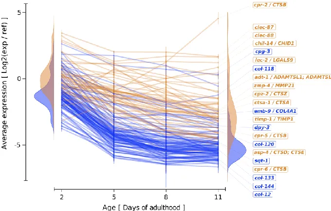
matrisome-annotator.permalink.cc/; [29]). About 150 out of these 1254 age-regulated
305
genes [35] are matrisome genes, comprising of 92 collagen genes (Figure 2A, 2B).
306
Most matrisome genes are expressed during development and growth, but their
307
expression rapidly declines during the reproductive phase (day 1-4 of adulthood;
308
Figure 2C). There are only three matrisome-associated genes that increase in
expression during aging (cpr-2, chil-14, lec-2; Figure 2C). CPR-2 is the cathepsin B
310
(CTSB) orthologue and might be involved in ECM degradation. Consistent with
311
observations in mammals, tissue inhibitor of metalloproteases, TIMP-1, expression is
312
also progressively lost during aging (Figure 2C). Taken together, we found a general
313
decline of matrisome gene expression during aging for the model organism C. elegans 314
(Figure 2C) that is similar to the progressive decline in collagen biosynthesis observed
315
in human skin [10] and across species [8,9]. A comprehensive assessment of not only
316
the transcriptional matreotype but also of the protein levels in mammalian tissues
317
during aging might be feasible by using an in-situ decellularization protocol coupled
318
with ECM proteomics [36]. This could reveal the longitudinal signature of the
319
matreotype during aging.
320
321
Matreotype during longevity 322
Comparing the gene expression or protein levels at a given chronological age between
323
wild type and long-lived animals revealed several molecular mechanisms at work,
324
which were then experimentally assessed for their functional importance for longevity.
325
For instance, reduced Insulin/IGF-1 signaling, upregulates genes involved in
326
antimicrobial, oxidative stress and xenobiotic responses, protein homeostasis, and
327
metabolism, all of which are required to promote healthy aging [37]. Re-analyzing the
328
expression profile of these long-lived mutants with reduced Insulin/IGF-1 signaling [9],
329
revealed that almost one-fifth, i.e., 79 of the 426 upregulate genes are matrisome
330
genes (See Supplementary Table 5 of [29]). Out of the 79 upregulated matrisome
331
genes, 48 are collagens and 15 are ECM proteases (cathepsins, astacin-like
332
metalloendopeptidases, MMPs) [29], suggesting an activation of collagen remodeling
333
[9]. Such a mobilization of matrisome genes also occurs through other
longevity-334
assurance pathways. Re-analyzing the proteomics dataset comparing long-lived germ
335
stem cell mutants (glp-1) with wild-type C. elegans [38], revealed an increase of 177
336
proteins including 25 matrisome proteins in long-lived C. elegans (Supplementary
337
Table 6 of [29]). The 25 matrisome proteins include two basement membrane-forming
338
laminins, ten collagens, one prolyl 4-hydroxylase (DPY-18), which is important for
339
collagen stability, and three ECM-remodeling enzymes (Supplementary Table 6 of
340
[29]), suggesting an increase in ECM turnover and homeostasis. Surprisingly, I could
341
not find any proteomics attempts comparing long-lived mice with wild type during
342
aging. Whether longevity-assurance pathways in mammals alter the matreotype
towards re-activating ECM homeostasis needs to be investigated. Taken together,
344
based on the data from C. elegans, it appears that longevity-assurance pathways
345
invest in collagen or ECM turnover to maintain a youthful matreotype.
346
347
Clinical implications for the matreotype 348
The physiological state of a cell or tissue is reflected in an unique ECM composition
349
or matreotype [2]. For instance, fibroblasts that become senescent or are
350
dedifferentiated into myofibroblasts or cancer-associated fibroblast express a distinct
351
set of ECM proteins. Based on the ECM signature or matreotype, it is even possible
352
to identify tumor type [39]. Thus, distinct matreotypes could be developed into
353
biomarkers or prognostic indicators for disease and health status with implications for
354
personalized medicine. Targeting ECM-cell surface receptors might provide an entry
355
point to remodel ECM and matreotype, since cell-surface receptors read-out ECM
356
stiffness and ECM properties to reprogram cells. Biologics or antibodies that act only
357
on the cellular surfaces could target and alter these ECM-cell surface receptors to
358
change intracellular signaling and to induce the desired gene expression program.
359
Currently, there are about 11 different targets being investigated in 27 clinical trials
360
with primary end points specific to ECM stiffness [40] that might reprogram the
361
matreotype.
362
363
Conclusions 364
Major efforts have revealed how proteins are maintained within cells and cellular
365
compartments and how longevity interventions improve protein homeostasis during
366
aging (Figure 1). Here, I propose that collagen homeostasis or ECM turnover is a
367
process that works efficiently when the organisms are young to maintain their somatic
368
tissue. Since collagen biosynthesis is costly and energy-intensive, upon reproduction
369
resources might be allocated to produce high quality off-springs. Moreover, ECM
370
turnover might be the repair mechanism to cleave-out, digest, and degrade damaged
371
ECM proteins from the matrix. This requires cellular contact, proper
372
mechanotransduction, and de-novo synthesis of ECM components from cells.
373
Damage to the ECM or to cells will start a vicious downwards spiral of ECM
374
fragmentation and cell-detachment leading to a progressive decline in cellular and
375
ECM homeostasis during aging. Longevity interventions might also maintain protein
376
homeostasis of extracellular proteins reflected in changes of the matreotype (Figure
1). Thus far, we have taken the first steps to define the matrisome and the matreotypes
378
during aging and longevity (Figure 2). Since the matreotype reflects the cellular, tissue,
379
and disease status, quantifying and defining the matreotype could become a valuable
380
biomarker for health assessment. Rejuvenating the matreotype might systemically
381
rejuvenate cellular and tissue functions. Identifying druggable targets and
382
understanding how to trigger rejuvenation of the aged matreotype has broad
383
implications for clinical applications.
384
Acknowledgments 386
I thank Cyril Statzer for making the figures, Katrien De Bock, Gabriele Ewald, Nancy
387
Hynes, and members of the Ewald lab for discussion and comments on the
388
manuscript. My inspiration for the term matreotype came from reading about the
389
matrisome by Alexandra Naba and Richard O. Hynes and from Ruedi Aebersold’s
390
definition of proteotype. Furthermore, this work was also inspired by Fritz Verzár
391
(1886–1979), the founder of this journal and a pioneer in investigating collagens in
392
aging research ( https://www.unibas.ch/en/Research/Uni-Nova/Uni-Nova-128/Uni-393
Nova-128-An-almost-forgotten-pioneer.html). Standing on the shoulders of giants; I
394
apologize for omitting or not citing individual original work and simply referring to
395
reviews due to reference limitation. This work was supported by the Swiss National
396
Science Foundation [163898].
397
Figures 399
400
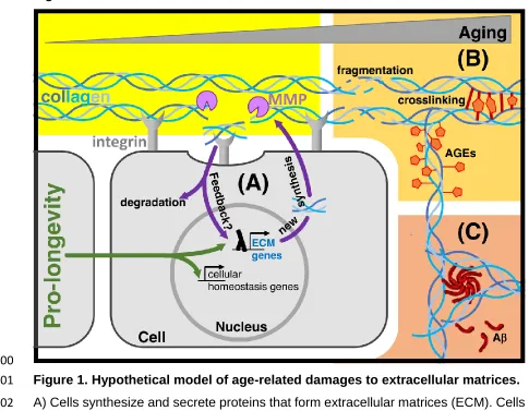
Figure 1.Hypothetical model of age-related damages to extracellular matrices. 401
A) Cells synthesize and secrete proteins that form extracellular matrices (ECM). Cells
402
are anchored to the ECM via cell-surface receptors, such as integrins. Integrins and
403
other receptors transduce mechanical information via biochemical intracellular
404
signaling cascades or cytoskeleton linkages to the nucleus. Matrix metalloproteases
405
(MMP) cleave collagens and other proteins from the ECM. Pro-longevity interventions
406
not only improve cellular protein homeostasis, but might also improve extracellular
407
protein homeostasis.
408
B) During aging, the ECM becomes fragmented, glycated, modified by advanced
409
glycation end products (AGE), and/or crosslinked.
410
C) Aggregation prone peptides, such as amyloid beta (A), accumulate in extracellular
411
matrices.
412
414
Figure 2. Changes in the matreotype during aging. 415
A) Schematic representation of the entire matrisome of C. elegans.
416
B) The aging matreotype consists of 150 differentially expressed matrisome genes
417
during C. elegans aging.
418
C) Longitudinal expression profile of the 150 matrisome genes undergoing significant
419
age-dependent expression changes.
420
Core matrisome and corresponding subcategories in shades of blue and
matrisome-421
associated categories in shades of organ. Expression dataset from Budovskaya et al., 422
Cell 2008 re-analyzed with the C. elegans Matrisome Annotator ( http://ce-matrisome-423
annotator.permalink.cc/ developed by Cyril Statzer (Teuscher et al., Matrix Biology 424
Plus 2019).
References 426
1. Hynes RO: The extracellular matrix: not just pretty fibrils. Science 2009 Nov
427
27;326:1216–1219.
428
2. Bonnans C, Chou J, Werb Z: Remodelling the extracellular matrix in
429
development and disease. Nat Rev Mol Cell Biol 2014 Dec;15:786–801.
430
3. Frantz C, Stewart KM, Weaver VM: The extracellular matrix at a glance.
431
Journal of Cell Science 2010 Dec 15;123:4195–4200.
432
4. Birch HL: Extracellular Matrix and Ageing; in : Biochemistry and Cell Biology of
433
Ageing: Part I Biomedical Science. Singapore, Springer Singapore, 2019, pp
434
169–190.
435
5. Toyama BH, Hetzer MW: Protein homeostasis: live long, won't prosper. Nat
436
Rev Mol Cell Biol 2013 Jan;14:55–61.
437
6. Kjaer M, Langberg H, Miller BF, Boushel R, Crameri R, Koskinen S, et al.:
438
Metabolic activity and collagen turnover in human tendon in response to
439
physical activity. J Musculoskelet Neuronal Interact 2005 Mar;5:41–52.
440
7. Fisher GJ, Quan T, Purohit T, Shao Y, Cho MK, He T, et al.: Collagen
441
fragmentation promotes oxidative stress and elevates matrix
442
metalloproteinase-1 in fibroblasts in aged human skin. American Journal Of
443
Pathology 2009 Jan;174:101–114.
444
8. de Magalhães JP, Curado J, Church GM: Meta-analysis of age-related gene
445
expression profiles identifies common signatures of aging. Bioinformatics 2009
446
Apr 1;25:875–881.
447
9. Ewald CY, Landis JN, Porter Abate J, Murphy CT, Blackwell TK:
Dauer-448
independent insulin/IGF-1-signalling implicates collagen remodelling in
449
longevity. Nature 2015 Mar 5;519:97–101.
450
10. Shuster S, Black MM, McVitie E: The influence of age and sex on skin
451
thickness, skin collagen and density. Br J Dermatol 1975 Dec;93:639–643.
452
11. Gutiérrez-Fernández A, Soria-Valles C, Osorio FG, Gutiérrez-Abril J,
453
Garabaya C, Aguirre A, et al.: Loss of MT1-MMP causes cell senescence and
454
nuclear defects which can be reversed by retinoic acid. EMBO J 2015 Jul
455
14;34:1875–1888.
456
12. Vafaie F, Yin H, O'Neil C, Nong Z, Watson A, Arpino J-M, et al.:
Collagenase-457
resistant collagen promotes mouse aging and vascular cell senescence. Aging
458
Cell 2013 Sep 19;13:121–130.
459
13. Hernandez L, Roux KJ, Wong ESM, Mounkes LC, Mutalif R, Navasankari R, et
460
al.: Functional coupling between the extracellular matrix and nuclear lamina by
461
Wnt signaling in progeria. Dev Cell 2010 Sep 14;19:413–425.
14. Bae EJ, Lee HJ, Rockenstein E, Ho DH, Park EB, Yang NY, et al.:
Antibody-463
Aided Clearance of Extracellular -Synuclein Prevents Cell-to-Cell Aggregate
464
Transmission. J Neurosci 2012 Sep 26;32:13454–13469.
465
15. Urushitani M, Ezzi SA, Julien J-P: Therapeutic effects of immunization with
466
mutant superoxide dismutase in mice models of amyotrophic lateral sclerosis.
467
Proc Natl Acad Sci USA 2007 Feb 13;104:2495–2500.
468
16. Selkoe DJ: The cell biology of beta-amyloid precursor protein and presenilin in
469
Alzheimer's disease. Trends Cell Biol 1998 Nov;8:447–453.
470
17. Erikson GA, Bodian DL, Rueda M, Molparia B, Scott ER, Scott-Van Zeeland
471
AA, et al.: Whole-Genome Sequencing of a Healthy Aging Cohort. Cell 2016
472
May 5;165:1002–1011.
473
18. Morikiri Y, Matsuta E, Inoue H: The collagen-derived compound collagen
474
tripeptide induces collagen expression and extends lifespan via a conserved
475
p38 mitogen-activated protein kinase cascade. Biochemical and Biophysical
476
Research Communications 2018 Nov 10;505:1168–1173.
477
19. Liang J, Pei X-R, Wang N, Zhang Z-F, Wang J-B, Li Y: Marine collagen
478
peptides prepared from chum salmon (Oncorhynchus keta) skin extend the life
479
span and inhibit spontaneous tumor incidence in Sprague-Dawley Rats. J Med
480
Food 2010 Aug;13:757–770.
481
20. Liang J, Pei X, Zhang Z, Wang N, Wang J, Li Y: The protective effects of
long-482
term oral administration of marine collagen hydrolysate from chum salmon on
483
collagen matrix homeostasis in the chronological aged skin of Sprague-Dawley
484
male rats. J Food Sci 2010 Oct;75:H230–8.
485
21. Tsuruoka N, Yamato R, Sakai Y, Yoshitake Y, Yonekura H: Promotion by
486
collagen tripeptide of type I collagen gene expression in human osteoblastic
487
cells and fracture healing of rat femur. Biosci Biotechnol Biochem 2007
488
Nov;71:2680–2687.
489
22. Choi HR, Cho KA, Kang HT, Lee JB, Kaeberlein M, Suh Y, et al.: Restoration
490
of senescent human diploid fibroblasts by modulation of the extracellular
491
matrix. Aging Cell 2011 Feb;10:148–157.
492
23. Sun Y, Li W, Lu Z, Chen R, Ling J, Ran Q, et al.: Rescuing replication and
493
osteogenesis of aged mesenchymal stem cells by exposure to a young
494
extracellular matrix. The FASEB Journal 2011 May;25:1474–1485.
495
24. Hendrix MJC, Seftor EA, Seftor REB, Kasemeier-Kulesa J, Kulesa PM,
496
Postovit L-M: Reprogramming metastatic tumour cells with embryonic
497
microenvironments. Nat Rev Cancer 2007 Apr;7:246–255.
498
25. Carlson BM, Faulkner JA: Muscle transplantation between young and old rats:
499
age of host determines recovery. Am J Physiol 1989 Jun;256:C1262–6.
26. Carlson BM, Dedkov EI, Borisov AB, Faulkner JA: Skeletal muscle
501
regeneration in very old rats. J Gerontol A Biol Sci Med Sci 2001
502
May;56:B224–33.
503
27. Castellano JM, Mosher KI, Abbey RJ, McBride AA, James ML, Berdnik D, et
504
al.: Human umbilical cord plasma proteins revitalize hippocampal function in
505
aged mice. Nature 2017 Apr 27;544:488–492.
506
28. Naba A, Clauser KR, Ding H, Whittaker CA, Carr SA, Hynes RO: The
507
extracellular matrix: Tools and insights for the “omics” era. Matrix Biol 2016
508
Jan;49:10–24.
509
29. Teuscher AC, Jongsma E, Davis MN, Statzer C, Gebauer JM, Naba A, et al.:
510
The in-silico characterization of the Caenorhabditis elegans matrisome and
511
proposal of a novel collagen classification. Matrix Biology Plus 2019 Mar
512
11;:1–13.
513
30. Aebersold R, Mann M: Mass-spectrometric exploration of proteome structure
514
and function. Nature 2016 Sep 15;537:347–355.
515
31. Naba A, Clauser KR, Hynes RO: Enrichment of Extracellular Matrix Proteins
516
from Tissues and Digestion into Peptides for Mass Spectrometry Analysis. J
517
Vis Exp 2015;:e53057.
518
32. Wick G, Grundtman C, Mayerl C, Wimpissinger T-F, Feichtinger J, Zelger B, et
519
al.: The Immunology of Fibrosis. Annu Rev Immunol 2013;31:107–135.
520
33. Teuscher AC, Statzer C, Pantasis S, Bordoli MR, Ewald CY: Assessing
521
Collagen Deposition During Aging in Mammalian Tissue and in Caenorhabditis 522
elegans. Methods Mol Biol 2019;1944:169–188. 523
34. Teuscher AC, Ewald CY: Overcoming Autofluorescence to Assess GFP
524
Expression During Normal Physiology and Aging in Caenorhabditis elegans.
525
BIO-PROTOCOL 2018 Jul 20;8. DOI: 10.21769/BioProtoc.2940
526
35. Budovskaya YV, Wu K, Southworth LK, Jiang M, Tedesco P, Johnson TE, et
527
al.: An elt-3/elt-5/elt-6 GATA Transcription Circuit Guides Aging in C. elegans.
528
Cell 2008 Jul 25;134:291–303.
529
36. Mayorca-Guiliani AE, Madsen CD, Cox TR, Horton ER, Venning FA, Erler JT:
530
ISDoT: in situ decellularization of tissues for high-resolution imaging and
531
proteomic analysis of native extracellular matrix. Nat Med 2017 Jun
532
12;23:890–898.
533
37. Ewald CY, Castillo-Quan JI, Blackwell TK: Untangling Longevity, Dauer, and
534
Healthspan in Caenorhabditis elegans Insulin/IGF-1-Signalling. Gerontology
535
2018;64:96–104.
536
38. Pu Y-Z, Wan Q-L, Ding A-J, Luo H-R, Wu G-S: Quantitative proteomics
537
analysis of Caenorhabditis elegans upon germ cell loss. J Proteomics 2017
538
Mar 6;156:85–93.
39. Socovich AM, Naba A: The cancer matrisome: From comprehensive
540
characterization to biomarker discovery. Seminars in Cell & Developmental
541
Biology 2019 May;89:157–166.
542
40. Lampi MC, Reinhart-King CA: Targeting extracellular matrix stiffness to
543
attenuate disease: From molecular mechanisms to clinical trials. Sci Transl
544
Med 2018 Jan 3;10:eaao0475.
545