Article
1
Computational insight into the effect of natural
2
compounds on the destabilization of preformed
3
amyloid-β(1-40) fibrils
4
Francesco Tavanti*, Alfonso Pedone and Maria Cristina Menziani*
5
Department of Chemical and Geological Sciences, University of Modena and Reggio Emilia, Via G. Campi,
6
103 – Modena, Italy.
7
* Correspondence: [email protected], [email protected]
8
Abstract: One of the principal hallmarks of Alzheimer’s disease (AD) is related to the aggregation
9
of amyloid-β fibrils in an insoluble form in the brain, also known as amyloidosis. Therefore, a
10
prominent therapeutic strategy against AD consists either in blocking the amyloid aggregation
11
and/or destroying the already formed aggregates. Natural products have shown significant
12
therapeutic potential as amyloid inhibitors from in vitro studies as well as in vivo animal tests. In
13
this study, the interaction of five natural biophenols (curcumin, dopamine,
(-)-Epigallocatechin-3-14
gallate, Quercetin, and Rosmarinic acid) with the amyloid-β(1-40) fibrils has been studied through
15
computational simulations. The results allowed the identification and characterization of the
16
different binding modalities of each compounds and their consequences on fibril dynamics and
17
aggregation. It emerges that the lateral aggregation of the fibrils is strongly influenced by the
18
intercalation of the ligands, which modulate the double-layered structure stability.
19
Keywords: Molecular dynamics simulation; biophenols; natural compounds; amyloid fibrils;
20
Alzheimer’s disease; ligand-protofiber interactions.
21
22
1. Introduction
23
The pathological hallmark of AD is the extracellular accumulation of insoluble proteinaceous
24
deposits called amyloid fibrils[1] that induce cytotoxicity. The formation of mature amyloid fibrils
25
(Aβ) proceeds through a nucleation dependent process, where monomers and oligomers aggregate
26
together forming β-sheets rich protein structures. The most common fibrils are 40) and
Aβ(1-27
42) that are composed by 40 and 42 amino acids, respectively, and are characterized by β-strand units
28
aligned perpendicular to the main fibril axis[2]. Destabilization and clearance of amyloid aggregates
29
by small molecules is one of the promising approaches towards the development of AD therapies[4].
30
In the last years, epidemiological studies on the effects of the diet against AD and dementia
31
suggested that the high intake of flavonoids and polyphenols found in fruits and vegetables reduces
32
the risk of AD and cognitive impairments, and several natural molecules have been identified to
33
promote cognitive health and to interfere with the amyloidogenic activity in AD[11].
34
A detailed knowledge of how these molecules interact with Aβ fibrils is a prerequisite for the
35
design of new efficient drugs. Unfortunately, despite intensive researches, the experimental
36
characterization of full-length Aβ oligomers/inhibitor complexes at a high level of resolution remains
37
a great challenge.
38
Atomistic computer simulations are well suited to provide molecular-level details of amyloid
39
oligomer and fibril interactions with ligands, helping in the future development and characterization
40
of druggable modalities[12]. Basically four aspects of the flavonoids-amyloid interaction have been
41
studied by computational methods: 1) the effect of ligands on the conformational transitions of Aβ
42
monomers from initial random coil or α-helix into β-sheet structures[13,14] and ligand-mediated
43
conformational change on Aβ dimer[15] by means of Replica Exchange Molecular Dynamics (REMD)
44
simulations; 2) the effect of ligands on the aggregation of Aβ(17–36) using Coarse-Grained
45
Simulations[16]; 3) effect of ligands on the conformation and stability of amyloid-beta mutants[17] by
46
Molecular Dynamics (MD) simulations; 4) preferential binding sites of ligands and their effect on
47
amyloid structure-dynamics[18] on Aβ fragments and full-length single Aβ protofilament[19–25] by
48
means of Docking experiments, MD simulations and Free Energy calculations.
49
Although recently a few studies devoted their attention to the interaction of ligands (mainly
50
markers for amyloid detection ref. [26–28]) with multiple Aβ protofilaments, to the best of our
51
knowledge this aspect has not been investigated thoroughly for natural polyphenol ligands, except
52
for curcumin[19].
53
In this study, the binding modalities of five natural biophenols (curcumin, dopamine,
(-)-54
Epigallocatechin-3-gallate, Quercetin, and Rosmarinic acid) with single Aβ(1-40) protofilament and
55
double-layer oligomers aggregates will be studied through atomistic computational simulations in
56
order to explore structural changes in aggregate pathways upon binding.
57
First, putative binding sites on the Aβ(1-40) protofibril will be explored by replica exchange
58
molecular dynamics (REMD) simulations. Then, binding free energies (Gbind) will be computed on
59
the complexes to determine the thermodynamically favourite binding modalities. Finally, the
60
structural effects caused by the binding of polyphenols to two double-layer protofilament
61
polymorphs will be assessed. To this goal the determination of the stability of the sheet-to-sheet
62
associations of the double-layered organizations with and without the polyphenols will be computed
63
by means of the Potential of Mean Force (PMF) methodology.
64
2. Methods
65
2.1 Molecular Dynamics Simulations
66
Molecular dynamics simulations were performed with GROMOS 54a7 force field[29]. The
67
structural model of amyloid fibrils was retrieved from the PDB[30] (PDB ID: 2LMN[31]). From this
68
structure, an Aβ monomer was isolated and the missing N-terminal peptide region of Aβ(1-40)
69
monomer (1DAEFRHDS8) was built using the Molefacture plugin in the VMD package[32] as random
70
coils as predicted by both the Jpred web server[33] and by the Modeller package[34] for protein
71
secondary structure assignments. Standard protonation states corresponding to pH 7 were assigned
72
to ionisable residues. The Aβ(1-40)protofibril was composed by repeating 10 monomeric units along
73
its principal axis obtaining a continuous structure 5nm long.
74
The force field assigned to each ligand in their standard protonation states at pH 7 was built in
75
the GROMACS format[35] by using the Automated Topology Builder[36,37]web server.
76
The simulation box (7.5x9.7x8.0xnm) contains one Aβ(1-40)protofibril composed by repeating
77
10 monomeric units, one ligand placed in a random position with respect to the fibril, and about
78
30000 simple point charge water molecules[38]. Counter ions (Na+ and Cl-) were added at random
79
locations to neutralize the systems, with ion concentration of 150 mM, close to the physiological
80
value.
81
All the simulations were carried out at physiological temperature (310K) and pressure of 1 bar.
82
The systems were first equilibrated for 2 ns in the NVT ensemble, then 10n runs were carried out in
83
the NPT ensemble. The temperature was controlled using a velocity-rescaling thermostat with a
84
coupling time of 0.1ps. During equilibration the Berendsen barostat was used to control the pressure,
85
while during the production run the Parrinello-Rhaman barostat was used with coupling time of 2ps
86
and an isothermal compressibility of 4.5 × 10⁻⁵ bar−1 and the timestep used was 2.0fs.The
particle-87
mesh Ewald algorithm was used to calculate long-range electrostatics[39],[40] with a fourth-order
88
cubic interpolation, a grid spacing of 0.16nm, and a real-space cutoff of 1nm. Both Van Der Waals
89
and neighbour list cutoffs describing short-range interactions were set to 1.0 nm. A production run
90
of 50 ns was used to identify the ligand binding sites (paragraph 2.2), whereas trajectories of 100 ns
91
were necessary for the computation of the stability of the different protofibrils polymorphs
92
(paragraph 2.4). Data analysis were performed using the GROMACS-5.0.4 package[41].
93
2.2 Ligand binding sites
95
Temperature Replica Exchange MD (REMD) simulations were used to define the most probable
96
interacting sites of each compound with the Aβ(1-40) protofibrils. The temperatures used for replicas
97
were obtained by the work of Patriksson and van der Spoel[42] and are reported below: 300.00, 301.16,
98
302.32, 303.49, 304.66, 305.83, 307.01, 308.19, 309.38, 310.57, 311.76, 312.96, 314.16, 315.37, 316.57,
99
317.78, 319.00, 320.22, 321.44, 322.66, 323.89, 325.12, 326.36, 327.60, 328.85, 330.09, 331.34, 332.60,
100
333.86, 335.12, 336.39, 337.66, 338.93, 340.21.
101
An acceptance ratio of 20% was chosen as previously suggested by Ngo et al.[43]. Each REMD
102
simulation replica was equilibrated with an NVT and an NPT ensemble with the same parameters as
103
for MD simulations. Then a 50ns run (i.e. the production run) was performed for each replica,
104
exchanges between neighbouring replicas were checked every 500 steps corresponding to 1ps[43].
105
The 50ns simulations were used for data analysis.
106
2.3 Ligand binding energy
107
The Molecular Mechanics-Poisson-Boltzmann surface area (MM_PBSA) method [44] was used
108
to calculate the binding energy of each ligand to the protofibril. This method is based on the
single-109
trajectory approach. Thus, 100 snapshots collected consecutivelly over the course of the 50ns
110
simulations, once the ligand reached a stable binding (i.e. rmsd of its center of mass < 5 Å, Figure S1)
111
were used. The binding free energy (ΔGbinding) is described as the free energy difference between the
112
complex, Gcomplex, and the summation of the free energy of protein, Gprotein, and ligand, Gligand:
113
(1)
The free energy of each molecule is given by
114
(2)
Where T and S represent the temperature and entropy, respectively, and the mechanical energy, EMM,
115
of the solute in the gas phase is given by the summation of bond, angles, dihedrals, Van der Waals
116
and electrostatic terms:
117
(3)
The solvation energy, Gsolvation, is calculated as follows:
118
(4)
Where the non-polar solvation term, Gsurf, is approximated on the solvent-accessible-surface area
119
(SASA) derived from the Shrake-Rupley numerical method[45]
120
(5)
with γ=0.0072kcal/mol Å2 and β=0[46].
121
The term comprising the electrostatic potential between the solute and the solvent, GPB, is calculated
122
using the continuum solvent approximation[47] by the APBS package[48].
123
The entropy term, TΔS, is computed using the Quasi-harmonic formula[49].
124
2.4 Aβ(1-40) oligomer double-layered structures
125
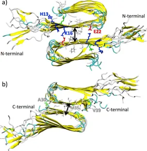
Two possible double-layered structures were build by stacking the -sheets of each monomer
126
onto each other in an antiparallel fashion[50,51], as shown in Figure 1. The C-terminal–C-terminal
127
and N-terminal–N-terminal interfaces were, thus, obtained. The inter-sheet distance was computed
128
G
binding=
G
complex-
(
G
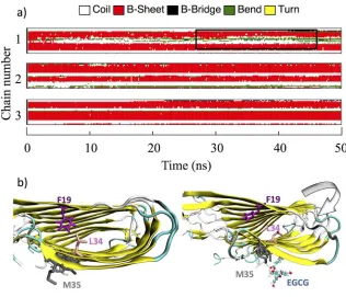
protein+
G
ligand)
G
=
E
MM+
G
solvation-
T
S
E
MM=
E
bond+
E
angle+
E
digedral+
E
electr+
E
VdWG
solvation=
G
surf+
G
PBas the distance between the centres of mass of the two β-sheets that are in contact. The amino acids
129
that were considered for the calculations of the centre of mass are H13, H14, Q15, K16, L17, V18, F19,
130
F20, A21, E22 for the N-terminal–N-terminal interface (β-1 β-sheets) (Figure 1a) and A30, I31, I32,
131
G33, L34, M35, V36, G37, G38, V39 for the C-terminal–C-terminal interface (β-2 β-sheets) (Figure 1b).
132
133
Figure 1. Cartoon representation of double-layered structures of Aβ(1-40) oligomers facing through
134
their β-1, in a), and β-2 β-sheets, in b). Fibrils are coloured accordingly to their secondary structures.
135
Amino acids at the interface are explicitly represented (colour code: blue for positive charged, red for
136
negatively charged, and white for hydrophobic amino acid residues). Black arrows roughly represent
137
the inter-sheet distance.
138
In order to evaluate the influence of the ligands on the stability of the different protofibrils
139
polymorphs the Potential of Mean Force (PMF) method implemented in the GROMACS program
140
was used[52,53].
141
The backbone of protofibril(1) was restrained in its starting position, while a force increasing
142
with time, was assigned to the centre of mass of protofibril(2). Three directions were taken into
143
account, as shown in Figure 2: the x-axis (i.e. outward), the y-axis (i.e. lateral) and the z-axis (i.e.
144
vertical). For each ligand and for both protofibril contact modes (β-1 and β-2 β-sheets), three runs
145
were performed using as starting configurations the ones at 90, 95 and 100 ns, ensuring good
146
sampling. The starting force used at the beginning of the simulation was 1000 kJ/mol nm2 and the
147
149
Figure 2: pulling directions applied to protofibril(2) during the calculation of the forces needed for
150
double-layered destruction: x-axis (i.e. outward: shift of the protofibril(2) along its secondary axes),
151
the y-axis (i.e. lateral, shift of the protofibril(2) along its primary axes) and the z-axis (i.e. vertical,
152
progressive removal of protofibril(2)).
153
3. Results and Discussion
154
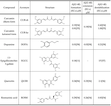
The five natural compounds studied are listed in Table 1, together with their effective
155
concentrations (EC50) for the formation, extension and destabilization of preformed Aβ(1-40)
(fAβ(1-156
40)).
157
The overall in vitro activities of CUR and ROSM are similar [54]. Moreover, in vivo observations
158
suggest that curcumin may be beneficial even after the disease has developed, reducing amyloid
159
levels and plaque burden of aged mice with advanced amyloid accumulation[55]. Quercetin (QUER)
160
shows moderate in vitro preformed fAβ(1-40) destabilization effects with respect to CUR [56].
(-)-161
epigallocatechin-3-gallate (EGCG) is undergoing phase II–III clinical trials as inhibitor of Aβ
162
fibrillogenesis. It decreases plaque burdens in the brain, and reduces soluble and insoluble preformed
163
fAβ(1-40)s [57]. Finally, Dopamine (DOPA) proved to be a potent anti-amyloidogenic agent at all the
164
different levels of formation, extension of amyloid fibrils and destabilization of preformed fAβ(1–
165
40)s [58].
166
Heterogeneity in the experimental conditions (i.e. peptide concentrations, incubation condition
167
and procedure of fAβ preparation) used in different laboratories or different experiments in the same
168
lab gives rise of discrepancy in effective EC50 concentrations, thus preventing a quantitative
169
rationalization of the observed experimental trend by means of the results of the computational
170
simulations. However, some interesting qualitative structure-activity relationships could be
171
considered, as shown in the following.
172
Table 1. The effective concentrations (EC50) of the ligands studied for the formation, extension and
174
destabilization of fAβ(1-40).
175
Compound Acronym Structure
Aβ(1-40) formation (EC50) μM
Aβ(1-40) extension (EC50)
μM
Aβ(1-40) destabilization
(EC50) μM
Curcumin
diketo form CUR-di
0.19[54]
0.81[55] 0.19[54]
0.42[54] 1.00[55] Curcumin
ketoenol form CUR-ke
Dopamine DOPA 0.01[58] 0.03[58] 0.21[58]
(-)-
Epigallocatechin-3-gallate
EGCG 0.18[11] - 15*[57]
Quercetin QUER 0.24[56] 0.25[56] 2.1[56]
Rosmarinic acid ROSM 0.29[54] 0.26[54] 0.83[54]
* referred to Aβ(1-42) fibrils.
176
3.1 Putative Binding sites and Binding Free Energies
177
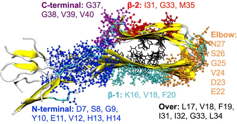
Six main binding sites have been highlighted by means of the REMD method applied to the
178
ligands considered. They are located at the surface of the protofibril:
179
1. β-1 β-sheet corresponding to the AA sequence 16KLVFFAEDV24,
180
2. β-2 β-sheet corresponding to the AA sequence 31IIGLMVG37,
181
3. Elbow connecting the two β-sheets in correspondence of the AA sequence 22EDVGSN27,
182
4. top of the protofibril Over the two β-sheets of the terminal A(1-40) monomer,
183
5. disordered tails located at the N-terminal,
184
6. end of the β-2 β-sheet, on the C-terminal (entry of the cleft).
185
187
Figure 3: ball-and-stick representation of the ligand binding sites obtained by REMD. Amino-acids
188
(one-letter code) involved in the interactions are reported for each binding site with different colours.
189
For each binding site, amino acids that make persistent interactions (in this work, an interaction
190
is considered as persistent if the amino acid residue remains in contact with the ligand for at least
191
60% of the total simulation time) with the ligands and that contributes more than 1 kcal/mol to the
192
binding energy are highlighted in Figure 3. The probability of the occupancy of each site is shown in
193
Figure 4a.
194
195
Figure 4: Probability of occupancy of each binding site (a), and binding free energy (b) for each ligand
196
It is interesting to note the different occupancy preferences of the two curcumin’s forms. The
198
CUR-di predominantly interacts with the N-terminal, whereas CUR-ke is mainly found at the β-2
199
site.
200
Multiple binding sites have been previously described in the literature for curcumin derivatives
201
and others related compounds. In particular, the β-2 site has been very recently targeted in a
202
combined computational and experimental study by Battisti et al.[22] aimed at the design of
203
curcumin-like amyloid beta peptides inhibitors. Binding to N-terminal and Over position have been
204
observed for curcumin and other ligands by means of site map analysis by Kundaikar et al.[59].
205
Moreover, the -1 binding site has previously been suggested as possible binding site for curcumin
206
on the basis of solid-state NMR experiments[60] and computational studies on Aβ hexapeptide
207
16KLVFFA21 and full length Aβ fibrils[19,22].
208
Although a few studies in the literature proposed the cavity formed by the two β-sheets and the
209
turn as possible binding site for curcumin[24,25] and other compounds such as Orange-G[26] this site
210
is never occupied by the ligands considered in the present study. However, small portions of
CUR-211
ke, EGCG, QUER and ROSM ligands occasionally can penetrate this cavity during the dynamic
212
simulations runs, when they are interacting with the A(1-40) protofibril in the Over position.
213
By considering the probability of the occupancy of each binding sites (Figure 4a) together with
214
the corresponding Binding Free Energies (Figure 4b) it emerges that:
215
- CUR-di and CUR-ke. CUR-ke, the predominant form in aqueous solution on the basis of the
216
recent results obtained by Manolova et al.[61], shows a strong propensity to dock at the β-2 site and
217
realizes at this site strong interactions (Gbind > -20 kcal/mol) with the fAβ(1-40) fibril. However,
218
moderate to strong (-10 < Gbind > -20 kcal/mol) free energies of binding are found for all the binding
219
sites with the exception of the N-terminal one.
220
-DOPA shows a preference for docking the β-2 and N-ter sites. However, by considering the
221
free energy of binding, it does not show selectivity among the six sites studied, realizing
222
moderate to weak interactions G
bind< -10 kcal/mol) with all of them.
223
- EGCG targets preferentially the N-ter and β-2 sites, and, secondarily, the Elbow and β-1 sites.
224
However, this ligand is able to realize strong binding with all the possible six sites. The most stable
225
complexes (Gbind > -20 kcal/mol) are obtained at the β-2, and β-1 sites. The ability of EGCG to bind
226
to the N-terminal amino acids (1-16 residues) is confirmed by results obtained by isothermal titration
227
calorimetry experiments[62]. Moreover, recent findings by solution NMR indicate that EGCG
228
preferentially binds to A oligomers and shield them at the -1 and -2 sites,[63] where it remodels
229
the oligomer surface altering the interactions with the monomers.
230
- QUER is found almost equally distributed between the β-1 and “over” sites with significant
231
lower probability for the other sites. However, it realizes moderate binding free energies (Gbind ~
-232
10 kcal/mol) in all sites, with the most stable complexes (Gbind ~ -20 kcal/mol) involving the β-2, and
233
β-1 sites. These results are in agreement with the finding of a computational study recently reported
234
by Ren et al.[20] for a structurally homologous compound, genistein. They showed that genistein
235
prefers to bind the β-sheet grooves to interfere with their self-aggregation.
236
-ROSM has higher probability for docking at the N-ter and β-1 sites, but realizes the most stable
237
interactions with moderate binding free energies at the β-2 and β-1 sites. Indeed, NMR investigations
238
suggest that a ROSM hairpin-like structure would allow the intercalation into the Aβ oligomers
239
structure, at the interprotofilament (β−β zippers) interface[6].
240
Thus, taking the error in the computation of the Gbind into account, it can be stated that the β-2
241
grove is a common structural target for all the ligands studied; at this site the ligands realize their
242
most stable interactions with residues M35, G33, and I31. The β-1 site is also targeted for energetically
243
favoured complexes, realized mainly by the interaction with the K16, V18, F20 residues.
244
These regions are particularly interesting since they constitute the junction between
245
protofilaments in common Aβ(1-40) polymorphs[31,64]. Several recent computational studies
246
employing different multiple protofilament structures and a variety of ligands, used as markers for
247
preferential bind sites[26–28]. Binding of ligands at these sites can interfere with the formation or
249
induce the disruption of the aggregates, as discussed in the next section.
250
In agreement with the previous studies on related compounds [16, 54], the binding free energies
251
obtained for these complexes are driven by more favourable non-polar interactions rather than by
252
electrostatic ones. (Figure S2).
253
Visual inspection of all MD trajectories shows that the random-coil N-terminal 1DAEFRHDS8
254
sequence does not alter appreciably the conformation and the usual behaviour of the rest of the fibril,
255
despite its high flexibility promote the nomadism of the ligands that bind preferentially to D7 and
256
S8. Moreover, overall the binding of the ligands do not disturb the structural integrity and secondary
257
structure of the Aβ protofibrils, their overall U-shaped conformations being retained with or without
258
interacting ligands.
259
However, in a few cases (EGCG, CUR-ke and QUER) when the ligands, during the dynamic run,
260
migrate from the Over site to the β-2 β-sheet in proximity to M35, a perturbation of the fibril
261
secondary structure in the terminal monomers laying at the head of the protofibril is observed. This
262
perturbation, observed in the time of the simulations expecially in the elbow region, induces a bent
263
in the long fibril axis that can impair the process of fibril elongation.
264
Figure 5 explains the phenomenon for the complex formed by EGCG and the Aβ protofibrils.
265
The side-chain of M35, interacting with the ligand, chaperons it in the search for the best interactions
266
in the β-2 groove, causing a bending of the protofibril and altering the Aβ protofibril secondary
267
structure in the Elbow region. Moreover, the dynamics of the M35 side-chain, induced by the
268
interacting ligand, disrupts the hydrophobic interaction between L34 and F19, which is found to
269
influence a broad range of different processes including the initiation of fibrillation, oligomer
270
stability, fibril elongation, and cellular toxicity[65]. In addition, it is worth undelaying that M35 itself
271
is also known to be responsible for the hierarchical assembly of amyloid fibrils.
272
273
Figure 5: a) Evolution of the Aβ(1-40) secondary structure (computed with the GROMACS DSSP tool)
274
upon EGCG binding on the β-2 β-sheet grove. The perturbation induced at the monomers lying at the
275
head of the protofibril is highlighted by a black box. For clarity safe, only the top three Aβ(1-40)
276
monomers are showed. b) Conformation of F19 and L34 before (left) and after (right) the interaction
277
of EGCG with M35.
278
3.2 Influence of the ligands on the stability of the Aβ(1-40) oligomer double-layered structures
280
The effect of the ligands on the stability of the protofibril double-layered structures has been
281
quantified by the calculation of the forces (PMF) for protofibril(1)/ligand-protofibril(2) unbinding.
282
On the basis of the binding site preferences discussed in the previous section, the intercalation of the
283
ligands into the C-terminal–C-terminal and N-terminal–N-terminal interfaces of the protofibrils have
284
been considered. Moreover, three possible ways for complex disruption have been examined by
285
applying the forces along the x-axis (i.e. outward: shift of the protofibril(2) along its secondary axes),
286
the y-axis (i.e. lateral, shift of the protofibril(2) along its primary axes) and the z-axis (i.e. vertical,
287
progressive removal of protofibril(2)), as shown in Figure 2.
288
The results are reported in Table 2, together with the force need to separate the pristine
289
protofibril-protofibril aggregation, taken as control.
290
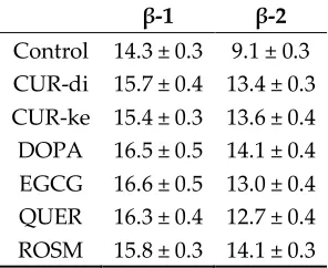
It is worth noting that the C-terminal–C-terminal interface of the double-layered Aβ-sheets
291
consists of highly hydrophobic patches of I31, I41, and M35, with an average intermolecular distances
292
between the two -sheets of ~ 9.1 Å (see Table 3), whereas the N-terminal–N-terminal interface
293
consists of both hydrophobic patches of V18 and F20 and K16–E22 salt bridges with an average
294
intermolecular distances of ~ 14.3 Å, in agreement with previous computational studies on Aβ17-42
295
[66] and on different segmental polymorphs (A 35-42, A 16-21, A 27-32) modelled by Berhanu
296
et al.[67]. These characteristics determine the stability of the β-sheet-β-sheet interfaces, which is
297
significantly higher for the N-terminal–N-terminal arrangement with respect to the
C-terminal–C-298
terminal one, as results from the PMF for protofibril(1)-protofibril(2) unbinding, at least for the
299
vertical and outward directions (Table 2).
300
Table 2: Computed force (expressed in kJ/mol) needed for protofibril(1)-protofibril(2) (control) and
301
protofibril(1)/ligand-protofibril(2) unbinding along the x, y and z-axis.
302
Overall, the binding of the ligands to -sheet--sheet interfacial pockets located between two
303
protofilaments produces a reduction of the stability of the protofibril dimeric structures. However,
304
this cannot be directly correlated to the increasing in the intermolecular distances between the two
305
interacting protofibril. In fact, for the N-terminal–N-terminal interface, the distance increases upon
306
ligand binding, is in the order of 2 Å, while for the C-terminal–C-terminal one, initially characterized
307
by a tight binding due to hydrophobic interactions, is ~ 4-5 Å. (Table 3).
308
Table 3: Inter-sheet distance in the Aβ(1-40) oligomer double-layered structures
309
β-1 β-2
Control 14.3 ± 0.3 9.1 ± 0.3 CUR-di 15.7 ± 0.4 13.4 ± 0.3 CUR-ke 15.4 ± 0.3 13.6 ± 0.4 DOPA 16.5 ± 0.5 14.1 ± 0.4 EGCG 16.6 ± 0.5 13.0 ± 0.4 QUER 16.3 ± 0.4 12.7 ± 0.4 ROSM 15.8 ± 0.3 14.1 ± 0.3
Force Direction Lateral (x-axis) Vertical (y-axis) Outward (z-axis)
Ligand/binding site β-1 β-2 β-1 β-2 β-1 β-2
On the other hand, the maximum destabilization of the double-layered Aβ-sheet aggregates is
310
observed for the -arrangements, when the forces are applied, along the vertical (y) and outward
311
(z), and lateral (x) axes, in the order.
312
The binding of ligands at the C-terminal–C-terminal interface results in a moderate
313
destabilization of the double-layered Aβ-sheet aggregates with respect to the lateral and vertical
314
modalities, whereas for the outward disruption it appears that the ligands have no effect or operate
315
a small stabilization of the complexes, the large errors obtained do not allow further lucubration.
316
It is worth noting that the inter-sheet separation produced by DOPA, the smallest ligand, is
317
larger or comparable to the one observed for more cumbersome ligands, and its effect on the
318
destabilization of the protofibril dimeric aggregates is also overall stronger than the other ligands.
319
4. Concluding remarks
320
The results of the sistematic computational study carried out on the interaction of five natural
321
biophenols with single Aβ(1-40) protofilament by means of REMD simulations allowed the
322
individuation of multiple binding sites for each ligand located at the surface of the protofibril nearby
323
the β-1 β-sheet, β-2 β-sheet, elbow connecting the two β-sheets, top of the protofibril, disordered
N-324
terminal, and C-terminal.
325
The REMD methodology used does not allow the biophenols to entry into the hydrophobic core
326
of the preformed protofibril, probably because the energy penalty associate with the penetration
327
process cannot be overcome using conventional MD. The absence of binding sites in the cavity of the
328
preformed protofibril prevent the study of destabilizing effects of the ligands by promotion of
329
disruption of the native backbone hydrogen bonds in the protofibril interior.
330
The MM_PBSA energetic analysis of the binding shows that the β-1 and β-2 binding sites at the
331
exposed surface of the Aβ(1-40) protofibrils, shared by all the five ligands studied, are
332
thermodynamically favoured. At these sites the anti-amyloid activity of biophenols consists in the
333
inhibition of fibrils thickening and elongation.
334
In fact, although no significant perturbation of the overall protofibril secondary structure is
335
observed in the periods of time studied, interesting conformational changes of the terminal peptides
336
with subsequent bending of the principal axis of the protofibril is induced by ligands that migrate
337
during the dynamic run from the Over binding site to the β-2 binding site. This effect is more
338
marked for EGCG, but is observed also for CUR-ke and QUER and may preclude the association of
339
an incoming Aβ peptide inhibiting the fibril elongation.
340
Moreover, ligand binding at the β-2 binding site may inhibit the amyloidogenic process by
341
shielding the M35, which is responsible for the hierarchical assembly of amyloid fibrils, and
342
disrupting the hydrophobic interaction between L34 and F19, which is found to influence a broad
343
range of different processes including the initiation of fibrillation, oligomer stability, fibril elongation,
344
and cellular toxicity.
345
Finally, the stability of the -sheet--sheet interfaces of the Aβ(1-40) oligomer double-layered
346
structures is significantly affected by the intercalation of the biophenols. The force needed for
347
disruption of the aggregates is halved by all the ligands binding the N-terminal-N-terminal interface,
348
when the forces are applied along the principal axis of the protofibril. The most remarkable effect is
349
observed for DOPA on the double-layered structure in the N-terminal-N-terminal arrangement
350
whatever force direction; whereas ROSM and EGCG exert a stronger destabilization at the
double-351
layered structure in the C-terminal-C-terminal arrangement.
352
Supplementary Materials: Figure S1.
353
Author Contributions: Conceptualization, Maria Cristina Menziani; Formal analysis, Francesco Tavanti;
354
Funding acquisition, Maria Cristina Menziani; Investigation, Francesco Tavanti; Methodology, Francesco
355
Tavanti; Project administration, Maria Cristina Menziani; Supervision, Maria Cristina Menziani and Alfonso
356
Pedone; Visualization, Francesco Tavanti; Writing – original draft, Francesco Tavanti; Writing – review &
357
editing, Alfonso Pedone.
358
Acknowledgments: FT acknowledges the grant FAR2015 from UNIMORE. The authors gratefully acknowledge
359
References
361
1. Knowles, T. P. J.; Vendruscolo, M.; Dobson, C. M. The amyloid state and its association with protein
362
misfolding diseases. Nat. Rev. Mol. Cell Biol. 2014, 15, 384–396, doi:10.1038/nrm3810.
363
2. Chiti, F.; Dobson, C. M. Protein Misfolding, Functional Amyloid, and Human Disease. Annu. Rev. Biochem.
364
2006, 75, 333–366, doi:10.1146/annurev.biochem.75.101304.123901.
365
3. Eisenberg, D.; Jucker, M. The amyloid state of proteins in human diseases. Cell 2012, 148, 1188–1203,
366
doi:10.1016/j.cell.2012.02.022.
367
4. Han, X.; He, G. Toward a Rational Design to Regulate β-Amyloid Fibrillation for Alzheimer’s Disease
368
Treatment. ACS Chem. Neurosci. 2018, 9, 198–210, doi:10.1021/acschemneuro.7b00477.
369
5. Biophenols pharmacology against the amyloidogenic activity in Alzheimer’s disease. Biomed. Pharmacother.
370
2017, 89, 396–413, doi:10.1016/j.biopha.2017.02.051.
371
6. Airoldi, C.; Sironi, E.; Dias, C.; Marcelo, F.; Martins, A.; Rauter, A. P.; Nicotra, F.; Jimenez-Barbero, J.
372
Natural Compounds against Alzheimer’s Disease: Molecular Recognition of Aβ1–42 Peptide by Salvia
373
sclareoides Extract and its Major Component, Rosmarinic Acid, as Investigated by NMR. Chem. – Asian J.
374
2013, 8, 596–602, doi:10.1002/asia.201201063.
375
7. Kim, H.; Park, B.-S.; Lee, K.-G.; Choi, C. Y.; Jang, S. S.; Kim, Y.-H.; Lee, S.-E. Effects of Naturally Occurring
376
Compounds on Fibril Formation and Oxidative Stress of β-Amyloid. J. Agric. Food Chem. 2005, 53, 8537–
377
8541, doi:10.1021/jf051985c.
378
8. Ngo, S. T.; Li, M. S. Top-leads from natural products for treatment of Alzheimer’s disease: docking and
379
molecular dynamics study. Mol. Simul. 2013, 39, 279–291, doi:10.1080/08927022.2012.718769.
380
9. Williams, P.; Sorribas, A.; R. Howes, M.-J. Natural products as a source of Alzheimer’s drug leads. Nat.
381
Prod. Rep. 2011, 28, 48–77, doi:10.1039/C0NP00027B.
382
10. Natural product-based amyloid inhibitors. Biochem. Pharmacol. 2017, 139, 40–55,
383
doi:10.1016/j.bcp.2017.04.004.
384
11. Porat Yair; Abramowitz Adel; Gazit Ehud Inhibition of Amyloid Fibril Formation by Polyphenols:
385
Structural Similarity and Aromatic Interactions as a Common Inhibition Mechanism. Chem. Biol. Drug Des.
386
2005, 67, 27–37, doi:10.1111/j.1747-0285.2005.00318.x.
387
12. Lemkul, J. A.; Bevan, D. R. The Role of Molecular Simulations in the Development of Inhibitors of Amyloid
388
β-Peptide Aggregation for the Treatment of Alzheimer’s Disease. ACS Chem. Neurosci. 2012, 3, 845–856,
389
doi:10.1021/cn300091a.
390
13. Liu, F.-F.; Dong, X.-Y.; He, L.; Middelberg, A. P. J.; Sun, Y. Molecular Insight into Conformational Transition
391
of Amyloid β-Peptide 42 Inhibited by (−)-Epigallocatechin-3-gallate Probed by Molecular Simulations. J.
392
Phys. Chem. B 2011, 115, 11879–11887, doi:10.1021/jp202640b.
393
14. Zhao, L. N.; Chiu, S.-W.; Benoit, J.; Chew, L. Y.; Mu, Y. The Effect of Curcumin on the Stability of Aβ Dimers.
394
J. Phys. Chem. B 2012, 116, 7428–7435, doi:10.1021/jp3034209.
395
15. Zhang, T.; Zhang, J.; Derreumaux, P.; Mu, Y. Molecular Mechanism of the Inhibition of EGCG on the
396
Alzheimer Aβ1–42 Dimer. J. Phys. Chem. B 2013, 117, 3993–4002, doi:10.1021/jp312573y.
397
16. Wang, Y.; Latshaw, D. C.; Hall, C. K. Aggregation of Aβ(17–36) in the Presence of Naturally Occurring
398
Phenolic Inhibitors Using Coarse-Grained Simulations. J. Mol. Biol. 2017, 429, 3893–3908,
399
doi:10.1016/j.jmb.2017.10.006.
400
17. Awasthi, M.; Singh, S.; Pandey, V. P.; Dwivedi, U. N. Modulation in the conformational and stability
401
attributes of the Alzheimer’s disease associated amyloid-beta mutants and their favorable stabilization by
402
curcumin: molecular dynamics simulation analysis. J. Biomol. Struct. Dyn. 2017, 1–16,
403
doi:10.1080/07391102.2017.1279078.
404
18. Chebaro, Y.; Jiang, P.; Zang, T.; Mu, Y.; Nguyen, P. H.; Mousseau, N.; Derreumaux, P. Structures of Aβ17–
405
42 Trimers in Isolation and with Five Small-Molecule Drugs Using a Hierarchical Computational
406
Procedure. J. Phys. Chem. B 2012, 116, 8412–8422, doi:10.1021/jp2118778.
407
19. Rao, P. P. N.; Mohamed, T.; Teckwani, K.; Tin, G. Curcumin Binding to Beta Amyloid: A Computational
408
Study. Chem. Biol. Drug Des. 2015, 86, 813–820, doi:10.1111/cbdd.12552.
409
20. Ren, B.; Liu, Y.; Zhang, Y.; Cai, Y.; Gong, X.; Chang, Y.; Xu, L.; Zheng, J. Genistein: A Dual Inhibitor of Both
410
Amyloid β and Human Islet Amylin Peptides. ACS Chem. Neurosci. 2018,
411
doi:10.1021/acschemneuro.8b00039.
412
21. Taguchi, R.; Hatayama, K.; Takahashi, T.; Hayashi, T.; Sato, Y.; Sato, D.; Ohta, K.; Nakano, H.; Seki, C.;
413
β aggregation inhibition and antioxidant properties. Eur. J. Med. Chem. 2017, 138, 1066–1075,
415
doi:10.1016/j.ejmech.2017.07.026.
416
22. Battisti, A.; Piccionello, A. P.; Sgarbossa, A.; Vilasi, S.; Ricci, C.; Ghetti, F.; Spinozzi, F.; Gammazza, A. M.;
417
Giacalone, V.; Martorana, A.; Lauria, A.; Ferrero, C.; Bulone, D.; Rosalia Mangione, M.; Biagio, P. L. S.;
418
Grazia Ortore, M. Curcumin-like compounds designed to modify amyloid beta peptide aggregation
419
patterns. RSC Adv. 2017, 7, 31714–31724, doi:10.1039/C7RA05300B.
420
23. Espargaró, A.; Ginex, T.; Vadell, M. del M.; Busquets, M. A.; Estelrich, J.; Muñoz-Torrero, D.; Luque, F. J.;
421
Sabate, R. Combined in Vitro Cell-Based/in Silico Screening of Naturally Occurring Flavonoids and
422
Phenolic Compounds as Potential Anti-Alzheimer Drugs. J. Nat. Prod. 2017, 80, 278–289,
423
doi:10.1021/acs.jnatprod.6b00643.
424
24. Ngo, S. T.; Li, M. S. Curcumin Binds to Aβ1–40 Peptides and Fibrils Stronger Than Ibuprofen and
425
Naproxen. J. Phys. Chem. B 2012, 116, 10165–10175, doi:10.1021/jp302506a.
426
25. Ngo, S. T.; Fang, S.-T.; Huang, S.-H.; Chou, C.-L.; Huy, P. D. Q.; Li, M. S.; Chen, Y.-C. Anti-arrhythmic
427
Medication Propafenone a Potential Drug for Alzheimer’s Disease Inhibiting Aggregation of Aβ: In Silico
428
and in Vitro Studies. J. Chem. Inf. Model. 2016, 56, 1344–1356, doi:10.1021/acs.jcim.6b00029.
429
26. Kawai, R.; Araki, M.; Yoshimura, M.; Kamiya, N.; Ono, M.; Saji, H.; Okuno, Y. Core Binding Site of a
430
Thioflavin-T-Derived Imaging Probe on Amyloid β Fibrils Predicted by Computational Methods. ACS
431
Chem. Neurosci. 2018, doi:10.1021/acschemneuro.7b00389.
432
27. Murugan, N. A.; Halldin, C.; Nordberg, A.; Långström, B.; Ågren, H. The Culprit Is in the Cave: The Core
433
Sites Explain the Binding Profiles of Amyloid-Specific Tracers. J. Phys. Chem. Lett. 2016, 7, 3313–3321,
434
doi:10.1021/acs.jpclett.6b01586.
435
28. Peccati, F.; Pantaleone, S.; Riffet, V.; Solans-Monfort, X.; Contreras-García, J.; Guallar, V.; Sodupe, M.
436
Binding of Thioflavin T and Related Probes to Polymorphic Models of Amyloid-β Fibrils. J. Phys. Chem. B
437
2017, 121, 8926–8934, doi:10.1021/acs.jpcb.7b06675.
438
29. Schmid, N.; Eichenberger, A. P.; Choutko, A.; Riniker, S.; Winger, M.; Mark, A. E.; van Gunsteren, W. F.
439
Definition and testing of the GROMOS force-field versions 54A7 and 54B7. Eur. Biophys. J. EBJ 2011, 40,
440
843–856, doi:10.1007/s00249-011-0700-9.
441
30. Berman, H. M.; Westbrook, J.; Feng, Z.; Gilliland, G.; Bhat, T. N.; Weissig, H.; Shindyalov, I. N.; Bourne, P.
442
E. The Protein Data Bank. Nucleic Acids Res. 2000, 28, 235–242, doi:10.1093/nar/28.1.235.
443
31. Petkova, A. T.; Yau, W.-M.; Tycko, R. Experimental Constraints on Quaternary Structure in Alzheimer’s
β-444
Amyloid Fibrils†. Biochemistry (Mosc.) 2006, 45, 498–512, doi:10.1021/bi051952q.
445
32. Humphrey, W.; Dalke, A.; Schulten, K. VMD: visual molecular dynamics. J. Mol. Graph. 1996, 14, 33–38, 27–
446
28.
447
33. Drozdetskiy, A.; Cole, C.; Procter, J.; Barton, G. J. JPred4: a protein secondary structure prediction server.
448
Nucleic Acids Res. 2015, 43, W389–W394, doi:10.1093/nar/gkv332.
449
34. Eswar, N.; Webb, B.; Marti-Renom, M. A.; Madhusudhan, M. S.; Eramian, D.; Shen, M.-Y.; Pieper, U.; Sali,
450
A. Comparative protein structure modeling using Modeller. Curr. Protoc. Bioinforma. 2006, Chapter 5, Unit
451
5.6, doi:10.1002/0471250953.bi0506s15.
452
35. Pronk, S.; Páll, S.; Schulz, R.; Larsson, P.; Bjelkmar, P.; Apostolov, R.; Shirts, M. R.; Smith, J. C.; Kasson, P.
453
M.; van der Spoel, D.; Hess, B.; Lindahl, E. GROMACS 4.5: a high-throughput and highly parallel open
454
source molecular simulation toolkit. Bioinforma. Oxf. Engl. 2013, 29, 845–854,
455
doi:10.1093/bioinformatics/btt055.
456
36. Koziara, K. B.; Stroet, M.; Malde, A. K.; Mark, A. E. Testing and validation of the Automated Topology
457
Builder (ATB) version 2.0: prediction of hydration free enthalpies. J. Comput. Aided Mol. Des. 2014, 28, 221–
458
233, doi:10.1007/s10822-014-9713-7.
459
37. Malde, A. K.; Zuo, L.; Breeze, M.; Stroet, M.; Poger, D.; Nair, P. C.; Oostenbrink, C.; Mark, A. E. An
460
Automated Force Field Topology Builder (ATB) and Repository: Version 1.0. J. Chem. Theory Comput. 2011,
461
7, 4026–4037, doi:10.1021/ct200196m.
462
38. Berendsen, H. J. C.; Postma, J. P. M.; Gunsteren, W. F. van; Hermans, J. Interaction Models for Water in
463
Relation to Protein Hydration. In Intermolecular Forces; Pullman, B., Ed.; The Jerusalem Symposia on
464
Quantum Chemistry and Biochemistry; Springer Netherlands, 1981; pp. 331–342 ISBN 978-90-481-8368-5.
465
39. Darden, T.; York, D.; Pedersen, L. Particle mesh Ewald: An N⋅log(N) method for Ewald sums in large
466
40. Essmann, U.; Perera, L.; Berkowitz, M. L.; Darden, T.; Lee, H.; Pedersen, L. G. A smooth particle mesh
468
Ewald method. J. Chem. Phys. 1995, 103, 8577–8593, doi:10.1063/1.470117.
469
41. Abraham M. J.; van der Spoel D.; Lindahl E.; Hess B.; and the GROMACS development team GROMACS:
470
High performance molecular simulations through multi-level parallelism from laptops to supercomputers.
471
GROMACS User Man. Version 504.
472
42. Patriksson, A.; Spoel, D. van der A temperature predictor for parallel tempering simulations. Phys. Chem.
473
Chem. Phys. 2008, 10, 2073–2077, doi:10.1039/B716554D.
474
43. Ngo, S. T.; Hung, H. M.; Tran, K. N.; Nguyen, M. T. Replica exchange molecular dynamics study of the
475
amyloid beta (11–40) trimer penetrating a membrane. RSC Adv. 2017, 7, 7346–7357,
476
doi:10.1039/C6RA26461A.
477
44. Kumari, R.; Kumar, R.; Lynn, A. g_mmpbsa—A GROMACS Tool for High-Throughput MM-PBSA
478
Calculations. J. Chem. Inf. Model. 2014, 54, 1951–1962, doi:10.1021/ci500020m.
479
45. Shrake, A.; Rupley, J. A. Environment and exposure to solvent of protein atoms. Lysozyme and insulin. J.
480
Mol. Biol. 1973, 79, 351–371, doi:10.1016/0022-2836(73)90011-9.
481
46. Sitkoff, D.; Sharp, K. A.; Honig, B. Accurate Calculation of Hydration Free Energies Using Macroscopic
482
Solvent Models. J. Phys. Chem. 1994, 98, 1978–1988, doi:10.1021/j100058a043.
483
47. Sharp, K. A.; Honig, B. Electrostatic interactions in macromolecules: theory and applications. Annu. Rev.
484
Biophys. Biophys. Chem. 1990, 19, 301–332, doi:10.1146/annurev.bb.19.060190.001505.
485
48. Baker, N. A.; Sept, D.; Joseph, S.; Holst, M. J.; McCammon, J. A. Electrostatics of nanosystems: Application
486
to microtubules and the ribosome. Proc. Natl. Acad. Sci. 2001, 98, 10037–10041, doi:10.1073/pnas.181342398.
487
49. Baron, R.; Hünenberger, P. H.; McCammon, J. A. Absolute Single-Molecule Entropies from
Quasi-488
Harmonic Analysis of Microsecond Molecular Dynamics: Correction Terms and Convergence Properties.
489
J. Chem. Theory Comput. 2009, 5, 3150–3160, doi:10.1021/ct900373z.
490
50. Tycko, R. Molecular structure of amyloid fibrils: insights from solid-state NMR. Q. Rev. Biophys. 2006, 39,
491
1–55, doi:10.1017/S0033583506004173.
492
51. Tycko, R. Amyloid Polymorphism: Structural Basis and Neurobiological Relevance. Neuron 2015, 86, 632–
493
645, doi:10.1016/j.neuron.2015.03.017.
494
52. Trzesniak Daniel; Kunz Anna-Pitschna E.; van Gunsteren Wilfred F. A Comparison of Methods to Compute
495
the Potential of Mean Force. ChemPhysChem 2006, 8, 162–169, doi:10.1002/cphc.200600527.
496
53. Roux, B. The calculation of the potential of mean force using computer simulations. Comput. Phys. Commun.
497
1995, 91, 275–282, doi:10.1016/0010-4655(95)00053-I.
498
54. Ono, K.; Hasegawa, K.; Naiki, H.; Yamada, M. Curcumin has potent anti-amyloidogenic effects for
499
Alzheimer’s β-amyloid fibrils in vitro. J. Neurosci. Res. 2004, 75, 742–750, doi:10.1002/jnr.20025.
500
55. Yang, F.; Lim, G. P.; Begum, A. N.; Ubeda, O. J.; Simmons, M. R.; Ambegaokar, S. S.; Chen, P. P.; Kayed, R.;
501
Glabe, C. G.; Frautschy, S. A.; Cole, G. M. Curcumin Inhibits Formation of Amyloid β Oligomers and Fibrils,
502
Binds Plaques, and Reduces Amyloid in Vivo. J. Biol. Chem. 2005, 280, 5892–5901,
503
doi:10.1074/jbc.M404751200.
504
56. Ono, K.; Yoshiike, Y.; Takashima, A.; Hasegawa, K.; Naiki, H.; Yamada, M. Potent anti-amyloidogenic and
505
fibril-destabilizing effects of polyphenols in vitro: implications for the prevention and therapeutics of
506
Alzheimer’s disease. J. Neurochem. 2003, 87, 172–181, doi:10.1046/j.1471-4159.2003.01976.x.
507
57. Bieschke, J.; Russ, J.; Friedrich, R. P.; Ehrnhoefer, D. E.; Wobst, H.; Neugebauer, K.; Wanker, E. E. EGCG
508
remodels mature α-synuclein and amyloid-β fibrils and reduces cellular toxicity. Proc. Natl. Acad. Sci. 2010,
509
107, 7710–7715, doi:10.1073/pnas.0910723107.
510
58. Ono, K.; Hasegawa, K.; Naiki, H.; Yamada, M. Anti-Parkinsonian agents have anti-amyloidogenic activity
511
for Alzheimer’s beta-amyloid fibrils in vitro. Neurochem. Int. 2006, 48, 275–285,
512
doi:10.1016/j.neuint.2005.11.001.
513
59. Kundaikar, H. S.; Degani, M. S. Insights into the Interaction Mechanism of Ligands with Aβ42 Based on
514
Molecular Dynamics Simulations and Mechanics: Implications of Role of Common Binding Site in Drug
515
Design for Alzheimer’s Disease. Chem. Biol. Drug Des. 2015, 86, 805–812, doi:10.1111/cbdd.12555.
516
60. Masuda, Y.; Fukuchi, M.; Yatagawa, T.; Tada, M.; Takeda, K.; Irie, K.; Akagi, K.; Monobe, Y.; Imazawa, T.;
517
Takegoshi, K. Solid-state NMR analysis of interaction sites of curcumin and 42-residue amyloid β-protein
518
61. Manolova, Y.; Deneva, V.; Antonov, L.; Drakalska, E.; Momekova, D.; Lambov, N. The effect of the water
520
on the curcumin tautomerism: A quantitative approach. Spectrochim. Acta. A. Mol. Biomol. Spectrosc. 2014,
521
132, 815–820, doi:10.1016/j.saa.2014.05.096.
522
62. Wang, S.-H.; Dong, X.-Y.; Sun, Y. Thermodynamic Analysis of the Molecular Interactions between Amyloid
523
β-Protein Fragments and (−)-Epigallocatechin-3-gallate. J. Phys. Chem. B 2012, 116, 5803–5809,
524
doi:10.1021/jp209406t.
525
63. Ahmed, R.; Melacini, G. A solution NMR toolset to probe the molecular mechanisms of amyloid inhibitors.
526
Chem. Commun. 2018, doi:10.1039/C8CC01380B.
527
64. Bertini, I.; Gonnelli, L.; Luchinat, C.; Mao, J.; Nesi, A. A New Structural Model of Aβ40 Fibrils. J. Am. Chem.
528
Soc. 2011, 133, 16013–16022, doi:10.1021/ja2035859.
529
65. Korn, A.; McLennan, S.; Adler, J.; Krueger, M.; Surendran, D.; Maiti, S.; Huster, D. Amyloid β (1-40) Toxicity
530
Depends on the Molecular Contact between Phenylalanine 19 and Leucine 34. ACS Chem. Neurosci. 2017,
531
doi:10.1021/acschemneuro.7b00360.
532
66. Zheng, J.; Jang, H.; Ma, B.; Tsai, C.-J.; Nussinov, R. Modeling the Alzheimer Aβ17-42 Fibril Architecture:
533
Tight Intermolecular Sheet-Sheet Association and Intramolecular Hydrated Cavities. Biophys. J. 2007, 93,
534
3046–3057, doi:10.1529/biophysj.107.110700.
535
67. Berhanu, W. M.; Hansmann, U. H. E. Structure and Dynamics of Amyloid-β Segmental Polymorphisms.
536
PLOS ONE 2012, 7, e41479, doi:10.1371/journal.pone.0041479.
537
68. Re, F.; Airoldi, C.; Zona, C.; Masserini, M.; Ferla, B. L.; Nicotra, N. Q. and F. Beta Amyloid Aggregation
538
Inhibitors: Small Molecules as Candidate Drugs for Therapy of Alzheimers Disease Available online:
539
http://www.eurekaselect.com/72179/article (accessed on Dec 18, 2017).
540
69. Landau, M.; Sawaya, M. R.; Faull, K. F.; Laganowsky, A.; Jiang, L.; Sievers, S. A.; Liu, J.; Barrio, J. R.;
541
Eisenberg, D. Towards a Pharmacophore for Amyloid. PLoS Biol 2011, 9, e1001080,
542
doi:10.1371/journal.pbio.1001080.
543
70. Sievers, S. A.; Karanicolas, J.; Chang, H. W.; Zhao, A.; Jiang, L.; Zirafi, O.; Stevens, J. T.; Münch, J.; Baker,
544
D.; Eisenberg, D. Structure-based design of non-natural amino-acid inhibitors of amyloid fibril formation.
545
Nature 2011, 475, 96–100, doi:10.1038/nature10154.
546
71. Jiang, L.; Liu, C.; Leibly, D.; Landau, M.; Zhao, M.; Hughes, M. P.; Eisenberg, D. S. Structure-based
547
discovery of fiber-binding compounds that reduce the cytotoxicity of amyloid beta. eLife 2013, 2, e00857,
548
doi:10.7554/eLife.00857.
549
72. Zheng, J.; Baghkhanian, A. M.; Nowick, J. S. A Hydrophobic Surface Is Essential To Inhibit the Aggregation
550
of a Tau-Protein-Derived Hexapeptide. J. Am. Chem. Soc. 2013, 135, 6846–6852, doi:10.1021/ja310817d.
551
73. Matthes, D.; Gapsys, V.; Brennecke, J. T.; Groot, B. L. de An Atomistic View of Amyloidogenic
Self-552
assembly: Structure and Dynamics of Heterogeneous Conformational States in the Pre-nucleation Phase.
553
Sci. Rep. 2016, 6, 33156, doi:10.1038/srep33156.