Review
1
Mitochondrial dynamics in stem cells and
2
differentiation
3
Bong Jong Seo
1,2, Sang Hoon Yoon
1,2and Jeong Tae Do
1,*
4
1 Department of Stem Cell and Regenerative Biotechnology, Konkuk Institute of Technology, Konkuk
5
University, Seoul, Republic of Korea
6
2 These authors contributed equally to this work.
7
* Correspondence: [email protected]; Tel.: +82-2-450-3673
8
9
Abstract:
Mitochondria are highly dynamic organelles that continuously change their shape. Their
10
main function is ATP production; however, they are additionally involved in a variety of cellular
11
phenomena, such as apoptosis, cell cycle, proliferation, differentiation, reprogramming, and aging.
12
The change in mitochondrial morphology is closely related to the functionality of mitochondria.
13
Normal mitochondrial dynamics are critical for cellular function, embryonic development, and
14
tissue formation. Thus, defect in proteins involved in mitochondrial dynamics that control
15
mitochondrial fusion and fission can affect cellular differentiation, proliferation, cellular
16
reprogramming, and aging. Here we review the processes and proteins involved in mitochondrial
17
dynamics and its various associated cellular phenomena.
18
Keywords:
Mitochondria; Mitochondrial dynamics; fusion; fission; pluripotency; differentiation
19
20
1. Introduction
21
Mitochondria are cytoplasmic organelles of cells and function as energy stations for adenosine
22
triphosphate (ATP) production. The major functions of mitochondria are aerobic energy production,
23
ROS production, calcium homeostasis, cellular signaling pathways, and synthesis and/or assembly
24
of cellular metabolites, such as fatty acids, amino acids, iron/sulfur clusters, pyrimidines, heme, and
25
steroid hormones [7-9]. Mitochondrial dysfunction causes aging, loss of synaptic nerve cells, and cell
26
death in many human neurological diseases [10,
11]. The shape of a mitochondrion is directly or
27
indirectly determined by several factors. The indirect determinants of mitochondrial shape include
28
several environmental conditions such as a low-oxygen [1], a high demand for energy [2] and
29
metabolites [3]. The shape of mitochondria is also directly regulated by mitochondrial
30
intermembrane proteins and their accessory proteins [4]. Numerous researchers have studied the
31
role of these proteins in various cell types in determining the shape of mitochondria. However, the
32
results of these studies are rather uninformative and lack an understanding of the underlying
33
mechanisms, especially in stem cells. In this review, we describe the basic mechanisms of the
34
proteins involved in mitochondrial dynamics. Furthermore, we focus on how these proteins affect
35
the cellular metabolism, reprogramming, differentiation, and aging.
36
37
2. Components determining the mitochondrial structure
38
Mitochondria are present in most cell types and tissues, and mitochondrial shape is changed
39
exquisitely by the process of fusion and fission. Mitochondrial movement and nuclear fission were
40
observed under an optical microscope nearly 100 years ago [5]. This process is involved in the
41
growth and division of mitochondria and is important in maintaining the number and functions of
42
mitochondria [6].
43
Mitochondrial fission is regulated by post-translational modification of the Drp1 protein,
44
including modification by phosphorylation, S-nitrosylation, small ubiquitin-like modifier
45
(SUMO)-ylation, ubiquitination, and O-GlcNAc modification (O-GlcNAcylation) in response to a
46
variety of cellular stimuli [12]. Mitochondrial fusion is a two-step process involving the
47
mitochondrial outer membrane (MOM) fusion, mediated by mitofusin proteins (Mfn1 and Mfn2)
48
[13-17] and mitochondrial inner membrane (MIM) fusion, mediated by Opa1, and could be possibly
49
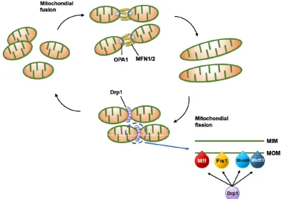
coupled [18-20]. We describe here the functions of proteins involved in mitochondrial fusion and
50
fission (Fig. 1).
51
52
53
Figure 1. Schematic illustration of the mitochondrial dynamics. Mitochondria dynamically change
54
their morphology through the cycle of fusion and fission. Main fusion factors are OPA1 and MFN1
55
and MFN2, which bind to the inner membrane (MIM) and outer membrane (MOM) of mitochondria.
56
Drp1 is major fission factor that bind to MOM and form ring-like structure around mitochondria
57
leading to the separation of mitochondria into two. Mff, Fis1, Mid49, and Mid51 function as adaptors
58
to recruit Drp1 to the MOM.
59
60
2.1.
Mitochondrial fission proteins
61
Mitochondrial division in a cell contributes to ensuring proper distribution and quality control
62
of mitochondria, which maintain a cell in a healthy state. A member of the dynamin family of
63
GTPases, dynamin-like 1 (Dnm1), which is also referred to as dynamin related protein 1 (Drp1), is a
64
major player of mitochondrial division [21-23]. Genetic and biochemical studies in yeast have shown
65
that Dnm1-mediated mitochondrial cleavage requires the tail-anchored MOM protein Fis1 and its
66
two adaptor protein Mdv1 or its paralogue Caf4, which connect Dnm1 to Fis1 [6,
24-31]. During
67
mitochondrial division, Fis1 transiently interacts with cytosolic Dnm1 by the
68
tetratricopeptide-repeat motif via the cytosolic adapter protein Mdv1/Caf4, indicating that Fis1
69
functions as a mitochondrial Dnm1 receptor [27]. However, Mdv1 and its homologs (Caf4, Num1,
70
and Mdm36) were not found in mammalian cells, indicating that only two proteins, Dnm1 and Fis1,
71
are conserved in all species that contain mitochondria [32]. Several fission-related proteins have been
72
identified in mammals, but the detailed mechanistic role in mitochondrial fission has not been
73
2.2.
Mitochondrial fission accessory proteins
75
Besides the major mitochondrial fission proteins, mitochondrial fission accessory proteins such
76
as mitochondrial fission factor (Mff), mitochondrial dynamics 49 (MiD49), mitochondrial dynamics
77
51 (MiD51), ganglioside-induced differentiation-associated protein 1 (GDAP1), and endophilins
78
additionally play crucial roles in mitochondrial fission.
79
Mff is a C-terminal-tail immobilized protein recently identified in a Drosophila RNA interference
80
(RNAi) library used to search for mitochondrial morphological changes. Mammalian mitochondria
81
also contain an orthologue of Mff, suggesting that Mff may be involved in the mitochondrial
82
division and fission in mammalian cells [33]. Mff overexpression caused mitochondrial
83
fragmentation, similar to Drp1 overexpression in mammalian cells [33-35]. Consistent with these
84
observations, in vitro and in vivo experiments have demonstrated that Mff transiently interacts with
85
Drp1 through the N-terminal cytoplasmic domain.
86
MiD51 and MiD49 variants, known as mitochondrial elongation factor 1 and 2 (MIEF1/2),
87
respectively, are MOM proteins identified by random cell localization screens of raw proteins that
88
cause unique distribution and changes in mitochondrial morphology [36]. MIEF1/2 form foci and
89
rings around mitochondria and directly recruit cytosolic Drp1 to the mitochondrial outer membrane
90
surface [37], serving as adaptors linking Drp1 and Mff [36]. Therefore, MIEF1/2 was suggested to
91
be a receptor for Drp1 and mediator of mitochondrial division (fission). MIEF1/2 knockdown by
92
RNAi resulted in the reduction of interaction of Drp1 with mitochondria, leading to mitochondrial
93
elongation. Surprisingly, overexpression of MIEF1/2 induced mitochondrial fission by sequestering
94
Drp1 protein activity [36, 37] . Zhao et al., on the other hand, claimed that the knockdown of MIEF1
95
by RNAi induces mitochondrial fragmentation. They concluded that MIEF1 functions as a Drp1
96
suppressor that inhibits GTPase-dependent fission activity of Drp1 and MIEF1 also has a role
97
independent of Mfn2 in the fusion pathway [38]. Given the discrepancy, more research concerning
98
MIEF1/2 has to be carried out.
99
GDAP1 is another mitochondrial division-related factor located on the MOM through the
100
C-terminal hydrophobic transmembrane domain, which pushes the bulk N-terminal domain to the
101
cytoplasm [39]. It is expressed in myelinating Schwann cells and motor and sensory neurons [40].
102
The GDAP1 mutation induced progression to peripheral nerve injury Charcot-Marie-Tooth disease,
103
with primary axonal damage and primary dehydration of the peripheral nerve [41]. GDAP1 mutants
104
found in patients with the Charcot-Marie-Tooth disease do not target mitochondria and lack
105
mitochondrial cleavage activity [42]. GDAP1-induced mitochondrial fragmentation is inhibited by
106
Drp1 knockdown or the expression of a dominant-negative Drp1-K38A mutation, indicating that
107
GDAP1 is a Drp1-dependent modulator of mitochondrial division [43].
108
Endophilins, fatty acyl transferases, were proposed to mediate membrane curvature changes
109
and participate in membrane cleavage during endocytosis and intracellular organelle biogenesis
110
[44]. They have an N-terminal Bar domain interacting with the membrane and a C-terminal SH3
111
domain mediating protein binding [45-48]. Endophilin B1 (also called Endo B1, Bif-1) was identified
112
by a yeast two-hybrid protein screen to bind to Bax, a proapoptotic Bcl-2 family member, and was
113
reported to be involved in apoptosis, mitochondrial morphogenesis, and autophagosome formation
114
[49-52].
115
2.3.
Mitochondrial fusion proteins
116
At the molecular level, mitochondrial fusion is a two-step process that requires coordinated
117
sequential fusion of the MOM and MIM [53-55]. In mammals, this process relies on the unique
118
mitochondrial sub-localization of the three fusion-related proteins: the MOM-located mitofusin 1
119
The mitofusin proteins Mfn1 and Mfn2 belong to the ubiquitous transmembrane GTPase
121
family, which is conserved from yeast to humans [57, 58]. Mfn1 and Mfn2 share about 80% similarity
122
and show the same structural motifs [15]. Their amino terminal GTPase domain contains five motifs,
123
each of which plays an important role in GTP binding and hydrolysis [59]. Notably, the proline-rich
124
region (PR) involved in protein-protein interactions is found only in Mfn2. Mfn1 and Mfn2
125
double-knockout (DKO) mice die prematurely during pregnancy due to insufficient mitochondrial
126
fusion in the placenta [17,
60]. Interestingly, double-mutant embryos die without any visible
127
developmental defect, suggesting the non-redundant function of Mfn1 and Mfn2 in embryonic
128
development. Indeed, Mfn1 mediates mitochondrial docking and fusion more efficiently than Mfn2,
129
presumably due to its high GTPase activity [61]. Furthermore, Mfnl is required to mediate
130
Opa1-induced mitochondrial fusion, but not Mfn2 [19].
131
Opa1 is also a dynamin family GTPase that promotes IMM fusion following OMM fusion [18,
132
62]. Cryo-immunogold EM analysis revealed that Opa1 is a mitochondrial intermembrane space
133
protein [63]. Opa1 function is controlled in part by proteolysis, by which Opa1 is cleaved and
134
mitochondrial fusion is blocked [64, 65]. Proteolytic inactivation of Opa1 could induce the change of
135
mitochondrial morphology, such as swelling and constriction of mitochondrial tubules and swollen
136
cristae [63]. In addition, Opa1 was suggested to help maintain cristae morphology like Mitofilin and
137
ATP synthase [66]. As cristae shape is important for the assembly of respiratory chain complexes
138
and respiratory efficiency, Opa1 may be essential for the proper assembly and function of the
139
electron transport supercomplex [20, 67].
140
2.4.
Mitochondrial fusion accessory proteins
141
Besides the three major mitochondrial fusion proteins, some accessory proteins, such as PINK1
142
(protein phosphatase and tensin homolog (PTEN)-induced kinase 1) and PARKIN, could affect the
143
mitochondrial fusion machinery. PINK1 is a ubiquitin kinase that phosphorylates ubiquitin and
144
subsequently activate the ubiquitin ligase PARKIN. PINK1 and PARKIN have been suggested as
145
inducing factors for mitophagy. When PINK1 is stabilized on the MOM of malfunctioning
146
mitochondria, PINK1 recruits ubiquitin E3 ligase kinase and autophagy receptors, which leads to
147
autophagosome biogenesis and subsequent catabolism by lysosomes [68]. PINK1 protein or PINK1
148
kinase has little activity in normal mitochondria [69]. However, when depolarization occurs in the
149
mitochondria, the PINK1 process is stopped and PINK1 accumulates and phosphorylates the
150
substrate proteins [70]. Healthy mitochondria actively degrade PINK1 to prevent mitophagic
151
destruction. However, damaged mitochondria could no longer trigger PINK1 degradation, and
152
resulted in the accumulation of PINK1 in mitochondria followed by the mitotic destruction of the
153
organelle [71].
154
Parkinson's disease can be caused by a mutation in Pink1 or Parkin, which may lead to the
155
accumulation of damaged mitochondria in neurons. Ultimately, damaged mitochondria in patients
156
with Parkinson's disease can kill cells through ROS or other toxic substances in dopaminergic
157
neurons [72].
158
PARKIN (also known as PARK2) is an E3 ubiquitin ligase recruited to MOM by PINK1 [73].
159
PARKIN ubiquitinates several mitochondrial proteins to stimulate mitophagy [74]. The
160
PARKIN-mediated mitophagy is also linked to mitochondrial fission because mitochondrial
161
fragmentation is essential for engulfment of mitochondria by autophagosomes [75]. PINK-PARKIN
162
pathway plays an important role in mitochondrial fusion [76,
77] though detailed mechanism
163
remains poorly understood. Mitochondria can structurally and functionally contact with other
164
intracellular organelles. Endoplasmic reticulum (ER) and mitochondria can communicate with each
165
other through mitochondria-associated membranes (MAMs). The MAM is an important regulator of
166
mitochondrial and cell functions, such as mitochondrial division, apoptosis, lipid and Ca2
+167
3. Cellular metabolism and mitochondrial dynamics
169
The well-known function of mitochondria is the production of energy in the form of ATP via
170
oxidative phosphorylation (OXPHOS), which occurs in the mitochondrial cristae [79]. Besides, the
171
diverse functions of mitochondria are intimately related with their morphology. However, the
172
relationships between mitochondrial dynamics and cellular metabolism are generally veiled because
173
of the complex mechanisms involved, involvement of multiple factors across the cellular
174
environment, cell type variation, and differences between metabolic cues [80]. This is evident from
175
the challenges faced by many researchers in identifying the machinery of mitochondrial dynamics
176
and metabolism in different cell types; many pioneering studies on mitochondrial dynamics from
177
yeast have tried to address these challenges.
178
3.1.
The mitochondrial morphologies and energy metabolism in various stem cells
179
Well-developed mitochondria are generally thought to produce energy, or ATP, more
180
efficiently than the immature, globular mitochondria. Since well-developed mitochondria have
181
complex cristae structures, they have a greater surface area to accommodate a larger number of
182
inter-membrane proteins for energy production [81]. In fact, some reports showed that fused and
183
interconnected mitochondrial structures are found in cells that depend mainly on OXPHOS for
184
energy production [82]. However, the cells which have non-fused spherical (immature form)
185
mitochondria have a tendency to produce energy mainly via glycolytic metabolism [83]. Therefore,
186
cell types containing poorly developed mitochondria mainly have OXPHOS-independent
187
metabolism. However, there are some exceptions to this, as seen in various stem cell types (Table. 1).
188
Actively proliferating cells, such as stem cells and cancer cells, use aerobic glycolysis for energy
189
production.
190
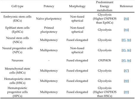
Table 1. Mitochondrial morphology and energy metabolism in various cell types
191
Cell type
Potency
Morphology
Predominant
Energy
metabolism
Reference
Embryonic stem cells
(ESCs)
Naïve pluripotency
Non-fused
spherical
Glycolysis
(Higher OXPHOS
than EpiSCs)
[84]
Epiblast stem cells
(EpiSCs)
Primed
pluripotency
Non-fused
spherical
Glycolysis [84]
Neural stem cells
(NSCs)
Multipotency Fused
elongated Glycolysis [85, 86]
Neural progenitor cells
(NPCs)
Multipotency
Non-fused
spherical
Glycolysis [85, 86]
Neurons -
Fused
elongated
OXPHOS
[85, 86]
Mesenchymal stem
cells (MSCs)
Multipotency Fused
elongated Glycolysis
[87]
Hematopoietic stem
cells (HSCs)
Multipotency Fused
elongated Glycolysis
[88]
Hematopoietic
progenitor cells
(HPCs)
Multipotency Fused
elongated
Glycolysis
(Higher OXPHOS
than HSCs)
[89]
There are two types of pluripotent stem cells (PSCs), naïve and primed PSCs. Naïve PSCs, such
193
as mouse embryonic stem cells (mESCs), are the in vitro counterparts of inner cell mass (ICM) of
194
preimplantation blastocyst and primed PSCs, such as mouse epiblast stem cells (mEpiSCs), are the in
195
vitro counterparts of epiblast of postimplantation embryos. The morphology of mEpiSCs is more
196
tubular and fused shape compared to naïve pluripotent state mESCs, but mESCs showed higher
197
OXPHOS activity than mEpiSCs [84].
198
Moreover, embryonic mouse NSCs have been found to depend on aerobic glycolytic
199
metabolism though they have a relatively fused mitochondrial network [86, 90]. On the other hand,
200
neurons terminally differentiated from NSCs rely on OXPHOS for energy metabolism, even if they
201
have a fused mitochondrial network similar to NSCs. Meanwhile, mouse NPCs whose
202
differentiation state is in between NSCs and neurons, had more fragmented mitochondria compared
203
with NSCs and neurons; however, they utilized aerobic glycolysis for major energy metabolism [86].
204
Metabolic shift from glycolysis to OXPHOS during the differentiation of NSCs occurred around the
205
time of transition from NSCs to intermediate progenitor cells [85].
206
MSCs, which have multipotent differentiation potential to all blood cell types, have
207
glycolysis-dependent energy metabolism [87]. They showed the relatively more tubular shape of
208
mitochondria that can further elongate upon differentiation, as observed in NSC differentiation [91].
209
HSCs mainly use glycolysis for energy metabolism [88]. However, further differentiated HPCs
210
were suggested to be more OXPHOS-dependent than HSCs [89]. This phenomenon might be a
211
response of HSCs in the hypoxic environment of bone marrow to limit the production of ROS from
212
the respiratory chain complexes in mitochondria [92]. Recently, Luchsinger et al. showed that the
213
differentiation of HSCs was accompanied by mitochondrial OXPHOS activation. HSCs express more
214
Mfn2 than differentiated hematopoietic lineages, indicating that they contain elongated
215
mitochondria as Mfn2 expression level is correlated with mitochondrial length [93].
216
3.2.
Warburg effect: survival strategy of proliferative glycolytic cells
217
As described above, most adult stem cells mainly used aerobic glycolysis for ATP production.
218
This kind of phenomenon found in stem cells is known as “Warburg effect” which was first
219
described in cancer cells [94]. Most cancer cells produce energy through a high rate of glycolysis
220
even when there is sufficient oxygen supply, a phenomenon termed the “Warburg effect”. The
221
precise mechanism of the Warburg effect remains unknown. This phenomenon was payed attention
222
in the process of cellular reprogramming, or induced pluripotent stem cell (iPSC) generation [95]
223
and metabolic switch from OXPHOS in mouse embryonic fibroblasts (MEFs) to glycolysis in
224
reprogrammed iPSCs. This phenomenon is commonly observed in various kinds of cancers which
225
display a highly proliferative state. Then, this raises the question of why proliferating cells choose an
226
inefficient pathway to produce energy? Cell division requires not only energy but also various kinds
227
of cellular constituents, such as nucleotides, amino acids, and lipids. Glycolysis along with pentose
228
phosphate pathway can account for cellular constituents as well as ATP [96]. Reduction in
229
mitochondrial metabolism may also allow in maintaining a low level of harmful free radicals such as
230
ROS. Therefore, glycolysis would be beneficial to the actively proliferating stem cells to self-renew
231
and maintain cell states [97].
232
3.3.
Metabolic regulation in mitochondria for the maintenance of pluripotent state
233
In addition to the energy metabolism, mitochondria also play a crucial role in the stemness of
234
PSCs. For example, PSCs showed a high level of uncoupling protein 2 (UCP2) protein [98], which is
235
located in the membrane between inter-membrane space and matrix, and functioned as metabolites
236
transportation to the out of the mitochondria, thereby regulating glucose and glutamine oxidation
237
Zhang
et al. reported that although glycolysis supported stemness of human PSCs under all
239
conditions, oxidative mitochondrial metabolism was also highly active in human PSCs when they
240
were cultured in a media containing lipid supplements [100]. This may highlight the importance of
241
cellular environment or culture condition, which could affect mitochondrial function and related
242
mitochondrial morphology in human PSCs.
243
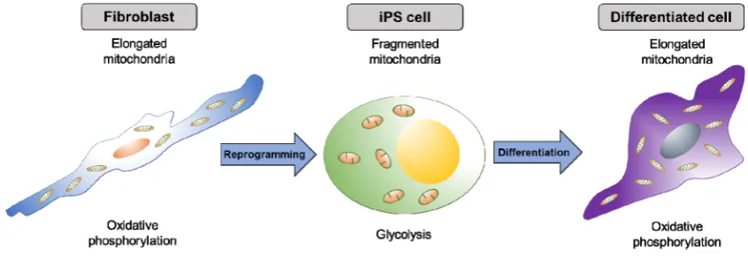
4. Mitochondrial dynamics in the reprogramming process
244
The proteins related to mitochondrial dynamics, such as fusion and fission, are interestingly
245
crucial for pluripotential reprogramming. During reprogramming (iPSC generation) and
246
re-differentiation of iPSCs, mitochondrial morphology dynamically changed (Fig. 2); mitochondria
247
became elongated during reprogramming and became globular-shaped after re-differentiation into a
248
neural lineage [101]. As the mitochondrial morphology changes dynamically during the process of
249
reprogramming, the metabolic profile switches from OXPHOS to glycolysis [101-104]. Several
250
studies suggested that the mitochondrial dynamics and energy metabolism are critical for the
251
reprogramming process.
252
253
Figure 2. Dynamic change of mitochondrial shape during the reprogramming and differentiation.
254
Elongated mitochondria in differentiated cells become spherical shape during the formation of iPS
255
cells. As iPS cells differentiate, mitochondria resort back to the elongated shape.
256
257
4.1.
Mitochondrial fission proteins affect pluripotential reprogramming
258
Here we will discuss how the mitochondrial fission proteins affect the reprogramming process.
259
Mdivi-1 treatment, which inhibits mitochondrial fission protein DRP1, was sufficient to suppress the
260
early stage of reprogramming of somatic cells [105]. Moreover, iPSCs lost pluripotency when
261
exposed to Mdivi-1, indicating that mitochondrial fission is important for gaining and maintaining
262
pluripotency.
263
Reduced expression 1 (REX1), which function in the maintenance of pluripotency, induces
264
phosphorylation of DRP1 at Ser616 and mitochondrial fission [106]. On the other hand, the
265
inhibition of the oncogenic mitogen-activated protein kinase (MAPK) cascade leads to robust
266
mitochondrial fusion via the loss of phosphorylation in DRP1 serine 616 through ERK1/2 protein
267
[107]. Therefore, Drp1 phosphorylation by ERK pathway is necessary for the pluripotential
268
reprogramming process [108]. Furthermore, inhibition of the accessory proteins, such as Gdap1,
269
Mid51, and Mff, which control Drp1 recruitment to the mitochondria, suppressed reprogramming
270
due to impaired mitochondrial fission. In particular, Gdap1-null cells displayed G2/M growth arrest
271
in cells undergoing reprogramming and affected the early phase in reprogramming [109].
272
Collectively, change in mitochondrial dynamics and cell cycle are crucial factors for efficient
273
4.2.
Mitochondrial fusion proteins affect pluripotential reprogramming
275
Mitochondrial fusion proteins can also affect reprogramming efficiency through a different
276
pathway from that of fission proteins. Son et al. revealed that depletion of mitochondrial fusion
277
proteins, such as Mfn1 and Mfn2, increased efficiency of somatic cell reprogramming into iPSCs as
278
well as maintained pluripotency [110]. They also showed that Mfn1 and Mfn2 depletion facilitates
279
the transition of OXPHOS to glycolytic metabolism, because Mfn1 and Mfn2 are inhibitors of
280
reprogramming as they directly bind to Ras and Raf and thus inhibit cell proliferation. Inhibition of
281
Mfn1 and Mfn2 also activated ROS-mediated hypoxia-inducible factor 1
α
(HIF1
α
) signaling at an
282
early stage and established the reprogramming favorable hypoxic condition [110].
283
5. Mitochondrial dynamics in the differentiation process
284
During the process of PSC differentiation, changes in mitochondrial morphology and
285
metabolite composition are essential among the various differentiated cell types [111]. As PSCs
286
mainly use glycolysis, and differentiated cells use OXPHOS for ATP production, respectively,
287
inhibition of mitochondrial OXPHOS during the differentiation of PSCs leads to defect in
288
differentiation and instead, supports the maintenance of pluripotency. In line with this, the proteins
289
related to mitochondrial dynamics also play a crucial role in the differentiation process.
290
5.1.
Mitochondrial fission proteins affect cellular differentiation
291
Several reports showed that Drp1-dependent mitochondrial fission is crucial for embryonic and
292
cellular differentiation in vivo and in vitro. Drp1-null mice showed defective trophoblast giant cells
293
and decreased cardiomyocyte beat rates and died around 11.5 dpc; however they showed normal
294
levels of intracellular ATP [112,
113]. Conditional knockout of Drp1 showed defect in cerebella
295
during postnatal development. Neural cell-specific Drp1 knockout mice displayed brain hypoplasia
296
and
in vitro culture of the forebrain showed a reduction in the number of neurites and abnormal
297
synapse formation [112, 113]. However, heterozygote knockout of Drp1 did not affect mitochondrial
298
and synaptic viability [114]. Kim et al. showed that inhibiting Drp1 activity induced morphological
299
change of migratory adult NSCs, which causes abnormal migration and prevention of neuronal
300
differentiation in the NSCs [115].
301
During cellular maturation, mitochondrial localization and distribution is under the control of
302
cellular states, such as cell division, migration etc. Therefore, the localization of mitochondria is
303
dynamically regulated during neuronal maturation and myogenic differentiation [116]. During
304
neuronal differentiation, mitochondria accumulate in the regions where high energy is required
305
such as growth cones at the early stage of differentiation and then localize at presynaptic terminals
306
following neuron maturation [115]. In myogenic differentiation, NO/cGMP control Drp1 localization
307
and activity and stimulate myogenesis through inhibition of Drp1-dependent mitochondrial fission
308
[116]. Furthermore, Mdivi-1 mediated Drp1 inhibition suppressed expression levels of key myogenic
309
regulatory factors (MRFs) such as MyoD and Myogenin in differentiating C2C12 cells, a mouse
310
muscle myoblast [117]. Likewise, myogenic differentiation of C2C12 myoblasts required
311
Drp1-mediated mitophagy [118]. Recent report also suggested that knock-down or inhibition of
312
Drp1 by using Mdivi-1 promotes differentiation into cardiac mesoderm lineage from human PSCs
313
[119].
314
Fis1 function in stem cells and differentiation has only been reported recently. Pei et al. showed
315
that gene expression level of Fis1 was specifically high in leukemia stem cells and it functions as a
316
crucial mitochondrial morphology regulator [120]. They also showed that loss of Fis1 impairs
317
mitochondrial dynamics and induces myeloid differentiation in acute myeloid leukemia.
318
5.2.
Mitochondrial fusion proteins affect cellular differentiation
320
Besides mitochondrial fission, mitochondrial fusion additionally executes crucial roles in
321
cellular differentiation process, especially in cardiac, neural, and mesenchymal differentiation. The
322
deletion of Mfn1 and Mfn2 in the mouse embryonic hearts impaired mouse heart development, and
323
ablation of Mfn2 or Opa1 in mouse ESCs resulted in defective cardiac differentiation of the ESCs
324
[121]. Gene expression profiling showed that mitochondrial morphology-related genes interacted
325
with calcineurin to regulate Notch1 signaling that control cardiac differentiation [121]. Similarly,
326
proteins that drive mitochondrial fusion, such as MFN (mitofusin) 1 and 2 and OPA1, are required
327
for the differentiation of stem cells into cells that depend on OXPHOS metabolism, like
328
cardiomyocytes and neurons [121, 122].
329
Mitochondrial dynamics is also involved in MSC differentiation, including adipogenesis,
330
osteogenesis, and chondrogenesis. Mitochondrial elongation (increase in Mfn1 and Mfn2 expression)
331
is correlated with the adipogenesis and osteogenesis, and mitochondrial fragmentation (increased
332
expression of Drp1, Fis1, and Fis2) is involved in condrogenesis. Consequently, knockdown of Mfn2
333
and the overexpression of a dominant negative form of Drp1 resulted in defective differentiation in
334
adipo- and osteogenesis, and chondrogenesis, respectively [91].
335
During the differentiation of human iPSCs into neurons, expression level of Mfn2 increased
336
with time after differentiation [122]. Knockdown of Mfn2 results in mitochondrial dysfunctions,
337
such as downregulated expression of complexes I and IV, and ATP levels, and impaired neuronal
338
differentiation. On the contrary, Mfn2 overexpression in NPCs promotes neuronal differentiation
339
with enhanced mitochondrial bioenergetics and functions. Taken together, many studies have
340
shown that mitochondrial fusion and fission play crucial roles in various cellular differentiation
341
processes through the control of bioenergetics, signaling pathways, or expression of tissue-specific
342
genes.
343
6. The Mitochondrial dynamics in aging
344
Mitochondria also play an important role in cellular aging and cell death associated with
345
necrosis, apoptosis, autophagy, and mitophagy through the modulation of redox by reduction
346
reaction mechanisms [123, 124]. The process of aging may be associated with the accumulation of
347
damages, such as production of metabolic by-products and ROS, accumulation of biological waste
348
products, telomere shortening, and dysregulation of metabolic pathways [125-127]. Most of these
349
aging factors are associated with mitochondrial dynamics and functions, indicating the close
350
relationship between aging and mitochondria.
351
6.1.
Mitochondrial ROS impact on cellular senescence-related mitochondrial dynamics
352
Abnormally elongated mitochondria are often observed in various senescent cells, implying
353
that mitochondrial dynamics may have a functional role in cell senescence and aging. This
354
phenomenon could be caused by the alteration of expression patterns of genes associated with
355
mitochondrial fission, such as Drp1 and Fis1, and with mitochondrial fusion, such as Mfn1 and
356
Mfn2. Mai et al. reported that the senescent human endothelial cells (HUVECs) showed reduced
357
expression levels of DRP1 and FIS1 that caused long interconnected mitochondria [128]. The loss of
358
DRP1 exacerbated endothelial cell dysfunction by inhibiting autophagic flux accompanied by
359
increasing mitochondrial ROS [129]. In addition, the regulation of ROS by mitochondrial fission is
360
dependent on protein disulfide isomerase A1 (PDIA1) in mouse endothelial cells; PDIA1-depleted
361
endothelial cells activated mitochondrial fission [130]. Recently, Leduc-Gaudet et al. also suggested
362
that the levels of mitochondrial dynamics-related proteins, including Mfn1, Mfn2, Opa1, and Drp1,
363
were not significantly different between young and aged skeletal muscles [131]. However, the ratio
364
cells; as skeletal muscle cells grew older, the ratio of Mfn2/Drp1 significantly increased [131]. On the
366
other hand, Debastian et al. suggested that the expression level of Mfn2 in skeletal muscle decreased
367
during aging [132]. Mfn2 deficiency in skeletal muscle caused the reduction of mitochondrial
368
respiration and elevation of oxidative stress, which was accompanied by the activation of the
369
transcription factor, HIF1
α
, to minimize the accumulation of damaged mitochondria [132]. Thus,
370
Mfn2 functions as a regulator in mitophagy and consequently controls the mitochondrial quality
371
control pathway. Moreover, the stress-responsive mitochondrial protein, Sirt4 was suggested to
372
have implications in aging. Similar to Mfn2-functioning in mitophagy, Sirt4 could promote
373
mitochondrial fusion by interacting with Opa1 and reduce mitophagy [133]. Overall, various aging
374
factors are interconnected, and of these, mitochondrial dysfunction and dynamics are the underlying
375
mechanism of cellular aging.
376
7. Conclusions
377
In this review, we have discussed the widespread involvement of mitochondria in various
378
cellular processes such as cell survival, cell cycle, proliferation, differentiation, reprogramming,
379
aging, and energy metabolism. The variety of functions carried out by mitochondria implicates that
380
the normal mitochondrial dynamics controlled by mitochondrial fusion/fission are critical for
381
human health. Clinically, defect in mitochondrial fusion/fission causes diseases including optic
382
atrophy, Charcot-Marie-Tooth disease, Parkinson's disease, and Alzheimer's disease [134-139]. Thus,
383
further understanding of mitochondrial dynamics is of paramount importance in elucidating
384
mechanisms of diseases at the cellular level and discovering novel therapies to cure associated
385
diseases.
386
387
Author Contributions: B.J.S., S.H.Y., and J.T.D. wrote the paper.
388
Funding: This research was supported by the Basic Science Research Program through the National Research
389
Foundation of Korea (NRF) funded by the Ministry of Science, ICT and Future Planning of the Republic of
390
Korea (grant nos. 2016M3A9B6946835 and 2015R15A1009701). This paper was written as part of Konkuk
391
University’s research support program for its faculty on sabbatical leave in 2018.
392
Conflicts of Interest: The authors declare no conflict of interest.
393
Abbreviations
394
MOM MIM
Mitochondrial outer membrane Mitochondrial inner membrane
MAM Mitochondria-associated membrane OXPHOS Oxidative phosphorylation
ESC Embryonic stem cell EpiSC Epiblast stem cell
NSC Neural stem cell
NPC Neural progenitor cell
MSC Mesenchymal stem cell
HSC Hematopoietic stem cell
HPC Hematopoietic progenitor cell PSC Pluripotent stem cell
iPSC Induced pluripotent stem cell MEF Mouse embryonic fibroblast HUVEC Human endothelial cell
References
395
1. Dunwoodie, S. L., The role of hypoxia in development of the Mammalian embryo. Developmental cell 2009,
396
2. Morrow, R. M.; Picard, M.; Derbeneva, O.; Leipzig, J.; McManus, M. J.; Gouspillou, G.; Barbat-Artigas, S.;
398
Dos Santos, C.; Hepple, R. T.; Murdock, D. G.; Wallace, D. C., Mitochondrial energy deficiency leads to
399
hyperproliferation of skeletal muscle mitochondria and enhanced insulin sensitivity. Proceedings of the National
400
Academy of Sciences of the United States of America 2017, 114, (10), 2705-2710.
401
3. Frezza, C., Mitochondrial metabolites: undercover signalling molecules. Interface focus 2017, 7, (2),
402
20160100.
403
4. Chen, H.; Chan, D. C., Mitochondrial dynamics--fusion, fission, movement, and mitophagy--in
404
neurodegenerative diseases. Human molecular genetics 2009, 18, (R2), R169-76.
405
5. Lewis, M. R.; Lewis, W. H., Mitochondria (and other cytoplasmic structures) in tissue cultures. Amer. J
406
Anat. 1915, 17, (3).
407
6. Okamoto, K.; Shaw, J. M., Mitochondrial morphology and dynamics in yeast and multicellular
408
eukaryotes. Annual review of genetics 2005, 39, 503-36.
409
7. Rizzuto, R.; Brini, M.; Murgia, M.; Pozzan, T., Microdomains with high Ca2+ close to IP3-sensitive
410
channels that are sensed by neighboring mitochondria. Science 1993, 262, (5134), 744-7.
411
8. Dimmer, K. S.; Scorrano, L., (De)constructing mitochondria: what for? Physiology 2006, 21, 233-41.
412
9. de Brito, O. M.; Scorrano, L., An intimate liaison: spatial organization of the endoplasmic
413
reticulum-mitochondria relationship. The EMBO journal 2010, 29, (16), 2715-23.
414
10. Zuchner, S.; Mersiyanova, I. V.; Muglia, M.; Bissar-Tadmouri, N.; Rochelle, J.; Dadali, E. L.; Zappia, M.;
415
Nelis, E.; Patitucci, A.; Senderek, J.; Parman, Y.; Evgrafov, O.; Jonghe, P. D.; Takahashi, Y.; Tsuji, S.;
416
Pericak-Vance, M. A.; Quattrone, A.; Battaloglu, E.; Polyakov, A. V.; Timmerman, V.; Schroder, J. M.; Vance, J.
417
M., Mutations in the mitochondrial GTPase mitofusin 2 cause Charcot-Marie-Tooth neuropathy type 2A.
418
Nature genetics 2004, 36, (5), 449-51.
419
11. Misko, A. L.; Sasaki, Y.; Tuck, E.; Milbrandt, J.; Baloh, R. H., Mitofusin2 mutations disrupt axonal
420
mitochondrial positioning and promote axon degeneration. The Journal of neuroscience : the official journal of the
421
Society for Neuroscience 2012, 32, (12), 4145-55.
422
12. Chang, C. R.; Blackstone, C., Dynamic regulation of mitochondrial fission through modification of the
423
dynamin-related protein Drp1. Annals of the New York Academy of Sciences 2010, 1201, 34-9.
424
13. Santel, A.; Fuller, M. T., Control of mitochondrial morphology by a human mitofusin. Journal of cell science
425
2001, 114, (Pt 5), 867-74.
426
14. Legros, F.; Lombes, A.; Frachon, P.; Rojo, M., Mitochondrial fusion in human cells is efficient, requires the
427
inner membrane potential, and is mediated by mitofusins. Molecular biology of the cell 2002, 13, (12), 4343-54.
428
15. Santel, A.; Frank, S.; Gaume, B.; Herrler, M.; Youle, R. J.; Fuller, M. T., Mitofusin-1 protein is a generally
429
expressed mediator of mitochondrial fusion in mammalian cells. Journal of cell science 2003, 116, (Pt 13), 2763-74.
430
16. Eura, Y.; Ishihara, N.; Yokota, S.; Mihara, K., Two mitofusin proteins, mammalian homologues of FZO,
431
with distinct functions are both required for mitochondrial fusion. Journal of biochemistry 2003, 134, (3), 333-44.
432
17. Chen, H.; Detmer, S. A.; Ewald, A. J.; Griffin, E. E.; Fraser, S. E.; Chan, D. C., Mitofusins Mfn1 and Mfn2
433
coordinately regulate mitochondrial fusion and are essential for embryonic development. The Journal of cell
434
biology 2003, 160, (2), 189-200.
435
18. Olichon, A.; Baricault, L.; Gas, N.; Guillou, E.; Valette, A.; Belenguer, P.; Lenaers, G., Loss of OPA1
436
perturbates the mitochondrial inner membrane structure and integrity, leading to cytochrome c release and
437
apoptosis. The Journal of biological chemistry 2003, 278, (10), 7743-6.
438
19. Cipolat, S.; Martins de Brito, O.; Dal Zilio, B.; Scorrano, L., OPA1 requires mitofusin 1 to promote
439
mitochondrial fusion. Proceedings of the National Academy of Sciences of the United States of America 2004, 101, (45),
440
15927-32.
441
20. Frezza, C.; Cipolat, S.; Martins de Brito, O.; Micaroni, M.; Beznoussenko, G. V.; Rudka, T.; Bartoli, D.;
442
Polishuck, R. S.; Danial, N. N.; De Strooper, B.; Scorrano, L., OPA1 controls apoptotic cristae remodeling
443
independently from mitochondrial fusion. Cell 2006, 126, (1), 177-89.
444
21. Smirnova, E.; Shurland, D. L.; Ryazantsev, S. N.; van der Bliek, A. M., A human dynamin-related protein
445
controls the distribution of mitochondria. The Journal of cell biology 1998, 143, (2), 351-8.
446
22. Smirnova, E.; Griparic, L.; Shurland, D. L.; van der Bliek, A. M., Dynamin-related protein Drp1 is
447
required for mitochondrial division in mammalian cells. Molecular biology of the cell 2001, 12, (8), 2245-56.
448
23. Ingerman, E.; Perkins, E. M.; Marino, M.; Mears, J. A.; McCaffery, J. M.; Hinshaw, J. E.; Nunnari, J., Dnm1
449
24. Hoppins, S.; Lackner, L.; Nunnari, J., The machines that divide and fuse mitochondria. Annual review of
451
biochemistry 2007, 76, 751-80.
452
25. Mozdy, A. D.; McCaffery, J. M.; Shaw, J. M., Dnm1p GTPase-mediated mitochondrial fission is a
453
multi-step process requiring the novel integral membrane component Fis1p. The Journal of cell biology 2000, 151,
454
(2), 367-80.
455
26. Otsuga, D.; Keegan, B. R.; Brisch, E.; Thatcher, J. W.; Hermann, G. J.; Bleazard, W.; Shaw, J. M., The
456
dynamin-related GTPase, Dnm1p, controls mitochondrial morphology in yeast. The Journal of cell biology 1998,
457
143, (2), 333-49.
458
27. Naylor, K.; Ingerman, E.; Okreglak, V.; Marino, M.; Hinshaw, J. E.; Nunnari, J., Mdv1 interacts with
459
assembled dnm1 to promote mitochondrial division. The Journal of biological chemistry 2006, 281, (4), 2177-83.
460
28. Karren, M. A.; Coonrod, E. M.; Anderson, T. K.; Shaw, J. M., The role of Fis1p-Mdv1p interactions in
461
mitochondrial fission complex assembly. The Journal of cell biology 2005, 171, (2), 291-301.
462
29. Tieu, Q.; Nunnari, J., Mdv1p is a WD repeat protein that interacts with the dynamin-related GTPase,
463
Dnm1p, to trigger mitochondrial division. The Journal of cell biology 2000, 151, (2), 353-66.
464
30. Lackner, L. L.; Nunnari, J. M., The molecular mechanism and cellular functions of mitochondrial division.
465
Biochimica et biophysica acta 2009, 1792, (12), 1138-44.
466
31. Lackner, L. L.; Horner, J. S.; Nunnari, J., Mechanistic analysis of a dynamin effector. Science 2009, 325,
467
(5942), 874-7.
468
32. Otera, H.; Ishihara, N.; Mihara, K., New insights into the function and regulation of mitochondrial fission.
469
Biochimica et biophysica acta 2013, 1833, (5), 1256-68.
470
33. Otera, H.; Wang, C.; Cleland, M. M.; Setoguchi, K.; Yokota, S.; Youle, R. J.; Mihara, K., Mff is an essential
471
factor for mitochondrial recruitment of Drp1 during mitochondrial fission in mammalian cells. The Journal of
472
cell biology 2010, 191, (6), 1141-58.
473
34. Otera, H.; Mihara, K., Molecular mechanisms and physiologic functions of mitochondrial dynamics.
474
Journal of biochemistry 2011, 149, (3), 241-51.
475
35. Otera, H.; Mihara, K., Discovery of the membrane receptor for mitochondrial fission GTPase Drp1. Small
476
GTPases 2011, 2, (3), 167-172.
477
36. Yu, R.; Liu, T.; Jin, S. B.; Ning, C.; Lendahl, U.; Nister, M.; Zhao, J., MIEF1/2 function as adaptors to recruit
478
Drp1 to mitochondria and regulate the association of Drp1 with Mff. Scientific reports 2017, 7, (1), 880.
479
37. Palmer, C. S.; Osellame, L. D.; Laine, D.; Koutsopoulos, O. S.; Frazier, A. E.; Ryan, M. T., MiD49 and
480
MiD51, new components of the mitochondrial fission machinery. EMBO reports 2011, 12, (6), 565-73.
481
38. Zhao, J.; Liu, T.; Jin, S.; Wang, X.; Qu, M.; Uhlen, P.; Tomilin, N.; Shupliakov, O.; Lendahl, U.; Nister, M.,
482
Human MIEF1 recruits Drp1 to mitochondrial outer membranes and promotes mitochondrial fusion rather
483
than fission. The EMBO journal 2011, 30, (14), 2762-78.
484
39. Wagner, K. M.; Ruegg, M.; Niemann, A.; Suter, U., Targeting and function of the mitochondrial fission
485
factor GDAP1 are dependent on its tail-anchor. PloS one 2009, 4, (4), e5160.
486
40. Pedrola, L.; Espert, A.; Valdes-Sanchez, T.; Sanchez-Piris, M.; Sirkowski, E. E.; Scherer, S. S.; Farinas, I.;
487
Palau, F., Cell expression of GDAP1 in the nervous system and pathogenesis of Charcot-Marie-Tooth type 4A
488
disease. Journal of cellular and molecular medicine 2008, 12, (2), 679-89.
489
41. Cassereau, J.; Chevrollier, A.; Bonneau, D.; Verny, C.; Procaccio, V.; Reynier, P.; Ferre, M., A locus-specific
490
database for mutations in GDAP1 allows analysis of genotype-phenotype correlations in Charcot-Marie-Tooth
491
diseases type 4A and 2K. Orphanet journal of rare diseases 2011, 6, 87.
492
42. Niemann, A.; Ruegg, M.; La Padula, V.; Schenone, A.; Suter, U., Ganglioside-induced differentiation
493
associated protein 1 is a regulator of the mitochondrial network: new implications for Charcot-Marie-Tooth
494
disease. The Journal of cell biology 2005, 170, (7), 1067-78.
495
43. Hakomori, S.; Igarashi, Y., Functional role of glycosphingolipids in cell recognition and signaling. Journal
496
of biochemistry 1995, 118, (6), 1091-103.
497
44. Modregger, J.; Schmidt, A. A.; Ritter, B.; Huttner, W. B.; Plomann, M., Characterization of Endophilin B1b,
498
a brain-specific membrane-associated lysophosphatidic acid acyl transferase with properties distinct from
499
endophilin A1. The Journal of biological chemistry 2003, 278, (6), 4160-7.
500
45. Hinshaw, J. E., Dynamin and its role in membrane fission. Annual review of cell and developmental biology
501
46. Schmidt, A.; Wolde, M.; Thiele, C.; Fest, W.; Kratzin, H.; Podtelejnikov, A. V.; Witke, W.; Huttner, W. B.;
503
Soling, H. D., Endophilin I mediates synaptic vesicle formation by transfer of arachidonate to lysophosphatidic
504
acid. Nature 1999, 401, (6749), 133-41.
505
47. Gad, H.; Ringstad, N.; Low, P.; Kjaerulff, O.; Gustafsson, J.; Wenk, M.; Di Paolo, G.; Nemoto, Y.; Crun, J.;
506
Ellisman, M. H.; De Camilli, P.; Shupliakov, O.; Brodin, L., Fission and uncoating of synaptic clathrin-coated
507
vesicles are perturbed by disruption of interactions with the SH3 domain of endophilin. Neuron 2000, 27, (2),
508
301-12.
509
48. Huttner, W. B.; Schmidt, A., Lipids, lipid modification and lipid-protein interaction in membrane
510
budding and fission--insights from the roles of endophilin A1 and synaptophysin in synaptic vesicle
511
endocytosis. Current opinion in neurobiology 2000, 10, (5), 543-51.
512
49. Cuddeback, S. M.; Yamaguchi, H.; Komatsu, K.; Miyashita, T.; Yamada, M.; Wu, C.; Singh, S.; Wang, H.
513
G., Molecular cloning and characterization of Bif-1. A novel Src homology 3 domain-containing protein that
514
associates with Bax. The Journal of biological chemistry 2001, 276, (23), 20559-65.
515
50. Takahashi, Y.; Karbowski, M.; Yamaguchi, H.; Kazi, A.; Wu, J.; Sebti, S. M.; Youle, R. J.; Wang, H. G., Loss
516
of Bif-1 suppresses Bax/Bak conformational change and mitochondrial apoptosis. Molecular and cellular biology
517
2005, 25, (21), 9369-82.
518
51. Karbowski, M.; Jeong, S. Y.; Youle, R. J., Endophilin B1 is required for the maintenance of mitochondrial
519
morphology. The Journal of cell biology 2004, 166, (7), 1027-39.
520
52. Takahashi, Y.; Coppola, D.; Matsushita, N.; Cualing, H. D.; Sun, M.; Sato, Y.; Liang, C.; Jung, J. U.; Cheng,
521
J. Q.; Mule, J. J.; Pledger, W. J.; Wang, H. G., Bif-1 interacts with Beclin 1 through UVRAG and regulates
522
autophagy and tumorigenesis. Nature cell biology 2007, 9, (10), 1142-51.
523
53. Scorrano, L., Keeping mitochondria in shape: a matter of life and death. European journal of clinical
524
investigation 2013, 43, (8), 886-93.
525
54. Malka, F.; Guillery, O.; Cifuentes-Diaz, C.; Guillou, E.; Belenguer, P.; Lombes, A.; Rojo, M., Separate
526
fusion of outer and inner mitochondrial membranes. EMBO reports 2005, 6, (9), 853-9.
527
55. Song, Z.; Ghochani, M.; McCaffery, J. M.; Frey, T. G.; Chan, D. C., Mitofusins and OPA1 mediate
528
sequential steps in mitochondrial membrane fusion. Molecular biology of the cell 2009, 20, (15), 3525-32.
529
56. Olichon, A.; Emorine, L. J.; Descoins, E.; Pelloquin, L.; Brichese, L.; Gas, N.; Guillou, E.; Delettre, C.;
530
Valette, A.; Hamel, C. P.; Ducommun, B.; Lenaers, G.; Belenguer, P., The human dynamin-related protein
531
OPA1 is anchored to the mitochondrial inner membrane facing the inter-membrane space. FEBS letters 2002,
532
523, (1-3), 171-6.
533
57. Mozdy, A. D.; Shaw, J. M., A fuzzy mitochondrial fusion apparatus comes into focus. Nature reviews.
534
Molecular cell biology 2003, 4, (6), 468-78.
535
58. de Brito, O. M.; Scorrano, L., Mitofusin 2: a mitochondria-shaping protein with signaling roles beyond
536
fusion. Antioxidants & redox signaling 2008, 10, (3), 621-33.
537
59. Bourne, H. R.; Sanders, D. A.; McCormick, F., The GTPase superfamily: conserved structure and
538
molecular mechanism. Nature 1991, 349, 117.
539
60. Chen, H.; McCaffery, J. M.; Chan, D. C., Mitochondrial fusion protects against neurodegeneration in the
540
cerebellum. Cell 2007, 130, (3), 548-62.
541
61. Ishihara, N.; Eura, Y.; Mihara, K., Mitofusin 1 and 2 play distinct roles in mitochondrial fusion reactions
542
via GTPase activity. Journal of cell science 2004, 117, (Pt 26), 6535-46.
543
62. Mishra, P.; Carelli, V.; Manfredi, G.; Chan, D. C., Proteolytic cleavage of Opa1 stimulates mitochondrial
544
inner membrane fusion and couples fusion to oxidative phosphorylation. Cell metabolism 2014, 19, (4), 630-41.
545
63. Griparic, L.; van der Wel, N. N.; Orozco, I. J.; Peters, P. J.; van der Bliek, A. M., Loss of the intermembrane
546
space protein Mgm1/OPA1 induces swelling and localized constrictions along the lengths of mitochondria. The
547
Journal of biological chemistry 2004, 279, (18), 18792-8.
548
64. Ehses, S.; Raschke, I.; Mancuso, G.; Bernacchia, A.; Geimer, S.; Tondera, D.; Martinou, J. C.; Westermann,
549
B.; Rugarli, E. I.; Langer, T., Regulation of OPA1 processing and mitochondrial fusion by m-AAA protease
550
isoenzymes and OMA1. The Journal of cell biology 2009, 187, (7), 1023-36.
551
65. Head, B.; Griparic, L.; Amiri, M.; Gandre-Babbe, S.; van der Bliek, A. M., Inducible proteolytic
552
inactivation of OPA1 mediated by the OMA1 protease in mammalian cells. The Journal of cell biology 2009, 187,
553
66. Rabl, R.; Soubannier, V.; Scholz, R.; Vogel, F.; Mendl, N.; Vasiljev-Neumeyer, A.; Korner, C.; Jagasia, R.;
555
Keil, T.; Baumeister, W.; Cyrklaff, M.; Neupert, W.; Reichert, A. S., Formation of cristae and crista junctions in
556
mitochondria depends on antagonism between Fcj1 and Su e/g. The Journal of cell biology 2009, 185, (6), 1047-63.
557
67. Cogliati, S.; Frezza, C.; Soriano, M. E.; Varanita, T.; Quintana-Cabrera, R.; Corrado, M.; Cipolat, S.; Costa,
558
V.; Casarin, A.; Gomes, L. C.; Perales-Clemente, E.; Salviati, L.; Fernandez-Silva, P.; Enriquez, J. A.; Scorrano, L.,
559
Mitochondrial cristae shape determines respiratory chain supercomplexes assembly and respiratory efficiency.
560
Cell 2013, 155, (1), 160-71.
561
68. Lazarou, M.; Sliter, D. A.; Kane, L. A.; Sarraf, S. A.; Wang, C.; Burman, J. L.; Sideris, D. P.; Fogel, A. I.;
562
Youle, R. J., The ubiquitin kinase PINK1 recruits autophagy receptors to induce mitophagy. Nature 2015, 524,
563
(7565), 309-314.
564
69. Narendra, D. P.; Jin, S. M.; Tanaka, A.; Suen, D.-F.; Gautier, C. A.; Shen, J.; Cookson, M. R.; Youle, R. J.,
565
PINK1 Is Selectively Stabilized on Impaired Mitochondria to Activate Parkin. PLOS Biology 2010, 8, (1),
566
e1000298.
567
70. Dorn, G. W., 2nd; Kitsis, R. N., The mitochondrial dynamism-mitophagy-cell death interactome: multiple
568
roles performed by members of a mitochondrial molecular ensemble. Circulation research 2015, 116, (1), 167-82.
569
71. Youle, R. J.; Narendra, D. P., Mechanisms of mitophagy. Nature Reviews Molecular Cell Biology 2010, 12, 9.
570
72. Chinta, S. J.; Andersen, J. K., Redox imbalance in Parkinson's disease. Biochimica et biophysica acta 2008,
571
1780, (11), 1362-7.
572
73. Panicker, N.; Dawson, V. L.; Dawson, T. M., Activation mechanisms of the E3 ubiquitin ligase parkin. The
573
Biochemical journal 2017, 474, (18), 3075-3086.
574
74. Lazarou, M.; Sliter, D. A.; Kane, L. A.; Sarraf, S. A.; Wang, C.; Burman, J. L.; Sideris, D. P.; Fogel, A. I.;
575
Youle, R. J., The ubiquitin kinase PINK1 recruits autophagy receptors to induce mitophagy. Nature 2015, 524,
576
309.
577
75. Jin, S. M.; Youle, R. J., PINK1- and Parkin-mediated mitophagy at a glance. Journal of cell science 2012, 125,
578
(Pt 4), 795-9.
579
76. Dagda, R. K.; Cherra, S. J., 3rd; Kulich, S. M.; Tandon, A.; Park, D.; Chu, C. T., Loss of PINK1 function
580
promotes mitophagy through effects on oxidative stress and mitochondrial fission. The Journal of biological
581
chemistry 2009, 284, (20), 13843-55.
582
77. Lutz, A. K.; Exner, N.; Fett, M. E.; Schlehe, J. S.; Kloos, K.; Lammermann, K.; Brunner, B.; Kurz-Drexler,
583
A.; Vogel, F.; Reichert, A. S.; Bouman, L.; Vogt-Weisenhorn, D.; Wurst, W.; Tatzelt, J.; Haass, C.; Winklhofer, K.
584
F., Loss of parkin or PINK1 function increases Drp1-dependent mitochondrial fragmentation. The Journal of
585
biological chemistry 2009, 284, (34), 22938-51.
586
78. Vance, J. E., MAM (mitochondria-associated membranes) in mammalian cells: lipids and beyond.
587
Biochimica et biophysica acta 2014, 1841, (4), 595-609.
588
79. Ernster, L.; Schatz, G., Mitochondria: a historical review. The Journal of cell biology 1981, 91, (3 Pt 2),
589
227s-255s.
590
80. Chen, H.; Chan, D. C., Mitochondrial Dynamics in Regulating the Unique Phenotypes of Cancer and
591
Stem Cells. Cell metabolism 2017, 26, (1), 39-48.
592
81. Zick, M.; Rabl, R.; Reichert, A. S., Cristae formation-linking ultrastructure and function of mitochondria.
593
Biochimica et biophysica acta 2009, 1793, (1), 5-19.
594
82. Rossignol, R.; Gilkerson, R.; Aggeler, R.; Yamagata, K.; Remington, S. J.; Capaldi, R. A., Energy substrate
595
modulates mitochondrial structure and oxidative capacity in cancer cells. Cancer research 2004, 64, (3), 985-93.
596
83. Collins, T. J.; Berridge, M. J.; Lipp, P.; Bootman, M. D., Mitochondria are morphologically and
597
functionally heterogeneous within cells. The EMBO journal 2002, 21, (7), 1616-27.
598
84. Zhou, W.; Choi, M.; Margineantu, D.; Margaretha, L.; Hesson, J.; Cavanaugh, C.; Blau, C. A.; Horwitz, M.
599
S.; Hockenbery, D.; Ware, C.; Ruohola-Baker, H., HIF1alpha induced switch from bivalent to exclusively
600
glycolytic metabolism during ESC-to-EpiSC/hESC transition. The EMBO journal 2012, 31, (9), 2103-16.
601
85. Beckervordersandforth, R.; Ebert, B.; Schaffner, I.; Moss, J.; Fiebig, C.; Shin, J.; Moore, D. L.; Ghosh, L.;
602
Trinchero, M. F.; Stockburger, C.; Friedland, K.; Steib, K.; von Wittgenstein, J.; Keiner, S.; Redecker, C.; Holter,
603
S. M.; Xiang, W.; Wurst, W.; Jagasia, R.; Schinder, A. F.; Ming, G. L.; Toni, N.; Jessberger, S.; Song, H.; Lie, D. C.,
604
Role of Mitochondrial Metabolism in the Control of Early Lineage Progression and Aging Phenotypes in Adult
605
86. Khacho, M.; Clark, A.; Svoboda, D. S.; Azzi, J.; MacLaurin, J. G.; Meghaizel, C.; Sesaki, H.; Lagace, D. C.;
607
Germain, M.; Harper, M. E.; Park, D. S.; Slack, R. S., Mitochondrial Dynamics Impacts Stem Cell Identity and
608
Fate Decisions by Regulating a Nuclear Transcriptional Program. Cell stem cell 2016, 19, (2), 232-247.
609
87. Chen, C. T.; Shih, Y. R.; Kuo, T. K.; Lee, O. K.; Wei, Y. H., Coordinated changes of mitochondrial
610
biogenesis and antioxidant enzymes during osteogenic differentiation of human mesenchymal stem cells. Stem
611
cells 2008, 26, (4), 960-8.
612
88. Simsek, T.; Kocabas, F.; Zheng, J.; Deberardinis, R. J.; Mahmoud, A. I.; Olson, E. N.; Schneider, J. W.;
613
Zhang, C. C.; Sadek, H. A., The distinct metabolic profile of hematopoietic stem cells reflects their location in a
614
hypoxic niche. Cell stem cell 2010, 7, (3), 380-90.
615
89. Suda, T.; Takubo, K.; Semenza, G. L., Metabolic regulation of hematopoietic stem cells in the hypoxic
616
niche. Cell stem cell 2011, 9, (4), 298-310.
617
90. Zheng, X.; Boyer, L.; Jin, M.; Mertens, J.; Kim, Y.; Ma, L.; Ma, L.; Hamm, M.; Gage, F. H.; Hunter, T.,
618
Metabolic reprogramming during neuronal differentiation from aerobic glycolysis to neuronal oxidative
619
phosphorylation. eLife 2016, 5.
620
91. Forni, M. F.; Peloggia, J.; Trudeau, K.; Shirihai, O.; Kowaltowski, A. J., Murine Mesenchymal Stem Cell
621
Commitment to Differentiation Is Regulated by Mitochondrial Dynamics. Stem cells 2016, 34, (3), 743-55.
622
92. Snoeck, H. W., Mitochondrial regulation of hematopoietic stem cells. Current opinion in cell biology 2017, 49,
623
91-98.
624
93. Luchsinger, L. L.; de Almeida, M. J.; Corrigan, D. J.; Mumau, M.; Snoeck, H. W., Mitofusin 2 maintains
625
haematopoietic stem cells with extensive lymphoid potential. Nature 2016, 529, (7587), 528-31.
626
94. Warburg, O., On respiratory impairment in cancer cells. Science 1956, 124, (3215), 269-70.
627
95. Takahashi, K.; Yamanaka, S., Induction of pluripotent stem cells from mouse embryonic and adult
628
fibroblast cultures by defined factors. Cell 2006, 126, (4), 663-76.
629
96. Vander Heiden, M. G.; Cantley, L. C.; Thompson, C. B., Understanding the Warburg effect: the metabolic
630
requirements of cell proliferation. Science 2009, 324, (5930), 1029-33.
631
97. Lisowski, P.; Kannan, P.; Mlody, B.; Prigione, A., Mitochondria and the dynamic control of stem cell
632
homeostasis. EMBO reports 2018, 19, (5).
633
98. Zhang, J.; Khvorostov, I.; Hong, J. S.; Oktay, Y.; Vergnes, L.; Nuebel, E.; Wahjudi, P. N.; Setoguchi, K.;
634
Wang, G.; Do, A.; Jung, H. J.; McCaffery, J. M.; Kurland, I. J.; Reue, K.; Lee, W. N.; Koehler, C. M.; Teitell, M. A.,
635
UCP2 regulates energy metabolism and differentiation potential of human pluripotent stem cells. The EMBO
636
journal 2011, 30, (24), 4860-73.
637
99. Vozza, A.; Parisi, G.; De Leonardis, F.; Lasorsa, F. M.; Castegna, A.; Amorese, D.; Marmo, R.; Calcagnile,
638
V. M.; Palmieri, L.; Ricquier, D.; Paradies, E.; Scarcia, P.; Palmieri, F.; Bouillaud, F.; Fiermonte, G., UCP2
639
transports C4 metabolites out of mitochondria, regulating glucose and glutamine oxidation. Proceedings of the
640
National Academy of Sciences of the United States of America 2014, 111, (3), 960-5.
641
100. Zhang, H.; Badur, M. G.; Divakaruni, A. S.; Parker, S. J.; Jager, C.; Hiller, K.; Murphy, A. N.; Metallo, C.
642
M., Distinct Metabolic States Can Support Self-Renewal and Lipogenesis in Human Pluripotent Stem Cells
643
under Different Culture Conditions. Cell reports 2016, 16, (6), 1536-1547.
644
101. Choi, H. W.; Kim, J. H.; Chung, M. K.; Hong, Y. J.; Jang, H. S.; Seo, B. J.; Jung, T. H.; Kim, J. S.; Chung, H.
645
M.; Byun, S. J.; Han, S. G.; Seo, H. G.; Do, J. T., Mitochondrial and metabolic remodeling during
646
reprogramming and differentiation of the reprogrammed cells. Stem cells and development 2015, 24, (11),
647
1366-73.
648
102. Prigione, A.; Fauler, B.; Lurz, R.; Lehrach, H.; Adjaye, J., The senescence-related mitochondrial/oxidative
649
stress pathway is repressed in human induced pluripotent stem cells. Stem cells 2010, 28, (4), 721-33.
650
103. Suhr, S. T.; Chang, E. A.; Tjong, J.; Alcasid, N.; Perkins, G. A.; Goissis, M. D.; Ellisman, M. H.; Perez, G. I.;
651
Cibelli, J. B., Mitochondrial rejuvenation after induced pluripotency. PloS one 2010, 5, (11), e14095.
652
104. Folmes, C. D.; Nelson, T. J.; Martinez-Fernandez, A.; Arrell, D. K.; Lindor, J. Z.; Dzeja, P. P.; Ikeda, Y.;
653
Perez-Terzic, C.; Terzic, A., Somatic oxidative bioenergetics transitions into pluripotency-dependent glycolysis
654
to facilitate nuclear reprogramming. Cell metabolism 2011, 14, (2), 264-71.
655
105. Vazquez-Martin, A.; Cufi, S.; Corominas-Faja, B.; Oliveras-Ferraros, C.; Vellon, L.; Menendez, J. A.,
656
Mitochondrial fusion by pharmacological manipulation impedes somatic cell reprogramming to pluripotency:
657
new insight into the role of mitophagy in cell stemness. Aging 2012, 4, (6), 393-401.
658
106. Son, M. Y.; Choi, H.; Han, Y. M.; Cho, Y. S., Unveiling the critical role of REX1 in the regulation of human