INVESTIGATION
Analyses of Dynein Heavy Chain Mutations Reveal
Complex Interactions Between Dynein Motor
Domains and Cellular Dynein Functions
Senthilkumar Sivagurunathan,*,1Robert R. Schnittker,* David S. Razafsky,*,2Swaran Nandini,† Michael D. Plamann,* and Stephen J. King*,†,3 *School of Biological Sciences, University of Missouri-Kansas City, Kansas City, Missouri 64110, and†Burnett School of Biomedical Sciences, College of Medicine, University of Central Florida, Orlando, Florida 32827
ABSTRACTCytoplasmic dynein transports cargoes for a variety of crucial cellular functions. However, since dynein is essential in most eukaryotic organisms, the in-depth study of the cellular function of dynein via genetic analysis of dynein mutations has not been practical. Here, we identify and characterize 34 different dynein heavy chain mutations using a genetic screen of the ascomycete fungusNeurospora crassa, in which dynein is nonessential. Interestingly, our studies show that these mutations segregate intofive different classes based on thein vivolocalization of the mutated dynein motors. Furthermore, we have determined that the different classes of dynein mutations alter vesicle trafficking, microtubule organization, and nuclear distribution in distinct ways and require dynactin to different extents. In addition, biochemical analyses of dynein from one mutant strain show a strong correlation between its in vitrobiochemical properties and the aberrant intracellular function of that altered dynein. When the mutations were mapped to the published dynein crystal structure, we found that the three-dimensional structural locations of the heavy chain mutations were linked to particular classes of altered dynein functions observed in cells. Together, our data indicate that thefive classes of dynein mutations represent the entrapment of dynein atfive separate points in the dynein mechanochemical and transport cycles. We have developedN. crassaas a model system where we can dissect the complexities of dynein structure, function, and interaction with other proteins with genetic, biochemical, and cell biological studies.
T
HE organization, survival, and function of eukaryoticcells depend on intracellular transport governed by the microtubule-based molecular motors cytoplasmic dynein and kinesin. Dynein carries out the inward transport of car-gos whereas kinesins are responsible for the outward move-ment. These motors, in addition to the transport and distribution of a wide variety of cargos, are also responsible for vital cellular processes ranging from mitosis to organelle
positioning to embryonic development (Schroeret al.1989;
Burkhardt et al. 1997; Rana et al. 2004; Kardon and Vale
2009). Although intracellular transport is necessary for the
function of all cells, polarized cells in particular have specific
transport needs due to their asymmetry and elongated shape.
These extraordinary requirements necessitate efficient
long-range microtubule-based transport mechanisms (Hirokawa
and Takemura 2005; Zhenget al.2008; Harada 2010). The
anterograde transport needs in these cells are satisfied by
a variety of kinesins but only a single cytoplasmic dynein
fulfills the retrograde transport requirements.
Cytoplasmic dynein is a megadalton-sized, multiprotein complex composed of two heavy chains (DHCs), and varying numbers of dynein intermediate chains (DICs), light inter-mediate chains, and light chains. The DHCs perform the ATPase motor and microtubule-binding functions and the other subunits couple dynein to dynactin and to cargoes
(Bielliet al.2001; Traeret al.2007; Caiet al.2010). The DHC
is an 4300- to 4600-amino-acid-long polypeptide and is
a member of the hexameric AAA (ATPaseAssociated with
various cellular Activities) family of ATPases (Iyer et al.
2004). The DHC can be further divided into two regions, Copyright © 2012 by the Genetics Society of America
doi: 10.1534/genetics.112.141580
Manuscript received April 30, 2012; accepted for publication May 21, 2012 Supporting information is available online at http://www.genetics.org/content/ suppl/2012/05/29/genetics.112.141580.DC1.
1Present address: Department of Biology, Indiana University, Bloomington, IN 47405.
2Present address: Department of Ophthalmology and Visual Sciences, Washington University School of Medicine, St. Louis, MO 63110.
a tail and a motor domain. The tail is involved in homodi-merization of DHC and interaction with the other dynein subunits whereas the motor domain performs ATP
hydrolysis-mediated microtubule translocation (Haburaet al.1999; King
2000; Tynanet al.2000).
The motor domain is composed of six repeats of ATP
modules designated as AAA1–AAA6. Thefirst four AAA
mod-ules contain a conserved phosphate binding loop (p-loop) motif that is implicated in nucleotide binding and hydrolysis
functions (Gibbons et al.1991; Ogawa 1991; Koonceet al.
1992; Mocz and Gibbons 1996; Moczet al.1998). The AAA1
module is the principal site of ATP hydrolysis as revealed by vanadate-mediated photocleavage and mutational analyses
(Gibbons et al. 1987; Silvanovich et al. 2003; Kon et al.
2004). Furthermore, data from the mutational analyses of AAA2–AAA4 p-loop motifs have revealed that these domains affect the microtubule (MT) interaction and ATP hydrolysis function of the motor to varying degrees, which implicates the nucleotide binding/hydrolysis functions of
these domains in dynein regulation (Silvanovich et al.
2003; Konet al.2004; Choet al.2008). DHC binds to
micro-tubules via a microtubule-binding stalk that extends from
AAA4 (Carter et al.2011; Konet al.2011, 2012). In
addi-tion, multiple studies have revealed the modulation of dy-nein activity by a variety of factors such as nucleotide status at different AAA modules, microtubule interaction, applied load, etc., indicating that different functions must be cou-pled through a less-understood, long-range intraprotein
communication mechanism (Gee et al.1997; Burgesset al.
2003, 2004; Konet al.2004, 2011; Malliket al.2004; Hook
et al.2005; Imamulaet al.2007; Robertset al.2009; Carter et al.2011).
To execute many of its cellular functions, dynein interacts
with other cellular factors such as dynactin (Kinget al.2003;
Schroer 2004). Dynactin interacts with dynein and acts as
a cargo adapter (Holleranet al. 1996; Steffen et al.1997)
and as a molecular tether that enhances the processivity of
the motor (King and Schroer 2000; Culver-Hanlon et al.
2006). In addition to dynactin, kinesin has also been known to cooperate with dynein to implement various cellular
func-tions (Januschkeet al.2002; Schusteret al.2011b). The role
of dynein in intracellular transport, the coordination of
dynein’s own multi-domain structure for efficient transport,
and dynein’s regulation by dynactin and kinesin is not
com-pletely understood despite several genetic, biochemical, and cell biological studies.
Cytoplasmic dynein inNeurospora crassaplays an
impor-tant role in a range of processes such as hyphal morphogenesis
(Riquelme et al. 2000), microtubule organization (Riquelme
et al. 2002), retrograde organelle transport (Seiler et al.
1999), and nuclear distribution (Plamann et al.1994; Bruno
et al. 1996). Despite dynein’s involvement in such key
pro-cesses, it is nonessential for the viability of N. crassa, which
makes large-scale mutational analysis practical. N. crassa
grows by apical extension of the hyphae, which is achieved by the regulated delivery of membranous organelles to the
hyphal tip and the recycling of membranes as well as the transport of endosomal cargoes to distal regions. These delivery mechanisms rely on microtubule-based motor
transport (Seiler et al. 1999; Steinberg 2007; Riquelme
et al. 2011). In the hyphal tips of filamentous fungi, the microtubules are arranged parallel along the longitudinal axis of the hyphae with the majority of plus ends closer to
the tips than the minus ends (Mourino-Perez et al. 2006;
Steinberg and Perez-Martin 2008). Dynein motors utilize this polarity to transport cargoes from the apical regions at the tips to the distal regions farther from the tip. Previous
studies (Plamannet al.1994; Brunoet al.1996) have
iso-lated mutant strains ofN. crassadefective in
dynein/dynac-tin function. These mutant strains exhibit very disdynein/dynac-tinct curled hyphal growth morphology referred to as the ropy phenotype.
Large-scale mutational analyses of dynein have not been previously feasible in most eukaryotes due to its indispens-able role in a variety of cellular processes. To overcome this
limitation, we utilized the filamentous fungusN. crassa as
our model system to characterize dynein in wild type and an array of ropy strains carrying DHC mutations. Our large-scale genetic study provides an important in-depth view of
the complex links between specific dynein motor domains
and particular functions of cytoplasmic dynein.
Materials and Methods
Isolation and initial genetic characterizations of DHC mutant strains
DHC mutant strains were isolated by employing a previously described genetic screen where mutations resulting in the
loss of dynein/dynactin function were identified as partial
suppressors of thecot-1temperature-sensitive (cot-1ts)
mu-tation (Plamannet al.1994; Brunoet al.1996; Tinsleyet al.
1996; Minkeet al.1999). TheN. crassa cot-1gene encodes
a serine/threonine protein kinase required for hyphal
elon-gation (Yardenet al.1992). Thecot-1tsmutation results in
normal radial colony growth similar to wild-type strains at
permissive temperatures (25) and in small (,1 mm)
colo-nies at restrictive temperatures (37) (Supporting
Informa-tion, Figure S1). Mutations resulting in a loss of dynein/
dynactin function partially suppress thecot-1tsgrowth defect
(Plamannet al.1994; Brunoet al.1996; Tinsleyet al.1996;
Minke et al. 1999). In brief, 105 conidia from the cot-1ts
strain were suspended in 30 ml of cooled Vogel’s minimal
agar media (50), plated, and incubated for 3–4 days at 37.
Spontaneous revertants displaying increased radial growth (3- to 5-mm colonies) with slightly curled hyphae were
identified, picked to slants, and incubated at 25. Typically,
one tofive partially suppressed mutants were identified per
likely to carry a mutation resulting in the ropy growth
phe-notype (Plamannet al.1994; Brunoet al.1996). The dynein
heavy chain gene (ro-1) is tightly linked with thecot-1gene,
and our analysis of 690 ropy mutants backcrossed to wild type indicated that 290 of these ropy mutants were dynein
heavy chain (ro-1) mutants.
The goal of this study was to examine the effects of DHC mutations where the DHC polypeptide is produced but has
deficient function. Therefore, anti-DHC antibodies were
used to screen by Western analysis the 290 ro-1 mutants
for those that still appeared to produce full-length protein
(Minke et al. 1999, 2000; Kumar et al. 2000; Lee et al.
2001). Seventy-six of the 290ro-1mutants produced what
appeared to be full-length DHC protein. However, the DHC is a very large polypeptide (500 kDa), and it was likely that frameshift or nonsense mutations that generate slightly
truncated proteins would be present within these 76 ro-1
mutants. To identify and eliminate these mutants from fu-ture consideration, we performed DNA sequence analysis of
the last 1500 bases of the ro-1 structural gene. Of the 76
mutants, 44 were identified as having frameshift or
non-sense mutation in this region, and we discarded all of these mutants but 2, which contained nonsense mutations that resulted in a slightly truncated DHC polypeptide (Figure
3). We sequenced the entire 14-kb ro-1 gene in all 34
strains used in this study, and we identified a single genetic
lesion in each of the 34ro-1mutants (Figure 3,Table S2).
Western blot analyses of a representative sample of the 34 mutant strains show that the mutant strains express varying amounts of DHC (Figure S2). We did not see a strong cor-relation between the amount of DHC expressed and any particular class of dynein mutant strains.
Strains, media, and growth conditions
Table S3 shows a list of primaryN. crassa strains used in these experiments and their genotypes. The DHC mutant strains examined in this study are presented in Figure 3 and Table S2. N. crassastrains were grown on Vogel’s minimal medium, supplemented with 1.5% sucrose (VSM). All reagents for media, supplements, and buffers used were purchased
from Fisher Scientific unless otherwise indicated. Radial
growth rates were determined from appropriate strains that
were centrally inoculated on 150-· 15-mm petri dishes and
grown for 16 hr at 25. After this initial growth period, fungal
colony diameters were measured at 8-hr intervals over the next 48 hr.
Generation of constructs and strains with dynein and dynactinfluorescent fusions
DIC-mCherry was created by fusing mCherry (CLONTECH)
to the C terminus of theN. crassadynein intermediate chain
(ro-6) by a combination of PCR techniques and cloned into
pBluescript to generate pDIC-mCherry. A 10-amino-acid
(GGGGS)2flexible linker (Wriggerset al.2005) was added
between the DIC and mCherry coding sequences. Enhanced
green fluorescent protein (EGFP)-p150 was created by fusing
EGFP (CLONTECH) to the N terminus of N. crassa dynactin
p150 (ro-3). All fusion protein constructs were cloned in-frame
and confirmed by DNA sequencing.
Transformation
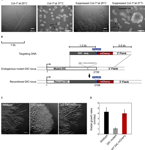
We PCR-amplified a 2.7-kb fragment encoding 1.8-kb DIC
sequence, mCherry fluorescent tag, and a 0.8-kb DIC
down-stream sequence from the pDIC-mCherry to generate a linear transformable DNA. This DNA fragment was integrated into
the native DIC locus (ro-6) by homologous recombination
(Figure S1). The DNA was transformed into aN. crassaDIC
mutant ropy strain that carried a 7-bp deletion at nucleotide
position 1695 (ro-6D1695–1702) in the DIC-coding sequence
(Davis 2000). DIC-mCherry positive colonies were identified
on the basis of (1) transformation-mediated rescue of the ropy phenotype of the DIC mutant strain leading to
wild-type growth and (2) detection of mCherryfluorescence. The
correct integration of DIC-mCherry onto the native locus
was verified by genome sequencing. The colony morphology
and radial growth rates of the DIC-mCherry strain were indistinguishable from the untransformed wild-type strain (Figure S1). A similar approach was used to generate an N-terminal EGFP fusion of the p150 subunit of dynactin. The DIC-mCherry strain was crossed with all the 34 DHC mutant strains using standard procedures (Davis 2000). Once the 34 DHC mutant strains with DIC-mCherry were isolated, genetic crosses were made with strains carrying
EGFP-p150, b-tubulin-GFP [Fungal Genetics Stock Center
(FGSC) #9520] and histone H1-GFP (FGSC #9518) to gen-erate progeny that carried DHC mutations and were
double-labeled (Table S2). A sequence encoding dual affinity tags
comprising a hexahistidine (HHHHHH) and a Strep-tag II (WSHPQFEK) tag (Schmidt and Skerra 2007) was fused to the C terminus of DIC using a strategy similar to the one employed to generate the DIC-mCherry strain.
Microscopy and imaging
For live-cell imaging, strains were grown on a slide coated
with a thinfilm of VSM agar for 16–18 hr at 25. Epifl
uo-rescence microscopy was performed using an Olympus BX50 microscope equipped with a mercury light source. Samples
were visualized using U Plan FI dry ·20 [0.50 numerical
aperture (na)] or oil immersion ·100 (1.3 na) objective
lenses. The GFP and mCherry fluorescence was detected
by appropriate emission and excitation filter sets. Images
and movies were captured with a SPOT RT-SE 18 camera sys-tem (Diagnostic Instruments) using SPOT software (v3.5.9).
The images were processed in Adobe Photoshop. Classifi
-cations of the dynein localization phenotypes were per-formed manually by visual examination of at least 50
intermediate magnifications as well as high-magnification
fluorescence micrographs from each strain studied.
Further-more, the spatial variations in dyneinfluorescence in relation
to the hyphal structure were confirmed by fluorescence
in-tensity profiles. The line-scan analysis tool in Metamorph
of fluorescence intensity (expressed as mean gray values)
profiles. The area of analysis was defined by a line width that
covered the width of the hyphae and a line length drawn
from the hyphal tip up to 120mm back from the tip. Fifteen
hyphae from each class were randomly chosen from images
taken at the same magnification and exposure settings for
fluorescence intensity determination. Fluorescence intensity
values from an untransfected control strain were used to
determine the background fluorescence that was then
subtracted from the values derived from other strains. The mean and the standard error were plotted using Kaleidagraph.
Distance measurements within hyphal tips were made
using SPOT software. Data were expressed as the mean6
SD. Fluorescence intensity of the comet tails from wild-type and class 3 mutant strains were determined using National Institutes of Health (NIH) ImageJ software. The
fluorescence intensity (expressed as mean gray values) of
individual comet tails was determined by manually select-ing a comet tail, measurselect-ing the mean gray value within the selection, and normalizing the data with the gray value measurements of regions close to corresponding comet tails.
The final fluorescence intensity measurement from each
strain was determined from the average of the gray values from measuring at least 40 individual comet tails from each strain.
Vesicle transport imaging experiments utilized N
-(3-tri-ethylammoniumpropyl)-4-(6-(4-(diethylamino) phenyl)
hexa-trienyl) pyridinium dibromide (trade name “FM 4-64,” Life
Technologies), which has no appreciablefluorescence unless
it is in a lipid membrane. Slide cultures of fungal strains were prepared in the same manner as described earlier. VSM liquid
media containing FM 4-64 was added to afinal concentration
of 5mM onto a growing colony, and a coverslip was placed on top. The hyphae were imaged in a time window between 5 and 15 min following dye treatment. Time-lapse image stacks (30-sec duration) were generated with 40-msec expo-sures captured every 200 msec. The individual image stacks were analyzed using Metamorph software to generate movement kymographs. The velocities, distances traveled, and the total number of the movements were determined from those kymographs using custom-designed software.
Cytoplasmic streaming-based transport was identified by
the slower movement of vesicles (,0.5mm/sec) in unison.
In this study, we did not further examine cytoplasmic streaming-based movement of vesicles. Individual vesicles were considered to have undergone a motility event if they exhibited a directed displacement at a velocity of at least 0.5mm/sec. Student’st-tests were used to examine significance between various experimental data sets. The motility index was calculated by taking the sum of the velocity multiplied by the distance of all motility events for a strain divided by the total length of the hyphae examined. Representative
live-cell movies of FM 4-64-labeled vesicle trafficking in
hy-phal tips from wild type (File S1), DHC deletion (File S2),
p150 deletion (File S3),Nkin (File S4), Class 1 (File S5),
Class 2 (File S6), Class 3 (File S7), Class 4 (File S8), and Class 5 (File S9) strains are included.
Benomyl treatment experiments were performed by
adding VSM liquid media containing 10 mM benomyl
(DuPont, Wilmington, DE) to a colony on a slide culture at room temperature. Benomyl treatment led to the
disrup-tion of microtubules in5 min after which the hyphae were
imaged. For controls, media containing DMSO was added to the colony, and imaging was performed as for benomyl treatment.
Purification of dynein from N. crassa
N. crassastrains expressing DIC-His-Strep affinity tags were homogenized with buffer A (35 mM PIPES, pH 7.0, 5 mM
MgSO4) supplemented with 1 mM EGTA, 0.5 mM EDTA,
1 mM DTT, 0.5 mM ATP, and protease inhibitor cocktail
(0.25 mM phenylmethanesulfonyl fluoride, 0.5 mg/ml
leu-peptin, 1.0mg/ml pepstatin A, 10mg/ml tosylphenylalanine
chloromethyl ketone, 10 mg/ml N-tosyl-L-lysine
chloro-methyl ketone, and 10 mg/ml p-toluenesulfonyl-L-arginine
methyl ester). After ultracentrifugation supernatant was loaded onto a SP Sepharose column (GE Healthcare, Uppsala, Swe-den), and bound protein was eluted with 500 mM KCl in buffer A. The eluted fraction was then loaded onto a HiTrap chelating
HP column (GE Healthcare) charged with CoCl2, and the
bound protein was eluted with 250 mM imidazole (Acros) in buffer A. The eluted protein was then loaded onto a StrepTrap
column (GE Healthcare), and the bound protein was finally
eluted with 2.5 mMD-desthiobiotin (Sigma) in buffer A
supple-mented with 1 mM EGTA, 0.5 mM EDTA, 1 mM DTT, and 10mM ATP.
Microtubule cosedimentation assay
PurifiedN. crassa dynein was incubated at 32for 20 min
with 5mM paclitaxel-stabilized microtubules in the presence
of 1 mM ATP, ADP, AMPPNP, or nucleotide-free conditions. Samples were subject to centrifugation, and the supernatant and pellet fractions were resolved by electrophoresis through 7.5% acrylamide gel, stained with Coomassie Brilliant Blue, and digitized using a Hewlett Packard Scanjet 7400c scanner. Digital images were analyzed using NIH ImageJ software and dynein heavy chain bands in the supernatant, and pellet
frac-tions were quantified densitometrically.
ATPase assay
The ATPase activity of N. crassa dynein was measured by
a radio thin-layer chromatography (TLC) assay (Gilbert and
Mackey 2000; Mesngonet al.2006). The reactions were
per-formed in BRB80 buffer (80 mM PIPES, 1 mM EGTA, 1 mM
MgCl2, pH 6.9) supplemented with 1 mM ATP and 10 mCi
a-32P-ATP (Perkin Elmer, Boston) in the absence or presence
of 5 mM paclitaxel-stabilized microtubules. The amount of
(Molecular Dynamics, Sunnyvale, CA). ATP and ADP spots
were quantified densitometrically using ImageQuant
soft-ware (Molecular Dynamics). The specific activity was
mea-sured by quantifying the concentration of dynein isolated
from wild-type and AAA3 E/Q strains relative to purified
bovine dynein.
Vanadate-mediated UV photolysis of N. crassa dynein
N. crassadynein was subject to vanadate-mediated
photol-ysis using standard conditions (Gibbons et al. 1987). The
major site of cleavage is termed as the V1 site, and the cleavage reaction components termed as the V1 condition
demand specific requirements such as UV irradiation, Mg2+,
and ATP. The V1 site cleavage leads to the formation of high- and low-molecular-weight UV fragments. Substitution
of Mg2+ with Mn2+as a divalent metal ion suppresses V1
cleavage (Gibbonset al.1987). The amount of cleavage
prod-ucts generated in the presence of magnesium (V1 Mg2+) or
manganese ions (V1 Mn2+) was used to measure the extent
of vanadate photolysis. Samples in cleavage buffer (80 mM
PIPES, pH 6.9) supplemented with 1 mM ATP, 2 mM MgCl2
or MnCl2, 500mM Na3VO4were irradiated with UV (365 nm)
light using UVL-56 BLAK-RAY lamp (UVP Inc., San Gabriel, CA) for 60 min on ice. Cleavage fragments were resolved by electrophoresis through 7.5% acrylamide gel, stained with Coomassie Brilliant Blue, and the gels were digitized. The
UV fragments were quantified by densitometry using NIH
ImageJ software.
Motility assay
In vitro bead-based motility assays were performed as
re-ported earlier (King and Schroer 2000; Mallik et al.2004;
Culver-Hanlon et al. 2006). Purified tubulin was used to
generate polymerized microtubules that were bound to
aflow chamber coated with poly-lysine. Unbound
microtu-bules were washed away by the addition of final dilution
buffer (33 mM PIPES, pH 7.0, 0.7 mM EGTA, 0.3 mM
MgSO4, 1 mM GTP, and 20mM paclitaxel). Dynein purified
from specifiedN. crassastrains was bound to polybead
car-boxylate microspheres (0.21-mm diameter; Polysciences,
Warrington, PA) by nonspecific adsorption at varying molar
ratios (20:1–40:1 dynein: bead) in the presence of 10 mm
ATP. The beads were added to theflow chamber and viewed
by video-enhanced differential interference contrast micros-copy. Custom-built image-processing software (Labview 6.1, National Instruments) was used for tracking the beads that exhibit directional motion. The run lengths and velocities of motility events were determined from the tracking data.
Results
Localization of dynein molecules to the hyphal tip in N. crassa colonies
To better understand the roles that cytoplasmic dynein motors normally play in polarized growth, we utilized
live-cell imaging approaches inN. crassa. We replaced the
endog-enous DIC with a version of the DIC that encoded a C-terminal mCherry tag (Figure S1). We then visualized DIC-mCherry
fluorescence, which we will refer to as“dyneinfluorescence”
throughout this article. The replacement of the single copy of the DIC gene by homologous recombination in the haploid genome ensured that DIC-mCherry was expressed under the control of its native promoter and that all DIC present in the strain was tagged. The presence of the mCherry tag had no effect on the normal function of the DIC as determined by hyphal morphology and colony growth rates (Figure S1),
confirming that DIC-mCherry-expressing cells have wild-type
dynein function.
When we observed dynein localization in actively growing
hyphae, we saw that the dynein fluorescence was
predomi-nantly found in the extreme tips of the hyphae (Figure 1A).
Thefluorescent signal tapered off dramatically within50mm
(average 45 611 mm) (Figure 3B), and the signal gradient
maintained its location at the hyphal tip even though the hy-phae itself was growing at several micrometers per minute. Very little substructure could be observed at intermediate
mag-nification across the hyphal tube.
At higher magnification, additional details of the hyphal
tip accumulation could be observed. Most images showed
mCherry fluorescence in both short linear tracks and
spherical structures in the region immediately following the hyphal tip (Figure 1B). The spherical structures were remi-niscent of endomembrane organelles that have been
de-scribed in previous studies (Bowman et al. 2009). The
short linear tracks resembled the comet tail tip accumulation of dynein at microtubule plus ends that has been reported by studies in other systems ranging from fungi to mammalian
neurons (Xianget al.1995; Vaughanet al.1999; Xianget al.
2000; Han et al. 2001; Ma and Chisholm 2002; Lee et al.
2003; Sheemanet al.2003; Zhanget al.2003, 2010, 2011;
Lenz et al.2006; Arimotoet al. 2011; Markuset al.2011;
Schuster et al.2011a). Apart from the spherical structures
and short linear tracks, no additional substructures were visible upon standard exposure settings or photobleaching
the bright hyphal tip dynein fluorescence (Figure S3). As
a control, we examined a DHC deletion strain and found
that the specific localization of the DIC-mCherry signal
was lost from hyphal tips, short linear tracks, and spherical structures (Figure 1).
To better determine if the short linear tracks were microtubule-based, we crossed the DIC-mCherry strain to
a strain expressingb-tubulin-GFP (Freitaget al.2004).
Pre-vious studies with these and similar strains have shown that abundant microtubules are predominantly arranged parallel to the longitudinal axis with plus ends extending into
regions near theN. crassahyphal tip. More complex
arrange-ments with microtubules of mixed polarity are found around the nuclei in the syncytial cytosol further back from the
hy-phal tips (Riquelmeet al.2002; Freitaget al.2004; Sampson
and Heath 2005; Uchida et al.2008). We found that dynein
of the tubulin GFP signals near the hyphal tips (Figure 2A). To further examine dynein localization with respect to microtubules, we treated growing colonies with benomyl, a cell-permeable microtubule-depolymerizing drug.
Treat-ment with 10mM benomyl for 5 min depolymerized
micro-tubules and resulted in a nonlinear, diffuse tubulin-GFP signal throughout the hyphae with occasional GFP puncta. Following microtubule depolymerization, the short linear tracks of the DIC-mCherry signal were abolished, and the high concentration of dynein at the hyphal tip began to expand distally along the hyphae (Figure 2B). Longer treatment times resulted in a loss of dynein signal at the hyphal tip, showing that the maintenance of the hyphal tip accumulation gradient requires a normal microtubule
net-work. Our data confirm that the short linear tracks are
asso-ciated with microtubule ends in a characteristic“comet-tail”
pattern.
Dynein localization requires conventional kinesin and dynactin
Multiple studies have shown that kinesin and dynactin play essential roles in the localization of dynein within cells
(Bradyet al.1990; Echeverri et al.1996; Waterman-Storer
et al.1997; Martinet al.1999; Vaughanet al.1999; Duncan
and Warrior 2002; Januschkeet al.2002; Kinget al.2003;
Zhanget al.2003; Ligonet al.2004; Theisset al.2005; Lenz
et al. 2006; Arimoto et al.2011). To determine if
conven-tional kinesin (Nkin) or dynactin have similar functions in
N. crassa, we crossed the DIC-mCherry strain to either a strain carrying repeat-induced point (RIP) mutations in
conventional kinesin (Seiler et al. 1997) or a dynactin
p150 (ro-3) null strain. In the Nkinmutant strain, hyphal
tip accumulation of dynein was completely abolished; regions closer to the tips were devoid of any dynein signal
but distal regions of the hyphae displayed a faint fl
uores-cence (Figure 1, A and B; Figure 3B). Similar to the Nkin
mutant strain, the p150 null strain also exhibited a loss of
hyphal tip dynein accumulation. However, unlike theNkin
mutant strain, a diffuse dynein signal was found along the entire length of hyphae (Figure 1, A and B; Figure 3B). As an alternative to analyzing deletion strains, we also examined colocalization between dynein and dynactin in a strain expressing both DIC-mCherry and EGFP-p150. Dynactin exhibited almost complete colocalization with dynein to the hyphal tip, to the comet tails, and to spherical structures near the hyphal tip (Figure S4). These data together illus-trate that the functions of both dynactin and kinesin are required for the correct cellular localization of dynein in N. crassahyphal growth.
Mutations in the DHC lead to multiple dynein mislocalization phenotypes
To further characterize dynein function within the hyphal
tip of N. crassa, we examined 34 DHC mutant strains
iso-lated from a genetic screen (seeMaterials and Methods). In
the current study, we crossed the mCherry-DIC allele into the 34 DHC mutant strains so that we could track dynein molecules in the mutant strains. All 34 strains lacked the bright hyphal tip accumulation observed in wild-type strains (Figure 1A). In addition, the remaining dynein molecules in
the DHC mutant strains were mislocalized into one of five
distinct classes of phenotypes that we termed classes 1–5 (Table 1).
The dynein localization signal in six class 1 mutant strains was found in distal long linear tracks arranged mostly parallel to the hyphal axis (Figure 1B). This signal pattern was readily distinguishable from the comet tails de-scribed earlier for wild-type dynein localization in length of Figure 1 Localization of dynein in mu-tant strains ofN. crassa. (A) Hyphae from the colony edge of different N. crassa
the track and location within the hyphae (Figure 2B). The dynein signal in these long linear tracks occurred in distal
regions 100 6 30 mm away from the tip and extended
even farther into distal regions far from the tips (Figure 1, A and B; Figure 3B). The four class 2 DHC mutant strains, in contrast to class 1 mutant strains, showed long linear tracks that occurred at apical regions close to the tip of the hyphae
that ran until75615mm back from the tip (Figure 1, A
and B; Figure 3B). A mutant strain that exhibited one of these two mutant localization phenotypes (class 1 or class
2) consistently showed the specified localization pattern and
did not exhibit the alternate class phenotype. The long
lin-ear tracks observed in both class 1 and class 2 likely reflect
dynein associated with microtubules, given that microtu-bules are the only structures that exhibit long parallel ar-rangement inside the hyphae.
The class 3 mutations were composed of nine strains that
each had residual DIC-mCherry fluorescence in hyphal tips
that was concentrated in comet tail structures (Figure 1, A and B; Figure 3B). The comet tails in the class 3 mutant strains appeared more numerous than those in the wild-type strain. Furthermore, measurements of individual comet tail
fluorescence intensities indicated a twofold increase in the
mCherryfluorescence in class 3 mutant comet tails in
com-parison with wild-type comet tails. The localization pattern observed with class 3 mutant dynein was most similar to wild type and may indicate enhanced microtubule plus-end association at the expense of hyphal tip accumulation.
The three class 4 mutant strains displayed phenotypes
where dynein accumulated distally as large “aggregates”
starting 85 6 20mm back from the hyphal tips (Figure
1, A and B; Figure 3B). Interestingly, these aggregates were
more or less evenly spaced, on average, 1165mm from one
another along the hyphae. At higher magnifications, these
strains revealed the presence of “aster-like”dynein signals
expanding from a central aggregate mass.
The remaining 12 DHC mutations in class 5 resulted in
the“disperse”localization of dynein along the hyphae
(Fig-ure 1, A and B). In these strains, we saw diffuse dynein
fluorescence of equal intensity throughout the hyphae
(Fig-ure 3B) that was reminiscent of what we observed with wild-type dynein in p150 deletion strains (Figure 1). We also observed a few punctate foci of dynein signal (Figure 1B).
effect of dynein disruption also observed altered
micro-tubule dynamics and organization (Koonce et al. 1999;
Riquelmeet al.2002; Uchidaet al.2008). In class 1 mutant
strains, microtubules in the distal regions (where dynein was found) had a thicker appearance than microtubules in the wild-type strain. Similarly, in class 2 mutant strains, the apical microtubules along which mutant dynein were found appeared thicker than in the wild type and other classes of DHC mutant strains. The thickness of the microtubules in class 1 and 2 indicates that microtubule bundling may have occurred. Class 3 mutant strains had disorganized microtu-bule networks with the microtumicrotu-bules showing excessive
cur-vature along the hyphal periphery. Class 4 mutant strains showed a unique aster-like microtubule pattern in regions away from the hyphal tip. Class 5 mutant strains had disor-ganized microtubule networks, and we observed many microtubules that were perpendicular to the hyphae in contrast to the normal parallel arrangement in wild-type hyphae.
We also found that the class 1, class 2, class 3, and class 4 mutant dynein localization patterns colocalized with subsets of visible microtubules (Figure 2A). The disperse distribution of dynein in class 5 DHC strains had no overlap with
micro-tubules (Figure 2A), and the spots of dynein fluorescence
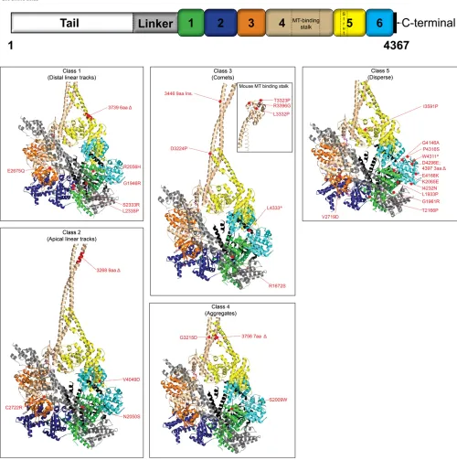
Figure 3 (A) Schematic representation of cytoplasmic dynein heavy chain from
N. crassa showing the mutations ana-lyzed. The relative positions of dynein heavy chain mutations are shown with corresponding amino acid changes. Amino acids (aa) are represented by single-letter codes. A “D” represents a deletion; “ins.” refers to insertion; and nonsense mutations are indicated by an asterisk. Numbers 1–6 indicate re-spective AAA domains. Color coding of the mutations represents grouping of different mutations to classes 1–5. (B) DIC-mCherry fluorescence intensity is plotted as a function of distance from the hyphal tip. Fluorescence signals in the wild-type strains (gray) exhibited an initial maximum closer to the hyphal tips and gradually tapered off at regions far from the tip. In class 1 (distal linear tracks) strains,fluorescence signals (me-dium blue) were low or absent at regions closer to tips but displayed increased signal intensities at distal regions from the tips. The class 2 (apical linear tracks) strains exhibited elevated
fluorescence intensities (black) closer to the tips that gradually tapered off as a function of distance from the hyphal tip. Class 3 (comet tail) strains also exhibited increasedfluorescence intensi-ties (green) closer to the tips. Class 4 (aggregate) strains show irregular in-creased fluorescence (purple) at distal regions. Thefluorescence intensity in class 5 (disperse) strains (brown) is slightly higher than thefluorescence signals of p150Dstrains (aqua), but both profiles show a uniform distribution along the entire length of the hyphae.Nkinmutant strains exhibit faintfluorescence (red) at distal regions. Data are shown as mean6SE (n= 15 for all classes).
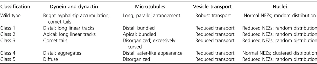
Table 1 Summary of dynein, dynactin, microtubule, and nuclear phenotypes in wild-type and mutant strains ofN. crassa
Classification Dynein and dynactin Microtubules Vesicle transport Nuclei
Wild type Bright hyphal-tip accumulation; comet tails
Long, parallel arrangement Robust transport Normal NEZs; random distribution
Class 1 Distal: long linear tracks Distal: bundled Reduced transport Reduced NEZs; random distribution Class 2 Apical: long linear tracks Apical: bundled Reduced transport Reduced NEZs; random distribution
Class 3 Comet tails Disorganized; excessively
curved
Reduced transport Reduced NEZs; random distribution
Class 4 Distal: aggregates Distal: aster-like appearance Reduced transport Normal NEZs; clustered distribution
present in the class 5 mutant strains were unaffected by benomyl treatment (Figure 2B). As expected for microtubule-dependent localization patterns, microtubule depolymerization with benomyl disrupted dynein localization to the long linear track, comet tail, and aggregate structures in mutant classes 1–4 strains (Figure 2B). After microtubule depolymerization, mu-tant class 1, class 2, and class 4 strains showed prominent punc-tate dynein signals that commonly overlapped with tubulin-GFP
puncta. The presence of overlapping punctatefluorescence
signals instead of diffuse signals as seen with wild type sug-gests that the mutant dynein altered microtubule-associated structures and remained bound to microtubule remnants in some way. In contrast, instead of comet tail localization, mutant class 3 strains showed diffuse dynein signals after benomyl treatment, suggesting complete dispersion of dy-nein in these strains. The variation in dydy-nein dispersion upon microtubule depolymerization indicates that there is heterogeneity in how dynein is interacting with microtu-bules in the different mutant strains. Taken together, these data reiterate the importance of dynein function in
microtu-bule organization and confirm our presumption that the
dynein in mutant class 1, 2, 3, and 4 strains are indeed associated with microtubules.
We examined the DHC mutant strains in a DIC-mCherry: EGFP-p150 background to see if the mutations altered the localization of dynactin. In all classes of DHC mutant strains, the dynactin signal was disrupted and there was a loss of the bright cloud of accumulation at the hyphal tip (Figure S4A). In class 5 mutant strains, the dynein and dynactin signals were each diffuse; therefore it was impossible to determine the extent, if any, of colocalization between dynein and dynactin. In all other mutant strains, dynactin distribution and dynein distribution were indistinguishable (Figure S4A; Table 1).
To test the possibility that dynactin function is required
for thefive different localization patterns that we observed,
we crossed representative strains from each of the five
mutant heavy chain classes by the p150 deletion strain (Figure S4B). The distal linear track dynein localization pat-tern observed in class 1 strains was not altered by the loss of dynactin function, showing that this phenotype is dynactin independent. In contrast, the typical dynein localization phenotypes in class 2 and class 3 strains were abolished when those strains were examined in a p150 deletion back-ground; instead, we observed the diffuse pattern normally found in class 5 strains. Interestingly, we found that the dynein localization pattern in class 4 strains was converted into a distal linear track phenotype similar to what was seen in class 1 strains. The diffuse dynein pattern observed in class 5 strains was not altered by the loss of dynactin func-tion. These data indicate that dynactin has a complex role in the localization of dynein within cells.
Vesicle transport is altered in the DHC mutant strains
We examined the effect of the DHC mutations on vesicle
transport in growing hyphae of N. crassa by utilizing the
lipophilic styryl dye FM 4-64. In addition to representative
strains from thefive classes of DHC mutations, we also
ex-amined DHC deletion and dynactin p150 deletion strains as
well as the RIPNkinmutant strain utilized earlier. Colonies
of wild-type and the various mutants strains were treated with FM 4-64, and hyphae at the colony edges were imaged with time-lapse microscopy to generate movies of endo-membrane vesicle transport. The dye was quickly taken up by the hyphae into individual vesicles, and, in the majority of wild-type hyphae, many of the vesicles accumulated near the hyphal tip. Vesicle accumulation was more rarely ob-served in the hyphal body of wild-type strains. A similar pattern of vesicle accumulation at hyphal tips was seen in all the mutant strains except the p150 deletion strain, where instead we saw a reversal in the location of accumulated vesicles with a majority of hyphae showing vesicle accumu-lation in the hyphal body instead of the hyphal tip (Table S1).
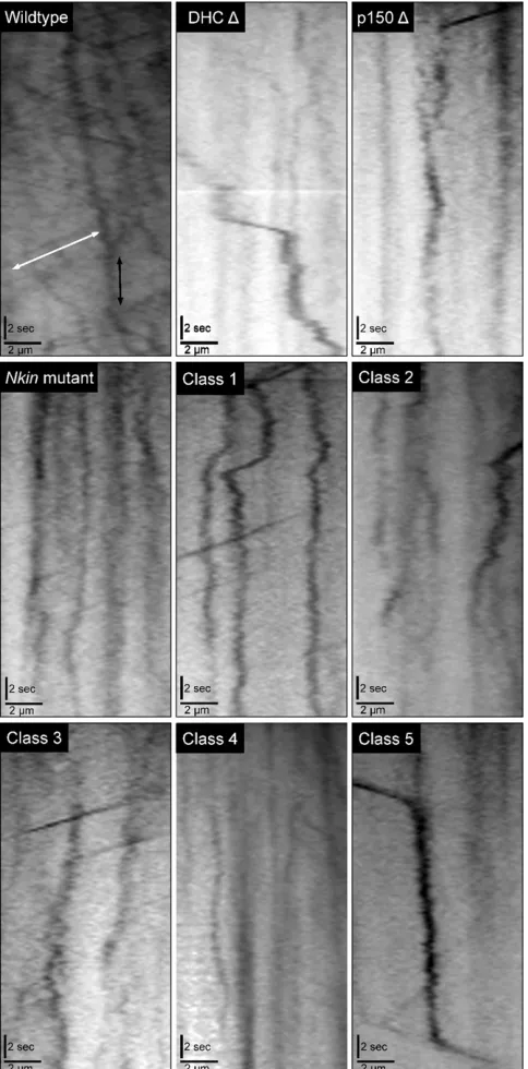
The FM 4-64-positive vesicles underwent two different types of motility in all hyphae: cytoplasmic streaming that slowly propelled vesicles in unison toward the hyphal tip and individual motility events that were much faster and were directed in either an inward (toward the hyphal base) or an outward (toward the hyphal tip) trajectory. We generated motility kymographs of the time-lapse movies to determine the relative numbers, velocities, and displace-ments of the individual motility events for wild type and all the mutants strains (Figure 4). We did not see large differ-ences in the percentage of inward or outward motility
events in the DHC deletion, Nkin, or in any other mutant
or wild-type strains (Table S1), as would be expected in a system where one direction of motor activity was lost when all the microtubules are aligned with the same polar-ity, as in an axon. This is likely due to the syncytial arrange-ment of scattered nuclei as foci for microtubule growth and organization in the hyphae, which means that microtubules that are not in the nuclear exclusion zone can be in either
orientation (Freitag et al.2004; Uchidaet al. 2008).
Simi-larly, we did not observe many differences between inward and outward transport in the wild-type or mutant strains (Table S1).
When we examined all of the motility events that occurred within active hyphae, we saw that there was a 30–80% decrease in the number of motility events that occurred in the mutant strains compared to the wild-type strain. Furthermore, we determined that all of the mutant
strains had a significant defect in the velocity of the motility
events (Table S1). Class 1 and class 5 strains did not show
a significant decrease in the distance traveled by FM 4-64
vesicles, but all other mutant strains exhibited significant
defects in vesicle displacement. To provide a single-term overall assessment of vesicle transport that occurred in the hyphae from the different strains, we generated a motility index that is a function of the number of movements and the
velocity and distance of those movements (seeMaterials and
strains, as well as the DHC deletion, p150 deletion, andNkin
mutant strains exhibited two- tofivefold decreases in their
motility index with respect to wild-type strains. Overall, the
FM 4-64 experiments illustrate that each of thefive classes
of DHC mutant strains exhibits transport phenotypes that
include decreases in the motility index and in the number of vesicles being actively transported by microtubule motors.
Nuclear distribution in strains expressing wild-type and mutant DHC
The role of dynein in the migration, distribution, and
position-ing of nuclei in cells has been well established (Plamannet al.
1994; Inoue et al.1998; Alberti-Seguiet al.2001; Duncan
and Warrior 2002).N. crassahyphae are multinucleate, and
these nuclei are normally found randomly distributed along the length of the hyphae except for a clear nuclear
exclu-sion zone (NEZ) within 25 mm of the hyphal tip (average
25 6 5 mm, n = 20) (Figure 5, A and B). The NEZ is
a common phenomenon observed in filamentous fungi
and has been reported previously in N. crassa (Freitag
et al.2004; Ramos-Garciaet al.2009).
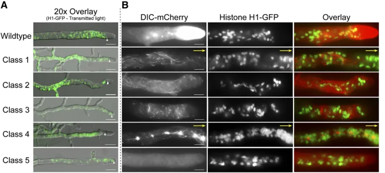
We examined the effect of DHC mutations on nuclear distribution in the mature hyphae of strains expressing DIC-mCherry and histone H1-GFP. Class 1, class 2, class 3, and class 5 mutant strains exhibited a reduction in the length of
the NEZ (NEZ size for class 1 = 1565mm; class 2 = 106
4mm; class 3 = 1265mm; and class 5 = 1165mm;n=
30 for all classes) (Figure 5A). It is highly likely that the altered microtubule organization observed with our DHC mutant strains contributed to NEZ size anomalies, as seen
in another report (Ramos-Garciaet al.2009). Although the
NEZ of the mutant class 1, class 2, class 3, and class 5 strains was altered, nuclei in these strains were randomly distrib-uted similar to wild-type strains (Figure 5). However, class 4 strains had a NEZ that was similar in size to that of wild-type strains yet the nuclei displayed dramatic clustering around the aggregates in distal regions with a few single nuclei extended into more apical regions up to the NEZ. This pat-tern suggests that the mutant dynein from class 4 strains may have a unique mechanism of interaction with both microtubules and nuclear cargoes. Our earlier experiments with class 4 mutations in combination with the loss of dynactin function also pointed out that class 4 strains be-haved differently that the other classes of mutant heavy chain strains. Collectively, these observations indicate that the different DHC mutations affect nuclear positioning to varying degrees (Table 1).
Biochemical analyses of wild-type and mutant dynein motors
To determine ifN. crassadynein is functionally equivalent to
dynein from other sources and to examine a possible
mech-anism of dynein mislocalization, we purified dynein from
a wild type as well as a representative DHC mutant strain. We chose to study the E2675Q mutation (hereafter referred to as AAA3 E/Q) from a class 1 mutant strain that exhibits the distal long linear track microtubule-associated dynein localization phenotype. This mutant strain was chosen from among the other DHC mutations because (1) the mutation is located on the Walker B domain of AAA3 that is implicated in ATP hydrolysis on the basis of mutational analysis on Figure 4 Kymograph analyses of vesicle transport in different strains.
various AAA family proteins (Whiteheart et al.1994; Babst et al.1998; Hartman and Vale 1999) and (2) previous stud-ies have shown that the disruption of dynein AAA3 ATP-binding/hydrolysis functions can alter ATPase activity at AAA1, microtubule interaction, and motility properties of
dy-nein (Silvanovichet al.2003; Konet al.2004; Reck-Peterson
and Vale 2004a,b; Choet al.2008). We sought to test if the
cellular localization phenotype of this mutant strain is a
re-flection of such altered biochemical properties of dynein.
To study dynein motors from these strains, we
de-veloped a new purification protocol (Figure S5A) that did
not rely upon nucleotide-dependent microtubule binding. This was because we expected that the AAA3 E/Q mutation would likely alter dynein mechanics in a way that would
prevent efficient motor purification with such conventional
approaches. Briefly, hexahistidine and Strep-tag II affinity
tags were fused to the C terminus of the DIC by employing a strategy similar to DIC-mCherry generation. Dynein
com-plexes were purified using this dual affinity tag (Figure
S5B).
Dynein interaction with microtubules is normally
modu-lated by different nucleotide conditions (Shpetner et al.
1988; Imamula et al.2007; Mizunoet al.2007). To
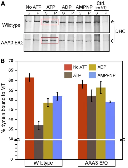
deter-mine if the AAA3 E/Q mutation affected this modulation, we examined the microtubule-binding behavior of wild-type and AAA3 E/Q dynein under varying nucleotide conditions using a microtubule-copelleting assay. As expected for wild-type dynein motors, the presence of 1 mM ATP resulted in a low percentage of dynein cosedimentation with microtu-bules (Figure 6, A and B). Under nucleotide-free, ADP, or
AMPPNP conditions, we observed a significant increase in
dynein cosedimentation with microtubules in comparison to the ATP condition (Figure 6, A and B). These observations are consistent with the nucleotide-sensitive microtubule-binding behavior exhibited by cytoplasmic dynein from
other sources (Shpetneret al.1988; Hayset al.1994; Imamula
et al. 2007). AAA3 E/Q dynein displayed a microtubule-dependent cosedimentation behavior similar to wild-type dynein under nucleotide-free, ADP, or AMPPNP conditions (Figure 6, A and B). However, in contrast to wild-type
dy-nein, the presence of ATP did not significantly lower the
cosedimentation of AAA3 E/Q dynein with microtubules (Figure 6, A and B). These results indicate that
nucleotide-sensitive microtubule-binding behavior of N. crassa
wild-type dynein is similar to microtubule-binding characteristics of cytoplasmic dynein from other sources and that the AAA3 E/Q mutation perturbs dynein so that its ability to bind microtubules is insensitive to the presence of ATP. The ATP-insensitive microtubule-binding ability of AAA3 E/Q is con-sistent with the rigor-like microtubule-binding reported for
similar AAA3 mutants in previous studies (Silvanovichet al.
2003; Konet al.2004; Choet al.2008). The results from the
AAA3 E/Q cosedimentation experiments suggest that the linear track phenotype observed in class 1 mutant strains may be a result of a rigor-like microtubule binding.
Dynein has an intrinsic basal ATPase activity that is
enhanced by the presence of microtubules (Shpetner et al.
1988; Silvanovichet al.2003; Konet al.2004). We
investi-gated the effect of the AAA3 E/Q mutation on both basal and microtubule-stimulated dynein ATPase activity. The basal ATPase activities of wild-type and AAA3 E/Q dynein
motors were significantly different from one another (Table
2). The ATPase rate of wild-type N. crassa dynein is 115
nmol ATP/mg dynein/min and in range with reports that probed the ATPase activity of cytoplasmic dynein from mam-malian sources (Ferro and Collins 1995; King and Schroer
2000; Mesngon et al. 2006). Surprisingly, the AAA3 E/Q
the ATPase activity approximately twofold in comparison to the corresponding basal activities (Table 2). This means that each motor sensed and responded to the presence of microtubules with a doubling in its own inherent ATPase rate. The enhanced ATPase rates (both basal and micro-tubule stimulated) observed with the AAA3 E/Q motor were unexpected on the basis of the nucleotide-insensitive microtubule-binding behavior that we observed (Figure 6, A and B).
Dynein undergoes a signature, site-specific UV photolysis
induced by vanadium ions (VO4)—a structural analog of
in-organic phosphate (Gibbonset al.1987). Vanadate-mediated
photolysis has been considered a defining characteristic of
dynein and can be applied as a tool to probe the conservation of active site (ATPase) conformation and any alterations to
this conformation (Grammeret al.1988; Gibbons and Mocz
1991). We performed vanadate-mediated photolysis
experi-ments on N. crassa dynein and found that the wild-type
dynein was susceptible to vanadate-mediated photocleavage
using a standard Mg2+condition (V1 Mg2+) (Figure 7A).
Quantification of the cleavage products showed that almost
50% of the total dynein was cleaved (Figure 7B). When we
substituted Mn2+instead of Mg2+under V1 conditions (V1
Mn2+), wild-type dynein vanadate cleavage was reduced
approximately twofold in comparison to the V1 Mg2+
con-dition (Figure 7, A and B). The reduction in cleavage upon
the substitution of the coordination metal (Mg2+) is in line
with observations from previous studies (Gibbons et al.
1987). Collectively, these observations demonstrate that the structural features responsible for V1 site cleavage are
conserved betweenN.crassadynein and dynein studied from
other sources (Gibbons et al. 1987; Schnapp and Reese
1989; Gatti et al.1994; Hayset al.1994; Allan 1995; Kon
et al.2004).
AAA3 E/Q dynein showed a V1 Mg2+cleavage pattern
similar to wild-type dynein (Figure 7, A and B). In contrast,
under V1 Mn2+conditions, AAA3 E/Q dynein did not show
as great a reduction in cleavage (Figure, 7 A and B). In addition, there was cleavage of AAA3 E/Q dynein at a second
site under V1 Mn2+conditions. The alteration of AAA3 E/Q
dynein V1 Mn2+cleavage patterns indicates that the AAA3
E/Q mutation alters the active site conformation of dynein. The alteration in vandate cleavage seen for AAA3 E/Q is related to the elevated ATPase activity that we observed (Table 2).
Next, we probed the motility properties ofN. crassa
wild-type and AAA3 E/Q dynein using an in vitro
microtubule-based bead motility assay. Beads coated with wild-type dynein exhibited motility as seen by the directional motion of the beads on microtubules. Wild-type dynein moved with
an average velocity of 0.7 mm/sec (Table 3), which is
con-sistent with the dynein velocities observed in previous
stud-ies (King and Schroer 2000; Tobaet al.2006; Ori-McKenney
et al.2010). The mean run length of wild-type dynein
(Ta-ble 3) was well within the range of in vivo dynein-based
motility events (Ma and Chisholm 2002; Pilling et al.
Figure 6 Effect of nucleotides on the cosedimentation of wild-type and AAA3 E/Q dynein with microtubules. (A) Representative silver-stained gel images showing microtubule cosedimentation of dynein isolated from wild-type and AAA3 E/Q strains under varying nucleotide conditions. Mixtures were centrifuged, and the supernatant (S) and pellet (P) fractions are shown. Regions boxed in red indicate dynein sedimentation in the presence of ATP. (B) Quantification of the percentage of dynein in pellet fraction in comparison to total dynein. Cosedimentation was quantified from three independent experiments. Data are shown as mean6SEM. Wild-type dynein cosedimentation in the presence of 1 mM ATP was significantly less than wild-type dynein cosedimentation in the other con-ditions examined (t-test,P,0.05). AAA3 E/Q dynein cosedimentation in the presence of ATP was not significantly different from that under the other conditions (t-test,P.0.05)
Table 2 ATPase activities of dynein isolated from wild-type and AAA3 E/Q strains
Dynein
Basal ATPase (nmol/min/mg dynein)
MT-stimulated ATPase
(nmol/min/mg dynein) Fold change
Wild type 11567 20163 1.7
AAA3 E/Q 418635 729660 1.7
2006; Ori-McKenneyet al.2010). When we examined beads
with AAA3 E/Q dynein, wefirst noted enhanced binding of
beads to microtubules in our motility chambers. Further analyses showed that none of these bound motors moved along microtubules. This inability to move is consistent with thein vivomicrotubule decoration phenotype (Figure 1B; Fig-ure 2A) and the nucleotide-insensitive microtubule-binding phenotypes (Figure 6, A and B).
Our studies were thefirst to examine the AAA3 Walker B
E/Q mutation in a whole dynein molecule, and our results are in general agreement with previous studies that showed
increased MT-binding affinity and altered dynein
localiza-tion in fruitfly and budding yeast (Silvanovichet al.2003;
Choet al.2008; Markuset al.2009). For example, one study
showed that UV-vanadate cleavage occurred at levels equal
to wild-type strains (Silvanovichet al.2003). Unfortunately,
that study did not have the resolution to determine if the cleavage products included altered species such as we
identified (Figure 7A). In contrast to our work, the ATPase
activity of tail-less artificially dimerized yeast AAA3 E/Q
dynein was reduced relative to wild type (Cho et al.2008)
whereas we saw an increase in the ATPase activity of
puri-fied native molecules (Table 2). This same report indicated
that the artificially dimerized yeast AAA3 E/Q dynein
exhibited extremely slow motility (0.0046mm/sec) along
axonemes while we failed to see any appreciable motility for AAA3 E/Q dynein along microtubules. The differences be-tween our data and some of the yeast dynein data may
re-flect either the use of tail-less motors (Cho et al. 2008;
Markus et al. 2009) or that yeast dynein is known to act
quite differently from fungal and higher-order organisms. In yeast, dynein does not transport endomembrane vesicles inside cells and instead is predominantly attached to the cell cortex for nuclear-positioning roles (Carminati and Stearns
1997; Cottingham and Hoyt 1997; Sheeman et al. 2003;
Markuset al.2009).
The properties exhibited byN. crassaAAA3 E/Q dynein,
such as the nucleotide-insensitive microtubule binding, en-hanced ATPase activity, and inability to translocate along microtubules are analogous to characteristics observed with kinesin and myosin motors carrying uncoupling mutations (Ruppel and Spudich 1996; Song and Endow 1998; Brendza et al. 2000; Heuston et al. 2010). Previous studies have shown that uncoupling mutations are able to disengage
a motor’s intradomain coordination mechanisms in a way
that results in constitutive cytoskeletal filament binding as
well as enzymatic and motility behaviors that are indepen-dent of each other (Ruppel and Spudich 1996; Song and
Endow 1998; Brendza et al. 2000; Heuston et al. 2010).
Our biochemical analyses of AAA3 E/Q dynein are consis-tent with those analogous studies and strongly suggest that the AAA3 E/Q mutation is an uncoupling mutation. Taken
together, our results indicate that the N. crassa wild-type
dynein exhibits motility properties comparable with dynein from other sources and that the AAA3 E/Q mutation uncou-ples the critical dynein motor functions of microtubule in-teraction and nucleotide hydrolysis.
Mapping of the DHC mutations onto the crystal structure
Recent crystal structures of the dynein heavy chain have shown many interesting features of the dynein molecules, including how the six AAA domains of the dynein ring are Figure 7 Effect of divalent metal ion on the vanadate-mediated V1
pho-tocleavage pattern of wild-type and mutant dynein. (A) Representative silver-stained gel images showing vanadate photocleavage of DHC from wild-type (left panels) and AAA3 E/Q (right panels) under V1 Mg2+and V1 Mn2+conditions. High- and low-molecular-weight UV cleavage products are indicated by“*”and“.”, respectively. Additional prominent cleav-age products found under AAA3 E/Q V1 Mn2+conditions are indicated by “<”. (B) Quantification of the percentage of dynein cleaved in compar-ison to the total dynein in the sample. Percentage cleavage was
quanti-fied from three independent experiments. Data are shown as mean6 SEM.
Table 3 In vitromotility characteristics of beads coated with dynein isolated from wild-type and AAA3 E/Q strains
Dynein MT binding Motility Velocity (mm/sec21) Distance (mm) n
Wild type + + 0.760.3 2.661.7 104
AAA3 E/Q +++ — 0.0 0.0 155
arranged with respect to each other, how the linker domain arches over the top of the motor ring, how the microtubule-binding domain (MTBD) stalk and its supporting strut
interact at a specific interface, and how multiple regions of
the AAA domains may be able to communicate nucleotide status and hydrolysis information to neighbor rings (Carter et al.2011; Konet al.2011; Konet al.2012; Schmidtet al. 2012). These same structures provided us with the oppor-tunity to map our mutations onto the DHC crystal structure (Konet al.2012) and to a portion of the microtubule-binding
stalk closer to the MTBD (Carteret al.2008) to gain a better
understanding of the structure/function relationships within the DHC. Our intention was to obtain a glimpse into how different motor domains may be utilized for particular dynein functions. The structural mutation map (Figure 8) shows
some intriguing correlations between the positions of thefive
classes of mutations and the dynein mislocalization pheno-types of those classes.
Class 1 mutations: The six class 1 (distal linear tracks) mutations were mapped to the p-loop of AAA1 (G1946R), helix 4 (H4) of AAA1 (R2056H), the pre-sensor 1 insert of AAA2 (S2333R, L2335P), Walker B of AAA3 (E2675Q), and
AAA5 strut coil 1 (3739 6-aaD) (Figure 8;Table S1). The
AAA1 p-loop (G1946R) and AAA3 Walker B (E2675Q) mutations have been reported to inhibit ATP binding at AAA1 and ATP hydrolysis at AAA3, respectively (Silvanovich et al.2003; Konet al.2004; Choet al.2008). The R2056H mutation is present in the second region of homology (SRH) domain, which has been implicated in sensing nucleotide status as well as in the stabilization of the transition state
of ATP hydrolysis (Ogura et al. 2004). The S2233R and
L3335P mutations are located on the pre-sensor I insert of the AAA2. Disruption of the pre-sensor I–linker interactions alters ATP-sensitive microtubule interaction as well as linker
motions (Konet al.2012), and pre-sensor I insert mutations
have been reported to affect ATPase kinetics (Burrowset al.
2009). The strut region has been proposed to relay rigid body motions between AAA4 and AAA5 domains into shear motions between the helices of the stalk coiled-coil. There-fore, it seems likely that the six-amino-acid deletion in the
strut mutation (3739 6-aa D) could interfere with the
nor-mal communication of nucleotide status between AAA1 and the microtubule-binding domain. The strut mutation 3739
6-aa D is intriguing in that a similar alteration of the strut
structure (Konet al.2012) results in a
microtubule-indepen-dent, elevated ATPase activity similar to that observed in the AAA3 Walker B (E2675Q) mutation in our study. A common theme that can be seen in all six of these class 1 mutations is that they are each located in an area that is intimately involved in the sensing of nucleotide status, hy-drolysis of nucleotides, or communication of information about nucleotide status.
Class 2 mutations: The four class 2 (apical linear track) mutations are located proximal to the sensor 1 of AAA1
(N2050S) and AAA3 (C2722R), in coiled coil 1 of the AAA4
microtubule-binding stalk (3268 9-aa D) and H1 of the
AAA6 domain (V4049D) domain (Figure 8;Table S1). Not
much can be inferred from the N2050S and C2722R muta-tions. However, sensor 1, which is a part of the SRH domain, has been reported to contact the Walker B domain and the
g-phosphate of the ATP and has been proposed to act as
a switch in mediating conformational changes (Davieset al.
2008). Mutations in the sensor 1 region in AAA family mem-bers have previously been shown to reduce the rate of ATP
hydrolysis (Song et al. 2000; Hattendorf and Lindquist
2002). This suggests that the apical microtubule decoration phenotype observed in these strains may be caused by a de-crease in the ATPase rates of the motor without the dramatic increase in the microtubule-binding behavior of dynein seen with class 1 mutations. A reduction in the ATPase activity
could lead to significantly increased dwell times of dynein
on microtubules (Figure 2A) and, by moving more slowly, could give rise to the observed apical microtubule mislocal-ization phenotype.
Class 3 mutations: Of the nine class 3 mutations (comet tails), two were located in the tail domain where structural data are not available (Y110S, W1308G), one was found in H11 of the linker (R1672S), one was at the extreme C
terminus of the molecule (L4333*), and the remainingfive
(D3224P, T3323P, L3332P, R3396G, and 3446 9-aa inser-tion) were present in the coiled coils or putative microtubule-binding interface of the microtubule-microtubule-binding domain (Figure 8; Table S1). It is easy to envision that alterations in the putative microtubule-binding interface or in the coiled-coils that con-nect and control the register of the interface to the rest of the dynein molecule could alter the binding of dynein to microtubule ends, enhancing the comet tails of dynein. For example, the MTBD mutations could (1) alter dynein– microtubule interaction mechanisms at the interaction interface and/or (2) alter the helix-sliding or stalk-tilting
mechanisms of allosteric communication (Carter et al.
2008; Konet al.2009, 2012). Although the tail domain is
known to be involved in the homodimerization of DHCs as
well as interaction with DIC, DLIC, and Lis1 (Pollocket al.
1998; Habura et al. 1999; Tynan et al. 2000; Tai et al.
2002; Ori-McKenney et al. 2010; Markus and Lee 2011),
there is no structural information that can help us decipher how the Y110S or W1308G mutations alter dynein activity and cause class 3 mutant dynein mislocalization. In contrast, the linker domain (where the R1672S mutation is located)
is known to make extensive contacts with AAA2 (Konet al.
2012) and AAA5 (Schmidtet al.2012) of the motor domain,
Class 4 mutations:The three class 4 (aggregate) mutations were found in H3 of the AAA1 domain (S2009W), coiled-coil 1 of the stalk (in an invariant glycine, G3215D), and the
strut coil 1 (3756 7-aaD) (Figure 8;Table S1). Two of these
mutations are located in positions at an interface between
the stalk and the strut (Carteret al.2011; Konet al.2011,
2012; Schmidt et al.2012). The strut/stalk interface is an
Additionally, only 12 amino acids within strut coil 1 separate
the 3756 7-aaDclass 4 mutation from the 3739 6-aaDclass
1 mutation. The identification of two different dynein
local-ization phenotypes by strut mutations located so close to each other suggests that the strut can be altered to disrupt different dynein motor functions.
Class 5 mutations:The 12 class 5 (diffuse) mutations were located throughout the primary structure of the dynein
motor (AAA1, AAA3, AAA5, and AAA6) (Figure 8; Table
S1). However, the structural location of these mutations in three-dimensional space is remarkable as 10 of the 12 are near each other, with most of those occurring at the
inter-faces of AAA1 and AAA6 (Figure 8;Table S1). The locations
of these mutations within the AAA domains do not provide further information on the potential relationship to altera-tions in ATP binding, hydrolysis, the relay of information, or other dynein functions.
Relationships between different classes of mutations
Examining the positions of the DHC mutations in relation to each other, it is possible to see that there is a range of possible relationships that can exist between mutations in the same or similar structures of the DHC. There are examples of mutations in the same structure having similar phenotypes (the majority of the MTBD mutations have class 3 phenotypes; S2333R and L2335P in the pre-sensor 1 insert of AAA2 have class 1 phenotypes). There are also examples of similar regions from different AAA domains having identical phenotypes. Mutations in helix 0 of AAA1 (L1933P) and AAA5 (I3591P) each have class 5 phenotypes, and mutations near sensor 1 of AAA1 (N2050S) and AAA3 (C2722R) each have class 2 phenotypes. In contrast, we also found examples of mutations in the same structure of the same domain having
different phenotypes. We identified two mutations where the
defect lies just proximal to sensor 1, yet the mutations can cause either class 2 (C2722R) or class 5 (V2719D)
pheno-types. Similarly, we identified two mutations that were each
in-frame deletions of the strut region yet resulted in either class 1 (3756 7-aa deletion) or class 4 (3739 6-aa deletion) phenotypes. We also note that the phenotypes that we
observed in the N. crassa AAA3 E/Q strain are not similar
to the phenotypes observed in the corresponding Aspergillus
nidulans AAA1 E/Q strain (Zhang et al. 2010). Surprising results like these will be important reminders of the com-plexity of dynein motors as comparisons are made bet-ween studies in different organisms and/or different AAA domains.
Discussion
In this study of dynein function in N. crassa, we utilized
afluorescent imaging approach to observe wild-type dynein
in growing polarized hyphae, we performed initial
charac-terizations of .30 DHC mutant strains, and we used
bio-chemical analyses to analyze in detail one of those mutant
strains. The combination of these approaches has provided us with multiple new insights into dynein function.
Wild-type dynein accumulated most prominently at the extreme tip of the hyphae; the dynein signal dropped as a gradient at positions farther away from the tip. This accumulation most likely represents a reservoir of dynein being retained at the hyphal tip for future cargo transport. In addition, wild-type dynein was also found on the ends of microtubules near the hyphal apex in the classic comet-tail
structures described in a variety of past studies (Xianget al.
1995, 2000; Vaughanet al.1999; Hanet al.2001; Ma and
Chisholm 2002; Zhanget al. 2003, 2010, 2011; Lenzet al.
2006; Arimoto et al. 2011; Markus et al. 2011; Schuster
et al.2011a).
By examining wild-type dynein in the absence of kinesin or dynactin function, we were able to determine how those
factors interact with dynein in N. crassa. We found that
kinesin and dynactin have different roles that affect dynein’s
ability to accumulate at hyphal tips and comet tails. The loss of kinesin function resulted in a diffuse dynein presence in distal regions of the hyphae with a concomitant absence of dynein in apical regions. Our data with defective kinesin strains show that dynein is actively recruited to the hyphal tips by the action of kinesin motors and that the absence of kinesin prevents dynein from effectively getting to the hy-phal tip region. This interpretation of our data is in agree-ment with work from others that illustrates an essential role for kinesin in the plus-end accumulation of dynein (Brady et al. 1990; Echeverri et al. 1996; Waterman-Storer et al.
1997; Martin et al. 1999; Vaughan et al. 1999; Duncan
and Warrior 2002; Januschkeet al.2002; Kinget al.2003;
Zhanget al.2003; Ligonet al.2004; Theisset al.2005; Lenz
et al.2006; Arimotoet al.2011).
The loss of dynactin function in a wild-type DHC strain resulted in a subtly different phenotype where diffuse dy-nein signal was present throughout both apical and distal regions of the hyphae. Our data support a model where dynein motors can be transported by kinesin to the hyphal tip in the absence of dynactin function (as seen by the presence of diffuse dynein signal along both apical and distal regions of the hyphae), but the dynein motors cannot then accumulate or interact with cargoes or microtubule
ends in the absence of functional dynactin (Quintyne et al.
1999; Quintyne and Schroer 2002). Double-mutant analyses with the dynactin p150 deletion allele in combination with
each of thefive classes of DHC mutations were able provide
more information about the role of dynactin in dynein
func-tion and to show differences between thefive classes of DHC
mutations. We found that DHC strains with class 1 muta-tions (distal microtubule tracks) had no change in dynein localization patterns in the absence of dynactin. On the basis
of the data that we have from in vitrocharacterizations of