
Abstract
WANG, GUANGQUAN. Identification of Peptide Ligands from Solid Phase
Combinatorial Peptide Libraries that Bind Staphylococcal Enterotoxin B (SEB). (Under
the direction of Dr. Ruben Carbonell.)
Staphylococcal enterotoxin B (SEB) is a primary toxin that causes food
poisoning. It also acts as a superantigen that interacts with the major histocompatibility
complex class II molecule (MHCII) and T cell receptor (TCR) to activate large amounts
of T cells leading to autoimmune diseases. Highly purified SEB is needed in research and
can be used as a standard in current detection methods for SEB. Inexpensive, robust
ligands with high affinity would be suitable replacements for antibodies in biosensors for
the detection of SEB. Affinity adsorption processes using short peptides as ligands show
great promise in purifying and detecting proteins in comparison with other methods due
to their high stability and low cost. We have found an affinity peptide ligand, YYWLHH,
from a solid phase combinatorial peptide library that can separate SEB from E. coli lysate
or BSA. Such a peptide ligand is a good candidate to replace antibodies in purification
and to develop new biosensors to detect SEB. In addition, the derived peptide ligand
IDENTIFICATION OF PEPTIDE LIGANDS FROM SOLID PHASE
COMBINATORIAL PEPTIDE LIBRARIES THAT BIND
STAPHYLOCOCCAL ENTEROTOXIN B (SEB)
By
GUANGQUAN WANG
A thesis submitted to the Graduate Faculty of North Carolina State University
in partial fulfillment of the requirements for the Degree of
Master of Science
DEPARTMENT OF CHEMICAL ENGINEERING
Raleigh2001
APPROVED BY
Biography
Guangquan Wang was born on April 20, 1972 in Boxing, Shandong Province,
China, where he attended elementary and high school. He went to Dalian University of
Technology in 1990 and obtained his B. S. degree in Chemical Engineering in 1994.
Guangquan began his graduate studies at the Chinese Academy of Sciences in August
1994. After one-year of course work at the Graduate School of the Chinese Academy of
Sciences in Beijing, he spent two years at the Institute of Chemical Physics at Dalian,
Chinese Academy of Sciences, to finish his M. S. in Chemical Engineering. He then
worked in the same institute as a research assistant for nearly two years.
In August of 1999, the author began his graduate studies in the Department of
Chemical Engineering at North Carolina State University. The author joined the
Acknowledgements
First, the author wishes to express his gratitude to his graduate advisor, Dr. Ruben
Carbonell, for providing the author a project that he really loves, and more importantly,
for his endless guidance to help the author overcome all the technical obstacles in this
thesis.
The author wishes to acknowledge the members of the Bioseparation group,
Honglue Shen, Patrick Gurgel, and Jeff Salm, for their kind help and ideas. In addition,
appreciation is also expressed to the secretaries, Carol Carroll, Peggy Wilkins, Sandra
Bailey and Ena Meng, for their help and compassion.
Finally, the author wishes to extend his deepest thanks to his wife, Yijing Yu, for
her love, understanding, and boundless support. My parents and family also deserve
Table of Contents
List of Tables
... viList of Figures
... viiIntroduction
... 1
Motivation
... 1
Staphylococcal enterotoxin B (SEB)
... 3Staphylococcal enterotoxins (SEs) ... 3
Characteristics of SEB ... 6
Detection of SEB ... 8
Purification of SEB ... 10
Affinity ligands from combinatorial peptide libraries
... 13Advantages of peptide ligands ... 13
Combinatorial libraries for peptide ligand detection ... 14
Phage-displayed random peptide libraries ... 14
One-bead-one-peptide libraries... 16
Construction of the one-bead-one-peptide libraries... 17
Screening of the one-bead-one-peptide libraries... 18
Protein purification by peptide ligands ... 22
Materials and Methods
... 26
Primary screening of the libraries
... 28
Secondary screening: binding confirmation in batch format
... 29Non-competitive format... 29
Competitive format ... 30
Tertiary screening: binding confirmation in column format
... 30Results
... 32
Primary screening
... 32
Secondary screening
... 33
Tertiary screening
... 36Discussion
... 40Conclusions
... 45List of Tables
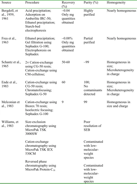
Table 1: Methods used in the purification of SEB in the literature ... 59
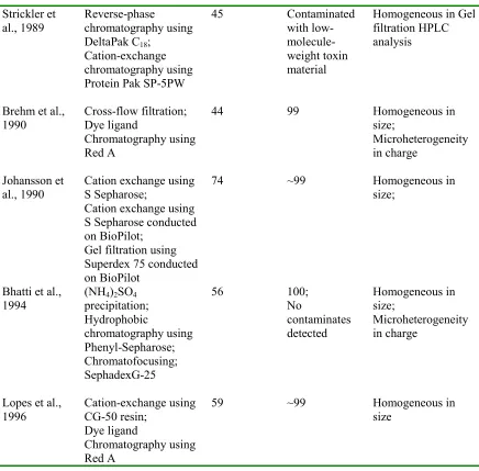
Table 2: Sequences deduced from the first library ... 61
Table 3: Sequence analysis of peptides from the first library... 62
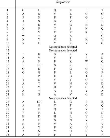
Table 4: Sequences deduced from the second library... 63
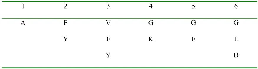
Table 5: Truncated sequences deduced from the second library ... 64
Table 6: Secondary screening of peptides from the first library... 65
Table 7: Secondary screening of peptides from the second library... 66
Table 8: Competitive secondary screening ... 67
Table 9: Secondary screening with blocked peptide ligands ... 68
Table 10: Effect of Tween on the binding of SEB to YYWFYY and YYWLHH ... 69
Table 11: Effect of salt in secondary screening ... 70
List of Figures
Figure 1: The 3D structure of SEB ... 78
Figure 2: Split synthesis of the one-bead-one-peptide library... 79
Figure 3: The binding of SEB to different columns using PBS as binding buffer ... 80
Figure 4: The binding of SEB to different columns using 0.5M sodium chloride in PBS
as binding buffer ... 81
Figure 5: The binding of SEB to different columns using 1M sodium chloride in PBS as binding buffer... 82
Figure 6: The effect of salt on SEB binding to a YYWLHH column... 83
Figure 7: The effect of salt on the binding of E. coli lysate to a YYWLHH column.... 84 Figure 8: The effect of salt on the separation of SEB from E. coli lysate using a
YYWLHH column... 85
Figure 9: The effect of salt on the binding of E. coli lysate to an amino column... 86 Figure 10: The effect of salt on the binding of E. coli lysate to an acetylated amino
column... 87
Figure 11: Purification of SEB from E. coli lysate using a YYWLHH column... 88
Figure 12: SDS-PAGE of the fractions from chromatography runs of the mixture of
1mg/ml E. coli lysate and 0.5mg/ml SEB using a YYWLHH column... 89 Figure 13: The effect of concentrations of E. coli lysate on the recovery of SEB using
a YYWLHH column ... 90
Figure 15: Purification of SEB from BSA using a YYWLHH column... 92
Figure 16: SDS-PAGE of the fractions from chromatography runs of the mixture of
5mg/ml BSA and 0.5mg/ml SEB using a YYWLHH column... 93
Introduction
Motivation
Staphylococcal enterotoxins (SEs) comprise a family of ten serologically distinct
toxins (labeled A-K), all of which are secreted by Staphylococcus aureus and share significant homology in primary structure. These toxins are primary causes of food
poisoning (Bergdoll, 1983). In addition, they act as superantigens to stimulate much more
T cells than conventional antigens do to enhance cytokine release (Swaminathan et al.,
1992; Marrack and Kappler, 1990; Johnson et al., 1991).
Due to the limited amount of SEs for intoxication (for example,
~5-20µg/Monkey), it is necessary to develop a sensitive and convenient analytical method to
detect such toxins. Furthermore, SEs contaminate other proteins produced from
Staphylococcal bacteria, such as protein A. Protein A plays an important role in
purification and therapeutic removal of IgG and IgG-containing immune complexes in
the treatment of certain cancers and autoimmune diseases (Balint et al., 1989). Thus an
extra step is needed to remove the trace SEs and reduce loss of activity and yield of
protein A. Homogeneous and highly purified preparations of SEs are required in studies
of the biological activity of SEs and the methods of identification and quantification of
SEs (Bhatti et al., 1994; Lopes et al., 1996). Hence an efficient and convenient
purification method is required to recover enterotoxins from fermentation broth and cell
culture. Affinity chromatography is used in laboratory and industrial scales to recover a
wide variety of biological molecules, especially when the concentration of product of
physical properties. Thus affinity chromatography is a promising method to detect,
remove or purify SEs.
The key in affinity chromatography is to create or find a proper adsorbent.
Ideally, a ligand should be specific, inexpensive, and stable during multiple operational
cycles. To recover the biological molecules while retaining their activity, the interaction
between ligands and target molecules should be moderate so that harsh elution conditions
can be avoided (Chase, 1983; Sproule et al., 2000). The primary affinity ligands used in
practice are immobilized metal atoms, dyes and polyclonal and monoclonal antibodies.
Both metal and dye ligands are pseudo-affinity ligands. These ligands tend to have lower
specificity than antibodies. In addition, dyes and metals have the potential to leach from
the column to contaminate the products. Monoclonal antibodies have been widely used in
protein purification because of their high affinity and specificity. However, monoclonal
antibodies must be purified extensively prior to use as affinity ligands. In addition,
immobilized antibodies are sensitive to operating conditions. Harsh conditions are
usually required to break antigen-antibody interactions in the elution step and even
harsher conditions are necessary (ex. 0.1M sodium hydroxide) to clean and generate the
columns prior to the next operating cycle. These conditions can denature the antibodies
over time so that only limited operating cycles can be carried out. Both the difficult
purification and relatively short life of antibodies result in high cost of antibodies as
affinity ligands. Furthermore, the leakage of the antibody into the product could elicit an
immune response (Huang and Carbonell, 1995; Bastek et al., 2000). As a result, low cost,
more robust ligands that have moderate interaction with the target could have widespread
been demonstrated to be good candidates as ligands in affinity chromatography (Lam et
al., 1991; Baumbach and Hammond, 1992; Huang and Carbonell, 1995).
SEB is the most widely studied member of the SEs (Swaminathan et al., 1992). It
is a good model of a toxin that can be used in biological warfare and other environmental
problems. We use SEB as target molecule to find affinity ligands from a solid phase
combinatorial peptide library, and then use such ligands to detect, remove and purify
SEB by affinity chromatography. In addition, the derived peptide ligands can help us
understand the interactions of SEB as a superantigen with the major histocompatibility
complex class II molecule (MHCII) and T cell receptor (TCR). As far as we are aware,
this is the first time an attempt has been made to identify affinity ligands for a toxin using
solid phase combinatorial peptide libraries.
Staphylococcal enterotoxin B (SEB)
Staphylococcal enterotoxins (SEs)
Staphylococcal enterotoxins (SEs) are a family of structurally related proteins that
are produced by Staphylococcus aureus (Bergdoll, 1983). These toxins function both as potent gastrointestinal toxins to cause toxic shock-like syndromes as well as
superantigens that stimulates non-specific T-cell proliferation to result in several allergic
and autoimmune diseases (Balaban and Rasooly, 2000). The SEs are single-chain simple
proteins (25-30kDa) which contain one disulfide loop in their molecule and are resistant
to temperature and pH denaturation (Bergdoll, 1983). To date ten SEs, designated as SEA
(Casman et al., 1963), SEB (Casman et al., 1963), SEC (Bergdoll et al., 1965; Reiser et
1998), SEH (Su and Wong, 1995), SEI (Munson et al., 1998), SEJ (Zhang et al., 1998)
and SEK (Orwin et al., 2001), have been identified and differentiated on the basis of their
reactions with specific antibodies. SEC is further divided into SEC1, SEC2 and SEC3
due to minor epitope variations. All these toxins share significant sequence homology.
Comparison of the sequences of the SEs indicates that SEA, SEE, and SED form one
group with an overall amino acid homology of 51%-81%, whereas SEB and SECs form
another group of toxins with sequence homology of 42%-67%. The homology between
the two groups is around 22-33% (Marrack and Kappler, 1990; Balaban and Rasooly,
2000). Among recently characterized SEs, SEJ has 52%-66% homology with the first
group, but SEI and SEH are more distantly related from both groups. Another enterotoxin
produced from Staphylococcus aureus, originally designated as SEF, has a distant
relationship with other SEs and now has been renamed as the Toxic Shock Syndrome
Toxin-1 (TSST1) (Bergdoll, 1985). Its 3D structure is remarkably similar to SEB (Kim et
al., 1994). All these toxins have similar mechanisms of pathogenesis.
As gastrointestinal toxins, SEs are the second most common cause of reported
food-borne illnesses. They usually contaminate some dairy food products, such as meat,
poultry, fish, milk and their products (Swaminathan et al., 1996; Balaban and Rasooly,
2000). Patients with SE poisoning may present with emesis, diarrhea, nausea, dizziness
and prostration. Some tests done on monkeys show that the amount necessary to cause
intoxication is very small, on the range of ~5-20µg/animal (Bergdoll, 1967). Little is
known about the mechanism of this intoxication at present. It is possible that emesis
occurs through stimulation of neural receptors in the abdomen by the toxins (Bayliss,
SEs are well understood to be superantigens. Superantigens cause immense T cell
activation that can be several orders of magnitude greater than that evoked by routine
antigens. This is due to their different modes of interaction with the major
histocompatibility complex class II molecule (MHCII) and T cell receptor (TCR) from
those of a routine antigen. Routine antigens have to be preprocessed by the
antigen-presenting cells (APC) into small peptides (13 to 17 residues in length). These antigenic
peptides bind to a cleft formed by two α helices of the α and β chains of the MHCII
molecule. The formed peptide-MHCII complex makes contact with all variable chains of
TCR and the fraction of T cells stimulated is limited by these interactions (Goldsby et al.,
2000; Swaminathan et al., 1992). Usually only 0.0001%~0.001% of T cells get activated
this way (Marrack and Kappler, 1987). Superantigens need no such preprocessing. They
bind as intact molecules but to a different part of the MHCII molecule than normal
antigenic peptides. The superantigen-MHCII complexes make contact only with the Vβ
chain of TCR. Different SEs have distinguishable specificities to TCR and MHC II even
though they fall into the same group based on sequence homology (Swaminathan et al.,
1996). The formation of MHCII-SE-TCR ternary complexes triggers the proliferation of
all T cells bearing particular types of Vβ elements. Since there are only a limited number
of Vβ elements in humans, a very large fraction (2~20%) of T cells get stimulated. The
consequent release of T cell-derived lymphokines such as interleukin-2 or tumor necrosis
factor may be involved in the mechanism of toxicity, causing fever, weight loss, and
osmotic imbalances that could lead to death (Swaminathan et al., 1992; Marrack and
is dissociable from that of T cell stimulation, both emesis and T cell stimulation are
dependent upon distinct but overlapping regions on SEs (Harris et al., 1993).
Characteristics of SEB
SEB is a monomer protein with a MW 28,366 Daltons and pI 8.6. The
three-dimensional structure of SEB determined by X-ray crystallography reveals a molecule
containing two domains composed of residues 1-120 and 127-239 respectively (Figure 1)
(Swaminathan et al., 1996; Papageorgiou et al., 1998). Domain 1 consists of two β sheets,
one formed by β1, β4 and β5 and the other by β2, β3, β4 and β5. It also contains three α
-helices, α1, α2 and α3. Domain 2 mainly consists of two α helices, α4 and α5 and a
twisted β sheet formed by β6, β7, β9, β10 and β12; it also includes two very short β
strands, β7 and β11. SEB binds to MHCII with high affinity (Kd ~10-6) (Scholl, et al.,
1989) and to T cells bearing Vβ3, 7, 8.1, 8.2, 8.3 and 17 in mice (White et al., 1989).
Based on the topology and mutational studies of SEB, the T-cell receptor-binding site on
SEB encompasses a shallow cavity formed by domains 1 and 2. The MHCII molecule
binds to an adjacent site, namely the α5 face of the SEB molecule. The active site for
emesis is thought to be in the α4 groove (Swaminathan et al., 1992).
SEB is one of the most heat-stable proteins. The activity loss due to heat is fast
initially and then levels off. Less than 50% activity is lost when the toxin is heated to
100°C at pH 7.3 for 5 mins (Schantz et al., 1965). Heat aggregation results in rapid loss
of activity at 70 to 80°C. But heating to 100°C can recover 35 to 40% immunological
activity because of the dissociation of aggregates at higher temperature. Therefore, heat
inactivated by heat can be recovered (Satterlee et al., 1969; Jamlang et al., 1971; Fung, et
al., 1973).
SEB is resistant to denaturation when it is exposed to denaturants, such as
guanidine hydrochloride and urea. Isothermal denaturation experiments show that
prolonged exposure (hours to days, depending on denaturant conditions) of SEB in
guanidine hydrochloride up to 6M and urea up to 9M is required for unfolding to reach
equilibrium. Refolding the denatured toxin to native protein after dilution of denaturant is
complete within minutes to a few hours. This indicates a very large activation energy for
unfolding and a comparatively small activation energy for refolding of SEB. The stable
structure of SEB is due to the disulfide loop that locks covalently two β sheets in a stable,
anti-parallel configuration (Warren, 1974, 1977). However, SEB can be denatured rapidly
and irreversibly when the pH is below 3.5. This is because SEB is a basic protein, and
hence low pH results in protonation of carboxylate groups that are involved in
maintaining the native SEB conformation. Such protonation leads to a conformational
change of SEB so that the local positive electrostatic free energy resulting from
protonation of the carboxylate groups can be reduced (Warren et al., 1974).
It is a challenge to deal with microheterogeneity of SEB when characterizing
SEB. Different preparation and purification methods give different SEB-associated
forms. It is generally agreed that only one component is synthesized within
Staphylococcus aureus strains. The hydrolysis of labile amide groups of glutamine and
asparagine residues in the protein converts the original component to less alkaline forms
of SEB by amidohydrolase in the fermentation broth. Thus there are series of isomers of
comparison between the amino acid sequence deduced from the DNA sequence, and the
amino acid sequence analyzed from purified SEB by Edman degradation, where most of
the differences involve aspartic acid and asparagines, and glutamic acid and glutamine
(Huang and Bergdoll, 1970; Jones and Khan, 1986). The distribution of the
multiply-charged SEB species is dependent on the operation conditions, especially temperature
and pH, in the preparation methods employed (Baird-Parker and Joseph, 1964; Schantz et
al., 1965; Chang and Dickie, 1970; Metzger et al., 1971; Spero et al., 1974). In addition,
polymerization of SEB usually develops during storage, and low ionic strength can
enhance that process (Jamlang et al., 1971).
Detection of SEB
The required detection level of enterotoxin in foods from outbreaks is
0.1~0.2µg/100g of food (Bergdoll, 1979). Most of the methods for detection of
enterotoxins are based on the use of antibodies prepared against the enterotoxins. One of
them is the gel diffusion method. Polyclonal antibodies prepared in rabbits using the
individual purified enterotoxins react with the enterotoxins in gels to give precipitin
reactions that are highly specific (Bergdoll, 1996). The used gels are generally
Ouchterlony gel plates and microslides. The normal sensitivity of gel diffusion methods
is around 0.1~0.5 µg/ml (Robbins et al., 1974; Meyer and Palmieri, 1980). The reversed
passive latex agglutination method (RPLA) is more sensitive than gel diffusion methods.
The antibody-coated latex particles agglutinate when brought in contact with the
enterotoxins. RPLA is sensitive enough to detect enterotoxins in most foods that cause
food poisoning (Weineke and Gilbert, 1987). However, enzyme-linked immunosorbent
enterotoxins. Several kits are available commercially (Fey et al., 1984; Park et al., 1994).
The antibody is treated with the sample and then the antibody-enterotoxin complex is
incubated with the antibody conjugate. The color developed from
enzyme-substrate reaction is directly proportional to the amount of enterotoxin in the sample.
Thus ELISA can be used directly on crude extracts from food or partially purified
samples. Most of the sorbents used to attach antibodies are microtiter plates, others
include polystyrene spheres, tubes, and dip sticks with wells. The sensitivity of an ELISA
is usually less than 1 ng/g food (Fey et al., 1984; Bergdoll, 1996). However, occasional
antibody cross-reactivity with unrelated antigens and the insensitivity with heat-treated
foods prevent definitive identification of the enterotoxin with the use of an ELISA (Park,
et al., 1992; Rasooly and Rasooly, 1998). Western blotting can overcome these major
problems because the Western procedure solubilizes denatured toxin, which may still be
biological active, and allows characterization of antigen that reacts with the antibody
(Rasooly and Rasooly, 1998). The most sensitive detection method (at pg level) is the
T-cell proliferation assay which measures the ability of enterotoxins to act as superantigen.
However, this assay is nonspecific since any enterotoxin in the sample can induce T-cell
proliferation. Thus one more step such as an ELISA is subsequently needed to identify
the enterotoxin (Rasooly et al., 1997). All these immunological methods share the same
disadvantage. They are relatively time-consuming, ranging from several hours up to
several days.
Biosensors based on immunological testing are an ideal alternative to the
immunological methods mentioned above. The limits of detection and sensitivity of
minutes. The detection mechanism of biosensors is based on the interaction between a
surface-immobilized receptor, mostly an antibody, and a solution-born analyte. The
consequential biological response of the antigen-antibody interaction is then translated
into an electronic output that can be analyzed (Rasooly and Rasooly, 1999; Nedelkov et
al., 2000). There are two common detection methods used in biosensors. One is based on
surface evanescent waves, which can be used to measure the changes in refractive index
due to antigen-antibody interactions at the surface of optical waveguides; these changes
can be recorded as a shift in resonance angle (Rasooly and Rasooly, 1999). The other is
based on surface plasmon resonance (SPR), where the change in the refractive index at
the surface results in a measured shift in the wavelength of light absorbed, or by light
diffraction (Nedelkov et al., 2000). The major problems of ELISAs, such as antibody
cross-reactivity with unrelated antigens and the insensitivity with heat-treated foods, can
also occur with biosensors. As in ELISA, the use of sandwich format immunoassay that
involves a secondary antibody could increase the specificity in a biosensor. A fiber-optic
biosensor system that includes a secondary fluorescent-labeled antibody can be used to
detect SEB with little cross-reactivity to SEA and SED (Tempelman et al., 1996; King et
al., 1999). Recently, biomolecular interaction analysis mass spectrometry (BIA-MS) has
been developed to differentiate specific antibody-enterotoxin binding from nonspecific
antibody-antigen binding in SEB detection, where a second step, matrix-assisted laser
desorption /ionization time-of-flight mass spectrometry, is used to identify SEB
(Nedelkov et al., 2000). These specific biosensor systems require complex instruments
and are not as convenient as ELISAs.
The determination of the biological activity of SEs requires highly purified and
homogeneous enterotoxins. Meanwhile, the methods of identification and quantification
of SEs, such as ELISA and biosensors, also require highly purified enterotoxins as
standards. Thus it is necessary to develop corresponding methods for the purification of
SEs. SEs are produced within Staphylococcus aureus strains and are released in the fermentation growth medium. SEA, SED and SEE belonging to the same group based on
sequence homology are synthesized and released into the growth medium near the
beginning of the exponential growth phase, whereas SEB and SECs belonging to another
group are produced during the late stationary phase (Baird-Parker, 1971; Spero et al.,
1987). Staphylococcus aureus strains, which release one dominant enterotoxin, and the corresponding growth conditions, are known for most of the SEs. With the use of a
highly SEB-productive strain, Staphylococcus aureus S-6, the concentration of SEB can
reach 270µg per ml culture supernant after 48-h fermentation (Donnelly et al., 1970;
Lopes et al., 1996).
Challenges presented in the purification of SEs from fermentation broth include:
1) contaminants with similar molecular weights and charge to the enterotoxin of interest,
such as other enterotoxins, protein A, proteases, staphylokinase, hemolysins or endotoxin
(Bhatti, et al., 1994); 2) microheterogeneity of SEs, which results from polymerization or
hydrolysis of labile amide groups. Different techniques have been used to purify SEs.
Most of these are developed from the purification of SEB. Only minor variants are
needed to apply these techniques to purify other enterotoxins because all SEs share a
great structure homology and have similar properties. The degree of purity and percent
methods developed to date seldom guarantee both (Table 1). Although multi-step
procedures give high purity, they usually give low yield of SEB and take a long time, and
cannot easily be adapted for large-scale purification. Chromatography methods give high
recovery and purity compared to chemical methods, such as acid or ethanol precipitation
(Table 1). Different chromatography methods, such as cation exchange, hydrophobic,
reverse-phase or chromatofocusing chromatography, have been used successfully to
recover SEB in large-scale. However, these methods are not specific enough to get highly
purified and homogeneous SEB. Additional steps, such as gel filtration, electrophoresis
or isoelectric focusing, are needed to polish the partially purified enterotoxin (Table 1).
Dye ligand affinity chromatography is a kind of psudo-affinity chromatography method,
which is more specific than the chromatography methods mentioned above. The
adsorption of enterotoxins to dye ligands results from a mixture of interactions consisting
of electrostatic, hydrophobic and hydrogen bond interactions. Large-scale purification of
SEA, SEB and SEC2 with one-step dye ligand chromatography by using Red A as ligand
has been demonstrated by Brehm et al (1990). But there are still some contaminants in
the preparation of SEB after dye ligand chromatography. A pre-purification step, such as
cation exchange, is recommended to remove a portion of the contaminants prior to Red A
chromatography (Lopes, et al., 1996). Thus more specific ligands are needed to capture
Affinity ligands from combinatorial peptide libraries
Advantages of peptide ligands
Affinity chromatography is the most efficient way to purify biomolecules. Many
biomolecules can be purified within one step using affinity chromatography with high
recovery. The key in affinity chromatography is to find a proper affinity ligand
corresponding to the molecules of interest. While monoclonal antibodies are the most
common ligands for laboratory uses, it is difficult to scale up these columns because
antibodies are quite costly and sensitive to operating conditions. Also, the leakage of the
antibodies under harsh elution and cleaning conditions can result in serous contamination
due to the immunogenicity of these proteins. Usually affinity chromatography with
monoclonal antibodies as ligands is used to polish the product in the last step of
purification. Dye ligands and immobilized metal ions are more stable than antibodies so
that they can stand harsh operation conditions, but both of them are lack of sufficient
specificity. In addition, dye ligands are toxic and there is severe leakage of immobilized
metal ions. Prior experiences using peptides as ligands ranging from 3 to 25 amino acids
has shown that peptides have affinities to molecules of interest that compare well with
dyes and immobilized metal ions. As opposed to monoclonal antibodies, small peptide
ligands are much more stable because they don’t require a specific tertiary structure to
maintain their biological activity. Small peptides are also not likely to cause immune
response in case of leakage into the products and it is easy to isolate peptides from the
products downstream. Peptides can be manufactured aseptically in large scale under
GMP (good manufacturing practices) conditions at relatively low cost. The interactions
conditions without damage to protein activity (Baumbach and Hammond, 1992; Huang
and Carbonell, 1995; Huang et al., 1996; Huang and Carbonell, 1999; Bastek et al.,
2000). In addition to being good candidate as ligands in affinity chromatography,
peptides are used widely to determine protein-protein interactions without a priori
information on protein structure (for example in epitope mapping).
Combinatorial libraries for peptide ligand detection
One of the challenges for use of peptide ligands is the identification of a sequence
that shows affinity and specificity to the target protein. The design of specific
complementary peptide sequence to the target protein has been demonstrated difficult
even when the structure of the target protein is known (Lawrence and Davis, 1992;
Saragovi et al., 1992). The development of combinatorial libraries has allowed screening
millions of peptide sequences to discover specific peptides that bind to the target protein.
Peptide libraries can be generated either biologically or synthetically. Several
combinatorial library methods have been described in the literature (Lam et al., 1997).
The most widely used biological libraries are phage-displayed libraries, while
one-bead-one-peptide libraries are the dominant libraries obtained directly from chemical synthesis.
Phage-displayed random peptide libraries
In phage-displayed peptide libraries, a random gene with a given length is
synthesized and then inserted into bacterial phage gene III. The corresponding peptide
coded by the inserted DNA is displayed at the N-terminal of the gene III protein (pIII) on
the phage surface. Each phage displays one kind of peptide sequence that is different
from other phages. Affinity peptides on phage that bind to the target protein are selected
incubated with the target protein that has already been immobilized on a Petri dish or
ELISA plates. Non-binding phages are washed out extensively, and then the bound
phages are eluted under harsh conditions. The eluted phages are then amplified on agar
medium and subjected to the next round of affinity purification. The tight-binding phages
are then cloned and propagated in Escherichia coli. The amino acid sequence of the peptide on the phage is deduced by sequencing the coded DNA in the phage gene III
(Scott and Smith, 1990; Devlin et al., 1990; Cwirla et al., 1990).
Ligands identified from phage libraries frequently interact with natural binding
sites on the target molecule and resemble the target’s natural ligands. Thus
phage-displayed random peptide libraries have been used to investigate protein-protein
interactions in a variety of contexts. For example, phage-displayed random peptide
libraries have been used to map the epitopes of monoclonal and polyclonal antibodies,
and to identify peptide ligands for receptors, receptor ligands, and folded domains within
larger proteins, such as several SH2, SH3 domains (Daniels and Lane, 1996; Zwick et al.,
1998). Recently, peptide ligands for some superantigens, for example, SEB and TSST-1,
have been determined with phage-displayed random peptide libraries (Sato et al, 1996;
Goldman et al., 2000). But biopanning with phage-displayed libraries is slow and
subjected to non-specific binding.
The most important aspects in the determination of peptide ligands using phage
display are the construction and maintenance of libraries with sufficient structural
diversity and the efficient selection of specific binding phage from nonspecific binding
phage. Phage-displayed random peptide libraries have been constructed to display
Once it is created, a phage library can be regenerated continuously and re-used unlike a
synthetic library. The problem in phage library construction is that the library may not be
truly random due to genetic bias in the creation of these libraries. The efficiency of
screening can be controlled by adjusting the washing conditions in the screening process
(D’Mello and Howard, 2001). But it is also possible that the specific peptide on phage
that is finally selected is not the original one from the library because of some biological
bias in amplification and propagation of the phage. Care must be taken to maintain the
diversity of the libraries. Usually the diversity of the original phage library is on the order
of 108 peptides. Selection must be avoided during the expansion and propagation of
library for phage with selective growth advantage (Daniels and Lane, 1996). In addition,
the resources of the peptide synthesis on phage are limited to the 20 natural amino acids
so that D-amino acids or other molecules cannot be used to increase the diversity of the
library.
One-bead-one-peptide libraries
Synthesized libraries are created on solid supports through organic chemistry.
There are several distinct combinatorial library methods (Lam et al., 1997). The
one-bead-one-peptide library method is used extensively in drug discovery processes due to
its unique features (Lebl et al., 1995). Compared to other methods, the synthesis of a
one-bead-one-peptide library is rapid with use of the “split synthesis” approach. Because one
bead has one unique peptide sequence, all of the beads can be tested concurrently but
independently. Once positive beads have been identified, the chemical structure of the
peptides on the beads can be directly determined by sequencing or by an encoding
attached on solid) or solution phase (i.e. peptide cleaved from solid support). As in
phage-displayed libraries, the screening of peptide ligands from one-bead-one-peptide
libraries involves three steps (Lam et al., 1997): (i) construction of the library, (ii)
screening the library with the target molecule, (iii) determination of the peptide sequence.
We will give a brief discussion of the application of one-bead-one-peptide libraries in
determining peptide ligands specific for a biomolecule of interest.
Construction of the one-bead-one-peptide libraries
The first one-bead-one–peptide library was synthesized by Lam et al. (1991)
using the “split synthesis” approach developed by Furka et al. (1991)(Figure 2). The resin
beads are divided equally into separate reaction vessels each with a single amino acid.
After the first amino acid is coupled to the resins, beads are repooled, mixed thoroughly,
and redistributed into separate reaction vessels. The next coupling step is then performed.
This divide-couple-recombine technique is repeated until the desired length of the peptide
library is reached. There are Xn random sequences in the library, where X is the number
of amino acids used for coupling, and n is the length of the library. Each resin bead
displays only one peptide sequence. Thus libraries of this type are called
“one-bead-one-peptide” libraries (Lam et al., 1991). Because other ligands besides naturally occurring
amino acids, such as D-amino acids, oligonucleotides, synthetic oligomers, proteins and
small molecules, also can be coupled to solid resins, the idea of a one-bead-one-peptide
library has been extended to one-bead-one-compound library (Lam et al., 1997). The
introduction of other compounds besides amino acids in combinatorial library
construction increases the diversity of the library in comparison with a phage-displayed
However, all synthetic methods have a practical limit on the size of the library as well as
the length of the peptides on beads, while peptides on phage can be fairly large.
The choice of the solid support is critical for the library construction and the
following application of the library. The biological signal released from the peptides on a
single bead quantitatively depends on the amount of the peptide on the bead. As a result,
the size and substitution homogeneity is of the utmost importance. Meanwhile, the resin
should resist the formation of clusters because cluster would prevent the statistical
distribution of resin beads and lower the number of structures created. In addition, resins
should be compatible with various organic and aqueous media. Solid beads with porous
structure are preferred. The high surface area that porous resins provide can attach more
ligands, facilitating bead sequencing and providing high capacity for their usage in
chromatography. Moreover, the pores should be large enough to eliminate diffusion
resistance especially when using large proteins as targets. In order to avoid nonspecific
binding between the solid matrices and proteins, hydrophilic resins are preferred. If the
peptide ligands will be used to purify protein in chromatography, the resins should have
enough mechanical rigidity to withstand the high pressure used in liquid chromatography.
A variety of polymer beads have been used to attach peptides in library construction,
including polyhydroxylated methacrylate, polydimethylacrylamide,
polyoxyethylene-grafted polystyrene, TentaGel and so on (Buettner et al., 1996; Lam et al., 1997).
Screening of the one-bead-one-peptide libraries
The identification of the specific peptide ligands to the target protein from a
one-bead-one-peptide library depends on the screening methods used. Both solid-phase and
library method. The most widely adopted method of screening is the “on-bead” binding
assay (Lam and Lebl, 1994, Lam et al., 1997). The target protein is incubated with the
library beads. The library beads with specific peptide sequence to the target protein bind
the target protein. The binding of the target to the bead-bound ligands is usually detected
by using a reporter group such as an enzyme, a radionuclide, a fluorescent probe, or a
color dye covalently attached to the target molecules. Alternatively, antibodies can also
be used in the detection scheme as in ELISA. The signals generated from these reporter
groups are proportional to the amount and density of peptides on the bead. Nonspecific
binding can result in high background resulting in some difficulty in determining the
affinity ligands. This usually is eliminated by using a high ionic strength buffer (e.g.
0.2-0.4M NaCl) to reduce purely electrostatic binding and blocking proteins (e.g. casein or
bovine serum albumin) and nonionic detergents (e.g. 0.1% Tween 20).
One of the most convenient screening methods is the enzyme-linked colorimetric
detection scheme. It has been used to discover the binding motifs for streptavidin (Lam et
al., 1991; Lam and Lebl, 1992), avidin (Lam and Lebl, 1992), monoclonal antibodies
(Lam et al., 1996), proteases (Lam et al., 1996), and MHC molecules (Smith et al., 1994).
The alkaline phosphatase coupled target protein is used to bind to library beads, and then
the substrate of alkaline phosphatase, 5-bromo-4-chloro-3-indolyl-phosphate (BCIP) is
added. The reacting beads turn turquoise, while the majority of the beads in the library
remain colorless. The positive turquoise beads are isolated and then sequenced by Edman
degradation. The enzyme-linked colorimetric detection method is extremely rapid, taking
a few hours to screen 107-108 beads. The problem with this method is that the enzyme
on beads. Radionuclide-labeled targets can be used to screen library beads to avoid this
problem. The radionuclide probes, such as 3H and 14C, are particularly small compared to
enzyme as reporter groups on the target, and it has been demonstrated that the labeled
target shows almost same biological properties as the natural target. The library is
incubated with the radiolabeled target protein, washed, and then suspended in agarose
gel. The slurry is poured onto a gel bond to form a monolayer so that all beads are
spatially separated. Exposure of the gel to autoradiography film can locate the positive
beads that are then isolated and sequenced. Several researchers have screened peptide
libraries using radiolabeled targets (Kassarjianet al., 1993; Turck, 1994; Nestler, et al.,
1996; Mondorf et al., 1998). The method developed by Mondorf et al. (1998) using 14C
offers high resolution and sensitivity. It has been used to identify affinity peptide ligands
for s-protein (Mondorf et al., 1998), fibringen (Mondorf et al., 1998), Alpha-1-proteinase
inhibitor (Bastek et al., 2000), α-lactalbumin (Gurgel et al., 2001) and recombinant factor
VIII (Chen et al., 2000). Immunostaining schemes similar to ELISA also can be used to
target the protein on beads. There are no modifications of the target using this method, so
the bead-bound ligands bind directly to the native protein, and not to any adducts.
However, the antibodies used in the detection system could bind to bead-bound ligands
besides the targets to bring the possibilities of interference and false positives. A two
color PEptide Library Immunostaining Chromatographic ANanlysis (PELICAN) has
been developed to determine beads specific for the target from those beads resulting from
antibody cross reactivity (Buettner et al., 1996). Peptide ligands used to purify protease
factor IX and fibrinogen were identified with the use of the PELICAN method (Buettner
dye-labeled targets or fluorescently dye-labeled target (Chen et al., 1993; Needels et al., 1993).
However, dyes always complicate the screening process by binding to many peptide
ligands directly, while autofluoresence of the library can make the library unsuitable for
this kind of screening process (Lam et al., 1997). In order to minimize the number of
false positive beads, two orthogonal screening methods can be used for one target. For
example, a dual-color detection scheme (Lam et al., 1995) and a cross-screening scheme
with enzyme-linked colorimetric and radiolabeled assays (Liu and Lam, 2000) have been
developed. In this way, many of the initially determined positive beads are eliminated,
and the chances to get the true positive beads are greatly enhanced.
One of the disadvantages of on-bead screening is the high peptide density
required for peptide sequencing on beads. This can lead to multiple-point attachment of
the target to the peptides so that nonspecific interaction between target and peptides will
be enhanced. Thus the selected peptide ligands may have less affinity and specificity to
the target. Screening of soluble peptide libraries can make the affinity ligands more
selective. The format of affinity chromatographic screening developed by Evans et al.
and Huang & Carbonell is suitable for screening peptide libraries due to its rapidity
(Evans et al., 1996; Huang and Carbonell, 1999). The targets are immobilized onto resins
and then packed into a chromatographic column. The soluble peptide libraries are
pumped into the column at a proper flow rate to ensure the peptides have enough time to
bind to the immobilized targets. Then the column is washed thoroughly with binding
buffer. The affinity peptide ligands bound to the targets are eluted and isolated by
reverse-phase chromatography. The fractions are then sequenced by Edman degradation
showing that the consensus sequence, NFVE that binds to the s-protein, is the same as
that found from the screening a phage displayed library (Huang and Carbonell, 1999).
Evans et al. used a similar system to recover the known epitope, YGGFL, for monoclonal
antibody (3E-7), and then determining the affinity ligands for bacterial
lipopolysaccharide (LPS, endotoxin) (Evans et al., 1996). Although this technique is
rapid and able to avoid false signals from nonspecific binding, it misses some affinity
peptide leads that generally cannot be missed by using on-bead screening. The kinetics of
the chromatography and the orientation of the immobilized targets, may limit the contact
between the immobilized targets and free peptide ligands. In addition, the methodologies
used in this technique are more complex than those in on-bead screening (Huang and
Carbonell, 1999).
Protein purification by peptide ligands
Although peptide ligands from phage library have been presented on
chromatographic support to purify proteins (Baumbach and Hammond, 1992; Huang and
Carbonell, 1995; Huang et al., 1996), it is possible that the microenvironment and the
orientation of the peptides on the chromatographic support could be very different from
that on phage. This can affect adversely the interactions between the peptide ligands and
the target (Buettner et al., 1996). It has been found that some peptides derived from phage
library only work when the peptide is an integral part of the phage coat protein and not
when isolated in free solution (Lowe, 2001). Chemically derived peptides are synthesized
and screened on solid beads. They can be used directly to purify protein by
chromatography. Thus one-bead-one-peptide libraries are widely used to discover peptide
The affinity and specificity of the peptide ligands from the screening of the
peptide library can be confirmed and further demonstrated using a chromatography
format. Some peptide ligands identified by library screening are bioselective to their
targets, while other peptide ligands are pseudo-affinity ligands behaving like dye ligands.
The study by Huang et al. shows that the peptide ligand, YNFEVL, is so specific to
s-protein that randomization of the peptide sequence destroys the binding (Huang and
Carbonell, 1995). A similar study on the binding of von Willebrand Factor to the peptide
RLRSFY shows that the randomization has little effect on the binding (Huang et al.,
1996). The pseudo-affinity peptide ligands are better used to capture the target at the first
step in purification (Bastek et al., 2000); other steps such as filtration are needed to polish
the products. The bioselective peptide ligands can be efficient for purifying the protein in
one step, but usually preparation prior to the affinity chromatography step is needed to
protect the ligands and maximize the efficiency of the affinity column. Basically, the
affinity of the selected peptide ligands depends on the screening of the peptide library.
Based on the behavior of the peptide ligands in purifying target in a column format, it is
possible to adjust the screening conditions to yield proper ligands for purification.
Ligand density can affect the interactions between the peptide ligands and the
target protein. If the binding is attributed to monovalent interactions, the capacity
increases when increasing the ligand density, while the association constant may remain
constant at low ligand density and decrease at high ligand density due to steric effects.
Thus there is an optimal density at which the peptide ligands have high capacity and an
acceptable extent of steric hindrance. If the binding is attributed to multivalent
constant. For highly specific ligands, increasing the ligand density may increase the steric
hindrance at the surface and make the binding less efficient, namely, decreasing the
association constant and the utilization of the ligands. Small protein molecules are
expected to have monovalent interactions with the adsorbent. The binding of s-protein to
a peptide sequence, YNFEVL, has been shown to be 1:1 specific (Smith et al., 1993).
Further adsorption isotherm measurements in a batch system have shown that the binding
capacity increases from 0.0466µmol/ml to 1.1650µmol/ml, while the peptide utilization
decreases from 96% to 40%, and the binding constant decreases from 1.2×10⁵ to 5.6×10⁴
M-1 when the peptide density increases from 0.05 to 3.0µmol/ml (Huang and Carbonell,
1995). It is more likely that the binding of large protein molecules to peptide ligands is
multivalent because there may have several interaction sites at their surfaces. The results
of binding of von Willebrand Factor (vWF) to a small peptide ligand, Ac-RVRSFYK,
immobilized on Toyopearl resin, shows that the association constant increases from
8.82×10⁵ to 2.06×10⁶ M-1, and the maximum capacity from 2.32 to 10.33 mg/ml when
the peptide density increases from 32 to 60 mg/ml (Huang et al., 1996).
Baumbach and Hammond (1992) first demonstrated the availability to use peptide
ligands as affinity ligands in large-scale chromatography processes by using streptavidin
as target. Since then, this technique has been successfully used to purify a variety of
proteins, such as s-protein (Huang and Carbonell, 1995), von willebrand factor (Huang et
al., 1996), fibrinogen (Buettner et al., 1997; Mondorf et al., 1998), factor IX (Buettner et
al., 1996), factor VIII (Necina et al., 1998; Amatschek et al., 2000; Chen et al., 2000),
trypsin (Makriyannis and Clonis, 1997), IgG (Verdoliva et al., 1995; Fassina et al., 1996),
anti-MUC1 antibodies (Murray et al., 1997; Murray et al., 1998), Alpha-1-proteinase inhibitor
Materials and Methods
Peptide synthesis
Two hexamer one-bead-one-peptide libraries were used in our test. Both libraries
were synthesized similarly. The first library was from Peptides International (Louisville,
KY) and was synthesized onto Toyopearl AF Amino 650M resin of 65µm in diameter
with 16 of 20 natural amino acids (except cysteine, methionine, isoleucine and
Tryptophan). The second hexamer peptide library was from Bayer Corporation (Raleigh,
NC) and was synthesized directly onto Toyopearl AF Amino 650M resin of 120µm in
diameter (TosoHass, Montgomeryville, PA) with 18 of the 20 natural amino acids
(excepting cysteine and methionine). Both libraries were synthesized using standard
fluorenylmethyloxycarbonyl (Fmoc) chemistry as described by Buettner et al. (Buettner
et al., 1996, 1998). The base resin is highly hydrophilic with pores of 1000A in diameter,
making it suitable for purifying proteins up to 5M Da in size. Briefly, a 1:1 mixture of
Fmoc-L-alanine to tBoc(tert.butyloxycarbonyl)-L-alanine was coupled to the amino
functionality on the resin. The tBoc group was released with TFA and the free amino
functionality was acetylated with acetic anhydride. No further peptide synthesis occurred
at these acetylated sites. Subsequently, the Fmoc protecting groups were released with
piperidine. Fmoc-protected amino acids were used to synthesize peptides onto resin by
using “split synthesis” until the last cycle was finished. The final substitution density
based on total amino acid analysis was 100µmol/g. Sequencing of the peptides was
performed at the Protein Technology Laboratory, Texas A&M University, College Park,
resin at a substitution density of 100µmol/g from Peptides International (Louisville, KY).
All the remaining amino groups used to couple peptides on the base resin were
acetylated.
Radiolabeling of SEB
Highly purified SEB of at least 95% purity (Toxin Technology, Sarasota, FL) was
labeled at 4°C with 14C by reductive methylation utilizing sodium cyanoborohydride and
14C-formaldehyde (NEN Life Science Products, Boston, MA) as described by Jentoft and
Dearborn (1983). The sodium cyanoborohydride was recrystallized prior to use due to
hydrate formation during storage. SEB was dissolved into 0.1M sodium phosphate buffer,
pH 7.0. Sodium cyanoborohydride and 14C-formaldehyde were added sequentially. The
14C-formaldehyde was added at a 2-fold molar excess over 5% of the total methylation
sites (68 total, 2×(33 lysines + N-terminus)) to achieve around 5% methylation of SEB.
Sodium cyanoborohydride was added at 10X the molarity of 14C-formaldehyde. The
reaction was performed at 4°C overnight. The labeled SEB was separated from 14
C-formaldehyde by using an EconoPac 10DG Desalting Column (Biorad, Hercules, CA)
equilibrated with 0.1M sodium phosphate buffer, pH 7.0. The radioactivity of each
fraction collected during the separation was determined using a 1500 Tri-Carb Liquid
Scitillation Analyzer (Packard, Meridian, CT) and CytoScint ES scintillation liquid (ICN,
Mesa, CA). The protein concentration of each fraction was quantified by the BCA Assay
(Pierce, Rockford, IL). The radioactivity yield after labeling was around 1014~1015
Primary screening of the libraries
A radiological detection approach described by Mondorf et al. was used to deduce
the peptides that bind SEB (Mondorf et al., 1998). 20mg of peptide library beads were
washed with 20% methanol for one hour, and then washed thoroughly with the binding
buffer, phosphate buffered saline (PBS) (Sigma, St. Louis, MO) containing 10mM
phosphate buffer, 2.7mM KCl, and 137mM NaCl, pH 7.4. The blocking reagent used for
the beads from the first library was a mixture of 1% casein (Pierce, Rockford, IL), 1%
BSA (Pierce, Rockford, IL) and E. coli lysate (1X over SEN in mass) (Promega,
Medison, WI), while the blocking reagent used for the beads from the second library was
1% casein or 0.5% casein plus 0.5% BSA. The library beads were first blocked with 1ml
of blocking reagent in PBS in a 1.7ml centrifuge tube for 2 hours on a rotating plate to
minimize nonspecific interactions between the library beads and 14C-labeled SEB.
Concentrated 14C-labeled SEB was added directly into the reaction slurries to reach a
concentration of 0.5µM and incubated for 2 hours at room temperature. Following the
incubation, the beads were transferred to a Poly-Prep chromatography column (Biorad,
Hercules, CA) and washed thoroughly with PBS with 0.05% Tween (Sigma, St. Louis,
MO) until the radioactivity of the flow through reached background levels. The washed
beads were suspended into 0.8% low melting agarose (Biorad, Hercules, CA) at 35°C and
divided equally into two aliquots in 50ml containers. Each of slurry was poured onto a
160×180 mm gelbond (BioWhittaker Molecular Applications, Rockland, ME) to form a
monolayer of beads. The gelbond was air-dried overnight in a hood and subsequently
Beads corresponding to the positive signals were identified by careful alignment of the
films and gel, and isolated with a scalpel under a microscope. The isolated beads were
each placed in a centrifuge tube with water and incubated for 10 minutes at 75°C in a
water bath, followed by sonication for 10 minutes to remove the agarose from bead. The
clean beads were suspended in methanol and sent to Protein Technologies Laboratories,
Texas A&M University, TX, for sequencing by Edman degradation using a
Hewlett-Packard G1005A.
Secondary screening: binding confirmation in batch format
Non-competitive format
Individual peptides deduced from primary screening were synthesized directly onto
Toyopearl AF Amino 650M resin at a substitution of 100µmol/g resin (Peptides
International, Louisville, KY). All remaining amino groups were acetylated. Beads were
washed with 20% methanol for one hour and subsequently rinsed thoroughly with PBS
buffer. 400µl of the slurry in PBS with a bead density of 1mg/20µl was transferred to a
0.5ml centrifugal filter with 0.45µm Durapore membrane (Millipore, Milford, MA).
Buffer was removed by centrifugation. Resins were incubated with 400µl of 1µM 14
C-SEB in PBS buffer at room temperature on a rotating plate for 2 hours, and then washed
with PBS buffer for 15 min, and then sequentially washed for one hour each with 400µl
of: 1M NaCl in PBS buffer, 2% acetic acid and 6M GdnHCl. Each solution after
centrifuge was collected into scintillation fluid for radioactivity counting. The total
in the same manner, allowing a total radioactivity balance. Toyopearl AF Amino 650M
resins and acetylated Toyopearl AF Amino 650M resins were treated the same as above
as controls.
Competitive format
Positive peptides identified from the non-competitive format studies were incubated
with a mixture of labeled SEB and a blocker protein to test the affinity of the peptides to
SEB. The procedure was similar to that of non-competitive format except using a mixture
of SEB and blocker instead of SEB, or adding blocker first to block the peptides for one
hour and then replacing blocker with the mixture. Two blockers including 1mg/ml BSA
(Pierce, Rockford, IL) and 1mg/ml casein (Pierce, Rockford, IL) were tested for their
effects on SEB binding. Toyopearl AF Amino 650M resins and acetylated Toyopearl AF
Amino 650M resins were investigated the same as above as controls.
Tertiary screening: binding confirmation in column format
To test the selectivity of the peptides that showed positive binding in the
secondary screening, the performance of the peptide ligands was investigated in a
chromatography column format. In order to minimize the usage of SEB due to its high
costs and toxicity, the peptide beads were packed into a 0.21×3cm column, a microbore
column from Alltech (Deerfield, IL) and tested on the Walters 616 LC system (Millipore,
Milford, MA) with a UV detector (Knauer, Germany) at 280 nm. The column was
pre-equilibrated with binding buffer at a flow rate of 0.15ml/min (173cm/hr). The following
program was used for each column run: From time 0 to 10 minutes, sample was injected
was then washed with binding buffer for 10 minutes and subsequently with 1M NaCl in
binding buffer for 20 minutes at a flow rate of 0.15ml/min. From time 40 to 60 minutes,
the bound protein was eluted by switching to 2% acetic acid at a flow rate of 0.15ml/min.
The column was re-equilibrated with 50 CVs (column volume) of binding buffer before
the next run. E. coli lysate (Promega, Medison, WI) and BSA(Pierce, Rockford, IL) were used as competitive protein mixtures to test the selectivity of the ligands to SEB in
chromatography. Using 0.5M sodium chloride in PBS as binding buffer, several solutions
of E. coli lysate combined with SEB were tested: 1mg/ml E. coli lysate, 1mg/ml E. coli lysate + 0.25mg/ml SEB, 1mg/ml E. coli lysate + 0.5mg/ml SEB, 1mg/ml E. coli lysate +
1mg/ml SEB, 1mg/ml SEB. Similar tests were done except using 5mg/ml BSA instead of
1mg/ml E. coli lysate. All peaks were collected and their composition determined by
Sodium-dodecyl-sulfate polyacrylamide gel electrophoresis (SDS-PAGE) (Invitrogen,
Carlsbad, CA). We also investigated the recovery of 0.5mg/ml of SEB from various
amount of E. coli lysate (1mg/ml, 2.5mg/ml, 5mg/ml, and 10mg/ml).
To investigate the effect of salt on the separation of SEB from E. coli lysate, another chromatography run was used. From time 0 to 10 minutes, sample was injected
through a 100µl loop (Thomson, Springfield, VA) at a flow rate of 20µl/min. The column
was then washed with binding buffer for 10 minutes and subsequently with 1M NaCl in
binding buffer for 35 minutes at a flow rate of 0.15ml/min. From time 55 to 75 minutes,
the bound protein was eluted by switching to 2% acetic acid at a flow rate of 0.15ml/min.
The column was re-equilibrated with 50 CVs (column volume) of binding buffer before
Results
Primary screening
Initial work in the identification of peptide ligand for SEB was performed by
using library 1, a one-bead-one-peptide library from Peptides International. This peptide
library was made of 16 of 20 natural amino acids (except cysteine, methionine, isoleucine
and Tryptophan) and the diameter of the library beads was 65µm on average. The
blocking agent used was a mixture of 1% casein and 1% BSA and E. coli lysate (10X
over SEB in mass). 0.5µM of labeled SEB was used to yield enough background for
alignment after 5 days of exposure of the beads to photographic films. There were no
positive beads whose signals were well above the background. Thirty-five beads that had
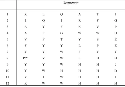
small positive signals were picked out and sequenced. The sequences are listed from the
N to C terminus as shown in Table 2. Because high density of peptide ligands would
cause significant high background due to nonspecific interaction, the peptide library used
here was a low-density library at a density of 100µmol/g resin. The amount of amino acid
at each cycle is less than 10pmol/bead, while the limitation of detection is
5~10pmol/bead by using Edman degradation at Texas A&M. This is a part of reason
resulting in difficulty in sequencing. In addition, the beads of 65µm are so small that it’s
hard to handle them while picking them out from gels and washing them to remove the
agarose. Thus another one-bead-one-peptide library (library 2) containing larger beads
was used next to determine peptide ligands for SEB.
The second library was synthesized as described by Buettner et al. using 18 of 20
peptides in each bead and hence yield stronger signals than small beads. Compared to the
first library, the second library produced positive signals well above the background, and
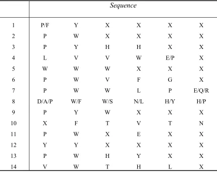
it was much easier to handle these larger beads. Around 1gram of library beads were
screened, which represents 5% of a whole peptide library. Unfortunately, many positive
beads selected for sequencing have truncated sequences due to the low peptide density.
Usually the first three amino acids near the N terminus can be sequenced, and tyrosine
(Y) and tryptophan (W) dominate these positions (Table 5). The initial yield of the first
amino acid on the bead ranges from 3.8 to 21.1pmol indicating that there is a large
distribution of peptide density on beads of this library. This peptide library had been used
previously to deduce peptide ligands for ovalbumin and prion protein in our lab. Thus the
problems associated with low peptide density and large distribution of peptide density
may result from long-time storage (received in Nov., 21, 1996). The peptide sequences
with all 6 amino acids sequenced are listed from the N to the C terminus in Table 4.
Aromatic amino acids dominate the first three positions at the N terminus in peptides
No.7 to No.12; especially there is a consensus, YYW, in sequences No. 7, 8, and 9. In
addition, there is another consensus, WHH, near the C terminus in sequences No.9 to
No.12, and a similar sequence to WHH, WLH, in sequence No.8. These sequences
indicate that aromatic amino acids may play a critical role for SEB binding.
Secondary screening
Secondary screening was used to confirm the peptide ligands from primary
screening. 14 of 35 peptide sequences deduced from the first library and 12 of peptide
sequences from the second library were synthesized directly onto Toyopearl AF Amino
groups at the bead surface were acetylated to avoid nonspecific binding. Because it was
difficult to call the first cycle of the peptide P/Y-Y-W-L-H-H, both PYWLHH and
YYWLHH were synthesized for secondary screening. All peptide ligands were incubated
with 1µM of 14C-labeled SEB for 2 hours to make sure equilibrium was established. The
bound SEB was eluted by using 1M NaCl, 2% acetic acid, and 6M GdnHCl. Nonspecific
electrostatic interactions were removed by using 1M NaCl. Stronger binding due to
affinity would be disrupted by using protein denaturant, 2% acetic acid and 6M GdnHCl.
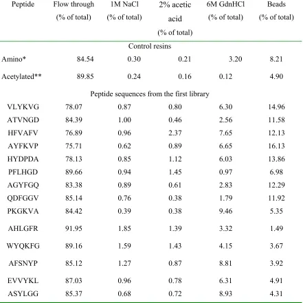
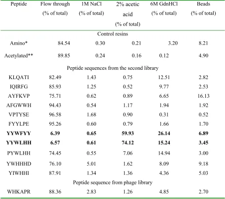
Results are shown in Table 6 and Table 7 with blank Toyopearl AF Amino 650M resin
and its acetylated form as controls. None of the sequences that deduced from the first
library can bind SEB significantly. This might have been expected because it was much
more difficult to pick up positive signals from background signals in primary screening.
There are only two peptide sequences, YYWFYY and YYWLHH (YYWHHG will be
tested in future), which can bind most of the SEB added, although other peptide
sequences such as FYYLPE, PYWLHH, YWHHHD and YIWHHI have similar structure
to these two. This indicates that the first three amino acids, YYW, may be special for
SEB recognition. Goldman E.R. et al. have deduced several peptide sequences that bind
SEB from a 12-mer phage-display library and there is a consensus, WHK, at the N
terminus of these sequences (Goldman E.R. et al., 2000)(Table 12). We selected one of
them, WHKAPRAPAPLL, and synthesized its first 6 amino acids near the N terminus,
WHKAPR, onto Toyopearl AF Amino 650M resin to test its ability to bind SEB. As
shown in Table 7, it is no better than the control resins. In order to choose an appropriate
concentration of SEB used in secondary screening, some of secondary screening
SEB was increased on both control resins and tested peptide ligands (data not shown). In
this case, a significant portion of the binding was due to the nonspecific interaction
between SEB and the solid support. Thus 1µM of SEB was set up for further
investigations so that the nonspecific binding onto the support can be neglected.
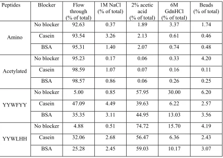
In order to know the selectivity of the positive peptide ligands, YYWFYY and
YYWLHH, competitive secondary screening was performed by adding competitive
proteins with SEB into binding buffer. Two kind of proteins, casein and BSA, which
were the blockers used in the primary screening, were tested. The results are presented in
Table 8 with Toyopearl AF Amino 650M resin and its acetylated form as controls. The
binding capacities of both YYWFYY and YYWLHH decreased with the existence of a
competitive protein. Casein was a stronger blocker than BSA to inhibit SEB binding. The
performance of YYWLHH was much better than that of YYWFYY. Even though the
concentration of competitive protein was 35 times higher than that of SEB, YYWLHH
was still able to bind more than 60% of SEB. This indicates that the amino acids close to
the resin also affected the performance of the peptide ligand significantly although steric
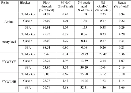
effects inhibited their contact with binding molecules. We further tested the affinity of
peptide ligands to SEB by blocking the peptides first using casein or BSA, then added
SEB directly to the SEB solution to see the performance of SEB binding. The fractions of
bound SEB were almost the same for YYWFYY and YYWLHH, but dropped
dramatically in comparison with those in noncompetitive secondary screening (Table 9).
This shows that casein and BSA also blocked the specific binding sites for SEB besides
blocking nonspecific binding sites, and SEB was not strong enough to displace bound
![Crystal structure of diaqua[5,10,15,20 tetrakis(4 bromophenyl)porphyrinato κ4N]magnesium](data:image/gif;base64,R0lGODlhAQABAIAAAP///wAAACH5BAEAAAAALAAAAAABAAEAAAICRAEAOw==)