Department of CIVIL, CSE, ECE, EEE, MECHNICAL Engg. and S&H of Muthayammal College of Engineering, Rasipuram, Tamilnadu, India
Copyright to IJIRSET www.ijirset.com 241
Texture and Shape Content Based MRI
Image Retrieval System
N. Kumaran #, Dr. R. Bhavani #
#
Department of Computer Science and Engineering, Annamalai University, Annamalai Nagar, Chidambaram, Tamilnadu, India
Abstract— More number of techniques about medical image
retrieval is used in medical domain. In our Content Based Medical Image Retrieval System, high level semantics of an image is very important. Regarding with this issue, we proposed a method using co-occurrences matrix to extract texture features and Canny Edge Detection is to extract shape features. Then K-means clustering algorithm and Euclidean distance measure are used to retrieve similar images for a query image in medical diagnosis. The performance of this system is efficient when compare to existing System.
Keywords
—
Canny Edge Detection; CBMIR,Co-occurrence Matrix; Euclidean Distance Measure; K-means Clustering
I. INTRODUCTION
large number of medical images are generated by hospitals and clinics every day (such as CT, X-ray and MRI).Such images constitute an important source of anatomical and functional information for diagnosis of diseases, medical research and education. It is well acknowledged that medical image database is key component in diagnosis and preventive medicine.
Content based medical image retrieval (CBMIR)[1][2][3] is the digital image searching problem in large database that makes use of contents of image themselves rather than relying on the textual information. Medical images generated in hospitals contain semantic information. This information can be used to retrieve the images. Under such circumstance, medical image retrieval has changed from previous text based method to the content based method or the method which combines both. According to the characteristic of medical images such as texture and shape features are the important low level features [4][5][6].
Quddus et.al [7] proposed a method, which reveals superior robustness performance with respect to accuracy, speed and multimodality in CBMIR. In their work, support vector machine (SVM) are used for identifying 3-D MR volume and performing semantic classification of human brain into various semantic region.
A novel scheme has been proposed by Jiann- Der Lee et.al [8] for image retrieval task using the feature
extracted directly from a compressed or uncompressed image. The texture information is first extracted by exploiting the multi resolution
nature of wavelet decomposition, which represents the horizontal, vertical and diagonal frequency distribution of an image. Then calculate the mean and standard deviation of wavelet coefficient of each sub-band as texture features. The technique of scale multiplication is analyzed in the framework of Canny edge detection proposed by Paul Bao et.al [9].
A scale multiplication function is defined as the product of the responses of the detection filter at two scales. Edge maps are constructed as the local maxima by thresholding the scale multiplication results. The detection and localization criteria of the scale multiplication are derived.
Reshma Chauudhari et. al [10] proposed an algorithm which incorporates the advantage of various other algorithms to improve the accuracy and the performance of retrieval. The accuracy of color histogram based matching can be increased by using color coherence vector for successive refinement. The speed of shape based retrieval can be enhanced by considering approximate shape rather than exact shape. In addition to this a combination of color and shape retrieval is also included to improve the accuracy of the result
A novel approach by successfully combining rotation invariant contourlet transform and Fourier descriptor was proposed by Arun K.S. et.al [11]. Rotation invariant contourlet transform is used for texture feature and Fourier descriptor extract shape feature.
Bag of feature approaches have become prominent for image retrieval and image classification task in the past decade. Jingyan et.al [12] proposed a novel visual word weighting method. The discriminative power of each visual word is analyzed by the sub-similarity function in the bin that corresponds to the visual word. Each sub-similarity function is then treated as a weak classifier. A strong classifier is learned by boosting method that combines that weak classifier.
This paper is organized as follows. Section II describes the overall architecture of the proposed retrieval technique. Section III describes the co-occurrence matrix
Department of CIVIL, CSE, ECE, EEE, MECHNICAL Engg. and S&H of Muthayammal College of Engineering, Rasipuram, Tamilnadu, India
and extraction of texture features. Section IV explains the extraction of shape feature using canny edge detection. Section V depicts the K-means clustering. Section VI illustrates about the Euclidean distance measure. Section VII express the experimental work and discuss the results obtained from experiments. Section VIII tells a summary of the paper.
II. PROPOSED METHOD
The block diagram of the proposed work is shown in the fig1. In this paper we extract texture and shape features using co-occurrence matrix [13] [14] and canny edge detection [15].
Fig. 1 Proposed CBMIR System
Then K-means clustering [16] and Euclidean distance measure [17] are used for best medical image retrieval
.
III.EXTRACTIONOFTEXTUREFEATURES Since we are interested in the statistical approach, first we make use of the most suitable texture features. The major advantage of using the texture attributes is obviously their simplicity The most common features used in practice are the measures derived from spatial gray tone co-occurrence matrix i.e., texture features such as contrast, angular second moment, entropy, energy, inertia, inverse difference moment etc. These features have been widely used in the analysis, classification and interpretation of medical images. They are defined by the equations as follows:
Entropy:
F3= -
i j
j
i
p
j
i
p
(
,
)
log(
(
,
))
Energy:
F4=
1 0 1 1
0
(
(
,
))
N
i N
j
p
i
j
2
Inertia:
F5=
1 0 1 10
(
)
N
i N
j
i
j
2
) , (i j p
Inverse difference moment:
F6=
1 0 1 1
0
(
(
,
))
N
i N
j
p
i
j
1+ (i-j)2
Average of Gray:
F7=
1 0 1 1
0
(
,
)]
[
N
i
N
j
p
i
j
i
Average of Difference:
F8=
1 1 0 1 0
(
,
)]
[
N
j
N
i
p
i
j
j
Gray non uniform:
F9=
1 0 1 1
0
(
,
)]
[
N
i N
j
p
i
j
2
Difference non-uniform:
F10=
1 1 0 1 0
(
,
)]
[
N
j N
i
p
i
j
2
Gray entropy:
F12=
1 0 1 1 0 1 10
(
,
)
lg
)
,
(
N i N j Nj
p
i
j
j
i
p
Difference Entropy:
F13=
110 1 0
1 0
(
,
)
lg
)
,
(
N j N i Ni
p
i
j
j
i
p
Correlation
F14= y
i j x
j
i
p
ij
(
)
(
,
)
y
x
IV.EXTRACTIONOFSHAPEFEATURES
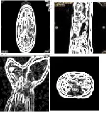
The Canny edge detection algorithm is known to many as the optimal edge detector. Canny's intentions were to enhance the many edge detectors already out at the time he started his work. Based on these criteria, the canny edge detector first smoothes the image for eliminate noise. It then finds the image gradient to highlight regions with high spatial derivatives. The algorithm then tracks along these regions and suppresses any pixel that is not at the maximum. By using canny edge detection we can find the edges in the following images is shown in fig.2.
Department of CIVIL, CSE, ECE, EEE, MECHNICAL Engg. and S&H of Muthayammal College of Engineering, Rasipuram, Tamilnadu, India
Copyright to IJIRSET www.ijirset.com 243
Fig. 2 Sample edge detection for brain, spine, and knee joint and abdomen MRI images.
By using canny edge detection the following shape features has been extracted:
Area:Area of selection in square pixels or in calibrated
square units. (e.g., mm2, μm2, etc)
Mean gray value: Ave rage gray value within the selection. This is the sum of the gray values of all the pixels in the selection divided by the number of pixels.
Standard deviation: Standard deviation of the gray values used to generate the mean gray value.
Center of mass: This is the brightness-weighted average of the x and y coordinates all pixels in the image or selection. These coordinates are the first order spatial moments.
Integrated density: The sum of the values of the pixels in the image or selection. This is equivalent to the product of Area and Mean Gray Value.
Median: The median value of the pixels in the image or selection.
Skewness: The third order moment about the mean. Kurtosis: The fourth order moment about the mean. The following fig.3 shows the sample output screen for extraction of texture and shape features.
Fig.3 Sample output screen for shape and texture feature extraction
V. K-MEANSCLUSTERING
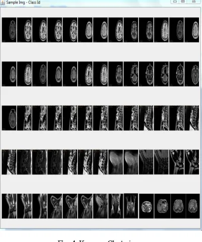
The K-means algorithm initializes 10 clusters by arbitrarily selecting one image to represent each cluster. Each of the remaining images is assigned to a cluster and the clustering criterion is used to calculate the cluster mean. These means are used as the new cluster points and each image is reassigned to the cluster t hat it is most similar to. This continues until there is no longer a change when the clusters are recalculated. The following fig.4 shows the clustering of database using cluster number as three.
VI.IMAGERETRIEVAL
The basis of many measures of similarity and dissimilarity is the Euclidean distance. The distance between vectors X and Y is defined as follows:
D
ij2
nv 1
(
X
viX
vj)
2
The Euclidean distance is calculated between the query image and the clustered images. The calculated distances are sorted in increasing order and display the first N images as the best similar MRI scan images for medical treatment.
VII. EXPERIMENTALRESULTS
The domain of medical imaging as a specific part of medicine is a very convenient environment for using a variety of CBIR systems. The main reason is the necessity of
.
Department of CIVIL, CSE, ECE, EEE, MECHNICAL Engg. and S&H of Muthayammal College of Engineering, Rasipuram, Tamilnadu, India
Fig. 4 K-means Clustering
effective content based analysis, extracting clinically relevant features out of the image and successful retrieval. This CBMIR (content based medical image retrieval) system consists of four major parts. The first one is feature extraction, where a set of features is generated to represent the content of each image in the database.. The second job is using K-means clustering for reducing robustness for retrieving images. Then the final and third task is to use Euclidean distance measure, where a distance between the query image and each class in the database is computed using their image feature values so that the N most similar images which are belonging to one class can be retrieved. The performance of both the shape and texture features, and combination of both features are compared and given in Table 1. To evaluate the retrieval efficiency of the proposed system, we use the performance measure, Recall and Precision.
Recall=Rr/T
Precision=Rr/Tr
TABEL: 1 Performance measure
MRI Images
Texture Shape Texture &Shape Precision Recall Precision Recall Precision Recall Brain 0.76 0.57 0.80 0.58 0.89 0.64 Spine 0.84 0.49 0.87 0.62 0.97 0.68
Knee joint 0.80 0.54 0.85 0.53 0.95 0.67
Abdomen 0.73 0.46 0.75 0.67 0.85 0.62
Where Rr is the number of relevant retrieved images, T is the total number of relevant items in an image database, and Tr is the number of all retrieved items. This method is implemented on a computer system using JAVA as the programming language and MS-Access as the back end. In this work we use around 450 MRI scan images as a database such as brain, abdomen, knee joint and spinal cord.The sample output screen is shown in Fig. 5.
Fig. 5 Sample output screen for spine MRI image retrieval
VIII. CONCLUSION
Content based medical image classification and retrieval based on texture, shape features and Euclidean distance measure are a field of diversity and have an enormous scope of application in medical fields. This method is applied around 450 MRI images such as brain, spine, abdomen, knee joint and retrieval efficiency is found with usual precision and recall analysis. We have planned to extend our work to various parts of the human body MRI scan images and test its performance in future.
REFERENCES
Department of CIVIL, CSE, ECE, EEE, MECHNICAL Engg. and S&H of Muthayammal College of Engineering, Rasipuram, Tamilnadu, India
Copyright to IJIRSET www.ijirset.com 245 [2] K.Wanjale, Tejas Borawake and Shahideep” Content Based Image
Retrieval for Medical Image Techniques and Storage Methods Review Paper”, International Journals of Computer Application, vol. 1, no.19, 2011
[3] Reshma Chauudhari and A.M Patil”Content Based Image Retrieval Using Color and Shape Features”, International Journal of Advanced Research in Electrical, Electronics and Instrumentation Engineering, vol.1, 2012.
[4] Bikesh Kr.Singh and G.R. Shinha” Content Based Retrieval of X-ray Images Using Fusion of Spectral Texture and Shape Descriptor”, International Conference on Advances in Recent Technologies in Communication and Computing, vol.978-0-7695-4201-0, 2010
[5] Shunren Xia, Weirong Mo, and Zanchao Zhang” A Content Based Retrieval System for Endoscopic Images”, International Journal of Information Technology, vol. 11 no.12, 2005.
[6] B.S Manjunath, Jens-rainer ohm, Vinod V. Vasudevan, and Akio yamanda” Color and Texture Descriptor”, IEEE Transactions on
Circuits and Systems for Video Technology, vol. 11, no.6, 2011. [7] Azhar Quddus and Ootman Basir” Semantics Image Retrieval in
Magnetic Resonance Brain Volumes”, IEEE Transactions on
Information in Biomedicine, vol.16, no.3, 2012. [8] Jiann-der lee and Li-peng Lou “Using Texture and Shape Features
to Retrieve Sets of Similar Medical Images”, IEEE Transaction on Biomedical Engineering, Applications Basis and Communication, vol.15 no.5, 2003
[9] Paul Bao, Lie Zhang, Xiaolin Wu “Canny Edge Detection Enhancement by Scale Multiplication “, IEEE Transaction on Pattern Analysis and Machine Intelligence, vol.27, no.9, 2005 [10] Reshma Chauudhari and A.M Patil ”Content Based Image
Retrieval Using Color and Shape Features”, International Journal of Advanced Research in Electrical, Electronics and Instrumentation Engineering, vol.1, 2012.
[11] Arun k. s, and Hema P Menon” Content Based Medical Image Retrieval by Combining Rotation Invariant Contourlet Features and Fourier Descriptors”, International Journals of Recent Trend in Engineering, vol.2, no.2, 2009.
[12] Jingyam Wang, Yongping Li, Ying Zhang, Chao Wang Honglan Xie, Guoling and Xin gao” Bag-of-Features Based Medical Image Retrieval via Multiple Assignment and Visual Words Weighting”, IEEE Transaction on Medical Imaging, vol. 30,no.11, 2011 [13] Peiqiang Zhang and Hongguang Zhu” Medical Image Retrieval
Based on Co-occurrence Matrix and Edge Histogram”, IEEE, vol. 978-1-61284-77-0, 2011 [14] David A. Clausi and Huang Deng ” Design-Based Texture Feature
Fusion Using Gabor Filters and Co-occurrence Probabilities”, IEEE, vol. 1057-7149, 2005.
[15] K.Lakshmi priya and D.chirstopher Durairaj ”Integration of morphological segmentation and Canny edge detection for iris regonotion”, IJCA vol.80-No.2 oct 2013
[16] Hui Xiong, Junjie Wu and Jain Chen “K-means Clustering verses Validation Measure: A data-distribution Prespective” IEEE, vol. 39, no. 2, April 2009
![Fig. 1 Proposed CBMIR System Then K-means clustering [16] and Euclidean distance](https://thumb-us.123doks.com/thumbv2/123dok_us/1536962.1188501/2.612.223.516.155.549/fig-proposed-cbmir-k-means-clustering-euclidean-distance.webp)