INVESTIGATION
Kinetochore Function and Chromosome Segregation
Rely on Critical Residues in Histones H3 and H4
in Budding Yeast
Tessie M. Ng,*,†Tineke L. Lenstra,‡Nicole Duggan,* Shuangying Jiang,§Steven Ceto,* Frank C. P. Holstege,‡Junbiao Dai,§Jef D. Boeke,** and Sue Biggins*,1 *Division of Basic Sciences, Fred Hutchinson Cancer Research Center, Seattle, Washington 98109,†Molecular and Cellular Biology Program, University of Washington, Seattle, Washington 98195,‡Molecular Cancer Research, University Medical Centre Utrecht, CG Utrecht 3584, The Netherlands,§School of Life Sciences, Tsinghua University, Beijing 100084, China, and **High Throughput Biology Center, Johns Hopkins University School of Medicine, Baltimore, Maryland 21205
ABSTRACTAccurate chromosome segregation requires that sister kinetochores biorient and attach to microtubules from opposite poles. Kinetochore biorientation relies on the underlying centromeric chromatin, which provides a platform to assemble the kinetochore and to recruit the regulatory factors that ensure the highfidelity of this process. To identify the centromeric chromatin determinants that contribute to chromosome segregation, we performed two complementary unbiased genetic screens using a library of budding yeast mutants in every residue of histone H3 and H4. In one screen, we identified mutants that lead to increased loss of a nonessential chromosome. In the second screen, we isolated mutants whose viability depends on a key regulator of biorientation, the Aurora B protein kinase. Nine mutants were common to both screens and exhibited kinetochore biorientation defects. Four of the mutants map near the unstructured nucleosome entry site, and their genetic interaction with reduced IPL1can be suppressed by increasing the dosage ofSGO1, a key regulator of biorientation. In addition, the composition of purified kinetochores was altered in six of the mutants. Together, this work identifies previously unknown histone residues involved in chromosome segregation and lays the foundation for future studies on the role of the underlying chromatin structure in chromosome segregation.
I
T is critical to understand the mechanisms that ensure accurate chromosome segregation because errors are asso-ciated with diseases such as cancer and can lead to cell death (Williams and Amon 2009; Compton 2011). Proper chromo-some segregation is a highly regulated process that requires the coordination of a number of events. After DNA replica-tion, sister chromatids are physically linked by cohesion (Oliveira and Nasmyth 2010; Nasmyth 2011). Kinetochores, the macromolecular complexes that assemble on centro-meric DNA, must biorient and attach to microtubules from opposite poles. Once bioriented attachments are made, sis-ter kinetochores come under tension due to microtubule-pulling forces on linked sister chromatids. Kinetochoreslacking tension trigger the spindle checkpoint until proper attachments are made (Nezi and Musacchio 2009). Once every pair of sister chromatids has made bioriented attach-ments at metaphase, cohesion is dissolved, allowing sister chromatids to segregate to opposite poles at anaphase.
A key regulator of biorientation is the conserved chro-mosomal passenger complex (CPC), an essential protein kinase complex that detects and corrects microtubule– kinetochore attachments that are not under tension (Lampson and Cheeseman 2011). Phosphorylation of kinetochore sub-strates by the CPC protein kinase Aurora B destabilizes such aberrant attachments, giving the cell another opportunity to make proper, bioriented attachments (Liu and Lampson 2009). The CPC localizes to the inner centromere (Cooke et al.1987), consistent with the model that tension between sister kinetochores stabilizes bioriented attachments by moving key substrates at the outer kinetochore away from the CPC (Tanaka et al. 2002; Fuller et al. 2008; Liuet al. 2009). However, the precise mechanism by which the CPC acts on attachments not under tension is still unclear Copyright © 2013 by the Genetics Society of America
doi: 10.1534/genetics.113.152082
Manuscript received April 8, 2013; accepted for publication September 5, 2013 Supporting information is available online athttp://www.genetics.org/lookup/suppl/ doi:10.1534/genetics.113.152082/-/DC1.
1Corresponding author: Fred Hutchinson Cancer Research Center, 1100 Fairview Ave.
(Maresca and Salmon 2010). The Aurora B kinase is also required for the spindle checkpoint when kinetochores lack tension (Biggins and Murray 2001), although it is controver-sial whether this function is due to its role in destabilizing kinetochore–microtubule attachments (Musacchio 2011).
Another conserved protein implicated in biorientation and the tension checkpoint is shugoshin. Although the shugoshin family is well known for its meiotic role in protecting centromeric cohesion (Watanabe 2005; Gutierrez-Caballero et al.2012), some family members also facilitate kinetochore biorientation and the checkpoint response to the lack of ten-sion during mitosis (Indjeianet al. 2005; Vauret al. 2005; Indjeian and Murray 2007; Kiburzet al.2008). A conserved requirement for shugoshin localization to centromeres and pericentromeres is the phosphorylation of H2A S121 by the Bub1 protein kinase (Kawashima et al. 2010). In budding yeast, shugoshin (Sgo1) recruitment to nucleosomes also requires residue G44 in H3, which resides near H2A S121 in the nucleosome structure (Luger et al. 1997; Luo et al. 2010). In many organisms, there is an interdependence be-tween shugoshin and Aurora B localization and activity (Dai et al.2006; Resnicket al.2006; Kawashimaet al.2007, 2010; Vanoosthuyseet al.2007; Kellyet al.2010; Wanget al.2010; Yamagishiet al.2010; Storchovaet al.2011), consistent with their close association with chromatin.
The underlying foundation of kinetochores is a specialized chromatin structure that creates the epigenetic mark for kinetochores and contributes to their assembly and function. While the bulk of the genome contains nucleosomes with 147 bp of DNA wrapped around two copies each of histone H2A, H2B, H3, and H4, the centromere contains a specialized histone H3 variant called Cenp-A (Maddox et al. 2012). In most organisms, Cenp-A nucleosomes are interspersed with H3 nucleosomes in the core centromere andflanked by H3 nucleosomes in heterochromatin (Blower et al. 2002; Cam et al. 2005). In budding yeast, there is a single Cenp-A nu-cleosome positioned at the centromere (Meluh et al. 1998; Furuyama and Biggins 2007; Krassovskyet al.2012), as well as additional Cenp-A in theflanking pericentromeric chroma-tin (Lawrimore et al. 2011; Henikoff and Henikoff 2012). While budding yeast pericentromeres lack heterochromatin, a conserved feature is the enrichment of cohesin andSgo1to promote kinetochore biorientation (Blat and Kleckner 1999; Tanaka et al. 1999; Kiburz et al. 2005, 2008; Eckert et al. 2007). In addition, evidence suggests that the pericentro-meric chromatin adopts a specialized intramolecular structure that is organized bySgo1and facilitates biorientation in bud-ding yeast (Yeh et al.2008; Haase et al.2012). Consistent with this, changes in pericentromeric chromatin composition lead to defects in the organization of inner kinetochore pro-teins and chromosome segregation (Chambers et al. 2012; Verdaasdonket al.2012).
While it is clear that a specialized chromatin structure facilitates the assembly and function of .38 core kineto-chore proteins and additional regulatory proteins (Stellfox et al.2012; Valenteet al.2012), the key determinants of this
chromatin structure have still not been fully elucidated. We therefore set out to identify histone H3 and H4 residues that contribute to chromosome segregation and kinetochore bio-rientation by performing two systematic genetic screens in budding yeast. Our work identifies key residues in both his-tones that were previously not known to regulate segrega-tion, some of which contribute toSgo1function. This work lays the foundation for future studies aimed at understand-ing the roles of centromeric and pericentromeric chromatin in chromosome segregation and genomic stability.
Materials and Methods
Screen to identify mutants sensitive to decreased IPL1 function
Individual mutations were integrated at the endogenous HHT2-HHF2locus as described previously (Daiet al.2008). H3 mutations were integrated into SBY9120 and H4 muta-tions into SBY9119. Correct integration was verified by PCR using the primers SB2409 and SB2410 for H3 mutants, and SB2409 and SB2411 for H4 mutants. Integrations were attempted at least three times before a given mutant was not pursued (Supporting Information, Table S1). The ab-sence of the endogenous wild-type (WT) locus was also confirmed using the primers SB2409 and SB2412. Fivefold serial dilutions of asynchronously growing cells were grown for 2–3 days on YPD plates in the presence and absence of 25mg/ml doxycycline or 15mg/ml benomyl. Primer sequen-ces are available upon request.
Chromosome loss assays
The yeast strain (JDY176) used for testing chromosome loss was derived from SBY8053, which contains an artificial chromosome III fragment with SUP11 and HIS3 markers (Hieter et al.1985). TheHHT1-HHF1-coding fragment, in-cluding the promoter, was knocked out to generate JDY168. The ura3-1mutation was corrected to obtain URA3strains followed by deletion of the ORF to generate an ura3Δ0 strain as described (Brachmann et al.1998). The resultant strain, JDY176, was used in subsequent studies.
calculate the percentage of chromosome loss in the first generation.
Microbial techniques and plasmid construction
Media and microbial techniques were as described (Sherman et al.1974; Roseet al.1990). All experiments were performed at 23° unless otherwise noted. In all synchronous cell-cycle experiments reported, 1 or 10mg/mla-factor (custom synthe-sized by United Biochemical Research, Inc., Seattle) was used to arrestbar1-1andBAR1cells in G1, respectively. Doxycycline (Sigma-Aldrich, St. Louis) was used at 25mg/ml. Yeast strains are listed inTable S3. High-copySGO1(pSB1780) or control [pRS425, (Sikorski and Hieter 1989)] plasmids were intro-duced into the histone mutant strains by transformation.
To generate a high-copy SGO1 plasmid with the LEU2 marker (pSB1780), the URA32-mm plasmid pMK573 (Luo et al. 2010) was digested with HpaI and AatII to remove URA3. TheLEU2gene was isolated from YEplac181 (Gietz and Sugino 1988) by digestion with the same restriction enzymes and ligated to the digested plasmid pMK573 to create pSB1780.
Flow cytometry
For the orc2-1 experiment, WT and orc2-1 strains were shifted to 37°for 3 hr. For the histone mutant strains, cells were grown at room temperature. After harvesting cells, they were fixed with 70% ethanol at room temperature. Fixed cells were then incubated in 0.2 mg/ml RNase A (Sigma-Aldrich) in 50 mM Tris–HCl, pH 8.0, for 4 hr at 37°and 2 mg/ml Proteinase K (Roche, Indianapolis) in 50 mM Tris, pH 7.5, for 1 hr at 50°. Cells were then incubated with 5 mM Sytox Green (Molecular Probes, Eugene, OR) in 50 mM Tris, pH 7.5. Data were collected and analyzed using Cell Quest software (BD Biosciences, San Jose, CA).
Microscopy, protein, and immunological techniques
Analysis of GFP-LacI was performed as described (Biggins et al. 1999). For all microscopy experiments, .200 cells were scored. The Bernoulli distribution was used to assess statistical significance at 95% confidence. Anaphase was an-alyzed by staining cells with 49,6-diamidino-2-phenylindole (Sigma-Aldrich) and identifying cells with two separated DNA masses. Protein extracts were made and immunoblot-ted as described (Minshullet al.1996). Quantitative immu-noblotting was performed with IRDye secondary antibodies from LI-COR at a 1:15,000 dilution. The immunoblots were imaged on a LI-COR imaging system, and the protein levels were quantified using the ImageJ program. The mean of three independent experiments is reported. Loading controls for all experiments were either anti-tubulin (Accurate Chemical and Scientific) used at 1:1000, or anti-PGK1 (Invi-trogen) at a 1:10,000 dilution. Centromeric minichromo-somes were purified and analyzed by immunoblotting as described previously (Akiyoshi et al. 2009). Anti-Spc105 polyclonal antibodies were used at a 1:1000 dilution (Akiyoshi et al. 2010), anti-FLAG monoclonal antibodies
(Sigma-Aldrich) were used at 1:3000, and anti-Cse4 poly-clonal antibodies at 1:500 (Pinskyet al.2003). Anti-Ndc80 (OD4, 1:10,000), anti-Ndc10 (OD1, 1:5,000), anti-Mif2 (OD2, 1:6,000), and anti-Ctf19 (OD10, 1:15,000) poly-clonal antibodies were a generous gift from Arshad Desai (Akiyoshiet al.2009).
Chromatin immunoprecipitation assays and quantitative real-time PCR
Chromatin immunoprecipitation (ChIP) was performed using antibodies againstCse4as described previously (Collins et al. 2005), and samples were quantified by quantitative real-time PCR (7900HT, ABI Prism). DNA samples were am-plified using a SYBR PCR mix (Applied Biosystems) at 95° for 10 min, 40 cycles of 95°for 15 sec, and 55°for 1 min using CEN3 (SB1253 and SB1254) and PHO5-specific pri-mers (SB3063 and SB3064). PCR amplification efficiency and linearity were determined using serial dilutions of sam-ples. Standard curves were generated for every PCR reaction and used for quantification of bound DNA that was expressed as the percentage of input DNA. Sequences of PCR primers are available upon request.
Nucleosome structures
Nucleosome structures were prepared with PyMOL (http:// www.pymol.org/).
Expression profiling
Each mutant strain [derivatives of JDY86 as previously described (Dai et al. 2008)] was profiled four times from two independently inoculated cultures and harvested in early mid-log phase in SC medium with 2% glucose. Sets of mutants were grown alongside corresponding H3 and H4 WT cultures (single-copy H3 or H4) and processed in paral-lel. Dual-channel 70-mer oligonucleotide arrays were employed with a common reference WT RNA. All steps after RNA isolation were automated using robotic liquid handlers. These procedures werefirst optimized for accuracy (correct fold-change) and precision (reproducible result), using spiked-in RNA for calibration (van de Peppel et al. 2003). After quality control, normalization, and dye-bias correction (Margaritis et al.2009), statistical analysis for mid-log cul-tures was performed for each mutantvs.the WT cells grown alongside using Limma. The reported fold-change is an av-erage of the replicate mutant profilesvs.the H3 or H4 WT. Microarray data have been deposited in ArrayExpress under accession no. E-MTAB-1242 and in the Gene Expression Omnibus database under accession no. GSE39903.
Results
Identification of H3 and H4 residues important for chromosome segregation
and H4 mutants that exhibit chromosome loss. We used a previously constructed library of mutants where every H3 and H4 amino acid was systematically changed to alanine and the modifiable residues were changed to both alanine and a charged residue (Dai et al.2008). Deletions within the amino-terminal tails of each histone were also assayed. A cassette containing the mutants was introduced into the primary H3 and H4 locus,HHT2-HHF2, in a strain containing a deletion of the secondary H3 and H4 locus, hht1-hhf1Δ. The strain also contained an artificial chromo-some fragment that allowed us to assay the frequency of chromosome loss using a colony color-sectoring assay (Hieter et al. 1985). Mutants that exhibited increased sec-toring were quantified for the percentage of chromosome loss per generation. Together, 26 mutants in H3 and 15 mutants in H4 exhibited an increased frequency of chromo-some loss compared to WT (Figure 1, A and B).
Histone mutations can cause pleiotropic effects, so we performed a second screen to identify the histone residues that might specifically contribute to kinetochore biorienta-tion. We isolated mutants that require the full activity of the Ipl1/Aurora B protein kinase for viability. To do this, we used a previously characterized doxycycline-repressible
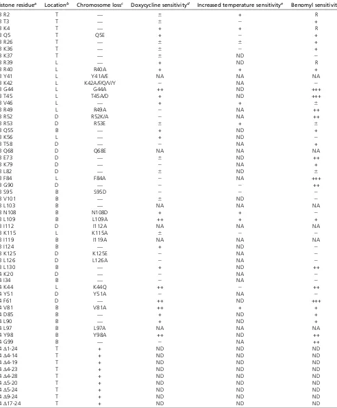
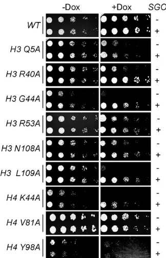
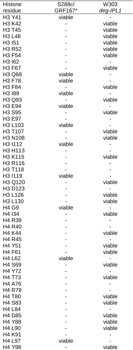
degron allele, deg-ipl1, that targets the protein for degra-dation by the proteasome (Nget al.2009). AlthoughIPL1is essential, doxycycline addition does not severely inhibit the growth of deg-ipl1 cells (Figure 1C), indicating that these cells retain enoughIpl1function to support viability. How-ever, deg-ipl1 is lethal when combined with other nones-sential mutants such as the mcm21 kinetochore mutant, indicating that it is a hypomorphic allele (Ng et al. 2009). We therefore introduced each alanine substitution mutation in H3 and H4 into a deg-ipl1 strain containing a deletion of the secondary copy of H3 and H4, hht1 -hhf1Δ. We note that 24 of the alanine mutants, including 6 of the residues identified in the chromosome loss screen (H3 Y41, H3 Q68, H3 L103, H3 I112, H3 I119, H4 L97), could not be generated in the deg-ipl1 strain background, suggesting synthetic lethality, and could not be further pur-sued (Table S1). The remaining mutants were analyzed for growth in the presence and absence of doxycycline, and 29 mutants that exhibited some degree of sensitivity to down-regulation ofIPL1compared to WT strains were identified (Table 1).
We focused on the mutants that were identified in both screens and are therefore most likely to be important for
Figure 1 Identification of H3 and H4 mutants with chro-mosome segregation defects. (A and B) The frequency of chromosome loss for the indicated histone H3 (A) and H4 (B) mutants identified in the colony-sectoring assay is plot-ted [all strains are derived from JDY176 (Table S3)]. (C) Fivefold serial dilutions of strains containing the indicated histone mutant in the presence or absence of deg-ipl1
(SBY719, SBY9837–SBY9847, SBY9880–SBY9888) were plated with (+) or without (2) doxycycline. Note that two different parent cassettes (WT H3 and H4) were used to generate mutants in the corresponding histone, and various pairs of images were cropped to assemble the
Table 1 Summary of mutant phenotypes identified in thedeg-ipl1and chromosome loss screens
Histone residuea Locationb Chromosome lossc Doxycycline sensitivityd Increased temperature sensitivitye Benomyl sensitivityf
H3 R2 T — 6 + R
H3 T3 T — 6 2 +
H3 K4 T — + + R
H3 Q5 T Q5E + 2 +
H3 R26 T — 6 6 +
H3 K36 T — 6 2 +
H3 K37 T — 6 ND 2
H3 R39 L — + ND R
H3 R40 L R40A + + +
H3 Y41 L Y41A/E NA NA NA
H3 K42 L K42A/F/Q/V/Y 2 NA 2
H3 G44 L G44A ++ ND +++
H3 T45 L T45A/D + ND +++
H3 V46 L — + + 6
H3 R49 L R49A 2 NA ++
H3 R52 D R52K/A 2 NA ++
H3 R53 D R53E 6 + 6
H3 Q55 B — + ND +
H3 K56 L — + ND 2
H3 T58 D — 2 NA +
H3 Q68 D Q68E NA NA NA
H3 E73 D — 6 ND ++
H3 K79 D — 2 NA +
H3 L82 D — 6 ND 6
H3 F84 L F84A 2 NA +++
H3 G90 D — 2 2 ++
H3 S95 B S95D 2 2 2
H3 V101 B — 6 ND 2
H3 L103 B — NA NA NA
H3 N108 B N108D + + 2
H3 L109 B L109A ++ + +
H3 I112 D I112A NA NA NA
H3 K115 L K115A 6 2 2
H3 I119 B I119A NA NA NA
H3 I124 B — + ND 2
H3 K125 D K125E 2 NA 2
H3 L126 D L126A 2 NA 2
H3 L130 B — + ND ++
H4 K20 D — 2 NA 2
H4 I34 B — 2 NA 2
H4 K44 L K44Q ++ 2 ++
H4 Y51 D Y51A 2 NA 2
H4 F61 D — ++ ND +++
H4 V81 B V81A ++ + +
H4 D85 B — + ND +
H4 L90 B — + ND +
H4 L97 B L97A NA NA NA
H4 Y98 B Y98A ++ ND ++
H4 G99 B — 2 NA ++
H4Δ1-24 T + ND ND ND
H4Δ4-14 T + ND ND ND
H4Δ4-19 T + ND ND ND
H4Δ4-23 T + ND ND ND
H4Δ4-28 T + ND ND ND
H4Δ5-20 T + ND ND ND
H4Δ5-24 T + ND ND ND
H4Δ9-24 T + ND ND ND
H4Δ17-24 T + ND ND ND
NA, not applicable because the histone mutant could not be generated in thedeg-ipl1strain background. ND, not determined.
aAll histone residues assayed were mutated to alanine. In some cases, additional mutants were also assayed as indicated in the chromosome loss column. bLocations as defined in DAIet al.(2008): T, tail; L, lateral; B, buried; D, disk.
cThe indicated histone mutant exhibited increased chromosome loss relative to WT. d“+”indicates sensitivity;“2”indicates no sensitivity.
chromosome segregation. The corresponding residues are H3 Q5, H3 R40, H3 G44, H3 T45, H3 R53, H3 N108, H3 L109, H3 K115, and H4 K44, H4 V81, and H4 Y98. However, the H3 K115A mutant cells grew extremely slowly (data not shown), and the H3 T45A mutant cells were previously reported to have replication defects (Baker et al.2010), so we did not continue to analyze them. The remaining nine mutants were assayed for the severity of their growth defect with or without the deg-ipl1 allele by plating serial dilutions in the absence and presence of doxycycline (Figure 1C). In the absence ofIPL1 downregu-lation, all of the mutants grew well except H4 K44A and H4 Y98A. In the presence of doxycycline, the H3 R40A, H3 G44A,H3 R53A,H3 N108A,H3 L109A,H4 K44A, and H4 V81A mutants exhibited a strong or complete loss of via-bility, whereas theH3 Q5A andH4 Y98Amutants showed a weak dependence on full IPL1function. All of the iden-tified residues are conserved, and mapping them onto the nucleosome structure shows that H3 R40, H3 G44, H3 R53, and H4 K44 cluster near the nucleosome entry/exit site, whereas H3 N108, H3 L109, H4 V81, and H4 Y98 are buried residues (Figure 1D) (Lugeret al.1997).
Analysis of replication and segregation in the histone mutant strains
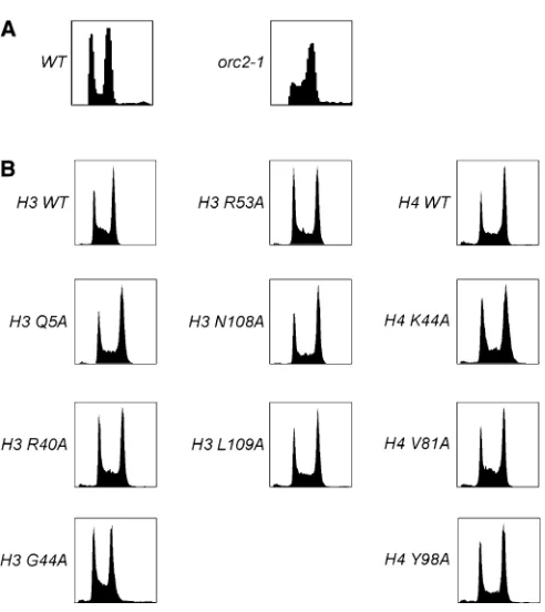
Because chromosome-loss phenotypes can be a result of either replication or segregation defects, we performed fluorescence-activated cell sorting (FACS) on the histone mutants to analyze replication. As a control, we analyzed theorc2-1temperature-sensitive mutant that is defective in replication and shows an accumulation of cells with DNA content between 1N and 2N (Figure 2A). The deg-ipl1 hht1-hhf1Δstrains containing WT or mutant H3 or H4 were grown in the absence of doxycycline and processed for FACS (Figure 2B). None of the mutants exhibited a strong delay in S-phase, although subtle replication defects may exist that cannot be detected due to the resolution of this assay.
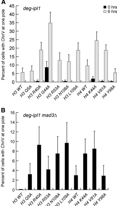
We next directly assayed chromosome segregation in each mutant strain by analyzing a fluorescently marked chromo-some (Straightet al.1996). Asynchronous cultures ofdeg-ipl1 strains containing GFP-marked Chromosome IV (ChrIV), and the H3 and H4 mutations were grown in doxycycline to re-press IPL1 for 6 hr. Cells that had proceeded through ana-phase (segregated DNA to opposite poles) were scored for segregation ofChrIVto a single pole (missegregation) or op-posite poles (accurate segregation) (Figure 3A). The stron-gest segregation defects occurred in the H3 R40A, H3 G44A, H3 L109A, H4 K44A, and H4 V81A mutant strains, which all exhibited.15% missegregation ofChrIVwithin 6 hr ofIPL1 downregulation. The H3 R53A and H3 N108A mutant strains showed a .10% missegregation defect after 6 hr, whereas there were no significant segregation defects in the H3 Q5A or H4 Y98A strains. Strikingly, the levels of chromosome mis-segregation in each mutant strain parallel the growth defects whenIPL1is downregulated (see Figure 1B), suggesting that the loss of viability is due to chromosome missegregation.
We next attempted to analyze segregation in a synchronous cycle by releasing cells from a G1 arrest into doxycyline. However, many of the histone mutants exhibited a transient delay in the onset of anaphase when released from G1 as indicated by the reduced percentage of cells with DNA masses at opposite poles relative to WT cells (data not shown). We reasoned that this delay could be due to spindle checkpoint activation, which would give sister chromatids additional time to biorient. We therefore examinedChrIVsegregation in deg-ipl1 mad3Δ strains containing the histone mutations during a synchronous cell cycle. As expected, the absence of the spin-dle checkpoint reduced the delay in anaphase onset because .50% of the cells segregated their DNA to opposite poles within 80 min post-G1 release, similar to WT cells.ChrIV seg-regation was monitored at the time point (100 or 120 min after G1 release) when the highest percentage of cells had DNA masses at opposite poles (Figure 3B). Similar to ourfi nd-ings on asynchronous cells, there were significant segregation defects (.8%) in the H3 R40A, H3 N108A, H3 L109A, and the H4 K44A mutant strains. By extrapolation, a missegregation frequency of 8% for a single chromosome means that,26% of the mutant cells would be able to segregate all 16 chromo-somes properly, consistent with the strong growth defects ob-served in these mutant strains. There were minor defects in the
H3 Q5A and H3 R53A mutant strains and no observable defect in the H4 Y98A strain. Whereas the H4 V81A strain appeared to have a missegregation defect, .4% of G1-arrested cells exhibited two GFP foci, indicative of aneuploidy, as compared to,1.3% in all other strains.H3 G44Amutant cells were not quantified in this experiment because.5% of cells exhibited two GFP foci in the G1 arrest, indicating preexisting aneuploidy that makes accurate quantification impossible.
Expression of segregation genes is not significantly altered in the histone mutants
To determine whether the segregation defects may be due to altered transcription of segregation genes, we performed DNA microarray expression analysis on each mutant. RNA was prepared from asynchronous cultures of WT or histone mutant strains (withoutdeg-ipl1). The complementary RNA was then labeled and hybridized to 70-mer oligonucleotide microarrays. Each mutant was analyzed for gene expression
changes .1.7-fold up or down and with aP-value ,0.01. We analyzed the list (Table S2) for genes known to be in-volved in kinetochore function or chromosome segregation and did not find any mutant that significantly altered any chromosome segregation genes. We note that H3 G44A, H4 K44A, and H4 V81A exhibit aneuploidy based on their gene expression profile, consistent with their segregation defects. Together, the microarray data suggest that the segregation defects in the histone mutants are not due to the altered transcription of one or more genes required for kinetochore function.
Cse4 localization to centromeres is normal in the histone mutants
Because the kinetochore assembles on a specialized chro-matin structure, we reasoned that the histone mutants might alter the chromatin at and/or around centromeres, thus disrupting chromosome segregation. Yeast centromeres contain a single well-positioned nucleosome that contains the specialized histone H3 variantCse4(Stoleret al.1995; Meluhet al.1998; Furuyama and Biggins 2007; Krassovsky et al.2012). We therefore tested whether totalCse4 levels are altered by any of the histone mutants by performing quantitative immunoblotting on crude lysates from WT and histone mutant strains with antibodies against Cse4. We compared the ratio of Cse4to Pgk1, a loading control, and found thatCse4levels are close to WT (610%) in most of the strains. However,Cse4levels were decreased by 13% in the H3 L109A mutant and 29% in the H4 V81A mutants and increased by 17% in the H3 R53A mutant strain (Figure 4A). The microarray data did not reveal any significant change in the transcription of CSE4in these mutants, indi-cating that the lower protein levels are likely due to a post-transcriptional effect. Cse4 levels are tightly regulated by proteolysis, so the H4 V81A mutant may alter the function or accessibility of the ubiquitin ligase that regulates Cse4 (Collins et al. 2004; Hewawasam et al. 2010; Ranjitkar et al. 2010). Although it is unclear how H3 mutants would alter Cse4levels, they may affect the ability of Cse4to in-corporate into euchromatin, which could affect the accessi-bility ofPsh1to degradeCse4.
We next asked whetherCse4incorporation at the centro-mere was affected in any of the histone mutant strains. Although most of the mutants that we identified are in H3, changes in the nucleosomes surrounding the centro-mere could lead to changes inCse4incorporation at centro-meres. In addition, it was recently reported that H3 also localizes to centromeres (Lochmann and Ivanov 2012), al-though the resolution of the assay used cannot discriminate between localization at the core centromere and surround-ing nucleosomes. To analyze Cse4 localization at centro-meres, we performed ChIP on the deg-ipl1 histone mutant strains grown in doxycycline for 6 hr. The cells contained a nonessential minichromosome so that we could analyze Cse4 in the context of a plasmid in addition to the en-dogenous centromere (Akiyoshi et al. 2009). Cse4 was
immunoprecipitated and the amount of DNA bound to Cse4 was analyzed by standard PCR with primers to the centromere. There was no obvious change in any of the strains (data not shown), so we analyzed a subset of them using quantitative real-time PCR with primers to the cen-tromeres or a control locus, PHO5 (Figure 4B). This revealed a slight decrease inCse4bound to the centromere in the H4 V81A mutant that had lowerCse4levels in the lysate. There was also a slight decrease in the level ofCse4 at the endogenous centromere in the H3 R53A mutant that had higher levels of total Cse4, although this was not ap-parent on the minichromosome. We have not previously detected changes in centromere-bound Cse4 when the gene is overexpressed (Collins et al. 2004), so this result is likely related to changes in the chromatin due to the H3 mutation rather than the alteredCse4levels. The remain-ing mutants showed no significant differences in the level ofCse4at either the endogenous or minichromosome cen-tromere when compared to WT. Together, these data dem-onstrate that the histone mutants do not significantly alter Cse4localization to the centromere.
Kinetochore stability is altered in the histone mutant strains
We next asked whether overall kinetochore integrity is normal in the mutants by purifying centromeric minichromosomes (Akiyoshiet al.2009). In contrast to ChIP techniques that
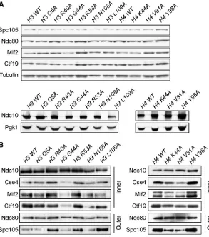
require a cross-linking step prior to immunoprecipitation, the minichromosome purification technique isolates native material and can therefore reveal subtle changes in kinet-ochore stability. A centromeric minichromosome contain-ing LacO sequences was introduced into each deg-ipl1 histone mutant that also expressedLacI-Flag, and the cells were grown asynchronously in doxycycline for 6 hr. We did not detect major alterations in the total protein levels of representative core kinetochore proteins in the lysates pre-pared from each histone mutant strain other than a slight reduction in Ndc10 levels in the H3 L109A mutant strain (Figure 5A). We therefore immunoprecipitated LacI-Flag and analyzed the purified minichromosome samples for the levels of copurifying kinetochore proteins. The Ndc10 protein is a component of the CBF3 complex that binds directly to the CDEIII element of the yeast centromere (Lechner and Carbon 1991). BecauseNdc10binding is un-likely to be affected by changes in neighboring nucleo-somes (Cho and Harrison 2012), we compared the relative levels of representative components spanning the inner (Mif2, Ctf19, and Cse4) and outer kinetochore (Spc105 and Ndc80) to Ndc10 levels (Figure 5B). In the H3 Q5A, H3 N108A, and H4 Y98A histone mutant strains, the levels of kinetochore proteins bound to minichromosomes were similar to WT. However, in the H3 G44A mutant, there was a decrease in all components, consistent with previous
Figure 4 Analysis of Cse4 levels and localization to the centromere in the H3 and H4 mutants. (A) The indicated
work that showed that this residue contributes to segregation (Luo et al. 2010). The H4 K44A and H4 V81A mutants appeared to copurify somewhat lower levels of the inner kinet-ochore componentsCse4,Mif2, andCtf19, revealing an overall defect in inner kinetochore stability. Because the levels ofCse4 at the centromere were not significantly different from WT in these mutants when assayed by ChIP (see Figure 4B), our data are consistent with the possibility that there is a subtle defect in kinetochore stability that is revealed during the purification of the minichromosome. Consistent with this, the Spc105outer kinetochore protein was also reduced in these two H4 mutants. We note that, although H4 K44 is a key residue inSet2 meth-ylation of H3 K36 (Duet al.2008), the latter residue was not identified in our screens. Surprisingly, the H3 R40A, H3 R53A, and H3 L109A mutants appeared to have a stronger associa-tion of one or more kinetochore proteins relative to Ndc10 than WT. While the underlying mechanism is not clear, it was recently reported that a deletion of theCnn1kinetochore protein leads to a more robust association between outer ki-netochore proteins (Bock et al. 2012).Cnn1 is the budding yeast ortholog of the chromatin-associated Cenp-T protein (Nishinoet al.2012; Schleifferet al.2012), raising the possi-bility that its function is altered in these histone mutants. Re-gardless of the mechanism, the kinetochore appears to be more robust to purification in the presence of these mutations.
High-copy SGO1 can alleviate defects in some histone mutant strains
Pericentromeric chromatin recruits the Sgo1protein to facili-tate biorientation and the tension checkpoint. Because both Ipl1 and Sgo1 have roles in kinetochore biorientation and the tension checkpoint (Bigginset al.1999; Biggins and Murray 2001; Indjeianet al.2005; Indjeian and Murray 2007), we considered the possibility that the histone mutants have defects in Sgo1function. Consistent with this, we identified the H3 G44 residue near the nucleosome entry/exit site that is re-quired for Sgo1localization to pericentromeric chromatin in our screen (Luoet al.2010). In addition, we also identified two other residues near the nucleosome entry exit site (H3 R40 and H4 K44) as well as two buried residues that could affect this region (H3 N108 and H3 L109). High-copySGO1can suppress the mitotic defects in an H3 G44S mutant (Luoet al.2010), so we tested whetherSGO1overexpression has an effect on the histone mutants that we identified. We analyzed the growth of deg-ipl1histone mutant strains containing a high-copySGO1 plasmid in the presence and absence of doxycycline (Figure 6A). We found that H3 R40A, H3 G44A, H3 N108A, H3 L109A, and H4 K44A were all suppressed to varying degrees while the growth of the other mutants was not affected. These data strongly suggest that Sgo1 function is compromised in these histone mutants and may be the underlying mecha-nism that leads to defects in segregation and biorientation.
Strikingly, all of these residues are close to the nucleosome/ entry exit site, suggesting that we have further defined structural constraints of the nucleosome required forSgo1 localization and/or function at centromeres.
Discussion
In sum, we utilized an H3 and H4 mutant library to identify residues that ensure the fidelity of chromosome segregation. Although many screens for histone mutations that affect genomic stability have been reported (Smith et al. 1996; Hyland et al. 2005; Matsubaraet al. 2007; Daiet al.2008; Sakamoto et al. 2009; Kawashima et al. 2011; Yu et al. 2011), none have been specifically directed at measuring chro-mosome segregation frequencies or sensitivity to Ipl1/Aurora downregulation. While this work was in progress, Kawashima et al. (2011) reported a systematic screen of all histone mutants for sensitivity to the microtubule-depolymerizing agents benomyl and thiabendazole. There was little overlap of mutants identified in their study with those identified here, with the exception of H3 G44A and H4 Y98A. The lack of overlap could potentially be due to off-target effects of the drugs.
Our work identified the H3 residues Q5, R40, G44, R53, N108, and L109 and the H4 residues K44, V81, and Y98 as important for segregation and biorientation. While it is unclear how all of the residues that we identified contribute to these processes, five of the mutants reside near the nucleosome entry/exit site and can be suppressed by increasing the dosage of SGO1, consistent with the Sgo1 -binding site spanning this region of the nucleosome. An attractive hypothesis is that the DNA at the entry/exit site of the nucleosome may come under tension when chores biorient, thus signaling to the cell that the kineto-chores have achieved biorientation. Consistent with this, tension-dependent changes in budding yeast pericentro-meric chromatin structure have been observed by micros-copy (Haase et al. 2012; Verdaasdonk et al. 2012). The localization ofSgo1to this region may therefore be coupled to its ability to trigger the spindle checkpoint when the peri-centromeric chromatin is not under tension.Sgo1andBub1 modulate pericentromeric chromatin structure in response to microtubule dynamics (Haaseet al.2012), so it is possible that the histone mutations that we have identified alter a specific structural property associated with pericentro-meres. We attempted to analyze chromatin structure in the pericentromere region in the absence of tension and Sgo1, but the resolution of the assay that we used was not sensitive enough to detect any changes (data not shown). In addition, we were not able to detect significant changes in Sgo1 localization to pericentromeres by ChIP (data not shown). An important future direction will be to determine how the interaction betweenSgo1and nucleosomes mech-anistically contributes to biorientation. It will also be impor-tant to understand how the other histone muimpor-tants that we identified contribute to chromosome segregation and thus
maintain genomic stability. It was recently shown that his-tones in the pericentromere are turned over at a higher rate than the arms, so one possibility is that the mutants that we identified alter histone dynamics within the pericentro-mere (Verdaasdonket al.2012). Our work provides a foun-dation for further mechanistic studies aimed at understanding the role of centromeric and pericentromeric chromatin in chromosome segregation.
Acknowledgments
We thank Shawna Miles and Laura Lee for help with the FACS analysis; Jairo Rodriguez, Toshi Tsukiyama, and the Biggins lab for helpful discussions and advice; Minhao Kuo and Arshad Desai for reagents; and Toshi Tsukiyama for comments on the manuscript. T.M.N. was supported in part by Public Health Service, National Research Service Award T32 GM07270 from the National Institute of General Medical Sciences. T.L.L. and F.C.P.H. are supported by The Netherlands Organization of Scientific Research. This work was also supported by National Institutes of Health grant GM078079 to S.B. and U54RR020839 to J.D.B.
Literature Cited
Akiyoshi, B., C. R. Nelson, J. A. Ranish, and S. Biggins, 2009 Quantitative proteomic analysis of purified yeast kinet-ochores identifies a PP1 regulatory subunit. Genes Dev. 23: 2887–2899.
Akiyoshi, B., K. K. Sarangapani, A. F. Powers, C. R. Nelson, S. L. Reichowet al., 2010 Tension directly stabilizes reconstituted kinetochore-microtubule attachments. Nature 468: 576–579. Baker, S. P., J. Phillips, S. Anderson, Q. Qiu, J. Shabanowitzet al.,
2010 Histone H3 Thr 45 phosphorylation is a replication-as-sociated post-translational modification inS. cerevisiae. Nat. Cell Biol. 12: 294–298.
Biggins, S., and A. W. Murray, 2001 The budding yeast protein kinase Ipl1/Aurora allows the absence of tension to activate the spindle checkpoint. Genes Dev. 15: 3118–3129.
Biggins, S., F. F. Severin, N. Bhalla, I. Sassoon, A. A. Hymanet al., 1999 The conserved protein kinase Ipl1 regulates microtubule binding to kinetochores in budding yeast. Genes Dev. 13: 532– 544.
Blat, Y., and N. Kleckner, 1999 Cohesins bind to preferential sites along yeast chromosome III, with differential regulation along armsvs.the centric region. Cell 98: 249–259.
Blower, M. D., B. A. Sullivan, and G. H. Karpen, 2002 Conserved organization of centromeric chromatin inflies and humans. Dev. Cell 2: 319–330.
Bock, L. J., C. Pagliuca, N. Kobayashi, R. A. Grove, Y. Oku et al.,
2012 Cnn1 inhibits the interactions between the KMN
com-plexes of the yeast kinetochore. Nat. Cell Biol. 14: 614–624. Brachmann, C. B., A. Davies, G. J. Cost, E. Caputo, J. Li et al.,
1998 Designer deletion strains derived from Saccharomyces
cerevisiaeS288C: a useful set of strains and plasmids for
PCR-mediated gene disruption and other applications. Yeast 14: 115–132.
Cam, H. P., T. Sugiyama, E. S. Chen, X. Chen, P. C. FitzGeraldet al., 2005 Comprehensive analysis of heterochromatin- and RNAi-mediated epigenetic control of thefission yeast genome. Nat. Genet. 37: 809–819.
Chambers, A. L., G. Ormerod, S. C. Durley, T. L. Sing, G. W. Brown
et al., 2012 The INO80 chromatin remodeling complex
pre-vents polyploidy and maintains normal chromatin structure at centromeres. Genes Dev. 26: 2590–2603.
Cho, U. S., and S. C. Harrison, 2012 Ndc10 is a platform for inner kinetochore assembly in budding yeast. Nat. Struct. Mol. Biol. 19: 48–55.
Collins, K. A., S. Furuyama, and S. Biggins, 2004 Proteolysis con-tributes to the exclusive centromere localization of the yeast Cse4/CENP-A histone H3 variant. Curr. Biol. 14: 1968–1972. Collins, K. A., A. R. Castillo, S. Y. Tatsutani, and S. Biggins,
2005 De novo kinetochore assembly requires the centromeric histone H3 variant. Mol. Biol. Cell 16: 5649–5660.
Compton, D. A., 2011 Mechanisms of aneuploidy. Curr. Opin. Cell Biol. 23: 109–113.
Cooke, C. A., M. M. Heck, and W. C. Earnshaw, 1987 The inner centromere protein (INCENP) antigens: movement from inner centromere to midbody during mitosis. J. Cell Biol. 105: 2053– 2067.
Dai, J., B. A. Sullivan, and J. M. Higgins, 2006 Regulation of mitotic chromosome cohesion by Haspin and Aurora B. Dev. Cell 11: 741–750.
Dai, J., E. M. Hyland, D. S. Yuan, H. Huang, J. S. Bader et al., 2008 Probing nucleosome function: a highly versatile library of synthetic histone H3 and H4 mutants. Cell 134: 1066–1078. Du, H. N., I. M. Fingerman, and S. D. Briggs, 2008 Histone H3 K36 methylation is mediated by a trans-histone methylation pathway involving an interaction between Set2 and histone H4. Genes Dev. 22: 2786–2798.
Eckert, C. A., D. J. Gravdahl, and P. C. Megee, 2007 The enhance-ment of pericentromeric cohesin association by conserved
kinet-ochore components promotes high-fidelity chromosome
segregation and is sensitive to microtubule-based tension. Genes Dev. 21: 278–291.
Fuller, B. G., M. A. Lampson, E. A. Foley, S. Rosasco-Nitcher, K. V. Le
et al., 2008 Midzone activation of aurora B in anaphase
pro-duces an intracellular phosphorylation gradient. Nature 453: 1132–1136.
Furuyama, S., and S. Biggins, 2007 Centromere identity is
speci-fied by a single centromeric nucleosome in budding yeast. Proc. Natl. Acad. Sci. USA 104: 14706–14711.
Gietz, R. D., and A. Sugino, 1988 New yeast-Escherichia coli shut-tle vectors constructed with in vitro mutagenized yeast genes lacking six-base pair restriction sites. Gene 74: 527–534. Gutierrez-Caballero, C., L. R. Cebollero, and A. M. Pendas,
2012 Shugoshins: from protectors of cohesion to versatile adaptors at the centromere. Trends Genet. 28: 351–360. Haase, J., A. Stephens, J. Verdaasdonk, E. Yeh, and K. Bloom,
2012 Bub1 kinase and Sgo1 modulate pericentric chromatin
in response to altered microtubule dynamics. Curr. Biol. 22: 471–481.
Henikoff, S., and J. G. Henikoff, 2012 “Point” centromeres of
Saccharomyces harbor single centromere-specific nucleosomes.
Genetics 190: 1575–1577.
Hewawasam, G., M. Shivaraju, M. Mattingly, S. Venkatesh, S. Martin-Brownet al., 2010 Psh1 is an E3 ubiquitin ligase that targets the centromeric histone variant Cse4. Mol. Cell 40: 444–454.
Hieter, P., C. Mann, M. Snyder, and R. W. Davis, 1985 Mitotic stability of yeast chromosomes: a colony colour assay that meas-ures nondisjunction and chromosome loss. Cell 40: 381–392. Hyland, E. M., M. S. Cosgrove, H. Molina, D. Wang, A. Pandey
et al., 2005 Insights into the role of histone H3 and histone
H4 core modifiable residues in Saccharomyces cerevisiae. Mol. Cell. Biol. 25: 10060–10070.
Indjeian, V. B., and A. W. Murray, 2007 Budding yeast mitotic chromosomes have an intrinsic bias to biorient on the spindle. Curr. Biol. 17: 1837–1846.
Indjeian, V. B., B. M. Stern, and A. W. Murray, 2005 The centro-meric protein Sgo1 is required to sense lack of tension on mi-totic chromosomes. Science 307: 130–133.
Kawashima, S. A., T. Tsukahara, M. Langegger, S. Hauf, T. S.
Kita-jimaet al., 2007 Shugoshin enables tension-generating
attach-ment of kinetochores by loading Aurora to centromeres. Genes Dev. 21: 420–435.
Kawashima, S. A., Y. Yamagishi, T. Honda, K. Ishiguro, and Y.
Watanabe, 2010 Phosphorylation of H2A by Bub1 prevents
chromosomal instability through localizing shugoshin. Science 327: 172–177.
Kawashima, S., Y. Nakabayashi, K. Matsubara, N. Sano, T. Enomoto
et al., 2011 Global analysis of core histones reveals
nucleoso-mal surfaces required for chromosome bi-orientation. EMBO J. 30: 3353–3367.
Kelly, A. E., C. Ghenoiu, J. Z. Xue, C. Zierhut, H. Kimura et al., 2010 Survivin reads phosphorylated histone H3 threonine 3 to activate the mitotic kinase Aurora B. Science 330: 235–239. Kiburz, B. M., D. B. Reynolds, P. C. Megee, A. L. Marston, B. H. Lee
et al., 2005 The core centromere and Sgo1 establish a 50-kb
cohesin-protected domain around centromeres during meiosis I. Genes Dev. 19: 3017–3030.
Kiburz, B. M., A. Amon, and A. L. Marston, 2008 Shugoshin pro-motes sister kinetochore biorientation inSaccharomyces
cerevi-siae. Mol. Biol. Cell 19: 1199–1209.
Krassovsky, K., J. G. Henikoff, and S. Henikoff, 2012 Tripartite organization of centromeric chromatin in budding yeast. Proc. Natl. Acad. Sci. USA 109: 243–248.
Lawrimore, J., K. S. Bloom, and E. D. Salmon, 2011 Point centro-meres contain more than a single centromere-specific Cse4 (CENP-A) nucleosome. J. Cell Biol. 195: 573–582.
Lechner, J., and J. Carbon, 1991 A 240 kd multisubunit protein complex, CBF3, is a major component of the budding yeast centromere. Cell 64: 717–725.
Liu, D., and M. A. Lampson, 2009 Regulation of kinetochore-mi-crotubule attachments by Aurora B kinase. Biochem. Soc. Trans. 37: 976–980.
Liu, D., G. Vader, M. J. Vromans, M. A. Lampson, and S. M. Lens, 2009 Sensing chromosome bi-orientation by spatial separation of aurora B kinase from kinetochore substrates. Science 323: 1350–1353.
Lochmann, B., and D. Ivanov, 2012 Histone H3 localizes to the centromeric DNA in budding yeast. PLoS Genet. 8: e1002739. Luger, K., A. W. Mader, R. K. Richmond, D. F. Sargent, and T. J.
Richmond, 1997 Crystal structure of the nucleosome core par-ticle at 2.8 A resolution. Nature 389: 251–260.
Luo, J., X. Xu, H. Hall, E. M. Hyland, J. D. Boeke et al.,
2010 Histone H3 exerts a key function in mitotic checkpoint control. Mol. Cell. Biol. 30: 537–549.
Maddox, P. S., K. D. Corbett, and A. Desai, 2012 Structure, as-sembly and reading of centromeric chromatin. Curr. Opin. Genet. Dev. 22: 139–147.
Maresca, T. J., and E. D. Salmon, 2010 Welcome to a new kind of tension: translating kinetochore mechanics into a wait-anaphase signal. J. Cell Sci. 123: 825–835.
Margaritis, T., P. Lijnzaad, D. van Leenen, D. Bouwmeester, P. Kemmeren et al., 2009 Adaptable gene-specific dye bias cor-rection for two-channel DNA microarrays. Mol. Syst. Biol. 5: 266.
Matsubara, K., N. Sano, T. Umehara, and M. Horikoshi, 2007 Global analysis of functional surfaces of core histones with comprehensive point mutants. Genes Cells 12: 13–33. Meluh, P. B., P. Yang, L. Glowczewski, D. Koshland, and M. M.
Smith, 1998 Cse4p is a component of the core centromere of
Saccharomyces cerevisiae. Cell 94: 607–613.
Minshull, J., A. Straight, A. Rudner, A. Dernburg, A. Belmontet al., 1996 Protein phosphatase 2A regulates MPF activity and sister chromatid cohesion in budding yeast. Curr. Biol. 6: 1609–1620. Musacchio, A., 2011 Spindle assembly checkpoint: the third de-cade. Philos. Trans. R. Soc. Lond. B Biol. Sci. 366: 3595–3604. Nasmyth, K., 2011 Cohesin: A catenase with separate entry and
exit gates? Nat. Cell Biol. 13: 1170–1177.
Nezi, L., and A. Musacchio, 2009 Sister chromatid tension and the spindle assembly checkpoint. Curr. Opin. Cell Biol. 21: 785–795. Ng, T. M., W. G. Waples, B. D. Lavoie, and S. Biggins,
2009 Pericentromeric sister chromatid cohesion promotes
kinetochore biorientation. Mol. Biol. Cell 20: 3818–3827. Nishino, T., K. Takeuchi, K. E. Gascoigne, A. Suzuki, T. Horiet al.,
2012 CENP-T-W-S-X forms a unique centromeric chromatin
structure with a histone-like fold. Cell 148: 487–501.
Oliveira, R. A., and K. Nasmyth, 2010 Getting through anaphase: splitting the sisters and beyond. Biochem. Soc. Trans. 38: 1639– 1644.
Pinsky, B. A., S. Y. Tatsutani, K. A. Collins, and S. Biggins,
2003 An Mtw1 complex promotes kinetochore biorientation
that is monitored by the Ipl1/Aurora protein kinase. Dev. Cell 5: 735–745.
Ranjitkar, P., M. O. Press, X. Yi, R. Baker, M. J. MacCoss et al., 2010 An E3 ubiquitin ligase prevents ectopic localization of the centromeric histone H3 variant via the centromere targeting domain. Mol. Cell 40: 455–464.
Resnick, T. D., D. L. Satinover, F. MacIsaac, P. T. Stukenberg, W. C. Earnshawet al., 2006 INCENP and Aurora B promote meiotic sister chromatid cohesion through localization of the Shugoshin MEI-S332 inDrosophila. Dev. Cell 11: 57–68.
Rose, M. D., F. Winston, and P. Heiter, 1990 Methods in Yeast Genetics. Cold Spring Harbor Laboratory Press, Cold Spring Har-bor, NY.
Sakamoto, M., S. Noguchi, S. Kawashima, Y. Okada, T. Enomoto
et al., 2009 Global analysis of mutual interaction surfaces of
nucleosomes with comprehensive point mutants. Genes Cells 14: 1271–1330.
Schleiffer, A., M. Maier, G. Litos, F. Lampert, P. Hornung et al., 2012 CENP-T proteins are conserved centromere receptors of the Ndc80 complex. Nat. Cell Biol. 14: 604–613.
Sherman, F., G. Fink, and C. Lawrence, 1974 Methods in Yeast Genetics. Cold Spring Harbor Laboratory Press, Cold Spring Har-bor, NY.
Sikorski, R. S., and P. Hieter, 1989 A system of shuttle vectors and yeast host strains designed for efficient manipulation of DNA in
Saccharomyces cerevisiae. Genetics 122: 19–27.
Smith, M. M., P. Yang, M. S. Santisteban, P. W. Boone, A. T. Goldstein et al., 1996 A novel histone H4 mutant defective in nuclear division and mitotic chromosome transmission. Mol. Cell. Biol. 16: 1017–1026.
Stellfox, M. E., A. O. Bailey, and D. R. Foltz, 2012 Putting CENP-A in its place. Cell. Mol. Life Sci.
Stoler, S., K. C. Keith, K. E. Curnick, and M. Fitzgerald-Hayes, 1995 A mutation inCSE4, an essential gene encoding a novel chromatin-associated protein in yeast, causes chromosome non-disjunction and cell cycle arrest at mitosis. Genes Dev. 9: 573– 586.
Storchova, Z., J. S. Becker, N. Talarek, S. Kogelsberger, and D.
Pellman, 2011 Bub1, Sgo1, and Mps1 mediate a distinct
pathway for chromosome biorientation in budding yeast. Mol. Biol. Cell 22: 1473–1485.
Straight, A. F., A. S. Belmont, C. C. Robinett, and A. W. Murray, 1996 GFP tagging of budding yeast chromosomes reveals that protein-protein interactions can mediate sister chromatid cohe-sion. Curr. Biol. 6: 1599–1608.
Tanaka, T., M. P. Cosma, K. Wirth, and K. Nasmyth, 1999 Identification of cohesin association sites at
centro-meres and along chromosome arms. Cell 98: 847–858.
Tanaka, T. U., N. Rachidi, C. Janke, G. Pereira, M. Galova et al.,
2002 Evidence that the Ipl1-Sli15 (Aurora kinase-INCENP)
complex promotes chromosome bi-orientation by altering kinet-ochore-spindle pole connections. Cell 108: 317–329.
Valente, L. P., M. C. Silva, and L. E. Jansen, 2012 Temporal con-trol of epigenetic centromere specification. Chromosome Res. 20: 481–492.
van de Peppel, J., P. Kemmeren, H. van Bakel, M. Radonjic, D. van Leenenet al., 2003 Monitoring global messenger RNA changes in externally controlled microarray experiments. EMBO Rep. 4: 387–393.
Vanoosthuyse, V., S. Prykhozhij, and K. G. Hardwick,
2007 Shugoshin 2 regulates localization of the
chromo-somal passenger proteins infission yeast mitosis. Mol. Biol. Cell 18: 1657–1669.
Vaur, S., F. Cubizolles, G. Plane, S. Genier, P. K. Rabitsch et al., 2005 Control of Shugoshin function duringfission-yeast mei-osis. Curr. Biol. 15: 2263–2270.
Verdaasdonk, J. S., R. Gardner, A. D. Stephens, E. Yeh, and K.
Bloom, 2012 Tension-dependent nucleosome remodeling at
the pericentromere in yeast. Mol. Biol. Cell 23: 2560–2570. Wang, F., J. Dai, J. R. Daum, E. Niedzialkowska, B. Banerjeeet al.,
2010 Histone H3 Thr-3 phosphorylation by Haspin positions Aurora B at centromeres in mitosis. Science 330: 231–235. Watanabe, Y., 2005 Shugoshin: guardian spirit at the centromere.
Curr. Opin. Cell Biol. 17: 590–595.
Williams, B. R., and A. Amon, 2009 Aneuploidy: Cancer’s fatal
Yamagishi, Y., T. Honda, Y. Tanno, and Y. Watanabe, 2010 Two histone marks establish the inner centromere and chromosome bi-orientation. Science 330: 239–243.
Yeh, E., J. Haase, L. V. Paliulis, A. Joglekar, L. Bond et al., 2008 Pericentric chromatin is organized into an intramolecu-lar loop in mitosis. Curr. Biol. 18: 81–90.
Yu, Y., M. Srinivasan, S. Nakanishi, J. Leatherwood, A. Shilatifard
et al., 2011 A conserved patch near the C terminus of histone
H4 is required for genome stability in budding yeast. Mol Cell Biol 31: 2311–2325.
GENETICS
Supporting Information
http://www.genetics.org/lookup/suppl/doi:10.1534/genetics.113.152082/-/DC1
Kinetochore Function and Chromosome Segregation
Rely on Critical Residues in Histones H3 and H4
in Budding Yeast
Tessie M. Ng, Tineke L. Lenstra, Nicole Duggan, Shuangying Jiang, Steven Ceto, Frank C. P. Holstege, Junbiao Dai, Jef D. Boeke, and Sue Biggins
Table S1 Viability of histone mutants in various strain backgrounds. Mutants that were not tested (-) because viable transformants could not be generated in the indicated strains.
Histone residue
S288c/ GRF167*
W303
deg-IPL1
H3 Y41 viable -
H3 K42 - viable
H3 T45 - viable
H3 L48 - viable
H3 I51 - viable
H3 R52 - viable
H3 F54 - viable
H3 I62 - -
H3 F67 - viable
H3 Q68 viable -
H3 F78 viable -
H3 F84 - viable
H3 I89 viable -
H3 Q93 - viable
H3 E94 viable -
H3 S95 - viable
H3 E97 - -
H3 L103 viable -
H3 T107 - viable
H3 N108 - viable
H3 I112 viable -
H3 H113 - -
H3 K115 - viable
H3 R116 - -
H3 T118 - -
H3 I119 viable -
H3 Q120 - viable
H3 D123 - -
H3 L126 - viable
H3 L130 - viable
H4 G9 viable -
H4 I34 - viable
H4 R39 - -
H4 R40 - -
H4 K44 - viable
H4 R45 - -
H4 Y51 - viable
H4 F61 - viable
H4 L62 viable -
H4 S69 - viable
H4 Y72 - -
H4 T73 - viable
H4 A76 - -
H4 R78 - -
H4 T80 - viable
H4 S83 - viable
H4 L84 - -
H4 D85 - viable
H4 Y88 - viable
H4 L90 - viable
H4 K91 - -
H4 L97 viable -
H4 Y98 - viable
LITERATURE CITED
Dai, J., Hyland, E.M., Yuan, D.S., Huang, H., Bader, J.S., and Boeke, J.D. (2008). Probing nucleosome
Table S2 The expression of segregation genes is not significantly altered in the histone mutants. Significantly up- and downregulated genes (fold change > 1.7 and p < 0.01) of different histone mutants as determined by microarray analysis. The up- and downregulated genes in this list do not overlap with genes involved in kinetochore function or chromosome segregation.
Table S3 Yeast strains used in this study.
Strain Genotype
SBY9119 MATa ura3-1 leu2-3,112 his3-11:pCUP1GFP12-LacI12:HIS trp1-1:lacO:TRP1 can1-100 ade2-1:7-tetOp-Ub-R-IPL1:ADE2 BAR1 ipl1∆KAN hht1-hhf1∆HYG
SBY9120 MATa ura3-1 leu2-3,112 his3-11:pCUP1GFP12-LacI12:HIS trp1-1:lacO:TRP1 can1-100 ade2-1:7-tetOp-Ub-R-IPL1:ADE2 BAR1 ipl1∆KAN hht1-hhf1∆HYG ura3∆NAT
JDY168 MATa, ura3-1, leu2-3,112, his3-112, trp1-1, can1-100, ade2-1, bar1-1, SUP11 CFIII(CEN3, YPH983):HIS3 hht1-hhf1::NatMX4
JDY176 MATa, ura3Δ0, leu2-3,11, his3-11, trp1-1, can1-100, ade2-1, bar1-1, SUP11 CFIII(CEN3, YPH983):HIS3 hht1-hhf1::NatMX4
All subsequent H3 strains are derivatives of SBY9120 and H4 strains of SBY9119.
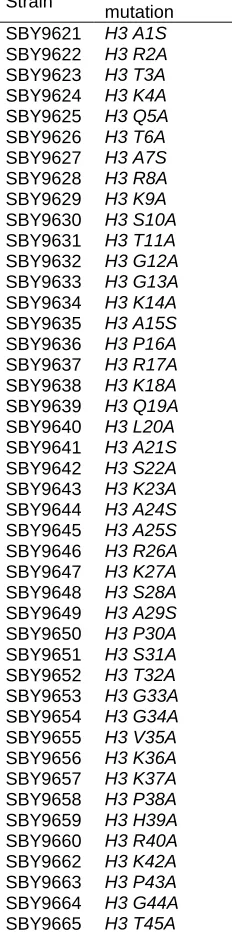
Strain Histone mutation SBY9621 H3 A1S
SBY9622 H3 R2A
SBY9623 H3 T3A
SBY9624 H3 K4A
SBY9625 H3 Q5A
SBY9626 H3 T6A
SBY9627 H3 A7S
SBY9628 H3 R8A
SBY9629 H3 K9A
SBY9630 H3 S10A
SBY9631 H3 T11A
SBY9632 H3 G12A
SBY9633 H3 G13A
SBY9634 H3 K14A
SBY9635 H3 A15S
SBY9636 H3 P16A
SBY9637 H3 R17A
SBY9638 H3 K18A
SBY9639 H3 Q19A
SBY9640 H3 L20A
SBY9641 H3 A21S
SBY9642 H3 S22A
SBY9643 H3 K23A
SBY9644 H3 A24S
SBY9645 H3 A25S
SBY9646 H3 R26A
SBY9647 H3 K27A
SBY9648 H3 S28A
SBY9649 H3 A29S
SBY9650 H3 P30A
SBY9651 H3 S31A
SBY9652 H3 T32A
SBY9653 H3 G33A
SBY9654 H3 G34A
SBY9655 H3 V35A
SBY9656 H3 K36A
SBY9657 H3 K37A
SBY9658 H3 P38A
SBY9659 H3 H39A
SBY9660 H3 R40A
SBY9662 H3 K42A
SBY9663 H3 P43A
SBY9664 H3 G44A
SBY9666 H3 V46A
SBY9667 H3 A47S
SBY9668 H3 L48A
SBY9669 H3 R49A
SBY9670 H3 E50A
SBY9671 H3 I51A
SBY9672 H3 R52A
SBY9673 H3 R53A
SBY9674 H3 F54A
SBY9675 H3 Q55A
SBY9676 H3 K56A
SBY9677 H3 S57A
SBY9678 H3 T58A
SBY9679 H3 E59A
SBY9680 H3 L60A
SBY9681 H3 L61A
SBY9683 H3 R63A
SBY9684 H3 K64A
SBY9685 H3 L65A
SBY9686 H3 P66A
SBY9687 H3 F67A
SBY9689 H3 R69A
SBY9690 H3 L70A
SBY9691 H3 V71A
SBY9692 H3 R72A
SBY9693 H3 E73A
SBY9694 H3 I74A
SBY9695 H3 A75S
SBY9696 H3 Q76A
SBY9697 H3 D77A
SBY9699 H3 K79A
SBY9700 H3 T80A
SBY9701 H3 D81A
SBY9702 H3 L82A
SBY9703 H3 R83A
SBY9704 H3 F84A
SBY9705 H3 Q85A
SBY9706 H3 S86A
SBY9707 H3 S87A
SBY9708 H3 A88S
SBY9709 H3 G90A
SBY9710 H3 A91S
SBY9711 H3 L92A
SBY9712 H3 Q93A
SBY9713 H3 S95A
SBY9714 H3 V96A
SBY9715 H3 A98S
SBY9716 H3 Y99A
SBY9717 H3 L100A
SBY9718 H3 V101A
SBY9719 H3 S102A
SBY9720 H3 F104A
SBY9721 H3 E105A
SBY9722 H3 D106A
SBY9723 H3 T107A
SBY9724 H3 N108A
SBY9725 H3 L109A
SBY9726 H3 A110S
SBY9727 H3 A111S
SBY9728 H3 A114S
SBY9729 H3 K115A
SBY9731 H3 Q120A
SBY9732 H3 K121A
SBY9733 H3 K122A
SBY9734 H3 I124A
SBY9735 H3 K125A
SBY9736 H3 L126A
SBY9737 H3 A127S
SBY9738 H3 R128A
SBY9739 H3 R129A
SBY9740 H3 L130A
SBY9741 H3 R131A
SBY9742 H3 G132A
SBY9743 H3 E133A
SBY9744 H3 R134A
SBY9745 H3 S135A
SBY9746 H4 S1A
SBY9747 H4 G2A
SBY9748 H4 R3A
SBY9749 H4 G4A
SBY9750 H4 K5A
SBY9751 H4 G6A
SBY9752 H4 G7A
SBY9753 H4 K8A
SBY9754 H4 L10A
SBY9755 H4 G11A
SBY9756 H4 K12A
SBY9757 H4 G13A
SBY9758 H4 G14A
SBY9759 H4 A15S
SBY9760 H4 K16A
SBY9761 H4 R17A
SBY9762 H4 H18A
SBY9763 H4 R19A
SBY9764 H4 K20A
SBY9765 H4 I21A
SBY9766 H4 L22A
SBY9767 H4 R23A
SBY9768 H4 D24A
SBY9769 H4 N25A
SBY9770 H4 I26A
SBY9771 H4 Q27A
SBY9772 H4 G28A
SBY9773 H4 I29A
SBY9774 H4 T30A
SBY9775 H4 K31A
SBY9776 H4 P32A
SBY9777 H4 A33S
SBY9778 H4 I34A
SBY9779 H4 R35A
SBY9780 H4 R36A
SBY9781 H4 L37A
SBY9782 H4 A38S
SBY9783 H4 G41A
SBY9784 H4 G42A
SBY9785 H4 V43A
SBY9786 H4 K44A
SBY9787 H4 I46A
SBY9788 H4 S47A
SBY9789 H4 G48A
SBY9790 H4 L49A
SBY9791 H4 I50A
SBY9793 H4 E52A
SBY9919 H4 E53A
SBY9794 H4 V54A
SBY9795 H4 R55A
SBY9796 H4 A56S
SBY9797 H4 V57A
SBY9798 H4 L58A
SBY9799 H4 K59A
SBY9800 H4 S60A
SBY9801 H4 F61A
SBY9802 H4 E63A
SBY9803 H4 S64A
SBY9804 H4 V65A
SBY9805 H4 I66A
SBY9806 H4 R67A
SBY9807 H4 D68A
SBY9808 H4 S69A
SBY9809 H4 V70A
SBY9810 H4 T71A
SBY9811 H4 Y72A
SBY9812 H4 T73A
SBY9813 H4 E74A
SBY9814 H4 H75A
SBY9815 H4 K77A
SBY9816 H4 K79A
SBY9817 H4 T80A
SBY9818 H4 V81A
SBY9819 H4 T82A
SBY9820 H4 S83A
SBY9821 H4 D85A
SBY9822 H4 V86A
SBY9823 H4 V87A
SBY9824 H4 Y88A
SBY9825 H4 A89S
SBY9826 H4 L90A
SBY9827 H4 R92A
SBY9828 H4 Q93A
SBY9829 H4 G94A
SBY9830 H4 R95A
SBY9831 H4 T96A
SBY9832 H4 Y98A
SBY9833 H4 G99A
SBY9834 H4 F100A
SBY9835 H4 G101A
SBY9836 H4 G102A
Strain Genotype
SBY4 SBY719
MAT ura3-1 leu2-3,112 his3-11 trp1-1 can1-100 ade2-1 bar1-1
MATa ura3-1 leu2-3,112 his3-11:pCUP1GFP12-LacI12:HIS chr8:CEN-lacO:TRP1 can1-100 ade2-1 bar1-1
SBY9837 MATa ura3-1 leu2-3,112 his3-11:pCUP1GFP12-LacI12:HIS chr8:CEN-lacO:TRP1 can1-100 ade2-1:7-tetOp-Ub-R-IPL1:ADE2 bar1-1 lys2∆ ipl1∆KAN hht1-hhf1∆HYG HHT2-HHF2 synthetic:URA3
SBY9838 MATa ura3-1 leu2-3,112 his3-11:pCUP1GFP12-LacI12:HIS chr8:CEN-lacO:TRP1 can1-100 ade2-1:7-tetOp-Ub-R-IPL1:ADE2 bar1-1 lys2∆ ipl1∆KAN hht1-hhf1∆HYG hht2-Q5A-HHF2 synthetic:URA3 PDS1-myc18:LEU2
SBY9839 MATa ura3-1 leu2-3,112 his3-11:pCUP1GFP12-LacI12:HIS chr8:CEN-lacO:TRP1 can1-100 ade2-1:7-tetOp-Ub-R-IPL1:ADE2 BAR ipl1∆KAN hht1-hhf1∆HYG hht2-R40A-HHF2
synthetic:URA3
ade2-1:7-tetOp-Ub-R-IPL1:ADE2 BAR lys2∆ ipl1∆KAN hht1-hhf1∆HYG hht2-G44A-HHF2 synthetic:URA3
SBY9841 MATa ura3-1 leu2-3,112 his3-11:pCUP1GFP12-LacI12:HIS chr8:CEN-lacO:TRP1 can1-100 ade2-1:7-tetOp-Ub-R-IPL1:ADE2 bar1-1 lys2 ipl1∆KAN hht1-hhf1∆HYG hht2-R53A-HHF2 synthetic:URA3
SBY9842 MATa ura3-1 leu2-3,112 his3-11:pCUP1GFP12-LacI12:HIS chr8:CEN-lacO:TRP1 can1-100 ade2-1:7-tetOp-Ub-R-IPL1:ADE2 bar1-1 lys2∆ ipl1∆KAN hht1-hhf1∆HYG hht2-N108A-HHF2 synthetic:URA3
SBY9843 MATa ura3-1 leu2-3,112 his3-11:pCUP1GFP12-LacI12:HIS chr8:CEN-lacO:TRP1 can1-100 ade2-1:7-tetOp-Ub-R-IPL1:ADE2 BAR lys2∆ ipl1∆KAN hht1-hhf1∆HYG hht2-L109A-HHF2 synthetic:URA3 PDS1-myc18:LEU2
SBY9844 MATa ura3-1 leu2-3,112 his3-11:pCUP1GFP12-LacI12:HIS chr8:CEN-lacO:TRP1 can1-100 ade2-1:7-tetOp-Ub-R-IPL1:ADE2 bar1-1 lys2∆ ipl1∆KAN hht1-hhf1∆HYG HHT2-HHF2 synthetic:URA3
SBY9845 MATa ura3-1 leu2-3,112 his3-11:pCUP1GFP12-LacI12:HIS chr8:CEN-lacO:TRP1 can1-100 ade2-1:7-tetOp-Ub-R-IPL1:ADE2 bar1-1 lys2∆ ipl1∆KAN hht1-hhf1∆HYG HHT2-hhf2-K44A synthetic:URA3
SBY9846 MATa ura3-1 leu2-3,112 his3-11:pCUP1GFP12-LacI12:HIS chr8:CEN-lacO:TRP1 can1-100 ade2-1:7-tetOp-Ub-R-IPL1:ADE2 bar1-1 ipl1∆KAN hht1-hhf1∆HYG HHT2-hhf2-V81A synthetic:URA3
SBY9847 MATa ura3-1 leu2-3,112 his3-11:pCUP1GFP12-LacI12:HIS chr8:CEN-lacO:TRP1 can1-100 ade2-1:7-tetOp-Ub-R-IPL1:ADE2 bar1-1 lys2∆ ipl1∆KAN hht1-hhf1∆HYG HHT2-hhf2-Y98A synthetic:URA3
SBY9848 MATa ura3-1 leu2-3,112 his3-11:pCUP1GFP12-LacI12:HIS chr8:CEN-lacO:TRP1 can1-100 ade2-1:7-tetOp-Ub-R-IPL1:ADE2 bar1-1 lys2∆ ipl1∆KAN hht1-hhf1∆HYG HHT2-HHF2 synthetic:URA3 mad3∆HIS
SBY9849 MATa ura3-1 leu2-3,112 his3-11:pCUP1GFP12-LacI12:HIS chr8:CEN-lacO:TRP1 can1-100 ade2-1:7-tetOp-Ub-R-IPL1:ADE2 bar1-1 lys2∆ ipl1∆KAN hht1-hhf1∆HYG hht2-Q5A-HHF2 synthetic:URA3 mad3∆HIS
SBY9850 MATa ura3-1 leu2-3,112 his3-11:pCUP1GFP12-LacI12:HIS chr8:CEN-lacO:TRP1 can1-100 ade2-1:7-tetOp-Ub-R-IPL1:ADE2 BAR ipl1∆KAN hht1-hhf1∆HYG hht2-R40A-HHF2
synthetic:URA3 mad3∆HIS
SBY9851 MATa ura3-1 leu2-3,112 his3-11:pCUP1GFP12-LacI12:HIS chr8:CEN-lacO:TRP1 can1-100 ade2-1:7-tetOp-Ub-R-IPL1:ADE2 BAR lys2∆ ipl1∆KAN hht1-hhf1∆HYG hht2-G44A-HHF2 synthetic:URA3 mad3∆HIS
SBY9852 MATa ura3-1 leu2-3,112 his3-11:pCUP1GFP12-LacI12:HIS chr8:CEN-lacO:TRP1 can1-100 ade2-1:7-tetOp-Ub-R-IPL1:ADE2 bar1-1 ipl1∆KAN hht1-hhf1∆HYG hht2-R53A-HHF2 synthetic:URA3 mad3∆HIS
SBY9853 MATa ura3-1 leu2-3,112 his3-11:pCUP1GFP12-LacI12:HIS chr8:CEN-lacO:TRP1 can1-100 ade2-1:7-tetOp-Ub-R-IPL1:ADE2 bar1-1 ipl1∆KAN hht1-hhf1∆HYG hht2-N108A-HHF2 synthetic:URA3 mad3∆HIS
SBY9854 MATa ura3-1 leu2-3,112 his3-11:pCUP1GFP12-LacI12:HIS chr8:CEN-lacO:TRP1 can1-100 ade2-1:7-tetOp-Ub-R-IPL1:ADE2 BAR ipl1∆KAN hht1-hhf1∆HYG hht2-L109A-HHF2 synthetic:URA3 mad3∆HIS
SBY9855 MATa ura3-1 leu2-3,112 his3-11:pCUP1GFP12-LacI12:HIS chr8:CEN-lacO:TRP1 can1-100 ade2-1:7-tetOp-Ub-R-IPL1:ADE2 bar1-1 ipl1∆KAN hht1-hhf1∆HYG HHT2-HHF2
synthetic:URA3 mad3∆HIS
SBY9856 MATa ura3-1 leu2-3,112 his3-11:pCUP1GFP12-LacI12:HIS chr8:CEN-lacO:TRP1 can1-100 ade2-1:7-tetOp-Ub-R-IPL1:ADE2 bar1-1 lys2∆ ipl1∆KAN hht1-hhf1∆HYG HHT2-hhf2-K44A synthetic:URA3 mad3∆HIS
SBY9857 MATa ura3-1 leu2-3,112 his3-11:pCUP1GFP12-LacI12:HIS chr8:CEN-lacO:TRP1 can1-100 ade2-1:7-tetOp-Ub-R-IPL1:ADE2 bar1-1 ipl1∆KAN hht1-hhf1∆HYG HHT2-hhf2-V81A synthetic:URA3 mad3∆HIS
SBY9858 MATa ura3-1 leu2-3,112 his3-11:pCUP1GFP12-LacI12:HIS chr8:CEN-lacO:TRP1 can1-100 ade2-1:7-tetOp-Ub-R-IPL1:ADE2 bar1-1 ipl1∆KAN hht1-hhf1∆HYG HHT2-hhf2-Y98A synthetic:URA3 mad3∆HIS
SBY9870 MATa ura3-1 leu2-3,112 his3-11:pCUP1GFP12-LacI12:HIS trp1-1:lacO:TRP1 can1-100 ade2-1:7-tetOp-Ub-R-IPL1:ADE2 hht1-hhf1∆HYG HHT2-HHF2 synthetic:URA3 [pRS425, LEU2, 2
]
SBY9871 MATa ura3-1 leu2-3,112 his3-11:pCUP1GFP12-LacI12:HIS trp1-1:lacO:TRP1 can1-100 ade2-1:7-tetOp-Ub-R-IPL1:ADE2 hht1-hhf1∆HYG HHT2-HHF2 synthetic:URA3 [pSB1780;
SBY9874 MATa ura3-1::NAT leu2-3,112 his3-11:pCUP1-GFP12-LacI12:HIS3 trp1-1:lacO:TRP1 ipl1∆KAN can1-100 ade2-1:7-tetOp-Ub-R-IPL1:ADE2 hht1-hhf1∆HYG hht2-R40A-HHF2 synthetic:URA3 [pRS425, LEU2, 2 ]
SBY9875 MATa ura3-1::NAT leu2-3,112 his3-11:pCUP1GFP12-LacI12:HIS trp1-1:lacO:TRP1 ipl1∆KAN can1-100 ade2-1:7-tetOp-Ub-R-IPL1:ADE2 hht1-hhf1∆HYG hht2-R40A-HHF2 synthetic:URA3 [pSB1780; SGO1,LEU2, 2 ]
SBY9876 MATa ura3-1::NAT leu2-3,112 his3-11:pCUP1GFP12-LacI12:HIS trp1-1:lacO:TRP1 ipl1∆KAN
can1-100 ade2-1:7-tetOp-Ub-R-IPL1:ADE2 hht1-hhf1∆HYG hht2-G44A-HHF2 synthetic:URA3 [pRS425, LEU2, 2 ]
SBY9877 MATa ura3-1::NAT leu2-3,112 his3-11:pCUP1GFP12-LacI12:HIS trp1-1:lacO:TRP1 ipl1∆KAN
can1-100 ade2-1:7-tetOp-Ub-R-IPL1:ADE2 hht1-hhf1∆HYG hht2-G44A-HHF2 synthetic:URA3 [pSB1780; SGO1,LEU2, 2 ]
SBY9880 MATa ura3-1 leu2-3,112 his3-11:pCUP1GFP12-LacI12:HIS chr8:CEN-lacO:TRP1 can1-100 ade2-1 hht1-hhf1∆HYG hht2-Q5A-HHF2 synthetic:URA3
SBY9881 MATa ura3-1 leu2-3,112 his3-11:pCUP1GFP12-LacI12:HIS chr8:CEN-lacO:TRP1 can1-100 ade2-1 lys2∆ hht1-hhf1∆HYG hht2-R40A-HHF2 synthetic:URA3
SBY9882 MATa ura3-1 leu2-3,112 his3-11:pCUP1GFP12-LacI12:HIS chr8:CEN-lacO:TRP1 can1-100 ade2-1 lys2∆ hht1-hhf1∆HYG hht2-G44A-HHF2 synthetic:URA3
SBY9883 MATa ura3-1 leu2-3,112 his3-11:pCUP1GFP12-LacI12:HIS chr8:CEN-lacO:TRP1 can1-100 ade2-1 lys2∆ hht1-hhf1∆HYG hht2-R53A-HHF2 synthetic:URA3
SBY9884 MATa ura3-1 leu2-3,112 his3-11:pCUP1GFP12-LacI12:HIS chr8:CEN-lacO:TRP1 can1-100 ade2-1 lys2∆ hht1-hhf1∆HYG hht2-N108A-HHF2 synthetic:URA3
SBY9885 MATa ura3-1 leu2-3,112 his3-11:pCUP1GFP12-LacI12:HIS chr8:CEN-lacO:TRP1 can1-100 ade2-1 lys2∆ hht1-hhf1∆HYG hht2-L109A-HHF2 synthetic:URA3
SBY9886 MATa ura3-1 leu2-3,112 his3-11:pCUP1GFP12-LacI12:HIS chr8:CEN-lacO:TRP1 can1-100 ade2-1 hht1-hhf1∆HYG HHT2-hhf2-K44A synthetic:URA3 PDS1-myc18:LEU2
SBY9887 MATa ura3-1 leu2-3,112 his3-11:pCUP1GFP12-LacI12:HIS chr8:CEN-lacO:TRP1 can1-100 ade2-1 hht1-hhf1∆HYG HHT2-hhf2-V81A synthetic:URA3
SBY9888 MATa ura3-1 leu2-3,112 his3-11:pCUP1GFP12-LacI12:HIS chr8:CEN-lacO:TRP1 can1-100 ade2-1 hht1-hhf1∆HYG HHT2-hhf2-Y98A synthetic:URA3
SBY10182 MATa ura3-1 leu2,3-112:: pCMV-LacI-3FLAG::LEU2 his3-11 trp1-1 ade2-1:7-tetOp-Ub-R-IPL1:ADE2 bar1-1 ipl1∆KAN can1-100 LYS2 hht1-hhf1∆HYG hht2-R40A-HHF2
synthetic:URA3 [pSB963, WT Minichromosome ,CEN3, LacO, TRP1]
SBY10183 MATa ura3-1 leu2,3-112:: pCMV-LacI-3FLAG::LEU2 his3-11 trp1-1 ade2-1:7-tetOp-Ub-R-IPL1:ADE2 bar1-1 ipl1∆KAN can1-100 LYS2 hht1-hhf1∆HYG hht2-L109A-HHF2
synthetic:URA3 [pSB963, WT Minichromosome ,CEN3, LacO, TRP1]
SBY10184 MATa ura3-1 leu2,3-112:: pCMV-LacI-3FLAG::LEU2 his3-11 trp1-1 ade2-1:7-tetOp-Ub-R-IPL1:ADE2 bar1-1 ipl1∆KAN can1-100 LYS2 hht1-hhf1∆HYG HHT2-HHF2 synthetic:URA3 [pSB963, WT Minichromosome ,CEN3, LacO, TRP1]
SBY10185 MATa ura3-1 leu2,3-112:: pCMV-LacI-3FLAG::LEU2 his3-11 trp1-1 ade2-1:7-tetOp-Ub-R-IPL1:ADE2 bar1-1 ipl1∆KAN can1-100 LYS2 hht1-hhf1∆HYG HHT2-HHF2 synthetic:URA3 [pSB963, WT Minichromosome ,CEN3, LacO, TRP1]
SBY10186 MATa ura3-1 leu2,3-112:: pCMV-LacI-3FLAG::LEU2 his3-11 trp1-1 ade2-1:7-tetOp-Ub-R-IPL1:ADE2 bar1-1 ipl1∆KAN can1-100 LYS2 hht1-hhf1∆HYG hht2-N108A-HHF2
synthetic:URA3 [pSB963, WT Minichromosome ,CEN3, LacO, TRP1]
SBY10187 MATa ura3-1 leu2,3-112:: pCMV-LacI-3FLAG::LEU2 his3-11 trp1-1 ade2-1:7-tetOp-Ub-R-IPL1:ADE2 bar1-1 ipl1∆KAN can1-100 LYS2 hht1-hhf1∆HYG hht2-R53A-HHF2
synthetic:URA3 [pSB963, WT Minichromosome ,CEN3, LacO, TRP1]
SBY10188 MATa ura3-1 leu2,3-112:: pCMV-LacI-3FLAG::LEU2 his3-11 trp1-1 ade2-1:7-tetOp-Ub-R-IPL1:ADE2 bar1-1 ipl1∆KAN can1-100 LYS2 hht1-hhf1∆HYG hht2-G44A-HHF2
synthetic:URA3 [pSB963, WT Minichromosome ,CEN3, LacO, TRP1]
SBY10189 MATa ura3-1 leu2,3-112:: pCMV-LacI-3FLAG::LEU2 his3-11 trp1-1 ade2-1:7-tetOp-Ub-R-IPL1:ADE2 bar1-1 ipl1∆KAN can1-100 LYS2 hht1-hhf1∆HYG HHT2-hhf2-K44A
synthetic:URA3 [pSB963, WT Minichromosome ,CEN3, LacO, TRP1]
SBY10190 MATa ura3-1 leu2,3-112:: pCMV-LacI-3FLAG::LEU2 his3-11 trp1-1 ade2-1:7-tetOp-Ub-R-IPL1:ADE2 bar1-1 ipl1∆KAN can1-100 LYS2 hht1-hhf1∆HYG HHT2-hhf2-V81A
synthetic:URA3 [pSB963, WT Minichromosome ,CEN3, LacO, TRP1]
SBY10191 MATa ura3-1 leu2,3-112:: pCMV-LacI-3FLAG::LEU2 his3-11 trp1-1 ade2-1:7-tetOp-Ub-R-IPL1:ADE2 bar1-1 ipl1∆KAN can1-100 LYS2 hht1-hhf1∆HYG HHT2-hhf2-Y98A
synthetic:URA3 [pSB963, WT Minichromosome ,CEN3, LacO, TRP1]