1556-6811/08/$08.00⫹0 doi:10.1128/CVI.00476-07
Copyright © 2008, American Society for Microbiology. All Rights Reserved.
Immunization with a
Pseudomonas aeruginosa
1244 Pilin Provides
O-Antigen-Specific Protection
䌤
Joseph Horzempa,
1† Thomas K. Held,
2‡ Alan S. Cross,
2§ Dana Furst,
1Mohammed Qutyan,
1Alice N. Neely,
3and Peter Castric
1*
Department of Biological Sciences, Duquesne University, Pittsburgh, Pennsylvania 152821; Department of Bacterial Diseases,
Walter Reed Army Institute of Research, Washington, D.C. 203072; and Shriners Hospitals for Children, Cincinnati, Ohio 452293
Received 3 December 2007/Returned for modification 24 January 2008/Accepted 1 February 2008
The O antigen is both a major structural outer membrane component and the dominant epitope of most gram-negative bacteria. Pseudomonas aeruginosa 1244 produces a type IV pilus and covalently links an O-antigen repeating unit to each pilin monomer. Here we show that immunization of mice with pure pilin from strain 1244 by use of either the mouse respiratory model or the thermal injury model resulted in protection from challenge with a pilus-null O-antigen-producing 1244 mutant. These results provide evidence that the pilin glycan stimulates a protective response that targets the O antigen, suggesting that this system could be used as the basis for the development of a variety of bioconjugate vaccines protective against gram-negative bacteria.
With the widespread occurrence of antibiotic resistance among many bacterial pathogens (29, 33, 47), it is increasingly necessary to turn to vaccination to combat these disease-caus-ing organisms. An important strategy for vaccine design in-volves the synthesis of glycoconjugates, which are produced by chemically cross-linking bacterial surface polysaccharides to carrier proteins (2, 23). Saccharides linked to proteins invoke T-cell-dependent immunity (4), which involves a memory re-sponse, avidity maturation, and immunoglobulin class switch-ing (46). Conversely, bacterial polysaccharides alone are usu-ally associated with a T-cell-independent response, which does not involve the induction of immunological memory (27). However promising this technology is, expansion of the glyco-conjugate vaccine technology has been hampered by the ex-pense of purifying and characterizing surface polysaccharides of bacterial pathogens, the selection of an effective carrier protein, and maintenance of the quality and stability of cross-linked products (23).
Several antibacterial glycoconjugate vaccines are commer-cially available (23).Haemophilus influenzae type b glycocon-jugate vaccines (39), in which the surface oligosaccharide of this bacterium was conjugated to either diphtheria toxoid (3), tetanus toxoid (22), or a meningococcal outer membrane protein (15), were the first to be licensed (23). Currently, numerous investigations are under way to test and develop
polysaccharide-protein conjugate vaccines against bacterial pathogens, includingFrancisella tularensis(9),Escherichia coli
O157 (24),Shigella sonnei(36), and the opportunistic pathogen
Pseudomonas aeruginosa(11).
The O antigen is the dominant surface polysaccharide of the lipopolysaccharide (LPS) molecule of most gram-negative bac-teria, and consequently, antibodies directed against this cellu-lar component have been shown to protect against infection (9, 10, 13, 14, 17). Interestingly, P. aeruginosa 1244 covalently attaches a single serotype O7 O-antigen repeating unit to Ser148of the pilin (8), with no alternate glycoforms or evidence
of nonglycosylated forms of this protein (6). Pilin (encoded by thepilAgene) is the monomeric subunit of the type IV pilus, an immunogenic bacterial surface appendage utilized for ad-hesion and surface motility (26).P. aeruginosa1244 pilin gly-cosylation is mediated by the oligosaccharyltransferase PilO (5, 12). This type of O-linked, PilO-mediated pilin glycosylation is common amongP. aeruginosastrains (7, 25, 41). Because theP. aeruginosa 1244 protein glycosylation machinery covalently links an O-antigen subunit and pilin, studies have suggested the possibility for exploitation of this system to biologically produce glycoconjugate vaccines (8, 20, 21).
Metabolically, the pilin glycan originates in the O-antigen-biosynthetic pathway (12). In accordance, expression of exog-enous O-antigen gene clusters inP. aeruginosa1244 allowed pilin to be glycosylated with the heterologous O-antigen re-peating unit (12). Furthermore, expression of plasmid-borne
pilAO1244in nonserotype O7P. aeruginosastrains resulted in pilin glycosylation, in which the glycan consisted of the host’s O subunit (12). These experiments indicated that all nine P. aeruginosaO subunits tested and theE. coliO157 O subunit could serve as the 1244 pilin glycan (12). As a variety of O subunits could be conjugated to pilin, this indicated that the PilO glycan specificity was low, an extremely promising phe-nomenon in terms of exploiting the 1244 glycosylation machin-ery for the generation of glycoconjugate vaccines. Moreover, recent studies defined the substrate specificity of the 1244
* Corresponding author. Mailing address: Department of Biological Sciences, Duquesne University, 600 Forbes Avenue, Pittsburgh, PA 15282. Phone: (412) 396-6319. Fax: (412) 396-5907. E-mail: castric @duq.edu.
† Present address: Department of Microbiology and Molecular Ge-netics, University of Pittsburgh School of Medicine, Pittsburgh, PA 15261.
‡ Present address: HELIOS Klinikum Berlin-Buch, Robert-Ro ¨ssle-Klinik, Klinik fu¨r Ha¨matologie, Onkologie und Tumorimmunologie, Schwanebecker Chaussee 50, 13125 Berlin, Germany .
§ Present address: Center for Vaccine Development, University of Maryland School of Medicine, Baltimore, MD 21201.
䌤Published ahead of print on 13 February 2008.
590
on August 17, 2020 by guest
http://cvi.asm.org/
glycosylation reaction (20, 21). Those studies indicated that PilO recognizes the sugar at the reducing end of the O-antigen repeating unit precursor (21) and that successful transfer of this carbohydrate is contingent upon the positioning of Ser at the pilin C terminus, as this configuration is essential for rec-ognition by the glycosylation machinery (20). While no other specific pilin recognition features were present, the charge of the pilin surface must be compatible with the glycosylation apparatus (20). Application of these data may allow the use of O subunits of diverse gram-negative bacteria to be covalently linked to a variety of engineered proteins, including toxoids, likely expanding the potential breadth of protection. However, it is first necessary to test if theP. aeruginosa1244 glycosylated pilin can provide O-antigen-specific protection, which is the objective of this study.
The current investigation shows that a bacterial protein gly-cosylation system is capable of biologically producing effica-cious glycoconjugate vaccines. The work presented here dem-onstrates that the pilin glycan is a major immunogenic epitope, directing the production of antibodies against O antigen com-posed of analogous subunits. Vaccination with the 1244 pilin provided significant protection, with immunological specificity for the O polysaccharide, in two separate infection models. These results suggest that the pilin glycosylation system ofP. aeruginosa1244 may be useful for the biological production of anti-O-antigen glycoconjugate vaccines against a variety of harmful gram-negative bacterial pathogens.
MATERIALS AND METHODS
Animals.The mice and rabbit strains used in this study are listed in Table 1. Immunizations involving female New Zealand White rabbits were conducted by Covance Research (Denver, PA). All animal experiments complied with federal and institutional specifications regarding the use of animals in research.
Bacterial strains and media.The bacterial strains and plasmid used in this study are listed Table 1. For general culturing, the bacteria were grown aerobi-cally at 37°C on LB agar plates or in LB broth shaken at 250 rpm. LB broth was used for cell growth for the preparation of strain 1244 pili glycosylated with the O7 repeating unit. For cell growth for the preparation of strain 1244 pili glyco-sylated with the O6 repeating unit, CAYE (2% agar, 0.75% Casamino Acids,
0.15% yeast extract) was used. These media contained carbenicillin (250g/ml)
and/or tetracycline (50g/ml), as needed. When it was necessary, the medium
was supplemented with 5 mM isopropyl--D-thiogalactopyranoside (IPTG).
Isolation of pili and purification of pilin.The glycosylated 1244 pili used for
vaccination were produced fromP. aeruginosa1244N3/pPAC46, as described
previously (5, 12). Briefly, cell cultures were grown in LB broth supplemented with carbenicillin, tetracycline, and 5 mM IPTG at 37°C with shaking at 250 rpm for 14 h. The pili were isolated from the supernatant after centrifugation at
16,000⫻gfor 30 min at 4°C. The pili were purified by repeated precipitation in
the presence of 0.5 M NaCl and 3% polyethylene glycol 8000 (40, 41). When it was required, traces of LPS were removed by first depolymerizing purified pili in
1.0%-octylglucoside followed by gel filtration and chromatofocusing, as
previ-ously described in detail (8). No contaminating protein or LPS could be detected by polyacrylamide gel electrophoresis followed by silver staining or by Western blotting with an O-antigen-specific monoclonal antibody as a probe. The product of this procedure was referred to as chromatographically pure pilin (CPP). Samples of CPP bearing the glycan identical to the O7 LPS O-antigen subunit
(CPPO7) were subjected to overnight dialysis against 6 liters H2O at 4°C, during
which the subunits reaggregated into pilus-like, nonnative fibers (43). The LPS
level of this preparation was measured by using theLimulus Gel-Clot test
(Associates of Cape Cod). In these assays, in which strain 1244 LPS prepared by an established protocol (44) was used as a standard, a contamination level of less than 0.0004% (wt/wt) was detected. This material was lyophilized, resuspended in sterile phosphate-buffered saline (PBS), and filter sterilized. The bicinchoninic acid protein assay (Pierce) was used according to the manufacturer’s protocol to determine the protein concentration.
P. aeruginosa1244 pili glycosylated with the serotype O6 repeating unit were
produced in two ways. In one,P. aeruginosaPAK, which produces O-antigen
serotype O6 and a pilus which is antigenically distinct from that produced by strain 1244, was grown so that it contained pPAC46. This arrangement has previously been shown to produce 1244 pilin glycosylated with the O6 repeating
unit (12). In the second situation,P. aeruginosa9D2, which also produces
O-antigen serotype O6, was also grown so that it contained pPAC46. Although this strain produces limited amounts of pili, the primary structure of the fibers produced is nearly identical to that of the fibers of strain 1244, differing only at residue 92 (7). This residue is situated in a region found to be poorly immuno-genic (7, 8). Overexpression of pPAC46 resulted in large amounts of pilin glycosylated with the O6 repeating unit. For pilus production, a 5-ml LB culture inoculated with one of these strains containing pPAC46 was grown in the pres-ence of carbenicillin at 37°C for 8 h. This suspension was used to inoculate aluminum foil-covered 68- by 28- by 3-cm metal pans containing 500 ml of solidified CAYE agar medium, carbenicillin, and IPTG, after which it was incu-bated at 37°C for 14 h. Cells were removed by scraping and were suspended in 50 ml of 40 mM sodium phosphate buffer, pH 7.2, per pan. This cell suspension was subjected to vigorous stirring for 30 min at room temperature to detach the
pili and was then centrifuged at 16,000⫻gfor 30 min. Pili were isolated from the
TABLE 1. Animal strains, bacterial strains, and plasmid used in this study
Organism or plasmid Descriptiona Source or reference
Animals Mice
ICR Age, 9–16 wk Charles River Laboratories, Wilmington, MA
C3H/HeJ Age, 9–16 wk Jackson Laboratories, Bar Harbor, ME
BALB/c Female; age, 6–8 wk Hilltop Lab Animals, Scottdale, PA
CF-1 Female; wt, 22 to 25 g Carworth Farms, New York, NY
New Zealand White rabbits Female, adult Covance Research, Denver, PA
Bacterial strains P. aeruginosa
1244 Wild type, IATS O7 35
1244N3 1244rpoN(Tcr) 34
1244.47 1244pilA(Hgr) 20
PAK Wild type, IATS O6 7
9D2 Wild type, IATS O6 7
K. pneumoniaeB5055 Serotype O1:K2 19
Plasmid pPAC46 pMMB66EH withP. aeruginosa1244 pilAOAprCbr
5
a
IATS, International Antigenic Typing System.
on August 17, 2020 by guest
http://cvi.asm.org/
supernatant and purified by the procedure described above. CCP06was produced
as described above.
Protocols for determination of immunogenicity.Two New Zealand White rabbits were injected intradermally in the back at multiple sites with a total of 250
g of CPPO7with Freund’s complete adjuvant on day 1. On day 21, the rabbits
received subcutaneous (s.c.) and intramuscular boosts containing 125g of
CPPO7with Freund’s incomplete adjuvant (FIA). On day 42, the rabbits received
s.c. boosts containing 125g of pilin administered in the rear flanks. On day 63,
the rabbits were injected s.c. with 125g pilin with FIA. On day 84, the rabbits
received s.c. dorsal injections containing 100g pilin and FIA. The final boost
consisted of a s.c. injection in the neck region containing 100g pilin with FIA.
On days 31, 52, 73, 94, and 115, sera were collected and analyzed for antibody production (data not shown). On day 118, approximately 50 ml of serum was collected in the terminal bleed and was used for subsequent assays. Adsorption of this serum was carried as follows. Six 82-mm nitrocellulose circles (Schleicher & Schuell) were incubated with stirring overnight at room temperature with 1.6 mg heterologously glycosylated (serotype O6 O-antigen subunit) 1244 pili
(CPPO6) suspended in 15 ml deionized water. These circles were then treated
with blocking buffer (12). Ten milliliters of serum recovered from one of the animals was incubated in succession overnight at room temperature with each of these circles.
To evaluate the induction of murine antibodies corresponding with acute-pneumonia protection studies, we used the following immunization schedule (first/second dose): intranasal (i.n.)/i.n. or i.n./s.c. At each immunization, a total
dose of 5g of native 1244 pili diluted to the appropriate volume in sterile
physiologic saline was administered to ICR mice. The time interval between the doses was 7 days. For i.n. immunization, the mice were anesthetized intraperi-toneally with ketamine HCl (80 mg/kg of body weight; Aveco Co., Fort Dodge, IA) and xylazine HCl (8 mg/kg; Mobay Corporation, Shawnee, KA) prior to the
instillation of the pili. The pili were delivered i.n. in a final volume of 25l by the
use of individual sterile aerosol-resistant pipette tips for each mouse to prevent
contamination. The final volume for s.c. injection was 100l. Control mice
received sterile physiologic saline at exactly the same volume and by exactly the same route. At 3, 7, 10, and 14 days after the second dose of vaccine, the mice
were killed by CO2inhalation to obtain serum samples via cardiac puncture.
Preparation of challenge.Overnight broth cultures of P. aeruginosa1244,
Klebsiella pneumoniaeB5055, or the 1244 isogenicpilAmutant,P. aeruginosa
1244.47, were used to streak a lawn on Trypticase soy agar plates, which were incubated at 37°C for 12 to 18 h. The cells were suspended in sterile PBS, and a
spectrophotometer (A650) or a Klett colorimeter was used to estimate the cell
density on the basis of the previously determined counts of CFU. These cells were serially diluted and plated to determine viable-cell counts, which were used to more accurately determine the inoculum density.
Acute-pneumonia model.For the pilus-specific-protection studies, ICR mice were immunized by either the i.n./i.n. or the i.n./s.c. route with purified 1244 pili, as described above. Seven days after the second vaccination, anesthesia was administered and the mice received an i.n. challenge of a lethal dose
(approxi-mately 4.3⫻106CFU) ofP. aeruginosa1244 in a final volume of 50l (25l per
nostril). Morbidity, mortality, and body weight were monitored daily for 8 days. The control mice were vaccinated with sterile physiologic saline as described above. In order to determine persistence and dissemination, in a separate ex-periment immunized and challenged mice were killed at 4, 24, and 48 h after bacterial treatment. The presence of bacteria in bronchoalveolar lavage (BAL) fluid at 4 and 24 h postchallenge was determined by a single washing with sterile physiologic saline via a 25-gauge hypodermic needle inserted into the trachea. In addition, the lungs, livers, and spleens from the animals were excised aseptically, weighed, and homogenized at 4, 24, and 48 h. The CFU counts in BAL fluid and organ homogenates were determined by previously described methods (19). The
K. pneumoniaeB5055 control challenge employed the same procedure used for
P. aeruginosa1244 and used a dose of 1.4⫻104
CFU.
In the glycan-specific-protection studies, BALB/c mice were used, and the
vaccine consisted of CPPO7. CPPO7(9g in 20l PBS; 10l per nostril) was
administered i.n./i.n., as described above. As a control, mice received CPPO6
(purified fromP. aeruginosa9D2/pPAK46) to which purified strain 1244 LPS,
prepared as described above, was added to a level 0.0004% (wt/wt) of that of pilin. Seven days following the second vaccination, the mice were anesthetized
and were challenged with 3.8 times the 50% lethal dose (LD50; 4.9⫻107CFU)
of strain 1244.47, which was administered i.n. in 20l PBS (10l per nostril). In
a separate trial, mice immunized with CPPO6were challenged with 2.6 times the
LD50 (3.4⫻ 10
7
CFU) of strain 1244.47. The LD50of strain 1244.47 was
determined to be 1.3⫻107
CFU by using the probit analysis of StatPlus 2007 software (data not shown). Following the challenge, the mice were closely mon-itored for 96 h, during which time morbidity and mortality were recorded.
Burned-mouse model.Tests with the burned-mouse model were conducted as
described previously (28). On day 0, CPPO7was diluted to a concentration of 40
g/100l with saline and mixed 1:1 with FIA, resulting in a solution with a pilus
concentration of 20g/100l. A total of 27 mice were immunized with 100l of
this material via the s.c. route (in the back). A control group (containing the same number of animals) was treated in the same manner with the same solution, except that the pili were absent. This treatment was repeated on day 7. On day 14, the mice were subjected to a nonlethal thermal injury of 15% of the body surface. which caused host immunosuppression, and were challenged with a
subeschar injection of approximately 5⫻107CFU ofP. aeruginosa1244.47.
Additionally, eight untreated mice were challenged in the same manner. Follow-ing the challenge, mice were observed for 5 days, durFollow-ing which time morbidity and mortality were recorded.
ELISA.Antibody titers in serum and BAL fluid were determined by an en-zyme-linked immunosorbent assay (ELISA) with 96-well plates coated with pili
from eitherP. aeruginosa1244 or strain PAK/pPAC46 at a concentration of 2
g/ml (50l/well, with incubation overnight at 4°C) orP. aeruginosa1244 LPS at
a concentration of 1g/ml (100l/well, with incubation overnight at 4°C). After
the wells were treated with blocking buffer and washed, samples were serially diluted (twofold) in blocking buffer and incubated overnight in duplicate at 4°C. Bound antibodies were detected by using goat anti-mouse immunoglobulin G (IgG), alka-line phosphatase-labeled goat anti-mouse IgM, goat anti-mouse-IgA, or alkaalka-line phosphatase-labeled goat anti-rabbit secondary antibodies (Kirkegaard & Perry, Gaithersburg, MD) diluted appropriately in blocking buffer; and the plates were
developed withp-nitrophenylphosphate (Sigma, St. Louis, MO) in diethanolamine
buffer (pH 9.8). The reaction was stopped by adding 50l of 3 M NaOH to each well
after 30 min. The ELISA titers were defined as the dilution which gave a change in
theA405of 0.200 at room temperature. The backgroundA405in all control wells was
always less than 0.050.
Statistics.Differences in survival between the groups were analyzed by the log rank test. For comparison of CFU counts, the two-tailed Mann-Whitney U test was used. The statistical analyses were done with either Statistica software (version 4.5) for Windows, StatSoft software, or GraphPad Prism software (ver-sion 4.02).
RESULTS
Pilus vaccine protection from lethal challenge. It was im-portant to establish a model to determine an effective route of immunization and an appropriate challenge with wild-typeP. aeruginosa1244 as a precedent for subsequent studies. AsP. aeruginosais a leading cause of nosocomial pneumonia (30), the murine acute-pneumonia model was used to determine the protective efficacy of a vaccine composed of strain 1244 pili. An i.n./i.n. or i.n./s.c. (first/second dose) immunization schedule was used, with 7 days between doses. At each immunization, the mice received 5 g of native 1244 pili diluted in sterile physiologic saline. Serum from both i.n./i.n.- and i.n./s.c.-im-munized mice contained high titers of LPS-specific antibodies (IgG and IgM) (Table 2). These results indicated that a specific immune response was well established at 1 week following the second vaccination, as evidenced by a high serum IgG titer with specificity for the serotype O7 O antigen (Table 2). If this response could sufficiently mediate clearance of the infection, then strain 1244 glycosylated pili may provide O-antigen-spe-cific immunity (8).
Because a specific immune response was established 1 week following the second pilus immunization (Table 2), the mice were challenged with a lethal dose of wild-typeP. aeruginosa
1244 at this time point. The mice that received the pilus vac-cine i.n./i.n. exhibited significant survival compared to the sur-vival of the mice that received saline following an i.n. challenge of a lethal dose of strain 1244 (P⬍0.0001) (Fig. 1). Although the mice that received the pilus vaccine i.n./s.c. showed re-duced survival compared to the survival of the mice immunized with the pilus vaccine i.n/i.n., these mice still exhibited
on August 17, 2020 by guest
http://cvi.asm.org/
icantly increased survival compared to the survival of the sa-line-vaccinated mice (P⫽0.0030) (Fig. 1). Compared to the saline-immunized controls, mice immunized with either regi-men had significantly fewer bacteria in their BAL fluid at 4 h after infection (Fig. 2A). These data suggest that immunization with strain 1244 pili either prevents the adherence of the bac-teria to epithelial surfaces or promotes the clearance of the bacteria from the airways.P. aeruginosa1244 consistently dis-seminated from the site of infection, as evidenced by the cul-ture of bacteria from homogenates of extrapulmonary organs, but only mice immunized twice i.n. were capable of lowering the numbers of bacterial CFU in the lungs as well as keeping bacterial multiplication in the liver and spleen at steady levels between 4 and 24 h after challenge (Fig. 2B). In addition, a significant decrease in the bacterial CFU from the BAL fluid over time (4 h versus 24 h after bacterial challenge) was ob-served only when the animals were immunized i.n./i.n. (Fig. 2A). Mice immunized i.n./s.c. and saline-immunized control mice showed increasing CFU counts in the lungs, liver, and
spleen at 24 h after infection (Fig. 2B). At 48 h after infection, all saline-immunized mice were dead; however, the surviving mice immunized either i.n./i.n. or i.n/s.c. showed low CFU counts in all three organs investigated (Fig. 2B), indicating vaccine-induced clearance. Thus, survival correlated with de-creasing bacterial counts both locally and systemically over the initial 48 h of infection.
Analysis of vaccine specificity.It was necessary to investigate if the immune response elicited by immunization with P. aeruginosa 1244 pili conferred specific protection or if the protection was due to a generalized inflammatory response caused by the administration of pili or trace LPS contamina-tion. Therefore, by use of the respiratory model, mice that were immunized twice i.n. with strain 1244 pili were subse-quently challenged with a heterologous bacterium (K. pneu-moniaeB5055) that did not produce pili and that was
serolog-TABLE 2. Systemic antibody responses of mice specific forP. aeruginosa O7 LPS after immunization with strain 1244 pili
Routea Ig class
Mean ELISA antibody titer on the indicated
day after the second immunizationb
3 7 10 14
i.n./i.n. IgG 112 79.4 58.9 234
IgM 479 269 91.2 93.3
IgA ND ND ND ND
i.n./s.c. IgG 166 269 135 123
IgM 617 794 676 490
IgA ND ND ND ND
a
The routes denoted are for the first/second immunization, in which the time between each immunization was 7 days.
b
Mean ELISA antibody titer (dilution) which gave a change in theA405of
0.200 after 30 min. Each value shown is the mean of the individual titers from three mice. ND, none detected, as these values were below the limits of detection
(A405⫽0.050).
FIG. 1. Survival afterP. aeruginosa1244 challenge following immu-nization with pili in the acute-pneumonia model. Mice exhibited sig-nificantly increased survival when they were immunized i.n./i.n. or i.n./s.c. with strain 1244 pili compared to the survival of mice immu-nized with saline (control) following i.n. administration of a lethal dose of P. aeruginosa 1244. Results are shown as Kaplan-Meier survival curves, and differences in survival were calculated by log rank analysis. Results for i.n./i.n versus results for the control,P⬍0.0001; results for i.n./s.c. versus results for the control,P⫽0.0030; results for i.n./i.n. versus results for i.n./s.c.,P⫽0.1449.
FIG. 2.P. aeruginosa1244 growth in BAL fluid (A) or the lungs, livers, and spleens (B) of strain 1244 pilus-immunized and saline-immunized mice. The graphed values are medians, and error bars represent quartiles. Data are from two independent experiments (n⫽ 4 mice per group per experiment). (A)ⴱ,P⫽0.0062 for the counts in mice immunized i.n./i.n. andP⫽0.0015 for the counts in mice immu-nized i.n./s.c. compared to the counts for the corresponding times postinfection of saline-immunized (control) animals (Mann-Whitney U test). (B)P. aeruginosa1244 CFU counts from organ homogenates at 4, 24, or 48 h postinfection of mice immunized i.n./i.n., i.n./s.c., or with saline (control) are shown for all time points except for control mice at 48 h, as these mice were dead, making statistical analysis infeasible.
on August 17, 2020 by guest
http://cvi.asm.org/
ically distinct from strain 1244. Neither immunized nor control mice survived a lethal challenge withK. pneumoniae, indicating that vaccination with P. aeruginosa 1244 pili protects only against a homologous challenge (P⫽0.0027 andP⫽0.0009, respectively) (Table 3). These data suggest that the protection described here (Fig. 1 and 2) was a result of a specific immune response and was not due to nonspecific inflammation from the pilus vaccine. LPS contamination was not likely responsible for this protection, as a previous study showed that mice im-munized s.c. with upwards of 200% of the contaminant level of strain 1244 LPS present in the current pilus dose did not induce a detectible anti-LPS response (8).
Immunogenicity of the pilin glycan.To better ascertain the immunogenicity of the pilin glycan, it was necessary to remove traces of LPS contamination. This was accomplished by depo-lymerization of the pili in the presence of -octylglucoside, followed by chromatographic purification of the pilin produced (8). Detergent was removed upon dialysis of the purified pilin, which allowed reaggregation of the pilin subunits into
nonna-tive pilus-like fibers (43). TheLimulusassay showed that fol-lowing this chromatographic purification, reaggregated pilin was contaminated with LPS to a level of less than 0.0004% (wt/wt). This substance, referred to as CPPO7, was used to immunize rabbits. One serum sample that was produced was adsorbed with CPPO6in order to remove pilin protein-specific antibodies and retain those that were glycan specific. Adsorbed sera were unresponsive to CPPO6, while untreated sera had high titers of antibodies specific for these pili, suggesting that the adsorption had successfully removed pilin protein-specific antibodies (Fig. 3A). The sera that were adsorbed with CPPO6 had slightly lower titers of antibodies specific for CPPO7than untreated serum (Fig. 3B). This difference was probably due to the absence of a pilin protein-specific response. Recognition of glycosylated pili by a serum sample that lacked a pilin protein-specific response suggested that the strong antipilus reaction was directed to the pilin glycan. Both the adsorbed and the untreated sera exhibited equal responses to serotype O7 LPS, showing that animals immunized with CPPO7 are capable of eliciting a strong anti-LPS response (Fig. 3C). Notably, the dominant antibody type produced by these rabbits was IgG, as determined by ELISA. Here, a 4-log-unit difference between the IgG and the IgM titers was seen (E. Jewell and P. Castric, unpublished observations), suggesting the involvement of T cells in the antiglycan response. These results confirm that CPPO7 is capable of stimulating a specific B-cell response against both the pilin glycan and the structurally similar O-antigen repeating unit of LPS (8) and suggest that the pilin glycan is a major pilin epitope.
TABLE 3. Vaccine specificity
Immunizationa
No. of surviving mice/no. of total mice after challenge with:
P. aeruginosa
1244 K. pneumoniae
P. aeruginosa1244 pili 4/4 0/8
Saline 0/6 0/6
a
Mice were immunized i.n./i.n.
FIG. 3. ELISAs with sera from rabbits that had been immunized with CPPO7. Untreated or CPPO6-adsorbed rabbit serum was used as the
primary antibody, and CPPO7(A), CPPO6(B), or serotype O7 LPS (C) was used as the antigen. Error bars represent standard deviations.
on August 17, 2020 by guest
http://cvi.asm.org/
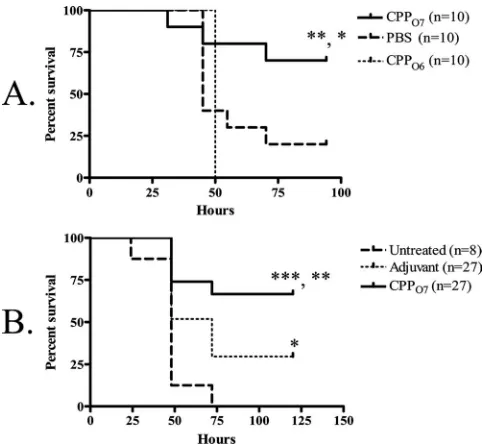
Immunization with CPPO7followed by a lethalP. aeruginosa 1244 pilA-null challenge. To determine the capacity of O-antigen-specific protection generated by a vaccine composed of CPPO7, we again employed the acute-pneumonia murine model but with apilA-negative strain (P. aeruginosa1244.47) as a challenge. This bacterial strain is an isogenic mutant of strain
P. aeruginosa1244 that does not produce pilin (20). However, strain 1244.47 produced wild-type levels of O7 LPS, as deter-mined by Western blot analysis with a serotype O7-specific monoclonal antibody as a probe (data not shown). Mice re-ceiving an i.n./i.n. administration of 9 g CPPO7 exhibited significant survival compared to the survival of PBS-immu-nized mice (P⫽0.0375) (Fig. 4A) following an i.n. challenge with a lethal dose of strain 1244.47, suggesting that this vaccine provides O-antigen-specific mucosal protection. Since CPPO7 may have contained a very small amount of remnant LPS following purification, the possibility remained that this mate-rial could have provided the protection seen here, as a previous study showed that intraperitoneal injection of 50 ngP. aerugi-nosaLPS could completely protect against a lethal challenge (32). To ensure that contaminant LPS did not provide the protection seen in the current study, mice were immunized i.n./i.n. with 9 g CPPO6 that had been supplemented with serotype O7 LPS at an amount equivalent to that in the CPPO7
vaccine. This LPS-supplemented CPPO6was therefore identi-cal to the CPPO7in both protein primary structure and
maxi-mum potential serotype O7 LPS contamination and differed only by the moieties of the glycan. Following an i.n. challenge with a lethal dose of strain 1244.47, the mice that were immu-nized with CPPO6had significantly reduced survival compared
to that for the mice immunized with CPPO7(P⫽0.0080) (Fig. 4A). These data indicate that the potentially minute levels of LPS present in CPPO7were not responsible for the protection observed (Fig. 4A). Moreover, this control confirmed that the general inflammation generated by the pilin vaccine was not conferring protection. Therefore, this mucosal protection was elicited by the O7 pilin glycan, which is identical in structure to the O-antigen subunit of the challenge strain. Notably, isolated bacterial clones from tissues did not produce pilin, as deter-mined by Western blot analysis with a pilin-specific monoclo-nal antibody as a probe, indicating that that this mutant did not revert to the wild type during infection (data not shown).
To determine if the vaccination with CPPO7could provide protective systemic immunity, we employed the burned-mouse model. Mice that were immunized twice with a mixture of 40
g CPPO7and FIA exhibited significant survival compared to
that for the mice that received the control (P⫽0.0042) (Fig. 4B) following a subeschar challenge with a lethal dose of strain 1244.47, indicating vaccine-induced, O-antigen-specific sys-temic protection. Because the challenge strain lacked pili but expressed the same O antigen as strain 1244, it is proposed that immunization with CPPO7can provide O-antigen-specific
pro-tection and that the pilin glycan is a protective epitope. It should be noted that significant survival was observed in adju-vant-treated mice compared to the survival of the untreated mice (Fig. 4B). This is likely due to an inflammatory response and the attenuation of strain 1244.47 due to the absence of pili. However, as the survival of the mice that received the pilus vaccine was significantly higher than that of the adjuvant-treated mice, this suggests that this additional protection was due to specific immunity.
DISCUSSION
Like P. aeruginosa 1244, other bacteria such as Campy-lobacter jejuniandNeisseria gonorrhoeaepossess molecular mech-anisms that mediate the covalent linkage of surface oligo-saccharides to proteins (1, 16, 42). Because of this peculiar phenomenon, all of these systems have been proposed to have the potential for use in the generation of bioconjugate vaccines (1, 8, 16). The current study was a necessary first step toward the assessment of the potential of a bacterial glycosylation system to generate an effective anti-O-antigen glycoconjugate vaccine. This work established that immunization with strain 1244 pilin, a glycoprotein in which the carbohydrate portion is an O subunit, provides O-antigen-specific protection against a
P. aeruginosachallenge. By using both theP. aeruginosa acute-pneumonia and the thermal-injury models, we present evi-dence suggesting that glycosylated pili provide O-antigen-spe-cific protection via the mucosal and systemic routes of immunity (Fig. 4). The results presented here are important because they show that a biological glycosylation system can be used to produce protective glycoconjugate vaccines that target the O antigen of a gram-negative bacterial pathogen. This
FIG. 4. Survival afterP. aeruginosa1244.47 challenge following im-munization with CPPO7 in the acute-pneumonia model (A) and
burned-mouse model (B). Results are shown as Kaplan-Meier survival curves, and differences in survival were calculated by log rank analysis. (A) Mice were immunized i.n./i.n. with either CPPO7, PBS, or CPPO6.
Mice immunized i.n./i.n. with CPPO7exhibited significantly increased
survival compared to the survival for mice immunized with PBS or CPPO6following a lethal dose of thepilAmutant, strain 1244.47.ⴱ,P⫽
0.0375 for the results for mice immunized with CPPO7 versus the
results for mice treated with PBS;ⴱⴱ,P⫽0.0080 for the results for mice immunized with CPPO7versus the results for mice immunized
with CPPO6. (B) Mice were immunized s.c. with either FIA mixed with
CPPO7or FIA alone or were untreated. Significant survival for mice
that received the pilus vaccine compared to the survival for adjuvant-treated mice (ⴱⴱ,P⫽0.0087) or untreated mice (ⴱⴱⴱ,P⫽0.0001) was observed.
on August 17, 2020 by guest
http://cvi.asm.org/
work provides a foundation for the development of the 1244 pilin glycosylation technology to produce vaccines against many gram-negative bacterial pathogens. Future studies should focus on expanding the application of this technology by testing the protective efficacies of vaccines composed of strain 1244 pilin glycosylated with O subunits from highly vir-ulent bacteria, such asE. coliO157. Fortuitously, O-antigen-biosynthetic genes are clustered on bacterial chromosomes (38), allowing ease of cloning (18). As shown previously, when
P. aeruginosa1244 expresses theE. coliO157 O-antigen bio-synthesis genes intrans, the pili from the strain produced are decorated with the O157 O subunit (12). The present study would suggest that O157-glycosylated pilin should protect against challenge; however, this remains to be determined.
In current chemically cross-linked glycoconjugate vaccines, two purification steps must be performed, including isolation of an appropriate carrier protein, in addition to refinement of the bacterial surface polysaccharide. For gram-negative bacte-ria, this involves separation of the LPS O antigen from the lipid A portion. Following purification, these molecules are sub-jected to coupling chemistry. Altogether, these procedures are time-consuming and expensive. However, the production of large amounts of glycosylated pili can be accomplished quickly, is relatively inexpensive, and requires only common laboratory procedures. Moreover, it is well known that pili are immuno-genic in humans, and their ability to generate protective im-munity is documented (31). The immunogenicity of pili is partly due to the polymeric display of subunits in the pilus fiber (37). The arrangement of the glycan in a polymeric form both in native pilus fibers and in reaggregated pilin subunits (43) might promote expedient B-cell proliferation due to the prox-imity of adjacent glycan epitopes. Previous data from our lab-oratory indicated that the pilin glycan is exposed along the surface of the pilus fiber and can easily be accessed by anti-bodies (41) and, presumably, by B-cell membrane-bound Ig. If glycan recognition by membrane-bound Ig molecules leads to the internalization and major histocompatibility complex class II presentation of pilin peptides, this may stimulate the in-volvement of helper T cells, resulting in the proliferation and differentiation of the B cells producing glycan- and O-antigen-specific Ig. Additional subsequent work should explore the use of a mucosal adjuvant, such as the oligonucleotide CpG1826 (48), as well as the effect of additional boosts, in an effort to increase glycan-specific antibody production and protection. It will also be important to discern which antibody types are essential for the success of this vaccine.
Future studies should focus on optimizing this biological system for the production of glycosylated pili to be used as vaccines. For instance, althoughP. aeruginosa1244 produces large yields of pili, the efficiency of pilus production may be increased by mutatingpilT(45). As PilT is a pilus motor pro-tein that mediates pilus retraction, mutational inactivation of this gene results in hyperpiliation (45). Additionally, a previous study has defined the pilin substrate of the 1244 glycosylation reaction, in which a C-terminal Ser or Thr and a compatible pilin surface charge are important (20). A gene encoding a normally nonglycosylated pilin (fromP. aeruginosaPA103) was mutated to contain these characteristic structures and was made capable of glycosylation by the 1244 machinery (20). Although the current study essentially showed that pilin is an
effective carrier protein for a glycoconjugate vaccine, other carriers, such as toxoids, may invoke a better response. It is therefore important to test the capacity of this glycosylation system to modify nonpilin proteins for use as vaccines. A re-cent study revealed that the glycan substrate recognition fea-tures lay within the reducing-end moiety of the O repeat (
-D-FucNAc) and that carbohydrates in this O-subunit position
from many gram-negative bacteria are structurally similar (21). However, it is essential that more O-antigen-biosynthetic clus-ters be cloned and their products tested for compatibility with the 1244 glycosylation machinery to maximize its vaccine pro-duction potential.
ACKNOWLEDGMENTS
This work was supported by a grant from the NIH to P.C. (grant AI054929).
We thank Antonio DiGiandomenico and Joanna Goldberg for in-struction on animal models and for insightful discussion. In addition, we thank Liang Yuan, and Kathleen C. Glazer for technical assistance with the acute-pneumonia model.
REFERENCES
1.Aas, F. E., A. Vik, J. Vedde, M. Koomey, and W. Egge-Jacobsen.2007.
Neisseria gonorrhoeaeO-linked pilin glycosylation: functional analyses define
both the biosynthetic pathway and glycan structure. Mol. Microbiol.65:607–
624.
2.Ada, G., and D. Isaacs.2003. Carbohydrate-protein conjugate vaccines. Clin.
Microbiol. Infect.9:79–85.
3.Anderson, P., M. E. Pichichero, and R. A. Insel.1985. Immunogens
consist-ing of oligosaccharides from the capsule ofHaemophilus influenzaetype b
coupled to diphtheria toxoid or the toxin protein CRM197. J. Clin. Investig.
76:52–59.
4.Avery, O., and W. Goebel.1931. Chemo-immunological studies on conju-gated carbohydrate-proteins. V. The immunological specificity of an antigen prepared by combining the capsular polysaccharide of type III
pneumococ-cus with foreign protein. J. Exp. Med.54:437–447.
5.Castric, P.1995.pilO, a gene required for glycosylation ofPseudomonas aeruginosa1244 pilin. Microbiology141:1247–1254.
6.Castric, P., F. J. Cassels, and R. W. Carlson.2001. Structural
characteriza-tion of thePseudomonas aeruginosa1244 pilin glycan. J. Biol. Chem.276:
26479–26485.
7.Castric, P. A., and C. D. Deal.1994. Differentiation ofPseudomonas
aerugi-nosapili based on sequence and B-cell epitope analyses. Infect. Immun.
62:371–376.
8.Comer, J. E., M. A. Marshall, V. J. Blanch, C. D. Deal, and P. Castric.2002.
Identification of thePseudomonas aeruginosa1244 pilin glycosylation site.
Infect. Immun.70:2837–2845.
9.Conlan, J., H. Shen, A. Webb, and M. Perry.2002. Mice vaccinated with the
O-antigen ofFrancisella tularensisLVS lipopolysaccharide conjugated to
bovine serum albumin develop varying degrees of protective immunity against systemic or aerosol challenge with virulent type A and type B strains
of the pathogen. Vaccine20:3465–3471.
10.Coughlin, R., and W. Bogard, Jr.1987. Immunoprotective murine monoclo-nal antibodies specific for the outer-core polysaccharide and for the
O-antigen ofEscherichia0111:B4 lipopolysaccharide (LPS). J. Immunol.139:
557–561.
11.Cryz, S. J., A. Lang, J. Wedgewood, J. Que, E. Furer, and U. Schaad.1997.
Immunization of cystic fibrosis patients with aPseudomonas aeruginosa
O-polysaccharide-toxin A conjugate vaccine. Behring Inst. Mitt.98:345–349.
12.DiGiandomenico, A., M. J. Matewish, A. Bisaillon, J. R. Stehle, J. S. Lam, and P. Castric.2002. Glycosylation ofPseudomonas aeruginosa1244 pilin:
specificity of glycan substrate. Mol. Microbiol.46:519–530.
13.DiGiandomenico, A., J. Rao, and J. B. Goldberg.2004. Oral vaccination of
BALB/c mice with Salmonella entericaserovar Typhimurium expressing
Pseudomonas aeruginosaO antigen promotes increased survival in an acute
fatal pneumonia model. Infect. Immun.72:7012–7021.
14.DiGiandomenico, A., J. Rao, K. Harcher, T. S. Zaidi, J. Gardner, A. N. Neely, G. B. Pier, and J. B. Goldberg.2007. Intranasal immunization with
heterologously expressed polysaccharide protects against multiple
Pseudo-monas aeruginosainfections. Proc. Natl. Acad. Sci. USA104:4624–4629. 15.Donnelly, J. J., R. R. Deck, and M. A. Liu.1990. Immunogenicity of a
Haemophilus influenzae polysaccharide-Neisseria meningitidis outer
mem-brane protein complex conjugate vaccine. J. Immunol.145:3071–3079.
16.Feldman, M., M. Wacker, M. Hernandez, P. Hitchen, C. Marolda, M. Kowarik, H. R. Morris, A. Dell, M. Valvano, and M. Aebi.2005. Engineering
on August 17, 2020 by guest
http://cvi.asm.org/
N-linked protein glycosylation with diverse O antigen lipopolysaccharide
structures inEscherichia coli. Proc. Natl. Acad. Sci. USA102:3016–3021.
17.Fulop, M., M. Mastroeni, M. Green, and R. Titball.2001. Role of antibody to lipopolysaccharide in protection against low- and high-virulence strains of
Francisella tularensis. Vaccine19:4465–4472.
18.Goldberg, J. B., K. Hatano, G. M. Meluleni, and G. B. Pier.1992. Cloning
and surface expression ofPseudomonas aeruginosaO antigen inEscherichia
coli. Proc. Natl. Acad. Sci. USA89:10716–10720.
19.Held, T., M. Trautmann, M. Mielke, H. Neudeck, S. J. Cryz, Jr., and A. Cross.1992. Monoclonal antibody againstKlebsiellacapsular polysaccharide
reduces severity and hematogenic spread of experimentalKlebsiella
pneu-moniaepneumonia. Infect. Immun.60:1771–1778.
20.Horzempa, J., J. E. Comer, S. Davis, and P. Castric.2006. Glycosylation
substrate specificity ofPseudomonas aeruginosa1244 pilin. J. Biol. Chem.
281:1128–1136.
21.Horzempa, J., C. R. Dean, J. B. Goldberg, and P. Castric.2006. Pseudomo-nas aeruginosa1244 pilin glycosylation: glycan substrate recognition. J.
Bac-teriol.188:4244–4252.
22.Jennings, H. J., and C. Lugowski.1981. Immunochemistry of groups A, B, and C meningococcal polysaccharide-tetanus toxoid conjugates. J. Immunol.
127:1011–1018.
23.Jones, C.2005. Vaccines based on the cell surface of carbohydrates of
pathogenic bacteria. An. Acad. Bras. Cienc.77:293–324.
24.Konadu, E., J. Parke, Jr., H. Tran, D. Bryla, J. Robbins, and S. Szu.1998.
Investigational vaccine forEscherichia coliO157: phase 1 study of O157
O-specific polysaccharide-Pseudomonas aeruginosarecombinant exoprotein
A conjugates in adults. J. Infect. Dis.177:383–387.
25.Kus, J., E. Tullis, D. Cvitkovitch, and L. L. Burrows. 2004. Significant
differences in type IV pilin allele distribution amongPseudomonas
aerugi-nosaisolates from cystic fibrosis (CF) versus non-CF patients. Microbiology
150:1315–1326.
26.Mattick, J. S.2002. Type IV pili and twitching motility. Annu. Rev.
Micro-biol.56:289–314.
27.Mond, J., A. Lees, and C. Snapper.1995. T cell-independent antigens type 2.
Annu. Rev. Immunol.13:655–692.
28.Neely, A., I. Holder, and G. Warden.1999. Then and now: studies using a burned mouse model reflect trends in burn research over the past 25 years.
Burns25:603–609.
29.Neu, H. C.1992. The crisis in antibiotic resistance. Science257:1064–1073. 30.Obritsch, M., D. Fish, R. MacLean, and R. Jung.2005. Nosocomial
infec-tions due to multidrug-resistantPseudomonas aeruginosa: epidemiology and
treatment options. Pharmacotherapy25:1353–1364.
31.Ohama, M., K. Hiramatsu, Y. Miyajima, K. Kishi, M. Nasu, and J. Kadota.
2006. Intratracheal immunization with pili protein protects against mortality
associated withPseudomonas aeruginosapneumonia in mice. FEMS
Immu-nol. Med. Microbiol.47:107–115.
32.Pier, G. B., H. F. Sidberry, and J. C. Sadoff.1978. Protective immunity induced in mice by immunization with high-molecular-weight polysaccharide fromPseudomonas aeruginosa. Infect. Immun.22:919–925.
33.Pootoolal, J., J. Neu, and G. Wright.2002. Glycopeptide antibiotic
resis-tance. Annu. Rev. Pharmacol. Toxicol.42:381–408.
34.Ramphal, R., L. Koo, K. S. Ishimoto, P. A. Totten, J. C. Lara, and S. Lory.
1991. Adhesion ofPseudomonas aeruginosapilin-deficient mutants to mucin.
Infect. Immun.59:1307–1311.
35.Ramphal, R., J. C. Sadoff, M. Pyle, and J. D. Silipigni.1984. Role of pili in
the adherence ofPseudomonas aeruginosato injured tracheal epithelium.
Infect. Immun.44:38–40.
36.Robin, G., Y. Keisari, R. Slepon, S. Ashkenazi, and D. Cohen.1999.
Quan-titative analysis of IgG class and subclass and IgA serum response toShigella
sonneiandShigella flexneri2a polysaccharides following vaccination with
Shigellaconjugate vaccines. Vaccine17:3109–3115.
37.Schaffer, C., M. Graninger, and P. Messner.2001. Prokaryotic glycosylation.
Proteomics1:248–261.
38.Schnaitman, C. A., and J. D. Klena.1993. Genetics of lipopolysaccharide
biosynthesis in enteric bacteria. Microbiol. Rev.57:655–682.
39.Schneerson, R., O. Barrera, A. Sutton, and J. B. Robbins.1980. Preparation,
characterization, and immunogenicity ofHaemophilus influenzaetype b
po-lysaccharide-protein conjugates. J. Exp. Med.152:361–376.
40.Silipigni-Fusco, J.1987. Studies on the role of somatic pili as virulence and
immunity factors in the pathogenicity ofPseudomonas aeruginosa.Ph.D.
thesis. University of Pittsburgh, Pittsburgh, PA.
41.Smedley, J., III, E. Jewell, J. Roguskie, J. Horzempa, A. Syboldt, D. Beer Stolz, and P. Castric.2005. Influence of pilin glycosylation onPseudomonas aeruginosa1244 pilus function. Infect. Immun.73:7922–7931.
42.Szymanski, C., and B. Wren.2005. Protein glycosylation in bacterial mucosal
pathogens. Nat. Rev.3:225–237.
43.Watts, T., D. Scraba, and W. Paranchych.1982. Formation of 9-nm filaments
from pilin monomers obtained by octyl-glucoside dissociation of
Pseudomo-nas aeruginosapili. J. Bacteriol.151:1508–1513.
44.Westphal, O., and K. Jann.1965. Bacterial lipopolysaccharides. Extraction
with phenol-water and further applications of the procedure, p. 83–91.In
R. L. Whistler (ed.), Methods in carbohydrate chemistry, vol. 5. Academic Press, New York, NY.
45.Whitchurch, C. B., M. Hobbs, S. P. Livingston, V. Krishnapillai, and J. S. Mattick.1991. Characterisation of aPseudomonas aeruginosatwitching mo-tility gene and evidence for a specialised protein export system widespread in
eubacteria. Gene101:33–44.
46.Wuorimaa, T., R. Dagan, M. Vakevainen, F. Bailleux, R. Haikala, M. Yaich, J. Eskola, and H. Kayhty.2001. Avidity and subclasses of IgG after immu-nization of infants with an 11-valent pneumococcal conjugate vaccine with or
without aluminum adjuvant. J. Infect. Dis.184:1211–1215.
47.Yonath, A.2005. Antibiotics targeting ribosomes: resistance, selectivity,
syn-ergism, and cellular regulation. Annu. Rev. Biochem.74:649–679.
48.Zuercher, A. W., M. P. Horn, H. Wu, Z. Song, C. J. Bundgaard, H. K. Johansen, N. Hoiby, P. Marcus, and A. B. Lang.2006. Intranasal immuni-sation with conjugate vaccine protects mice from systemic and respiratory
tract infection withPseudomonas aeruginosa.Vaccine24:4333–4342.