Scholarship@Western
Scholarship@Western
Electronic Thesis and Dissertation Repository
3-7-2017 12:00 AM
Rapid Regulatory and Effector Immune Responses in Toxic Shock
Rapid Regulatory and Effector Immune Responses in Toxic Shock
Syndrome
Syndrome
Peter Anthony Szabo
The University of Western Ontario
Supervisor
Dr. S. M. Mansour Haeryfar The University of Western Ontario
Graduate Program in Microbiology and Immunology
A thesis submitted in partial fulfillment of the requirements for the degree in Doctor of Philosophy
© Peter Anthony Szabo 2017
Follow this and additional works at: https://ir.lib.uwo.ca/etd
Part of the Immunology of Infectious Disease Commons
Recommended Citation Recommended Citation
Szabo, Peter Anthony, "Rapid Regulatory and Effector Immune Responses in Toxic Shock Syndrome" (2017). Electronic Thesis and Dissertation Repository. 4450.
https://ir.lib.uwo.ca/etd/4450
This Dissertation/Thesis is brought to you for free and open access by Scholarship@Western. It has been accepted for inclusion in Electronic Thesis and Dissertation Repository by an authorized administrator of
i
Toxic shock syndrome (TSS) is an acute, potentially fatal condition characterized by
high-grade fever, hypotensive shock and systemic inflammation. It is caused by exposure
to staphylococcal and streptococcal superantigens (SAgs), which can activate up to 50%
of T cells resulting in a hyperinflammatory ‘cytokine storm’ within hours. This
inflammatory cascade progresses to a life-threatening illness with alarming rapidity, and
SAg-exposed individuals can develop multi-organ failure within hours of onset of
symptoms. However, there are currently no available treatments that efficiently mitigate
the cytokine storm, which drives TSS immunopathology. Therefore, identifying and
understanding the critical components underlying this process should hold the key to
designing effective therapeutics to reduce TSS severity. In this thesis, I have utilized a
clinically relevant humanized HLA-DR4 transgenic (DR4tg) mouse model of TSS to
reveal the previously unrecognized roles of three rapid host responses in the initiation or
control of the cytokine storm. First, genetic and antibody-mediated depletion of invariant
natural killer T (iNKT) cells in DR4tg mice show that iNKT cells are pathogenic in TSS
and contribute to the cytokine storm. Targeting iNKT cell responses with the T helper
type-2 (Th2)-polarizing glycolipid agonist OCH also reduces TSS morbidity and
mortality. Second, I found that granulocytic myeloid-derived suppressor cells (MDSCs)
are rapidly recruited to the liver of DR4tg mice during TSS. These hepatic MDSCs
potently suppress SAg-induced T cell responses and may therefore mitigate tissue injury
in TSS. Lastly, I define the rapid production of interleukin-17A (IL-17A) by effector
memory T cells as a novel mechanism promoting immunopathology in TSS. Blockade of
ii
inflammatory mediators of TSS, suggesting that IL-17A contributes to the cytokine
storm. Importantly, the treatment of DR4tg mice with an IL-17A-neutralizing antibody
attenuates TSS-induced tissue damage, morbidity and mortality. Collectively, the results
presented in this thesis delineate the novel contributions of iNKT cells, MDSCs and
IL-17A to the early phase of TSS pathogenesis. Furthermore, my findings suggest that
therapeutic approaches targeting iNKT cells or IL-17A responses may be effective in
reducing TSS mortality.
iii
Keywords
Toxic shock syndrome, superantigen, cytokine storm, invariant natural killer T cells,
myeloid-derived suppressor cells, interleukin-17A, effector memory T cells, transgenic
iv
Co-Authorship Statement
The investigations presented in this thesis were predominantly carried out by Peter Szabo
under the supervision of Dr. S. M. Mansour Haeryfar. Details regarding authorship
contributions are listed below:
Chapter 1: Szabo, P. A.*, Anantha, R. V.*, Shaler, C. R., McCormick, J. K., and Haeryfar, S. M. 2015. CD1d- and MR1-Restricted T Cells in Sepsis. Front. Immunol. 6: 401.
Szabo P. wrote a first draft of sections relating to iNKT cells (a version of which appears
in Chapter 1) and animal models of sepsis. Anantha, R., listed as co-primary author,
wrote a first draft of sections relating to sepsis. Shaler, C., wrote a draft of sections
relating to mucosa-associated invariant T cells. Haeryfar, S.M.M. wrote the final version
of the manuscript text.
Chapter 3: Szabo, P. A., Rudak, P. T., Choi, J., Xu, S. X., Schaub, R., Singh, B., McCormick, J. K., Haeryfar, S. M. M. 2016. Invariant NKT Cells are Pathogenic in the HLA-DR4-Transgenic Humanized Mouse Model of Toxic Shock Syndrome and Can Be Targeted To Reduce Morbidity. J. Infect. Dis. Ahead of print.
Szabo, P. designed and performed experiments, analyzed data, and wrote the first draft of
the manuscript. Rudak, P., Choi, J., and Xu, S. aided in performing experiments. Schaub,
R. provided the NKT14 reagent. Singh, B., and McCormick, J. provided intellectual and
material support. Haeryfar, S.M.M. conceived the project, supervised all aspects of
research and wrote the final version of the manuscript text.
v
Szabo, P. designed and performed experiments, analyzed data and prepared the
manuscript. Goswami, A., who is listed as a co-primary author on this manuscript, aided
in experimental design, performed experiments and analyzed data. Memarnejadian, A.
and Mallett, C. aided in performing experiments. Foster, P. provided intellectual and
material support. Haeryfar, S.M.M. conceived the project, supervised all aspects of
research and wrote the final version of the manuscript text.
Chapter 5: Szabo P. A., Goswami A., Mazzuca D. M., Kim K., O’Gorman D. B.,
Hess D. A., Welch I. D., Young H. A., Singh B., McCormick J. K., Haeryfar S. M. M. 2017. Rapid and Rigorous IL-17A Production by a Distinct Subpopulation of Effector Memory T Lymphocytes Constitutes a Novel Mechanism of Toxic Shock
Syndrome Immunopathology. J. Immunol. 198: 2805-2818.
Szabo, P. designed and performed experiments, analyzed data and wrote the manuscript.
Goswami, A., Mazzuca, D., and Kim, K. aided in performing experiments. Hess, D.
provided material and intellectual support. Welch, I., performed histological scoring.
Young, H. provided intellectual support. Singh, B., and McCormick, J. provided
intellectual and/or material support. Haeryfar, S.M.M. conceived the project, supervised
vi
Acknowledgments
With sincere thanks to my supervisor and mentor Dr. S. M. Mansour Haeryfar, whose
unending support and guidance made this work possible. I credit his tremendous insight
and motivation for igniting my passion for research and the stimulating training
environment fostered in his lab for the countless techniques and skills I have learned
throughout my studies.
I would also like to thank my advisory committee, Drs. John McCormick and Bhagi
Singh for their continued intellectual and material support throughout my research and
for feedback on the writing of the manuscripts. A special thanks to Dr. John McCormick
for his critical feedback on the writing of this thesis.
I am extremely grateful for all the current and past members of the Haeryfar lab, whose
friendship and assistance was instrumental in the completion of this work. I am also
indebted to Delfina and Jin for their advice and patience in guiding me through the
technical aspects of research at the beginning of this endeavor.
Finally, a special thank you to both my father and my partner, Steph, for their unwavering
vii
Table of Contents
Abstract ... i
Keywords ... iii
Co-Authorship Statement... iv
Acknowledgments... vi
Table of Contents ... vii
List of Tables ... xii
List of Figures ... xiii
List of Abbreviations ... xv
Chapter 1 ... 1
Introduction ... 1
1.1 Preamble ... 2
1.2 Toxic Shock Syndrome ... 4
1.2.1 Epidemiology ... 6
1.2.2 Streptococcal TSS ... 7
1.2.3 Therapeutic Strategies ... 7
1.2.4 Superantigens ... 9
1.2.5 Superantigen Structure and Binding Properties ... 10
1.2.6 Superantigen-mediated T cell activation ... 11
1.2.7 Cytokine Storm ... 14
1.2.8 SAg-Induced Immunosuppression ... 18
1.2.9 Animal Models of TSS... 19
1.3 Invariant Natural Killer T Cells ... 26
1.3.1 Modes of Activation ... 27
viii
1.3.3 Experimental Models to Study iNKT Cell Functions ... 33
1.4 Myeloid-Derived Suppressor Cells ... 35
1.4.1 Definition ... 35
1.4.2 Mechanisms of Immunosuppression ... 38
1.4.3 Origin, Expansion and Functions ... 40
1.5 Interleukin-17A ... 44
1.5.1 The IL-17 Family ... 44
1.5.2 Sources of IL-17A ... 46
1.5.3 Signaling Pathway ... 47
1.5.4 Inflammation ... 48
1.6 References ... 51
Chapter 2 ... 92
Rationale and Specific Aims ... 92
2.1 Rationale ... 93
2.2 Specific Aims ... 95
2.2.1 Aim 1: To delineate the role of invariant natural killer T cells in TSS immunopathogenesis... 95
2.2.2 Aim 2: To determine the identity and function of a suddenly prominent population of hepatic myeloid cells in a humanized mouse model of TSS ... 96
2.2.3 Aim 3: To define the contribution of interleukin-17 to TSS immunopathology ... 97
2.3 References ... 98
Chapter 3 ... 100
InvariantNKT Cells are Pathogenic in the HLA-DR4-Transgenic Humanized Mouse Model of Toxic Shock Syndrome and Can Be Targeted to Reduce Morbidity ... 100
3.1 Introduction ... 101
ix
3.2.1 Ethics ... 103
3.2.2 Mice ... 103
3.2.3 Immunophenotyping ... 105
3.2.4 iNKT cell depletion ... 105
3.2.5 TSS induction and treatment ... 107
3.2.6 Statistical analysis ... 107
3.3 Results and Discussion ... 108
3.4 References ... 120
Chapter 4 ... 122
Swift Intrahepatic Accumulation of Granulocytic Myeloid-Derived Suppressor Cells in a Humanized Mouse Model of Toxic Shock Syndrome ... 122
4.1 Introduction ... 123
4.2 Materials and Methods ... 125
4.2.1 Ethics ... 125
4.2.2 Animals ... 125
4.2.3 Mouse model of TSS ... 125
4.2.3 Cytofluorimetric analyses ... 126
4.2.4 In vitro generation of mouse BMDCs ... 128
4.2.5 In vitro generation of human MDSCs ... 128
4.2.6 T cell suppression assays ... 128
4.2.7 Statistical analysis ... 129
4.3 Results and Discussion ... 130
4.4 References ... 141
x
Rapid and Rigorous IL-17A Production by a Distinct Subpopulation of Effector Memory T Lymphocytes Constitutes a Novel Mechanism of Toxic Shock Syndrome
Immunopathology ... 143
5.1 Introduction ... 144
5.2 Materials and Methods ... 147
5.2.1 Mice ... 147
5.2.2 Generation of bone marrow chimeras ... 149
5.2.3 Bacterial toxins ... 149
5.2.4 TSS mouse model and in vivo IL-17A neutralization ... 149
5.2.5 Human PBMC isolation and stimulation with SAgs ... 150
5.2.6 Human PBMC-dermal fibroblast co-culture ... 152
5.2.7 Isolation and cytofluorimetric analyses of mouse cells ... 153
5.2.8 Surface and intracellular staining of human cells and RNA flow cytometry .. 154
5.2.9 Statistical analyses ... 156
5.3 Results ... 157
5.3.1 IL-17A is rapidly produced by CD4+ TEM cells in a humanized mouse model of TSS ... 157
5.3.2 SAgs induce a rapid IL-17A response by human PBMCs in vitro ... 164
5.3.3 SAg-exposed human CD4+ TEM cells are rapid and potent producers of IL-17A. ... 168
5.3.4 IL-17RA blockade reduces inflammatory gene expression in SEB-exposed human PBMCs and secondarily activated dermal fibroblasts. ... 172
5.3.5 IL-17A is pathogenic in an in vivo model of TSS ... 179
5.4 Discussion ... 183
5.5 References ... 191
Chapter 6 ... 200
xi
6.1 Discussion ... 201
6.1.1 iNKT cells are pathogenic in TSS ... 201
6.1.2 MDSCs rapidly accumulate in the liver during TSS ... 204
6.1.3 IL-17A is pathogenic in TSS... 206
6.2 Conclusions and Implications For Future Studies ... 209
6.3 References ... 212
Appendices ... 216
Appendix A: Copyright approval ... 216
Appendix B: Human Ethics Approval ... 220
xii
List of Tables
Table 1.1: Clinical criteria for toxic shock syndrome... 5
Table 1.2: Common animal models of TSS ... 20
Table 3.1: PCR probes/primers used for genotyping and gene expression ... 104
Table 3.2: Fluorochrome-conjugated antibodies/tetramers used for iNKT cell studies . 106
Table 4.1: Fluorochrome-conjugated antibodies used for MDSC studies ... 127
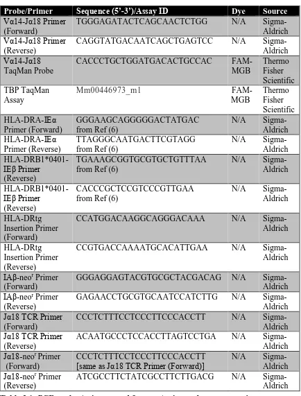
Table 5.1: PCR primers/probes used in IL-17A studies. *Primer was designed to span the insertion site of the HLA-DR4 transgenes in the mouse genome, thus enabling discrimination between homozygous and heterozygous transgenic mice. ... 148
Table 5.2: Antibodies/tetramers used in IL-17A studies ... 155
xiii
List of Figures
Figure 1.1: SAgs bypass conventional modes of antigen processing and presentation to activate a larger number of T cells. ... 13
Figure 3.1: Genetic or antibody-mediated depletion of iNKT cells reduces morbidity and
mortality in SEB-injected DR4tg mice. ... 110
Figure 3.2: DJ mice lack iNKT cells but harbor other major immune cell subsets. ... 111
Figure 3.3: TSS mortality is reduced in two separate models of iNKT cell deficiency. 114
Figure 3.4: iNKT cells are required for rapid inflammatory mediator production during
TSS and their TH2-polarization reduces TSS severity. ... 116
Figure 3.5: SEB-injected DJ mice exhibit a mild cytokine storm. ... 117
Figure 4.1: CD11b+Gr-1highLy-6C+ cells amass in the liver of DR4tg mice shortly after
SEB injection. ... 132
Figure 4.2: G-MDSCs accumulate selectively in the liver following systemic exposure to SEB. ... 133
Figure 4.3: Systemic exposure to SEB results in intrahepatic accumulation of CD11b+ cells
that concomitantly express Gr-1, Ly-6G and Ly-6C. ... 134
Figure 4.4: Hepatic CD11b+Gr-1high cells from SEB-treated DR4tg mice exhibit a
ring-shaped nucleus. ... 136
Figure 4.5: Mouse and human MDSCs suppress SEB-induced T cell proliferation. ... 138
Figure 4.6: SEB administration does not elevate Ki-67 expression by CD11b+Gr-1high cells in
the liver but lowers their frequency in the bone marrow. ... 139
Figure 5.1: Rapid IL-17A production by CD4+ TEM cells in a humanized mouse model of
TSS. ... 159
Figure 5.2: SEB-primed D17 mice launch a rapid IL-17A response and harbor an
enriched population of IL-17A-producing CD4+ TEM cells in their small intestine. ... 163
Figure 5.3: SAgs induce rapid IL-17A synthesis by human PBMCs. ... 166
Figure 5.4: CD4+ TEM cells are the major source of IL-17A in SAg-stimulated human
PBMCs. ... 170
xiv
Figure 5.6: Blockade of IL-17RA signaling attenuates SEB-induced inflammatory gene expression in human PBMCs and in downstream non-hematopoietic cells. ... 178
xv
List of Abbreviations
αGC α-galactosylceramide
act-D actinomycin-D
APC antigen-presenting cell
Bcl2 B cell lymphoma 2
CARS compensatory anti-inflammatory response syndrome
CCAC Canadian Council on Animal Care
CDR complementarity determining region
CEBP CCAAT/enhancer-binding protein
CFSE carboxyfluorescein succinimidyl ester
COX2 cyclooxygenase 2
D-gal D-galactosamine
DC dendritic cell
EGR2 early growth response 2
ERK extracellular signal-regulated kinase
FMO fluorescence minus one
G-CSF granulocyte-colony stimulating factor
GFP green fluorescent protein
G-MDSC granulocytic myeloid-derived suppressor cell
GAS group A streptococcus
GM-CSF granulocyte macrophage-colony stimulating factor
H&E haematoxylin and eosin
HLA human leukocyte antigen
xvi
IFN interferon
IL interleukin
IMC immature myeloid cell
iNKT invariant natural killer T
iNOS inducible nitric oxide synthase
ITAM immunoreceptor tyrosine-based activation motif
IVIG intravenous immunoglobulin
i.p. intraperitoneal
i.v. intravenous
JAK-STAT Janus kinase-signal transducers and activators of transcription
JNK c-Jun N-terminal kinase
mAb monoclonal antibody
M-CSF macrophage-colony stimulating factor
M-MDSC monocytic myeloid-derived suppressor cell
MAPK mitogen-activated protein kinase
MCP-1 monocyte chemoattractant protein-1
MDSC myeloid-derived suppressor cells
MFI mean fluorescence intensity
MHC major histocompatibility complex
MIP-2 macrophage inflammatory protein-2
NF-AT nuclear factor of activated T cells
NF-κB nuclear factor kappa-light-chain-enhancer of activated B cells
NKT natural killer T cells
xvii
PBS phosphate-buffered saline
PGE2 prostaglandin E2
PLC phospholipase C
PLZF promyelocytic leukaemia zinc finger protein
qPCR quantitative real-time polymerase chain reaction
R receptor
ROR retinoic acid receptor-related orphan receptor
ROS reactive oxygen species
RPL13a ribosomal protein L13a
SAg superantigen
SE staphylococcal enterotoxin
SEFIR SEF/IL-17R
SEL staphylococcal enterotoxin-like
SMEZ streptococcal mitogenic exotoxin Z
SPE streptococcal pyrogenic exotoxin
STSS streptococcal toxic shock syndrome
TBP TATA-binding protein
TCR T cell receptor
TEM T effector memory
Th1/2/17 T helper type-1/2/17
TIM3 T cell immunoglobulin and mucin domain containing-3
TIR Toll/IL-1R
TLR Toll-like receptor
xviii
TRAF tumor necrosis factor receptor associated factor
TRAIL tumor necrosis factor-related apoptosis-inducing ligand
Treg T regulatory
TSS toxic shock syndrome
TSST-1 toxic shock syndrome toxin-1
vNKT variant natural killer T
Chapter 1
Introduction
Parts of this chapter have been adapted and published in:
Szabo, P. A., Anantha, R. V., Shaler, C. R., McCormick, J. K., and Haeryfar, S. M. 2015.
CD1d- and MR1-Restricted T Cells in Sepsis. Front. Immunol. 6: 401.
1.1 Preamble
The immune system is an extraordinary protective entity capable of mounting rapid and
effective responses against virtually every type of pathogenic microorganism that we
encounter on a daily basis. Through an elaborate network of physical barriers,
anti-microbial molecules, and a myriad of cell types (e.g., neutrophils, natural killer cells,
monocytes, macrophages, dendritic cells, mast cells, basophils and eosinophils), the
innate arm of the immune system provides a non-specific front-line defense against
foreign invaders. Innate immune cells also help shape the nature of the highly specific
adaptive arm of immunity, where T and B cell responses to antigens from virtually all
encountered pathogens work together to limit or outright eliminate infection. This process
then gives rise to immunological memory, which promotes an immune response that is
both more rapid and greater in magnitude to pathogens that have been encountered
previously.
Not to be outdone, pathogenic microbes engage in an evolutionary ‘arms race’ with the
host immune system, developing sophisticated tactics for immune evasion to survive.
Through numerous virulence factors, pathogens exploit opportunities to bypass immune
defenses and colonize host tissues. The most successful of these pathogenic microbes are
typically known to be common causes of devastating disease in humans. The
Gram-positive coccus, Staphylococcus aureus, is among these pathogens and is currently the
most common cause of skin and soft-tissue infections worldwide (1). Interestingly,
around 20% of the human population appears to be persistently colonized by S. aureus
with another 30% being intermittent carriers (2). Most often, S. aureus carriage is
aureus causes severe, invasive and life-threatening infections including osteomyelitis,
endocarditis, pneumonia, enterocolitis, sepsis and toxic shock syndrome (TSS) (3).
One family of virulence factors, the staphylococcal superantigens (SAgs), is known to be
the direct causative agent of TSS (4). These potent exotoxins trigger the hyperactivation
of the immune system resulting in an avalanche of inflammatory mediators that cause
intense fever, hypotensive shock and organ damage. In essence, SAgs ‘short-circuit’ the
immune system, amplifying the immune response to the point of catastrophic collateral
damage to the host. Alarmingly, this process can progress to life-threatening disease
extremely rapidly, whereby multi-organ failure can occur in as little as 8-12 hours after
the onset of symptoms (5) and mortality rates can range as high as one-in-five (6).
Despite the high levels of morbidity and mortality in TSS, there is currently no treatment
for the overwhelming inflammation induced by SAgs. Identifying the underlying
immunological mechanisms that regulate or drive TSS pathogenesis may hold the key to
developing therapeutics to reduce disease severity. Here, I identify and define the role of
three previously unrecognized processes mediating TSS immunopathogenesis in its most
1.2 Toxic Shock Syndrome
A syndrome of disorientation, high fever, and scarlatiniform rash with rapid progression
to hypotensive shock and multi-organ dysfunction was first formally associated with
Staphyloccoccus aureus infection in 1978 (7). It was then that Todd and colleagues
coined the term ‘toxic shock syndrome’. In the early 1980s, TSS rose to public
consciousness following an outbreak in young females that was related to the usage of
high absorbency tampons (8-10). Several cases in men and non-menstruating women
were also reported during this time. A bacterial toxin from vaginal isolates of
Staphyloccoccus in TSS patients was identified as the cause of TSS (11, 12). These toxins
were later dubbed ‘superantigens’ due their ability to abnormally stimulate a large
fraction of T cells (13). As a result, SAgs trigger a massive inflammatory response
leading to the clinical symptoms of TSS (Table 1.1).
Cases of TSS are typically defined as either menstrual (14) or nonmenstrual (15) in
origin. Menstrual TSS occurs within the first few days before or after the beginning of a
woman’s menstrual cycle and is strongly associated with tampon usage due, at least in
part, to increases in oxygen levels in the normally anaerobic vaginal environment (16,
17). In the vast majority of menstrual TSS cases, vaginal colonization of S. aureus can be
readily detected (18). Nearly all cases of this form of TSS are caused by S. aureus strains
that produce the SAg toxic shock syndrome toxin-1 (TSST-1) (11, 12). Although the
exact reason for this association is unclear, it has been proposed that it may be a result of
the greater ability of TSST-1 to infiltrate mucosal surfaces compared to other SAgs (19,
20). A large proportion of mucosal S. aureus strains also more commonly produce
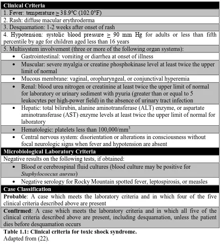
Clinical Criteria
1. Fever: temperature ≥ 38.9°C (102.0°F) 2. Rash: diffuse macular erythroderma
3. Desquamation: 1-2 weeks after onset of rash
4. Hypotension: systolic blood pressure ≥ 90 mm Hg for adults or less than fifth percentile by age for children aged less than 16 years
5. Multisystem involvement (three or more of the following organ systems):
• Gastrointestinal: vomiting or diarrhea at onset of illness
• Muscular: severe myalgia or creatine phosphokinase level at least twice the upper
limit of normal
• Mucous membrane: vaginal, oropharyngeal, or conjunctival hyperemia
• Renal: blood urea nitrogen or creatinine at least twice the upper limit of normal
for laboratory or urinary sediment with pyuria (greater than or equal to 5 leukocytes per high-power field) in the absence of urinary tract infection
• Hepatic: total bilirubin, alanine aminotransferase (ALT) enzyme, or aspartate
aminotransferase (AST) enzyme levels at least twice the upper limit of normal for laboratory
• Hematologic: platelets less than 100,000/mm3
• Central nervous system: disorientation or alterations in consciousness without
focal neurologic signs when fever and hypotension are absent
Microbiological Laboratory Criteria
Negative results on the following tests, if obtained:
• Blood or cerebrospinal fluid cultures (blood culture may be positive for
Staphylococcus aureus)
• Negative serology for Rocky Mountain spotted fever, leptospirosis, or measles
Case Classification
Probable: A case which meets the laboratory criteria and in which four of the five clinical criteria described above are present
Confirmed: A case which meets the laboratory criteria and in which all five of the clinical criteria described above are present, including desquamation, unless the patient dies before desquamation occurs
In contrast to menstrual TSS, a nonmenstrual form can originate from any infection or
colonization of SAg-producing strains of S. aureus. Non-menstrual TSS can occur in a
variety of clinical settings including skin and soft-tissue infections (15, 23), surgical and
post-partum wound infections (24, 25), after influenza infections (26) and burns (27-29).
Additional categories also include recalcitrant erythematous desquamating syndrome
(30), purpura fulminans (31), extreme pyrexia syndrome (32) and TSS-like
staphylococcal enterocolitis (33), which are all associated with S. aureus SAgs.
Approximately half of nonmenstrual TSS cases are caused by TSST-1, with the other half
caused by the staphylococcal enterotoxins (SEs) A, B, and C (23, 34-36).
1.2.1 Epidemiology
At its peak in the early 1980s, the incidence of TSS was estimated to be as high as 13.7
cases per 100,000 (37). With the removal of high-absorbency tampons from the market
and increased public awareness, these rates dropped to 0.5 per 100,000 by the late 1980s
(24, 38). More recently, a population-based surveillance program between 2000 and 2006
indicated that the annual incidence of TSS remained stable at 0.52 per 100,000 and that
approximately half of the reported cases were non-menstrual in origin (39). Notably, the
investigators suggested that this might be an underrepresentation of the disease burden,
likely due to both the strict CDC case definition (Table 1.1) and current advances in
supportive therapy that prevent severe manifestations of the disease. Despite its relatively
low incidence, TSS has an overall case fatality rate of 2-4% (23, 24, 39). Alarmingly,
however, non-menstrual TSS mortality rates can reach as high as 12-22% in some studies
1.2.2 Streptococcal TSS
A significant number of Streptococcus pyogenes or invasive group A Streptococcus
(GAS) infections share clinical similarities to TSS, and were designated as ‘toxic
shock-like syndrome’ or streptococcal TSS (STSS) in the late 1980s (40-42). STSS patients
have invasive GAS infections that culminate in shock, multi-organ failure and
progressive soft tissue destruction (42). Unlike staphylococcal TSS, which is typically
secondary to a localized S. aureus infection, STSS is the result of an invasive GAS
infection where the majority of patients are bacteremic (42, 43). Streptococcal M types 1
and 3 have been most associated with invasive GAS infections and the SAgs
streptococcal pyrogenic exotoxins (Spe) A and C are often detected in streptococcal
isolates from STSS patients (40, 44, 45). Annual incidence rates of invasive GAS
infections in Europe are 1.5-5.2 per 100,000 and approximately 5-14.4% of these patients
developed STSS (46, 47). A study from 11 European countries (Strep-EURO) revealed
an overall 7-day case fatality rate of 19% for GAS infections and 44% in patients that
progressed to STSS (47).
1.2.3 Therapeutic Strategies
There are currently no specific therapeutics available for the treatment of TSS. The key to
successful management of the disease is the rapid identification of the source of infection,
appropriate and effective use of antibiotics, removal of tampon in menstrual TSS, and
supportive care (48). In the early stages of the disease, similar therapeutic interventions
as septic shock are applied, including haemodynamic support, vasopressors and/or
Based on the finding that individuals lacking effective neutralizing antibodies to SAgs
may be at increased risk of developing TSS (50-52), intravenous immunoglobulin (IVIG)
has been proposed as an adjunctive therapy (48). IVIG is a combination of pooled human
antibodies derived from hundreds to thousands of donors, which typically contain
antibodies against both staphylococcal and streptococcal SAgs (53). The effectiveness of
IVIG in improving clinical outcomes has been demonstrated in a number of studies
involving STSS (54-57); however, evidence for its effectiveness in staphylococcal TSS is
limited (48). It should also be noted that the mechanism of action for IVIG is unclear.
IVIG preparations are able to neutralize both staphylococcal and streptococcal SAg
activity in vitro, though SAgs from S. aureus require much higher doses of IVIG to be
effectively inhibited (58). IVIG can also directly opsonize GAS, promoting bacterial
clearance by enhancing phagocytosis (59). Lastly, IVIG has a general anti-inflammatory
effect mediated by immunoglobulin Fc domains binding to Fc receptors on immune cells
(60). While IVIG has been shown to suppress SEB-induced T cell activation, this effect is
largely maintained after SEB-specific antibodies are depleted from the IVIG
preparations, suggesting that a mechanism independent of toxin neutralization is in effect
(61).
Several other strategies to attenuate TSS morbidity have been examined including the
inhibition of SAg-receptor interactions, blockade of SAg signal transduction, and active
vaccination against staphylococcal/streptococcal cell wall components or the various
exotoxins themselves (62, 63). Although promising, these approaches are still in their
infancy and are currently undergoing preclinical investigation. Despite the significant
remain unanswered. A more complete understanding of the cellular interactions and
biological effects of SAgs may aid in the quest for a therapeutic invention to reduce TSS
severity.
1.2.4 Superantigens
SAgs are a unique group of protein exotoxins secreted by the vast majority of S. aureus
and GAS strains that are pathogenic to humans (64). S. aureus encodes a large number of
SAgs including TSST-1 and the SEs A, B, C, D, E, G, H, I, R and T (65). SEs were
originally defined by their emetic properties and ability to cause food poisoning when
ingested by humans or when administered orally to monkeys (66). A group of SE-like
(SEl) toxins that are structurally similar to SEs have also been defined, although these
toxins either do not induce, or have not yet been shown to induce emesis (65).
Interestingly, almost all staphylococcal SAgs are encoded on mobile genetic elements
including pathogenicity islands, plasmids or phages, suggesting an exchange of genetic
elements between different strains or even different bacterial species (4). GAS also
produces numerous SAgs including SpeA, C, G-M, streptococcal superantigen (SSA) and
streptococcal mitogenic exotoxin Z (SMEZ) (64). Similar to staphylococcal SAgs, most
streptococcal SAgs are encoded on mobile genetic elements.
It is important to note that SAgs are not specifically limited to S. aureus and GAS, nor to
bacteria in general. SAgs have also been documented in the human pathogens
Plasmodium falciparum (67), Clostridium perfringens (68), Toxoplasma gondii (69),
1.2.5 Superantigen Structure and Binding Properties
SAgs are generated as pro-toxins that require cleavage of a secretion signal to be
exported from the cell in a Sec-dependent manner and range in size from 22-29 kDa (65).
Crystal structures of SAgs show that they are commonly made of two major protein
domains, an N-terminal oligosaccharide/oligonucleotide binding fold with a β barrel
motif and a C-terminal β-grasp motif, which are connected together by a central α-helix
(72).
SAgs are capable of bypassing conventional mechanisms of antigen presentation and
directly cross-link T cell receptors (TCRs) on T cells with major histocompatibility
complex (MHC) class II molecules on antigen-presenting cells (APCs) in an unprocessed
form (73, 74). SAgs interact with MHC class II outside the antigen-binding groove (75)
and bind specific TCR β-chain variable domains (Vβ), inducing the activation and clonal
expansion of T cells bearing the appropriate Vβ segments (76). Each SAg has a unique
pattern of Vβ specificities resulting in a ‘Vβ T cell signature’ that can be used to identify
the SAg(s) a patient is exposed to (77). Several SAgs including TSST-1, SEA and SEB
also bind the co-stimulatory molecule CD28 on T cells, which is required for maximal T
cell activation (78).
Based on phylogenetic analyses and structural similarities, staphylococcal and
streptococcal SAgs can be classified into 5 evolutionary groups (65, 79). Group I consists
of TSST-1 as an evolutionarily distinct SAg, which has a unique primary amino acid
sequence compared to other SAgs. TSST-1 lacks a cysteine loop associated with emetic
activity (80), further distinguishing it from SEs. It also utilizes a low-affinity MHC class
antigenic peptide present within the MHC class II molecule (81, 82). TSST-1 is also
specific for Vβ2+ T cells in humans, as it recognizes specific amino acids within the
complementarity-determining region (CDR) 2 and framework region 3 (83). Group II
SAgs include SEB, SEC and SpeA. This group also uses a single low-affinity binding
domain to the α-chain of MHC class II; however, this binding appears to be entirely
peripheral to the antigenic peptide (84, 85). Notably, group II SAgs bind the TCR Vβ in a
manner that is less dependent on specific amino acid side-chains, allowing for a wider
range of Vβ specificities (86-88). Group III SAgs include SEA, which, along with group
II SAgs, are implicated in the majority of food poisoning cases (89). In addition to the
low-affinity binding site on the MHC class II α-chain used by groups I and II, group III
SAgs contain a second high-affinity binding site that is zinc-dependent, which engages
the MHC class II β-chain and extends over the bound peptide (90-94). It has been
suggested that the additional presence of the high-affinity binding site may increase the
efficiency of these SAgs to activate T cells and APCs at significantly lower
concentrations. Group IV SAgs consist exclusively of streptococcal SAgs, including
SPEC and SMEZ, which also bind MHC class II α-chain and β-chain similar to group III
SAgs (95, 96). Lastly, group V SAgs include SEI and the SEl toxins, which use similar
binding targets as group III and group IV SAgs (97).
1.2.6 Superantigen-mediated T cell activation
The mode of TCR engagement by SAgs is in stark contrast with that of conventional
antigens. Typically, T cell activation is initiated by the interaction between the TCR and
its cognate antigenic peptide that is presented in the context of self-MHC, called the rule
variability in TCRs generated by the stochastic joining of TCR α- and β-V(D)J segments,
at most only 0.01% of naïve T cells can be activated by a specific antigen (99). However,
SAgs ‘hijack’ the immunological synapse, and activate T cells in a TCR Vβ-dependent
manner, regardless of T cell antigen specificity. Since there are a limited number of TCR
Vβ regions expressed by T cells, approximately 50 in humans, SAgs can activate up to
20-50% of T cells (Fig. 1.1) (64, 65). Furthermore, SAgs do this in an exceptionally
potent manner, where as low as femtogram (10-15 g) quantities can stimulate human T
cells in vitro (100).
Downstream of the TCR, T cell signaling by SAgs is relatively similar to that of
conventional antigens, whereby the three canonical signals required for T cell activation
are comparable for both SAgs and conventional antigens. Signal one is induced by SAg
engagement of the TCR-CD3 complex, recruiting the Src family of protein tyrosine
kinases including Lck (101). Activated Lck phosphorylates the CD3 immunoreceptor
tyrosine-based activation motifs (ITAMs), recruits a variety of adaptor molecules, and
facilitates a series of phosphorylation events mediated by phospholipase C (PLC)-γ1 that
culminate in the activation of protein kinase C and increased intracellular Ca2+ (102).
Together, these signaling pathways lead to activation and translocation of the nuclear
factor of activated T cells (NF-AT) and the nuclear factor kappa-light-chain-enhancer of
activated B cells (NF-κB) to the nucleus, facilitating the process of T cell activation and
proliferation. For signal two, T cell co-stimulatory molecules (e.g. CD28) can either
engage with their receptors (e.g. CD80/86) on APCs or be directly stimulated by SAgs
(103). Co-stimulation is required for proper T cell activation and ensuing proliferation,
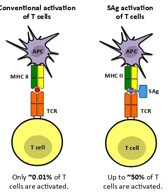
Figure 1.1: SAgs bypass conventional modes of antigen processing and presentation to activate a larger number of T cells.
Conventional protein antigens are taken up, processed into peptide fragments, and
presented to T cells in the peptide-binding groove of MHC class II by APCs. In contrast,
SAgs bind the lateral surfaces of MHC class II and the Vβ-domain of the TCR in
unprocessed form. Whereas conventional antigens activate up to ~0.01% of T cells, SAgs
may activate up to 50% of exposed T cells resulting in the overwhelming cytokine storm
in TSS. Adapted from (105).
SAg ac va on
of T cells
Conven onal ac va on
of T cells
Up to
~50%
of T
cells are ac vated.
Only
~0.01%
of T
cells are ac vated.
APC
APC
T cell
T cell
TCR
TCR
SAg
milieu present at the time of T cell activation and is essential for productive T cell
responses and polarization of T cell differentiation into distinct effector subtypes (106).
In 2006, Bueno et al. found that SAg-mediated T cell signaling did not strictly require
Lck activity and that a Gα11/PLC-β-mediated phosphorylation cascade can initiate an
Lck-independent signaling pathway (107). Interestingly, both Lckdependent and
-independent pathways can be utilized by SAgs, where either can lead to the activation of
transcription factors that ultimately facilitate T cell activation (108). These findings shed
light on how SAgs activate both CD4+ and CD8+ T cells, despite only cross-linking MHC
class II molecules. Since one function of the CD4/CD8 co-receptors is to recruit and
activate Lck, an Lck-independent pathway would allow for T cell activation to occur in
the absence of CD4/CD8 binding to MHC.
1.2.7 Cytokine Storm
The overwhelming immune activation caused by SAgs triggers a ‘cytokine storm’ – a
rapid and uncontrolled production of cytokines and inflammatory mediators from many
cell types including T cells, APCs, epithelial cells and endothelial cells. A variety of
inflammatory cytokines are rapidly produced within hours of SAg exposure, including
tumor necrosis factor α (TNFα), interleukin (IL)-1, IL-6, and interferon γ (IFNγ) (109,
110).
TNFα is a potent inducer of systemic inflammation and signals a range of cellular
responses including apoptosis, differentiation, proliferation, migration and cytokine
production (111, 112). TNFα signaling occurs through the TNF receptors (TNFR) 1 and
death domain) to activate the caspase 8, mitogen-activated protein kinase (MAPK), and
NF-κB pathways that mediate its pleiotropic effects. In endothelial cells, TNFα elicits
vasodilation, vascular permeability, and expression of pro-coagulant proteins such as
tissue factor to promote intravascular coagulation (113). Together with IL-1, TNFα is
likely the main mediator of inflammation-driven activation of coagulatory cascades in the
vasculature (114). The administration of TNFα alone causes septic shock-like state in
animals (115) and induces a systemic inflammatory response syndrome in humans
(116-118).
IL-1 can be subdivided into IL-1α and IL-1β that both signal through the IL-1 receptor
(IL-1R)-1 and accessory protein (IL-1RAcP) complex (119). IL-1 has similar and often
synergistic inflammatory effects with TNFα (120). IL-1β mediates pro-coagulant activity
and tissue factor expression in human monocytes when stimulated with SAgs (121). IL-1
and TNFα are often considered the two primary mediators of shock (122), where they act
synergistically to induce sustained hypotension, pulmonary edema and hemorrhage (123).
These overlapping functions may be due to the fact that both cytokines activate the
NF-κB signaling pathway, which plays critical role in the initiation and propagation of the
inflammatory cascade in TSS and septic shock (122, 124). This pathway promotes: i) the
expression of adhesion molecules and chemokines to recruit and activate neutrophils
causing endothelial cell injury, vascular permeability, and tissue damage; ii) expression
of tissue factor and other pro-coagulants initiating disseminated intravascular
coagulation; iii) induction of cyclooxygenase-2 (COX-2) and inducible nitric oxide
synthase (iNOS) pathways triggering vasodilator prostaglandins and nitric oxide,
production of inflammatory cytokines (e.g. TNFα, IL-1, IL-6 and IFNγ) thus amplifying
and propagating the inflammatory response. The extent of NF-κB activation has been
shown to correlate with mortality risk in several contexts, including septic shock (125).
IL-6 binds to the IL-6 receptor (IL-6R) and has many different biological effects
including the activation of T cells, initiation of the coagulation cascade, and neutrophil
activation and trafficking (126). Along with TNFα and IL-1, IL-6 is an endogenous
pyrogen, that is, a group of fever-inducing molecules that stimulate prostaglandin E2
(PGE2) synthesis, which acts on the hypothalamus to increase the thermostatic set point
(127). IL-6 is also a major mediator of the acute phase response, a systemic reaction to
inflammation characterized by the rapid production of acute phase reactants, such as
C-reactive protein, from the liver. These proteins trigger complement activation,
coagulatory cascades or promote leukocyte chemotaxis (128). Notably, serum IL-6
concentrations are markedly increased in TSS patients (129) and correlate with sepsis
severity and mortality (130). In spite of these many pro-inflammatory characteristics,
IL-6 can promote anti-inflammatory responses as well. While TNFα and IL-1 synergistically
induce IL-6 production, IL-6 can inhibit the release of TNFα and IL-1 and promote the
expression of several anti-inflammatory mediators, like IL-10 (131, 132). IL-6 responses
also appear to be protective in mouse models of endotoxemia (133) and streptococcal
infections (134), likely due to a suppressive effect on TNFα.
IFNγ signals through the interferon gamma receptor (IFNGR), consisting of subunits
IFNGR1 and IFNG2, and performs a wide variety of immunoregulatory roles including
innate immune cell activation, leukocyte trafficking, augmenting MHC class I and II
IFNγ induces the expression of the death receptor Fas, which triggers vascular cell
apoptosis (136, 137). Together with TNFα, IFNγ also disrupts ion transport and barrier
functions of epithelial cells leading to increased permeability (138). Indeed, IFNγ and
TNFα often have synergistic effects, where many IFNγ-induced genes are also induced
by TNFα, likely due to multiple avenues of cross-talk between signaling pathways (135).
A variety of chemokines are also rapidly produced in response to SAg stimulation (139,
140). TSST-1 can stimulate the production of chemokines directly from the human
vaginal epithelium (141). These chemoattractants facilitate the recruitment of neutrophils,
macrophages and T cells to the areas of inflammation and injury. In addition to
increasing the accessibility for SAgs to activate T cells, recruited immune cells can also
have direct tissue damaging effects. For example, cytokine-activated neutrophils produce
cytotoxic molecules and matrix metalloproteases once in peripheral tissues (142).
Recruited immune cells can also produce large amounts of inflammatory cytokines upon
activation, thus amplifying the cytokine storm. SAgs activate many non-immune cells
including fibroblasts, endothelial cells and epithelial cells (141, 143, 144) that produce
other inflammatory molecules (e.g. adhesion proteins, tissue proteases and reactive
oxygen species) in addition to cytokines, resulting in further tissue injury.
Overexpression of inflammatory cytokines in the cytokine storm results in widespread
inflammation, fever, disseminated intravascular coagulation, leukocyte migration,
vasodilation, capillary leak, shock, and tissue destruction. This process culminates in the
dysfunction of multiple organ systems, which, if severe, can lead to death. Two distinct
phases of multiple organ dysfunction have been proposed (145). In the early phase, the
endothelial cell damage, increased vascular permeability, swelling and extravasation. The
later phase of organ dysfunction begins as the inflammatory cells reach the interstitial
compartments of individual organs. Microvascular coagulopathy, dysregulated apoptosis
and tissue hypoxia cause damage to parenchymal cells, thus impairing organ function
(146). The reflux of inflammatory cytokines and other mediators back into circulation
then creates a positive feedback loop, amplifying this process.
1.2.8 SAg-Induced Immunosuppression
Following the hyperactivation of T cells, SAgs induce a state of immune dysregulation in
the body. Almost immediately after activation, surface TCR expression on SAg-reactive
T cells is reduced by 50% due to receptor endocytosis (147). The activation and
proliferation of SAg-reactive T cells is transient and the majority undergo apoptosis by
activation-induced cell death within days (148, 149). This creates a functional ‘hole’ in
the T cell repertoire and is sustained for a period of at least 4 weeks, when a 50%
reduction of T cells bearing SAg-reactive Vβ domains can still be observed (150).
Neonatal exposure to SAgs in mice also deletes virtually all SAg-reactive mature
thymocytes, in addition to the majority of immature thymocytes (73). Activated T cells
that escape deletion become anergic, or functionally unresponsive to in vitro
re-stimulation by SAgs (150, 151). However, whether this translates into anergy in vivo is
unclear. Multiple studies have demonstrated functional cytokine responses upon SAg
re-challenge, though SAg-reactive CD4+ T cells, and CD8+ T cells, to a lesser extent, show
impairments in their proliferative capacity (152-154). Exposure to SAgs also generates
immunosuppressive T regulatory cells (Tregs) (155-158), which may contribute to the
systemic inflammatory responses in TSS destabilize the Treg population, and their
expansion or adoptive transfer in mice exposed to SAg fail to mitigate immunopathology
(159).
In addition to causing T cell dysfunction, SAgs impair B cell-mediated humoral
immunity (160-162). Low or negative antibody titres for TSST-1 are observed in greater
than 90% of menstrual TSS patients, with the majority of patients failing to seroconvert
within two months (50). Some patients even remain seronegative even after recurrent
bouts of the disease. This is in contrast to a healthy population, where 85% of individuals
show antibody titres to TSST-1 at a level thought to be at least partially protective (52).
The failure to develop effective antibody-mediated immunity is likely due to impairment
in the T cell compartment during TSS. T cells anergized by SAgs can no longer provide
help for B cells to stimulate antibody production, thus impairing T cell-dependent B cell
responses (161, 162). SAgs also induce T cell-dependent B cell apoptosis (163-165).
Lastly, SAgs largely promote Th1-type cytokine responses. Therefore, the minimal
Th2-type cytokines present may be insufficient to support optimal B cell proliferation and
differentiation (124).
1.2.9 Animal Models of TSS
Preclinical animal models that simulate SAg-mediated immunopathology in humans have
greatly aided the characterization of molecular and cellular events in TSS. Despite our
current understanding, however, there has yet to be an effective therapy translatable to
the clinic. The careful and appropriate use of animal models of TSS (listed in Table 1.2)
remains our most essential tool for the development of novel treatment modalities to
Animal Model SAgs Defining Immunopathology Reference Mouse Sensitizing agents (D-gal/act D) SEA, SEB, TSST-1
TNFα-dependent lethal shock; severe
hemorrhagic hepatitis (166-170)
SAg + LPS
SEA, SEB, SEC, TSST-1
Rapid cytokine storm; hypothermia; intestinal apoptosis; lethal shock
(169, 171-175) HLA-transgenic SEB SpeA
Rapid cytokine storm; multi-organ inflammation; impaired gut permeability; lethal shock
(170, 176-178)
Aerosol/
i.n SEB
Rapid cytokine storm; pulmonary edema, hypothermia; liver neutrophil infiltration and
necrosis; lethal shock
(179-184)
Rabbit Mini-osmotic pump SEA, SEB, TSST-1 SpeA SpeC
Rapid cytokine storm; fever; conjuctival
hyperemia; anorexia, cachexia; lethal shock. (185-189)
SAg + LPS TSST-1, SEH
TNFα production; hyperemia; diarrhea; lethal shock
(190, 191) (192, 193)
i.v. SEA,
TSST1
IL-2, IFN, TNF production; fever; lethal shock (strain and sex dependent)
(191, 193-195)
Primate i.v. SEB Hypotension; cardiac output decline; decreased
blood oxygen content; lethal shock (196)
Aerosol SEB
IL-2, IL-6, IFNγ production; hypotension; fever; diarrhea; vomiting; pulmonary edema; lethal
shock
(197-199)
D-gal, D-galactosamine; act D, actinomycin D; LPS, lipopolysaccharide; tg, transgenic; i.n., intranasal; i.v., intravenous.
Mouse models are most often used to examine the underlying mechanisms of
SAg-induced morbidity, largely due to the variety of available immunological tools and
reagents, well-defined Vβ signatures, and relatively low cost of maintenance compared to
other animals. Problematically, mice are considerably more resistant to SAgs when
compared to humans as a result of a much lower binding affinity of SAgs to mouse MHC
class II in some strains (200). Therefore, sensitizing compounds such as actinomycin D
(167) or D- galacatosamine (D-gal) (166) must be used to amplify the effects of SAgs
and induce mortality. The severity of TSS in models using sensitizing agents varies
considerably and can skew inflammatory responses or disproportionately impact
individual organs. One of the most widely utilized models involves priming mice with
D-gal (166, 168, 169, 201, 202) followed by intraperitoneal administration of SAg to induce
a cytokine storm and other hallmark features of TSS. D-gal induces transcriptional arrest
in hepatocytes leading to extreme sensitivity to TNFα-induced apoptosis (203). When
used in combination with SAgs, D-gal induces severe TNFα-dependent hepatotoxicity
(204), a pathology not typically seen in human TSS (205). Although TNFα responsible
for the lethality in sensitized mice is derived, at least in part, from SAg-mediated T cell
activation (166, 206), the dominant event leading to sudden death in this model is likely
severe hepatitis. As expected, TNFα plays a key role in this model and anti-TNFα/β
neutralizing antibodies confer protection from lethality (166, 207). Conclusions from
these models must be drawn with full recognition that sensitizing agents cause cytokine
responses that do not necessarily reflect human disease. This problem is illuminated by
the notable failure of a TNFR fusion protein to prevent septic shock in humans, including
group receiving the highest-dose of the drug exhibited an increase in both disease
severity and mortality.
Another common mouse model utilizes bacterial lipopolysaccharide (LPS) as a
potentiating agent following SAg exposure (169, 171-174, 209, 210). SAgs are known to
substantially enhance LPS lethality due to a synergistic effect on the production of TNFα
(192). There is some evidence to suggest this synergy occurs in human TSS, as patients
with menstrual TSS show increased colonization of Gram-negative bacteria in the vaginal
flora (211) and low levels of LPS can be detected in acute-phase sera of TSS patients
(212). In mice, the injection of SAg followed by LPS results in rapid production of IL-1α,
IL-6, TNFα and IFNγ within hours, which is not observed in SAg-only controls (171).
Lethality in this model can be correlated to increased serum IL-6, TNFα, macrophage
inflammatory protein-2 (MIP-2) and monocyte chemoattractant protein-1 (MCP-1) by 8
hours and IFNγ and IL-2 by 21 hours after TSS induction (174). However, cytokine
responses and TSS mortality are highly influenced by LPS dosage as opposed to SAg.
Furthermore, exposure to SAg in human peripheral blood mononuclear cells (PBMCs)
strongly upregulates TLR4 expression and enhances production of inflammatory
cytokines by LPS independent of T cells (210). Thus, the use of LPS as a potentiating
agent may cause unnecessary confounding effects that are more reflective of
endotoxicosis than TSS. As the role of LPS in human TSS is undefined, the utility of this
mouse model and its relevance to TSS also remains to be determined.
In order to avoid potential pitfalls associated with sensitizing and potentiating agents in
mouse models of TSS, mice expressing human MHC class II, known as HLA-transgenic
SAgs to human leukocyte antigens (HLAs)-DQ and -DR, these transgenic mice respond
to much lower doses of SAg (216-219). Accordingly, our lab has previously
demonstrated robust cytokine responses in HLA-DR4tg mice injected with 50 μg of SEB
(220). Similarly, a single 50 μg dose of SEB is sufficient to induce a rapid inflammatory
cytokine response, inflammation in the liver, lungs, kidney, heart and small intestines,
and lethal shock in HLA-DR3 transgenic mice (170, 177). Importantly, in comparison to
D-gal sensitization where the disease is restricted to the liver, HLA-DR3 mice treated
with SAg alone show multi-organ involvement, similar to humans (170). The inhibition
of IFNγ in this model attenuates the cytokine storm, prevents severe intestinal pathology,
and protects against mortality (177). HLA transgenic mouse models have also been
successfully applied to STSS. HLA-DQ8 transgenic mice expressing human CD4 are
susceptible to SpeA-induced STSS that is characterized by rapid production of TNFα,
IFNγ and IL-6, elevated liver enzymes and lethal shock within 72 hours (178). A
potential limitation for using HLA-transgenic mice as disease models is that MHC class
II genes are among the most polymorphic genes in the genome and these inbred mouse
strains only mimic the responses of one specific haplotype. Furthermore, SAgs
preferentially bind some MHC class II molecules over others; SEB and TSST-1 bind
HLA-DR more efficiently than HLA-DQ for example (221), and thus care must be taken
to select the most appropriate transgenic strain for a given SAg. Nevertheless,
HLA-transgenic mice recapitulate many aspects of human TSS and therefore are one of the
most useful mouse models of the syndrome.
Several other mouse models have been established to investigate different aspects of TSS
inflammation was developed involving a single or double dose of SAg introduced
intranasally (179-184). TSS in this model culminates in severe lung inflammation,
systemic release of cytokines, hypothermia and death within days. However, this may
more accurately represent TSS resulting from the inhalation of aerosolized SEB in a
bioterrorism setting (222) rather than natural S. aureus infection. Intravaginal exposure to
SEB in HLA-transgenic mice causes a robust systemic inflammatory response and
leukocyte infiltration in the liver and lungs (223). Although almost all human menstrual
TSS cases are caused by TSST-1 and not SEB, further investigations in this model can be
targeted to understand and/or inhibit the mechanisms by which SAgs can cross mucosal
barriers and cause disease.
Historically, rabbits have been the ‘gold standard’ model for SAg-induced TSS as they
mimic almost all symptoms of human TSS after administration of staphylococcal and
streptococcal SAgs (185, 186, 191, 192, 194, 224, 225). Rabbits can also be implanted
with mini-osmotic pumps that release SAg continuously over the course of 7 days, more
accurately mimicking the mode of SAg exposure in a S. aureus infection (185-189).
Several rabbit models that utilize chambers to localize SAg-producing S. aureus have
been instrumental in identifying TSST-1 a causative agent of TSS (226-229). The
susceptibility to SAg-induced TSS varies among rabbit strains and may require LPS
sensitization to induce morbidity (191). Currently, the use of rabbits to model TSS is rare,
likely due to the technical and ethical considerations that make larger animals
unattractive to study. There are several drawbacks of using rabbits rather than mice, such
as increased housing and animal care costs, less availability of immunological reagents
In addition to mice and rabbits, several models of TSS utilizing larger animals have been
described. Intradermal exposure to SEB in non-human primates elicits skin reactions,
cutaneous mast cell degranulation and emesis (230). Non-human primates have also been
used to investigate immune responses to aerosolized SEB (197-199) and evaluate the
efficacy of vaccine candidates against lethal SEB challenge (231, 232). In the hopes of
more closely simulating human TSS, a porcine model was developed where piglets were
given SEB intravenously. Intoxicated piglets quickly developed vomiting, diarrhea,
temperature spikes, hypotension and multi-organ dysfunction (233). Lastly, goats have
been used to study the effects of both SEB and TSST-1-induced TSS, where intravenous
injection yields tachycardia, fever and various hematological changes (234). Investigating
TSS pathogenesis in larger animals is rare, due to both the high cost of animal
maintenance and significant ethical concerns surrounding their use as disease models
1.3 Invariant Natural Killer T Cells
Natural killer T (NKT) cells are a specialized group of innate-like T lymphocytes with
remarkable immunomodulatory properties. Named for their co-expression of both NK
cell markers (e.g. NK1.1 in mice and CD161 in humans) and αβ TCRs, NKT cells
possess specific reactivity to glycolipid antigens presented by the MHC class I-like
glycoprotein CD1d (235). CD1d is a member of the CD1 family of antigen-presenting
molecules that present lipids, rather than peptides, to non-MHC restricted T lymphocytes.
In humans, there are five members of CD1 molecules, CD1a-e, while mice only express
CD1d (236). CD1d is widely expressed on cells of hematopoietic origin, including typical
antigen-presenting cells like DCs, macrophages and B cells (237).
Two individual subsets of NKT cells can be defined based on the structural
characteristics of their TCRs in addition to their reactivity to glycolipid antigens. Type I
NKT cells, known as invariant NKT (iNKT) cells, express a canonical TCR composed of
a unique α-chain (Vα24-Jα18 in humans; Vα14-Jα18 in mice) paired with a limited
number of β-chains (Vβ11 in humans; Vβs 2, 7, or 8.2 in mice) (235). These cells exhibit
highly conserved reactivity to α-galactosylceramide (αGC) (238) and the invention of
αGC-loaded CD1d tetramers has enabled their unambiguous identification by flow
cytometry (239, 240). In mice, iNKT cells are either CD4+CD8- or CD4-CD8- (235), and
an additional CD8+ population exists in humans (241). iNKT cells also constitutively
express the activation/memory markers CD25, CD44 and CD69 on their cell surface,
consistent with a ‘memory-like’ phenotype. In contrast, type II NKT cells, known as
variant NKT (vNKT) cells, express a diverse αβ TCR repertoire and although they are
can be activated by the self-lipid sulfatide (242). The understanding of vNKT cell
responses in health and disease remains limited, largely due to a notable lack of robust
reagents to specifically identify and characterize vNKT cell function.
iNKT cells are widely distributed throughout the body and can be found in the thymus,
liver, bone marrow, spleen, lymph nodes and intestinal tract (239). They express a range
of chemokine receptors that mediate their homing to individual tissues and, unlike
conventional T cells, show limited recirculation under homeostatic conditions (243). In
mice, iNKT cells have been most commonly studied in the liver, where they represent up
to 20-30% of lymphocytes (239, 240). However, they are less frequent in all other tissues,
where they constitute ≤1% of T cells. In humans, iNKT cells are not highly enriched in
the liver and are 10-fold less frequent in most locations (244), except the human
omentum, where approximately 10% of T cells express the invariant Vα24-Jα18 TCR
(245). On average, iNKT cells represent ~0.1% of T cells in the peripheral blood of
humans, but this fraction can range from virtually undetectable to as high as 1% across
individuals (246, 247). The reason for this extreme variation among individuals as well as
the differences in abundance and tissue distribution between mice and humans is
unknown.
1.3.1 Modes of Activation
iNKT cells are activated in response to exposure to a wide range of pathogens, from
bacteria to viruses, protozoa and even fungi (248). Many of these microorganisms contain
lipid antigens, which can directly stimulate the iNKT cell TCR through their presentation
by CD1d on APCs. These exogenous antigens are usually glycerol-based lipids (e.g.


![Ethyl 2 {[(1Z) (3 methyl 5 oxo 1 phenyl 1,5 dihydropyrazol 4 ylidene)(phenyl)methyl]amino} 3 phenylpropanoate](data:image/gif;base64,R0lGODlhAQABAIAAAP///wAAACH5BAEAAAAALAAAAAABAAEAAAICRAEAOw==)