Trial Result in an Evolving Host Response That Affects Protective
Outcome
Christian Selinger,aNatasa Strbo,cLouis Gonzalez,cLauri Aicher,aJeffrey M. Weiss,aG. Lynn Law,aRobert E. Palermo,a,b Monica Vaccari,dGenoveffa Franchini,dEckhard R. Podack,cMichael G. Katzea,b
Department of Microbiology, University of Washington, Seattle, Washington, USAa; Washington National Primate Research Center, University of Washington, Seattle, Washington, USAb; Department of Microbiology and Immunology, Miller School of Medicine, University of Miami, Miami, Florida, USAc; Animal Models and Retroviral Vaccines, National Cancer Institute, Bethesda, Maryland, USAd
Using whole-blood transcriptional profiling, we investigated differences in the host response to vaccination and challenge in a rhesus macaque AIDS vaccine trial. Samples were collected from animals prior to and after vaccination with live, irradiated vac-cine cells secreting the modified endoplasmic reticulum chaperone gp96-Ig loaded with simian immunodeficiency virus (SIV) peptides, either alone or in combination with a SIV-gp120 protein boost. Additional samples were collected following multiple low-dose rectal challenges with SIVmac251. Animals in the boosted group had a 73% reduced risk of infection. Surprisingly, few changes in gene expression were observed during the vaccination phase. Focusing on postchallenge comparisons, in particular for protected animals, we identified a host response signature of protection comprised of strong interferon signaling after the first challenge, which then largely abated after further challenges. We also identified a host response signature, comprised of early macrophage-mediated inflammatory responses, in animals with undetectable viral loads 5 days after the first challenge but with unusually high viral titers after subsequent challenges. Statistical analysis showed that prime-boost vaccination signifi-cantly lowered the probability of infection in a time-consistent manner throughout several challenges. Given that humoral re-sponses in the prime-boost group were highly significant prechallenge correlates of protection, the strong innate signaling after the first challenge suggests that interferon signaling may enhance vaccine-induced antibody responses and is an important con-tributor to protection from infection during repeated low-dose exposure to SIV.
S
ince the identification of human immunodeficiency virus (HIV) as the primary cause of AIDS (1,2), substantial efforts have been put into developing a vaccine against the virus. Differ-ent vaccine strategies have been devised to generate specific cellu-lar or humoral responses, or combinations thereof, in order to prevent viral acquisition or disease progression. HIV cellular tro-pism, rapid escape mutations, and viral latency are major preven-tion and treatment hurdles (3–6). Here we used transcriptional profiling of host responses to vaccination and subsequent re-peated challenges to further understand the protective effects of a novel vaccination strategy based on the use of the endoplasmic reticulum resident chaperone gp96 (7). This heat shock protein, similar to soluble Hsfp and hsp65 (8,9), binds to and activates dendritic cells (10) and functions as a chaperone for tumor-se-creted peptides. The modified form of gp96, gp96-Ig, setumor-se-creted by tumors (11), results in an enhancement of tumor antigen cross-priming of CD8 cytotoxic T lymphocytes (CTLs) (12) and induces a mucosal immune response following intraperitoneal injection of transfected cells (13). When chaperoning simian immunodefi-ciency virus (SIV) peptides, gp96-Ig induces a strong multifunc-tional memory response in the rectal and vaginal mucosae of rhe-sus macaques (14).Starting from these observations, a vaccine approach was de-signed with the aim of combining the enhancement of CD8 CTL cross-priming elicited by gp96, the particular role of secreted gp96-Ig for loading a broad spectrum of SIV peptides to major histocompatibility complex class I (MHC I) molecules, and the CD4-dependent antibody responses elicited by the recombinant envelope protein rSIV-gp120. When used to vaccinate rhesus
ma-caques, live, irradiated vaccine cells secreting a modified gp96-Ig carrying SIV peptides (gp96SIVIg) in combination with a SIV-gp120 protein boost provided considerable protection against weekly mucosal challenge with the highly pathogenic strain SIVmac251(15). After seven challenges, the hazard ratio was 0.27, representing a 73% reduction in risk for viral acquisition. Impor-tantly, vaccination with either gp96SIVIg or gp120 protein alone provided no significant protection. However, gp96SIVIg vaccina-tion alone provided for potent cross-presentavaccina-tion of SIV antigens, generating CD8 CTLs but no antibody. Conversely, gp120 protein vaccination alone generated antibody but few CTLs. This argues that both cellular and humoral immune responses are required for protection.
In this study, we performed RNA profiling and functional genomic analyses of whole-blood samples obtained from our pre-vious vaccine trial (15). Our analyses revealed that monocyte/ macrophage- and neutrophil-mediated inflammatory response
Received3 July 2014 Returned for modification15 August 2014
Accepted26 September 2014
Published ahead of print1 October 2014
Editor:S. A. Plotkin
Address correspondence to Michael G. Katze, [email protected].
Supplemental material for this article may be found athttp://dx.doi.org/10.1128 /CVI.00455-14.
Copyright © 2014, American Society for Microbiology. All Rights Reserved.
doi:10.1128/CVI.00455-14
on August 17, 2020 by guest
http://cvi.asm.org/
signatures are predictive of an occurrence of delayed viremia, in contrast to effective protection from viral acquisition, achieved by prime-boost vaccination. Effective protection during multiple weekly low-dose challenges was associated with an evolving im-mune response specific to the persistently protected animals along the first three challenges. A strong interferon (IFN) response and a lack of inflammatory gene expression upon the first challenge, complemented by elements of humoral immunity, were followed by a resolved IFN response and the onset of adaptive immune responses, with predicted recruitment of phagocytic cells after the second challenge and a further increased inflammatory response after the third challenge. Taken together, the data in the present transcriptional analysis suggest that innate antiviral signaling in-duced by the first challenge contributes to persistent protection via a hypothesized enhancement of vaccine boost-elicited anti-body responses. The delay of viral acquisition is disrupted once proinflammatory signals become predominant during continued weekly challenges.
MATERIALS AND METHODS
Animals, vaccine cells, and study design.The present study used whole-blood samples from a previously described vaccine trial (15). Briefly, 36 Indian-origin, outbred, young-adult male and female rhesus macaques were divided into three groups of 12 animals each. Groups were balanced for Mamu-A*01 (three in each group), Mamu-B*08 (one in each group), and susceptible and resistant TRIM5␣alleles. There were no Mamu-B*17⫹animals. The prime group (PG) received gp96SIVIg vaccine 293
cells that were transfected with plasmids encoding gp96-Ig and SIVmac251
Rev-Tat-Nef, Gag, and gp160 (14). Animals were injected intraperitone-ally with 107irradiated gp96SIVIg vaccine cells, which secrete 10g of
gp96SIVIg per 24 h, in Hanks’ balanced salt solution. For the prime-boost
group (PBG), 100g of rSIV-gp120 protein was added to the vaccine cells. The control group received 293-gp96-Ig cells not transfected with SIV antigens. After a 32-week vaccination phase, which consisted of priming in weeks 0, 6, and 25 and (for the prime-boost group only) additional boosts in weeks 6 and 25, all animals were subjected to up to seven weekly low-dose intrarectal challenges, starting at week 33, with SIVmac251at a
dosage of 120 50% tissue culture infective doses (TCID50). Whenever an
animal had a detectable viral load (⬎50 copies of viral RNA/ml of plasma) at 5 days postchallenge, it was considered viremic, further challenges were suspended, and only viral load screening was continued on a weekly basis. Whole-blood samples (preserved in Paxgene tubes) were collected 2 weeks prior to the first prime, 1 week after the first prime, 2 weeks before and 1 week after the third prime, 1 week before the first challenge, 5 days into the corresponding study week for newly infected animals, and 4 days into the study week for viremic animals (also denoted as challenge 2 and challenge 3) (Fig. 1). The monkeys were housed and handled in accor-dance with the standards of the Association for the Assessment and Ac-creditation of Laboratory Animal Care International at Rockville Ad-vanced Bioscience Laboratories (Rockville, MD) (15).
RNA isolation and microarray processing.Total RNA was isolated from Paxgene tubes by use of Paxgene Blood RNeasy minikits (Qiagen) following the manufacturer’s protocol. RNA quality was assessed on an Agilent 2100 bioanalyzer, using the nanochip format, and only intact RNA was used for microarray analyses. We also used multidimensional scaling to screen for additional extreme sample outliers. One hundred nanograms of each RNA sample was hybridized to one Agilent 4⫻44K rhesus ma-caque array (GEO accession no.GPL17465), and processing of raw data was performed as previously described (16). The resulting data set was background corrected and quantile normalized using the R package limma (17). We filtered out low-intensity probes that were in the 20th percentile for at least three samples.
CD4ⴙand CD8ⴙT cell counts.CD4⫹and CD8⫹T cell counts were periodically determined from whole blood and by fluorescence-activated
cell sorter (FACS) analysis with a FACS/Lyse kit (BD Immunocytometry Systems, San Jose, CA), and analyses were performed using a FACSCalibur flow cytometer (BD Biosciences, San Jose, CA). The antibodies used were CD3 (SP34; BD Biosciences), CD4 (L200; BD Biosciences), anti-CD8 (DK25; DakoCytomation, Carpinteria, CA), and anti-CD20 (B9E9; Beckman Coulter, Fullerton, CA).
Statistical analysis.Differential expression was determined by com-paring either baseline (week⫺2) samples to group-matched samples dur-ing the vaccination phase or week 32 samples to group-matched samples during the challenge phase, based on a linear model fit with Bayesian correction for each probe, using the R package limma (17). There were at least 10 samples per group for each of the time points, except at week 32, when only 7 samples for the PG and 9 samples for the PBG passed quality control (QC). Criteria for differential expression were an absolute change of 1.4-fold (given the high absolute signal intensity after filtering) and aP value of⬍0.05, calculated using a moderatedttest.
Likelihood analysis of exposure-induced loss of immunization during repeated low-dose challenges with SIVmac251was carried out using the R
library regoes12pcb as detailed previously (18). The maximum log likeli-hoods of a geometric infection model fit (no challenge-induced changes in immunity) and a two-phase model fit (challenge-induced changes in im-munity between the first and following challenges) showed that in the prime-boost group, multiple exposures to SIV challenge induced a signif-icant loss of immunity between the first and following challenges. The right-tailed2test for maximal log likelihood differences gave aPvalue of
0.01 (PBG) for the geometric versus two-phase model. For the rescored survival data based on the assumption that SIV-positive animals with viral titers above the limit of detection had already been infected at the preced-ing challenge, thePvalue for the same test was 0.32. For graphical repre-sentation, we used the likelihood maximizers for the geometric model fit to illustrate that rescored survival data strongly supported a geometric infection model.
Functional enrichment analysis.Functional analysis of statistically significant gene expression changes was performed using the Ingenuity pathway analysis (IPA) knowledge base (Ingenuity Systems). For all gene set enrichment analyses, right-tailed Fisher’s exact test was used to calcu-late aPvalue determining the probability that each biological function or canonical pathway assigned to that data set was due to chance alone. All enrichment scores were calculated in IPA by using the probes that passed our QC filter as the background data set. AZscore was calculated to determine whether gene expression changes for known targets of each biofunction were consistent with what is expected from the literature (for Zvalues of⬎2, the biofunction is predicted to be significantly activated, and forZvalues of⬍2, the biofunction is predicted to be significantly inhibited). IPA network scoring was used to determine the highest-ranked networks associated with differentially expressed (DE) genes for prime-boost group animals in challenge 1 and challenge 2. These networks were merged into one graph and overlaid with log2fold change differences
between challenge 1 and challenge 2 data.
Microarray data accession number.Raw microarray data have been deposited in NCBI’s Gene Expression Omnibus and are accessible through GEO accession numberGSE59068.
RESULTS
To further define the correlates of protection against the acquisi-tion of infecacquisi-tion following vaccinaacquisi-tion, we assessed the global host response to vaccination and subsequent challenges through mi-croarray analysis of RNAs isolated from whole blood collected during a previous vaccine trial (15). In that study, rhesus ma-caques were vaccinated with live, irradiated vaccine cells secreting the modified endoplasmic reticulum chaperone gp96-Ig. The prime group (PG) was vaccinated with gp96-Ig carrying the pep-tides SIVmac251Rev-Tat-Nef, Gag, and gp160 (gp96SIVIg), and the prime-boost group (PBG) was vaccinated with p96SIVIg plus boosts with the recombinant envelope protein rSIV-gp120. The
on August 17, 2020 by guest
http://cvi.asm.org/
control group received gp96-Ig only (no SIV peptides). After a 32-week vaccination phase, all animals were subjected to up to 7 weekly low-dose mucosal challenges with highly pathogenic SIVmac251at a dosage of 120 TCID50. Transcriptional profiling was performed during the vaccination phase and following the first three challenges (Fig. 1A). Whenever an animal had a detectable viral load (⬎50 copies of viral RNA/ml of plasma) 5 days after any challenge, it was considered viremic, further challenges were sus-pended, and only viral load screening was continued on a weekly basis (15).
As noted previously (15), there were unusual patterns in viral titers. Specifically, although 11 of 12 animals vaccinated only with gp96SIVIg tested SIV negative after the first challenge, 7 of these animals showed very high viral titers when tested 5 days after the second challenge, suggesting that they had been infected 1 week earlier. This “delayed viremia” between the first and second chal-lenges was also seen in 4 animals from the prime-boost group.
Similar occurrences, but with a lower prevalence, took place for further challenges in all three study groups, including the control group. Assuming that these animals had already acquired SIV at the preceding challenge, survival analysis still demonstrated a sig-nificantly reduced risk of infection, with a hazard ratio of 0.31 (instead of 0.27), for the prime-boost group compared to the con-trol group (15). We therefore considered the aforementioned an-imals as having delayed viremia after the first challenge instead of being uninfected, and the animals were grouped accordingly for our transcriptional comparisons (Fig. 1A).
Minimal host response to the vaccination phase across vac-cination groups.We assessed the host transcriptional response throughout the vaccination phase to ascertain if there were vaccine-dependent differences. We determined the number of differen-tially expressed (DE) genes for each group by comparing all post-vaccination samples (weeks 1, 23, 26, and 32) within a group separately to the group-specific week⫺2 baseline samples. We
Wk32 Wk33 Wk34 Wk35
Ch1 Ch2 Ch3
Wk-2 Wk1 Wk23 Wk26
PBG PG
B.
1
9
2 0
2
0 5 7
5 4
8 0 4 7
Protected Delayed
viremic
CG PBG
PG NS*
681
NS* NS*
235
NS* 402
144
NS* 97
23
23 31
58
3 20
4
10 24
6
282
NS*
330
NS*
CG
3 1 6
6
viral challenge
PAXgene blood draw gp96SIV-Ig
gp96SIV-Ig + gp120 > 50 copies/ml blood
delayed viremic
protected
2 2
2
gp96-Ig
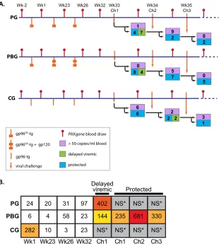
FIG 1Overview of study design and differential gene expression. (A) Schematic diagram of blood sampling schedule and challenge outcomes. Vaccination of the
prime group (PG), prime-boost group (PBG), and control group (CG) was performed at weeks 0, 6, and 25 (orange symbols). Beginning at week 33, animals were subjected to weekly low-dose challenges (orange arrows). Starting with 12 animals in each study group, the numbers in each tree branch indicate how many animals were SIV positive, delayed viremic, or protected after each challenge. Blood draws (red lines) for RNA isolation were performed at weeks⫺2, 1, 23, 26, and 32 and 5 days after each challenge. There were at least 10 samples per group for each of the time points, except at week 32, where only 7 samples for the PG and 9 samples for the PBG passed QC (see Materials and Methods). (B) Numbers of differentially expressed genes within each study group. Differential gene expression was determined by comparing vaccination-phase samples to their study-group-matched baseline at week⫺2. For the challenge phase, postchallenge samples were compared to their prechallenge baseline at week 32. Using a moderated, Bayesian-correctedttest in limma (see Materials and Methods), differential expression with aPvalue of⬍0.05 and group-averaged fold change to average baseline (fold change) of⬎1.4 was considered significant. The table shows numbers of DE genes in the three vaccination groups for each comparison. NS*, a nonsignificant number of samples was used for comparison. Colored boxes represent a heat map of the number of DE genes in each box.
on August 17, 2020 by guest
http://cvi.asm.org/
observed consistently modest numbers of DE genes during the vaccination phase, with rather inconclusive functional enrich-ment (Fig. 1B; see Table S1 in the supplemental material for com-parisons of DE genes for the PG and PBG at weeks 26 and 32 relative to week⫺2). Criteria for differential expression were an absolute change of⬎1.4-fold and aPvalue of⬍0.05, calculated using a moderatedttest (see Materials and Methods). It has pre-viously been described that the gp96-Ig vaccine approach (deliv-ered by the intraperitoneal route) induces a strong mucosal immune response, while systemic immune responses are not sig-nificantly upregulated (13,14). Therefore, the observed lack of gene expression in whole blood during the vaccination phase was not surprising. Although detection of significant gene expression changes affecting cell populations with low representation (such as dendritic cells) in whole blood is certainly hampered, the sig-nificance and novelty of using this noninvasive way to screen host responses in the present vaccine trials are given by the large differ-ences in the host transcriptional response after the first challenge between delayed-viremic and persistently protected animals: postchallenge comparisons to the postvaccination time point week 32 showed hundreds of DE genes for protected animals across all three challenges, as well as for delayed-viremic animals from challenge 1.
Large differences in host transcriptional response after the first challenge between delayed-viremic and persistently pro-tected animals.Given the aforementioned delayed viremia in sev-eral animals in both vaccine groups after the first challenge, we aimed to determine whether the presence of virus in delayed-viremic animals from the PG and PBG induced similar host re-sponses and how these rere-sponses compared to those of the unin-fected and protected animals in the control and prime-boost groups, respectively, after the first challenge. Genes that were dif-ferentially expressed compared to group-specific week 32 baseline controls were determined using the statistical cutoffs of an abso-lute change of⬎1.4-fold and aPvalue of⬍0.05. There were com-parable numbers of DE genes across these comparisons: 402 for delayed-viremic animals in the gp96SIVIg group, 144 for delayed-viremic animals in the prime-boost group, 235 for protected ani-mals in the prime-boost group, and 681 for uninfected control group animals.
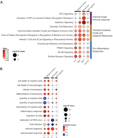
Enrichment in canonical pathways for each set of DE genes was performed. For the delayed-viremic animals in both vaccination groups, there was some enrichment in several pathways leading to or involving the inflammatory response, including granulocyte adhesion and diapedesis, TREM1 signaling, NF-B signaling, and Toll-like receptor (TLR) signaling (Fig. 2A). However, the path-way enrichment pattern differed between these two groups in that for several pathways involved in the transition between the innate and adaptive responses, the delayed-viremic animals in the prime-boost group had stronger enrichment, with a higher ratio of genes. In contrast, the protected animals in the prime-boost group had essentially no enrichment in pathways involved in a proinflamma-tory response, but they had a pronounced innate immune re-sponse, with enrichment of pathways such as IFN signaling (also observed in delayed-viremic PBG animals, not in delayed-viremic PG animals) and activation of IRF by cytosolic pattern recognition receptors.
To further investigate the differences between groups, we de-termined the functional enrichment of the DE genes that made up the major pathways that distinguished the groups (Fig. 2B). Quite
different biofunctions were triggered as a consequence of the first mucosal challenge. Among the largest disparities between pro-tected animals and those with delayed viremia were functional categories pertaining to viral infection and replication of virus, with a predicted inhibition of these functions in the protected animals and less statistically significant enrichment in the animals with delayed viremia, despite the noted similarity in enrichment of IFN signaling in PBG animals. As already noted from pathway analysis, delayed viremia was associated with a highly significant engagement of macrophage-related inflammatory response sig-naling. This was consistent with high activation scores for migra-tion and chemotaxis of myeloid cells. Also, the predicted decrease of the quantity of granulocytes (Fig. 2B) for delayed-viremic ani-mals, together with a strong activation of cell migration, points to an engagement of neutrophils in innate cellular defense mecha-nisms toward the challenge compartment. Based on transcrip-tional profiling after the first challenge, we identified strong pro-inflammatory responses as a hallmark of a lack of protection and innate antiviral defense mechanisms as unique signatures associ-ated with protection. Both the functional and canonical pathway enrichment profiles for the protected animals differed greatly from those of the control group, suggesting that the protected profile was a result of the vaccination regimen.
Evolving host responses in the protected group during mul-tiple challenges show strong but quickly resolved IFN signaling. Before addressing the question of which specific vaccination-in-duced host response factors contributed to persistent protection during the first three weekly viral challenges, we examined whether multiple challenges changed the rate of protection after each challenge. This question is particularly important in the con-text of vaccine studies. An effective vaccine eliciting memory re-sponses should result in protection rates that remain the same over time, despite repeated pathogen exposure.
Previously, viral copy numbers 5 days after challenge were used to determine if an animal had been infected (defined as⬎50 cop-ies of viral RNA/ml of blood). Using this criterion, the number of seropositive animals from the prime-boost group changed across the first three challenges, from 0 to 5 to 7 (Fig. 1A) (original sur-vival data are presented in reference15). On the other hand, as discussed in the preceding section, there is evidence that animals with viral load measurements above the limit of detection 5 days after infection had already acquired SIV at the preceding challenge in terms of their proinflammatory responses. We analyzed both the original and the rescored survival data for the prime-boost group and followed the statistical approach outlined by Regoes (18) to test in which case the survival data showed a statistical difference between a geometric model (no effect of challenge his-tory, corresponding to the null hypothesis) and a two-step model (effect of challenge history between challenges 1 and 2, corre-sponding to the alternative hypothesis).Pvalues were derived from a2test between the log likelihood maximizers of the two models. In the case of the original data, the null hypothesis was rejected (P⫽0.01), indicating that there was an effect of challenge history on future challenges. Importantly, when the delayed-vire-mic animals were rescored as infected, there was not a statistically significant effect of continuing exposure in the prime-boost group. This is graphically represented by a high accordance be-tween the log likelihood maximizer for the geometric model fit (infection probability per exposure of 0.32) (Fig. 3A, dashed line) and the actual survival curve. Also, this maximizer showed that
on August 17, 2020 by guest
http://cvi.asm.org/
prime-boost vaccination lowered the probability of infection by nearly half (from 0.52 for the control group to 0.32 for the prime-boost group), with time consistency over several challenges.
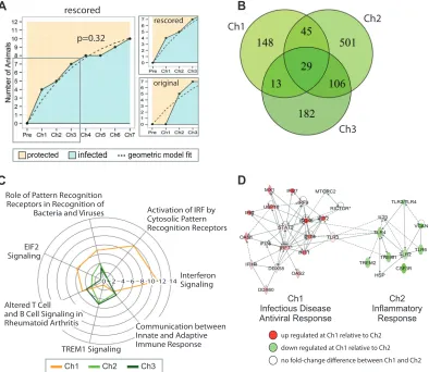
Even though the statistical analysis indicated that in the prime-boost group there was no effect of prior exposure, the RNA pro-files of the protected animals in this group differed slightly from one another after challenges 2 and 3 and differed vastly from the challenge 1 profiles. There were 330 and 681 DE genes in
compar-ing protected animals in challenges 2 and 3, respectively, to the week 32 baseline sample, in contrast to 235 DE genes with the protected animals in challenge 1 (absolute change of⬎1.4-fold andPvalue of⬍0.05). Additionally, there were significant num-bers of unique DE genes in each of the data sets (Fig. 3B). Results from functional enrichment analysis for canonical pathways of these sets of DE genes showed that only the protected animals from the prime-boost group had highly significant expression for
Antiviral Innate Immune response
Transition between Innate and Adaptive Responses
Pro-inflammatory Responses
EIF2 Signaling
Activation of IRF by Cytosolic Pattern Recognition Receptors
Interferon Signaling
Dendritic Cell Maturation
Communication between Innate and Adaptive Immune Cells
Granulocyte Adhesion and Diapedesis
TREM1 Signaling
NF-kB Signaling
Toll-like Receptor Signaling
PG PBG PBG Cont
rol
PG PBG PBG Cont
rol
Dela yed vir
emic
Prot ecte
d
SIV nega tive
Dela yed vir
emic
Prot ected
SIV nega tive
cell death of myeloid cells
cell death of macrophages
cellular homeostasis
differentiation of leukocytes
quantity of myeloid cells
quantity of granulocytes
chemotaxis of myeloid cells
inflammatory response
migration of cells
replication of RNA virus
antiviral response
antimicrobial response Viral infection
Role of Pattern Recognition Receptors in Recognition of Bacteria and Viruses
Altered T Cell and B Cell Signaling in Rheumatoid Arthritis
FIG 2Functional analysis of DE genes following challenge 1. (A) Canonical pathway enrichment analysis of the DE genes in SIV-negative animals in the prime
group (PG) and the prime-boost group (PBG) 5 days after the first challenge, with animals stratified into delayed-viremic and protected animals. Canonical pathways with the top five enrichment scores (given by the⫺log10Pvalue) in at least one gene list are shown (based on Fisher’s exact test, performed with IPA software). The size of each circle represents the enrichment score, and the color gradient represents the relative frequency of DE genes compared to the total number of genes associated with each pathway. X’s indicate that the respective pathway had no enrichment for the DE gene list of interest. Horizontal bars indicate the canonical pathways that were found predominately within either the delayed-viremic PG animals, the delayed-viremic PBG animals, or the protected PBG animals. Sizes of the nodes are proportional to the⫺log10Pvalue, and the color indicates the ratio of DE genes to the total number of genes in the category. (B) Biofunction enrichment analysis of the same lists of DE genes as those described in panel A. Based on Fisher’s exact test performed with IPA software, we considered nonredundant biological functions with the top 10 enrichment scores (given by⫺log10Pvalues) or absolute activationZscore values for at least one gene list. The size of each node is proportional to the⫺log10Pvalue, and the color indicates the activationZscore.
on August 17, 2020 by guest
http://cvi.asm.org/
EIF2 signaling, activation of IRF by cytosolic pattern recognition receptors, and IFN signaling after challenge 1 (Fig. 3C). These pathways included well-characterized antiviral response tran-scripts, such as ISG15, ISG20, IFIT1, IFIT3, DDX58, and OAS1, known to affect viral replication in the context of SIV infection (19–22). Further examination of host responses in persistently protected animals showed that after the second challenge, innate viral sensing in these animals was largely abated (Fig. 3C). TLR3 was differentially expressed only after the first challenge, and pro-inflammatory molecules, such as LCN2, TREM1, and SERPINA1, were upregulated⬎2-fold after the second challenge.
IPA network analysis further illustrates our findings (Fig. 3D), as the top-ranked challenge-specific networks show a clear con-trast between innate antiviral responses after the first challenge
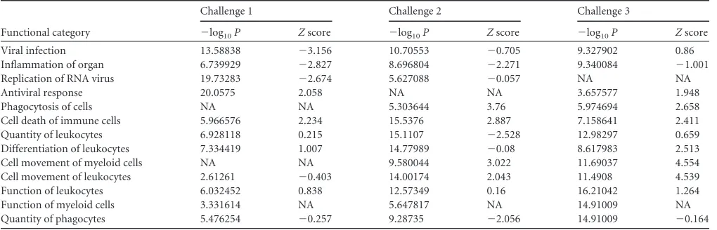
and an onset of proinflammatory signatures in conjunction with TLR2/4/6 expression. Functional analysis based on high-rankedZ scores (a |Z| value of⬎2 is required for significant predicted acti-vation) and enrichment scores (Table 1), showed that processes related to adhesion and movement of immune cells absent after the first challenge became more dominant after the second chal-lenge. Among the strongest contrasts in the prime-boost group between the first and second challenges was the predicted de-creased quantity of immune cells (downregulation of CD8A, PRKCH, and CCR7 and upregulation of SERPINA1 and LCN2), but also a significant increase in chemotaxis and cell movement of a large class of phagocytes after the second challenge (Table 1, challenge 2). Downregulation of MHC II molecules (HLA-DMB, HLA-DOB, HLA-DRA, and HLA-DR5) is an important hallmark
Ch1 Ch2
Ch3
D
Interferon Signaling Activation of IRF by Cytosolic Pattern Recognition Receptors Role of Pattern Recognition
Receptors in Recognition of Bacteria and Viruses
EIF2 Signaling
TREM1 Signaling
C
up regulated at Ch1 relative to Ch2
down regulated at Ch1 relative to Ch2
no fold-change difference between Ch1 and Ch2 infected
protected geometric model fit p=0.32
rescored
rescored
original
0 2 4 6 8 10 12 14
Ch1 Infectious Disease Antiviral Response
Ch2 Inflammatory
Response
Ch1 Ch2 Ch3
Altered T Cell and B Cell Signaling in
Rheumatoid Arthritis Communication between
Innate and Adaptive Immune Response
FIG 3Distinct host responses after each challenge in protected animals. (A) Challenge history and immunization for the prime-boost group. The number of
infected animals is indicated on theyaxis, and the number of challenges is indicated on thexaxis. The original survival data were rescored under the assumption that delayed-viremic animals were infected. ThePvalue refers to whether the survival data showed a statistical difference between the geometric model (no effect of challenge history, corresponding to the null hypothesis) and a two-step model (effect of challenge history between challenges 1 and 2, corresponding to the alternative hypothesis). This value was derived from a2test between the log likelihood maximizers of the two models. In the case of the original data, the null hypothesis was rejected (P⫽0.01) (data not shown). For the rescored data, thePvalue of 0.32 indicates that there was no difference, and therefore there was no effect of challenge history on immunity. The dashed lines represent the maximum likelihood of the fit to the survival data. The box represents the region of the graph that was enlarged in a comparison of the rescored to original data (graphs on the right). (B) Differential gene expression analysis based on statistical comparisons of study-group-matched baseline and challenge samples (fold change of⬎1.4-fold andPvalue of⬍0.05) showed a high frequency of differentially expressed genes in protected animals specific for each of the three challenges. (C) Canonical pathway analysis was performed using the IPA knowledge base. For each of the three lists of DE genes used for panel B, the top 4 enrichment scores (given by⫺log10Pvalues from Fisher’s exact test) within each set were considered and merged the resulting tables. The radial represents the enrichment score for each pathway across the three data sets. (D) Merging top-ranked networks between challenges 1 and 2 showed distinct differences in host response, with a highly upregulated antiviral response network at challenge 1 and, in contrast, a highly upregulated network of TLR and TREM signaling at challenge 2.
on August 17, 2020 by guest
http://cvi.asm.org/
of viral CTL avoidance (23), and this was observed only for ani-mals that succumbed to infection after the second challenge and was absent in persistently protected animals. By the third chal-lenge, the observed trend from strong innate signaling to adaptive immune response was further pronounced, with a more prevail-ing predicted activation of chemotaxis and cell movement of phagocytes by upregulation of the proinflammatory markers S100A8, S100A9, and TREM1 and upregulation of the cytopathic molecule MMP9.
As noted, the vaccine regimens were expected to induce a strong cell-mediated response for both vaccine groups, with an additional antibody arm for the prime-boost group. It was previ-ously shown that the presence of antibodies specific to SIV gp120 in blood serum prior to challenge was highly correlated (Pearson correlation coefficient of 0.84) with the number of challenges re-quired to establish infection for this group (15). The functional analysis of the prime-boost DE gene set identified elements that directly supported the anticipated humoral component through-out all three challenges, albeit with much less prevalence than the aspects involving IFN signaling. Differential gene expression showed a specific enrichment in the prime-boost group for B cell receptor signaling, phosphatidylinositol 3-kinase (PI3K) signaling in B lymphocytes, and antigen presentation, with downregulation of CD40 and CD79B and several MHC II molecules, such as HLA-DMB, HLA-DOB, and HLA-DRA. There was also increased ex-pression of TNFSF13B (BLysS), a cytokine of the TNF ligand fam-ily that is a B cell activator that promotes maturation and class switching.
In summary, we have established statistical evidence for effi-cient, time-homogenous vaccine protection throughout several challenges in terms of viral acquisition. In contrast, transcrip-tional profiling for protected animals revealed host responses that evolved from challenge to challenge, from an antiviral IFN sponse after the first challenge to cellular adaptive immune re-sponses after the second challenge and proinflammatory signa-tures after the third challenge.
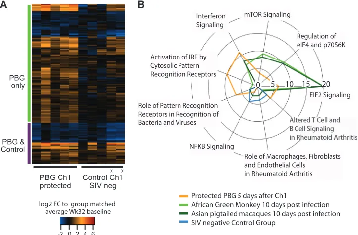
Comparative analysis of host responses in protected animals and natural hosts early after infection shows similar innate and anti-inflammatory signaling.The set of 235 genes that were dif-ferentially expressed after the first challenge in protected animals (Fig. 4A) showed a distinct cluster of innate antiviral immune
genes with upregulation of 4- to 8-fold after the first challenge (ISG15, IFI27, IFIT2, IFI6, IRF7, and MX1). To interrogate which part of this signature was associated with vaccine-related protec-tion, we compared it to SIV-negative control animals after the first challenge and found 171 differentially expressed genes unique to prime-boost-vaccinated animals (Fig. 4A, “PBG only”). Func-tional enrichment showed highly significant functions related to the predicted activation of cell death of immune cells and inhibi-tion of viral replicainhibi-tion and infecinhibi-tion only for this cluster. In con-trast, for the set of common genes (Fig. 4A, “PBG & Control”), we were not able to determine statistically significant activation pre-dictions.
Natural hosts, such as African green monkeys (AGM), acquire SIV with set-point viral load levels similar to those in macaques, without developing progression to chronic disease and its hall-mark of CD4⫹T cell depletion. The strong and quickly resolved IFN response has been found to be a hallmark of nonpathogenic infection, despite potent viral acquisition (24–26). In order to determine whether the observed IFN responses in protected PBG animals were specific to acquisition prevention (in both patho-genic and nonpathopatho-genic infection models), we compared tran-scriptional changes in blood 10 days after infection of AGM and Asian pigtail macaques (PM) (based on statistical analysis of data published in reference25) to host signatures of protected prime-boost group animals 5 days after the first challenge. Among the common, highly enriched pathways were EIF2K signaling (up-regulation of PKR and down(up-regulation of many ribosomal tran-scripts) and IFN signaling (Fig. 4B). Although pathogen receptor signaling was enriched for AGM, PM, and protected PBG signa-tures, closer inspection showed larger disparities. Whereas up-regulation of double-stranded RNA sensors (TLR3, RIG-I, and MD5) was present only in protected animals, TLR1 was solely expressed in natural hosts and in PM. As a control, we also com-pared these two signatures to that of SIV-negative control group animals. Despite undetectable viral loads 5 days after the first chal-lenge, none of the above-mentioned transcripts were differentially expressed in this group. In contrast, SIV-negative control group animals showed highly predicted activation of cell movement of myeloid cells and significant enrichment for inflammatory re-sponses by virtue of NF-B signaling. None of these categories
TABLE 1Functional enrichment scores for protected PBG animals following the first three challengesa
Functional category
Challenge 1 Challenge 2 Challenge 3
⫺log10P Zscore ⫺log10P Zscore ⫺log10P Zscore
Viral infection 13.58838 ⫺3.156 10.70553 ⫺0.705 9.327902 0.86
Inflammation of organ 6.739929 ⫺2.827 8.696804 ⫺2.271 9.340084 ⫺1.001
Replication of RNA virus 19.73283 ⫺2.674 5.627088 ⫺0.057 NA NA
Antiviral response 20.0575 2.058 NA NA 3.657577 1.948
Phagocytosis of cells NA NA 5.303644 3.76 5.974694 2.658
Cell death of immune cells 5.966576 2.234 15.5376 2.887 7.158641 2.411
Quantity of leukocytes 6.928118 0.215 15.1107 ⫺2.528 12.98297 0.659
Differentiation of leukocytes 7.334419 1.007 14.77989 ⫺0.08 8.617983 2.513
Cell movement of myeloid cells NA NA 9.580044 3.022 11.69037 4.554
Cell movement of leukocytes 2.61261 ⫺0.403 14.00174 2.043 11.4908 4.539
Function of leukocytes 6.032452 0.838 12.57349 0.16 16.21042 1.264
Function of myeloid cells 3.331614 NA 5.647817 NA 14.91009 NA
Quantity of phagocytes 5.476254 ⫺0.257 9.28735 ⫺2.056 14.91009 ⫺0.164
aNA, not available.
on August 17, 2020 by guest
http://cvi.asm.org/
were enriched for AGM, PM, or the protected prime-boost group animals.
DISCUSSION
Using the canonical multiple-low-dose-challenge scheme for nonhuman primate vaccine trials, the vaccination regimen stud-ied here showed a reduction of viral acquisition risk of 73% for the prime-boost group. Its unconventional design of using gp96-Ig-secreting cells carrying SIV peptides induced a strong multifunc-tional memory CTL response in the rectal lamina propria prior to challenge. In addition, elevated SIV gp120-specific antibody titers in blood serum 5 weeks prior to the first challenge are statistically significant correlates of protection for the prime-boost group an-imals (15). Despite the relatively high vaccine efficacy in the prime-boost group, protection from viral acquisition was only partial.
In the present genomic analyses, we identified specific host response factors that were required to carry prechallenge immune correlates over to protection after multiple challenges. Transcrip-tional profiling of whole blood demonstrated that persistent pro-tection upon viral challenge coincided with a strong IFN-medi-ated antiviral response, which abIFN-medi-ated after the second challenge. Similar patterns of IFN responses in the context of therapeutics against lentivirus infection have been shown to improve viral con-trol (27,28). It was also noted in a single-challenge nonhuman primate SIV vaccine trial that challenge outcome is correlated with the relative balance between SIV-specific IFN-␥-driven T cell re-sponses and nonspecific IFN-␥-driven inflammation (29). Loss of this balance is in line with the gene expression profiles of delayed-viremic prime-boost group animals, in which we observed an
early onset of inflammatory responses despite the presence of IFN signaling. The extent to which innate immunity can modulate or cooperate with adaptive immune activation to mount an antiviral immune response to HIV and SIV infections has been discussed (30). Within the context of an adenovirus type 5 (Ad5)-HIV vac-cine, postvaccine transcriptional changes in IFN signaling and myeloid cell trafficking are implicated in the induction and mag-nitude of HIV-specific CD8⫹T cell responses (31). In this study, although there was evidence of SIV-specific responses in terms of INF-␥secretion by CD8⫹T cells before challenge in both vaccine groups, the study outcome based on survival after viral challenge showed that these immune correlates of vaccination are not nec-essarily also immune correlates of protection (15). Also, for prime-boost group animals, the double-stranded RNA receptor TLR3 was expressed exclusively in protected animals after the first challenge, whereas the expression of surface TLR4 was unique to delayed-viremic animals after the first challenge. It was previously shown that only TLR3 enhances CD8 T cell responsesin vivoand that TLR2 and TLR4 activation may suppress pathogen-induced CD8 T cell responses (32). This suggests that TLR4-mediated sig-naling may possibly act as an antagonist to CD8⫹T cell responses during challenge, despite the presence of these responses prior to challenge. Furthermore, neutrophil and macrophage activation in animals with delayed viremia, in conjunction with upregulation of PD-L1, may be a cause of further inactivation of CD8⫹T cells (33).
Comparison of the host response of protected animals in the prime-boost group with that of SIV-infected natural hosts is of particular interest. The latter acquire SIV without developing im-munodeficiency and also show a strong type I IFN response at 10
PBG Ch1 protected
Control Ch1 SIV neg
Protected PBG 5 days after Ch1
African Green Monkey 10 days post infection Asian pigtailed macaques 10 days post infection SIV negative Control Group
log2 FC to group matched average Wk32 baseline PBG
only
PBG & Control
* *
0 5 10 15 20
mTOR Signaling
Regulation of eIF4 and p70S6K Interferon
Signaling
Activation of IRF by Cytosolic Pattern Recognition Receptors
Role of Pattern Recognition Receptors in Recognition of Bacteria and Viruses
NFKB Signaling
Role of Macrophages, Fibroblasts and Endothelial Cells
in Rheumatoid Arthritis
EIF2 Signaling
FIG 4Heat map of 235 DE genes in protected animals from the prime-boost group after the first challenge. (A) Individual fold changes (FC) relative to average
study-group-matched baseline samples after the first challenge. The gene set is partitioned into two clusters, based on their overlap with DE genes of SIV-negative control group animals. In both groups, data for only animals without unusually high viral loads after the first infection are shown. Asterisks highlight two control group animals which remained uninfected for at least three challenges. (B) Canonical pathway analysis was performed using the IPA knowledge base. For each of the four lists of DE genes (protected prime-boost animals after challenge 1, African green monkeys at 10 days postinfection, Asian pigtail macaques at 10 days postinfection, and SIV-negative control group animals 5 days after challenge 1), we considered the top three enrichment scores (given by⫺log10Pvalues for Fisher’s exact test and represented by the radius) within each set and merged the resulting tables.
on August 17, 2020 by guest
http://cvi.asm.org/
days postinfection, which is cleared approximately 3 weeks later (24,25,34), in contrast to the case in macaques. On the other hand, natural hosts lack high autologous neutralizing antibodies to SIV (35,36), and hence are not able to avoid SIV acquisition. Given the strong humoral immune correlates in protected ani-mals, we hypothesize that preexisting antibody responses may be enhanced sufficiently by direct stimulation of T and B cells by type I IFN (37,38). Also, the strong enrichment of EIF2 signaling after the first challenge in both the protected prime-boost group ani-mals and the SIV-infected natural hosts and PM provides further hypotheses for how the initial IFN-mediated antiviral response to SIV affects host restriction factors important for preventing or enhancing viral acquisition or target cell depletion (39).
Understanding HIV infection from a modeling perspective has relied on the argument that a basic mass-action law would faith-fully describe the evolution of infection in terms of the numbers of uninfected and infected CD4⫹T cells and the number of infective viral particles (40). Based on postchallenge data, there is evidence that for persistently protected animals, CD4⫹T cell counts re-mained stable at an intermediate level throughout several chal-lenges, whereas delayed-viremic or infected animals showed ei-ther very high or very low CD4⫹T cell levels prior to challenge and larger variations over time (Fig. 5). Also, prechallenge antibody data show a distinct pattern of elevated SIV gp120-specific anti-bodies (15). Clearly, larger numbers of naive target cells would increase the chance of establishing productive infection, and hav-ing smaller numbers of CD4⫹T cells during the challenge phase would decrease the probability of viral acquisition. Based on these data, we suggest that as long as antibody-secreting cells (ASCs)
can be efficiently activated (indicated by early upregulation of TNFSF13B) in a T helper cell-dependent manner, polyclonal an-tibody responses will delay viral acquisition during repeated chal-lenges. On the other hand, for animals with very small numbers of CD4⫹T cells, this response may become inefficient in that its induction of epitope-specific antibody production is too slow to prevent viral acquisition after challenge. This observation is cor-roborated by the stability and maintenance of tissue-resident CD4⫹T cells in the lamina propria for a long period in persistently protected animals.Figure 6shows percentages of CD4⫹CD3⫹ cells relative to CD3⫹cells at 11 weeks postinfection (or after the last challenge) and suggests that balancing CD4⫹T cell popula-tions during the early challenge phase may counteract the prema-ture exhaustion of resident T cells in the rectal mucosa. As indi-cated by transcriptomic profiling, a strong early IFN response after the first challenge can potentially act to sustain T helper cell-dependent humoral responses and substantially delay viral acquisition. Similar roles of highly efficient T helper cell function in mediating polyfunctional responses by virtue of IFN
produc-0 4
1 2 3
mean OD f
or SIV
mac 251 g
p120 an
tibody a
t w
eek 26
Number of CD3+/CD4+ c
ells per mm^3 blood
Pre-challenge humoral responses and post-challenge CD4+ T cell counts
3000
2000
1000
0
Wk32 Ch1 Ch2 Ch3
Wk26
Prime-boost group animals five days after first two challenges:
LOESS regression SE of regression delayed viremic/infected
protected
0 4
1 2 3
FIG 5Prechallenge immune correlates and postchallenge CD4⫹T cell counts
for prime-boost group animals during repeated low-dose challenges. Levels of SIV Env-specific antibodies are shown on the leftyaxis, and CD4⫹T cell count data are shown on the rightyaxis, starting at prechallenge week 32 and con-tinuing throughout three challenges. For both antibody titers and cell counts, colors stand for status within the first two challenges: turquoise represents animals which remained protected throughout the first two challenges, and red stands for animals which were either infected or delayed viremic during the first two challenges.
% of CD4+ T c
ells in rectal lamina propria,
11 weeks post infection/last challenge
Preservation of CD4+ T cells in rectal mucosa in vaccinated animals
PBG CG
P442
P=0.0151 P439
20 40 60
delayed viremic/infected protected
Prime-boost group animals five days after first two challenges:
FIG 6Preservation of rectal lamina propria CD4⫹lymphocytes in protected,
vaccinated animals with a strong IFN response after the first challenge. Ani-mals were immunized with gp96SIVor gp96Mock(prime group [PG] and con-trol group [CG], respectively) or with gp96SIVand gp120 (prime-boost group [PBG]) by the intraperitoneal route at weeks 0, 6, and 25. Pinch biopsy samples were harvested from each animal 11 weeks after infection (or after the last challenge for animals P439 and P442, which remained SIV negative after seven challenges). Rectal CD4⫹T cells were analyzed by flow cytometry after gating on live lymphocytes (CD3⫹cells), and the percentage of CD4-positive cells is shown. The individual data for each monkey were plotted. Turquoise repre-sents animals which remained protected throughout the first two challenges, and red stands for animals which were either infected or delayed viremic dur-ing the first two challenges. Data for animals P439 and P442 from the PBG are highlighted, as they remained SIV negative after seven consecutive challenges. Overall, PBG animals showed a high degree of preservation of CD4⫹T cells compared to controls (ttestPvalue of 0.0151).
on August 17, 2020 by guest
http://cvi.asm.org/
tion have been observed in elite controllers within a human cohort study (41).
In rescoring the survival data based on virological and host transcriptional evidence, we have presented statistical arguments in favor of protection rates remaining constant throughout mul-tiple exposures to SIV. In addition, whole-blood transcriptomic profiling demonstrated a short-term delay of viremia, with mac-rophage-mediated proinflammatory responses as the important hallmark, independently of the vaccine regimen. Macrophage ac-tivation and its proinflammatory consequences are associated with efficient viral replication in acute HIV/SIV infectionin vivo (42–44). On the other hand, early establishment of an anti-in-flammatory milieu is predictive of disease nonprogression in the case of natural hosts (45). In the context of the weekly mucosal low-dose challenge design, targeted transcriptional evaluation af-ter challenge may be used to deaf-termine whether animals with SIV levels below the limit of detection in blood should be suspended, at least temporarily, from further challenge.
Taken together, our results show that despite promising, vaccination-induced immune correlates, multiple low-dose chal-lenges can result in evolving host responses that affect the protec-tive outcome. Findings from our earlier study (15) indicate that gp96SIVIg induces a long-lasting memory response in gut mucosal sites, with the ability to rapidly undergo multiple rounds of pro-liferation in response to antigen, a hallmark of memory cells. In the context of this vaccine study, there is strong evidence that undetectable viral load was an insufficient characterization of pro-tection from viral acquisition on a subsequent challenge. Post-challenge host response profiling of vaccinated animals showed that whereas short-term seronegativity is characterized by strong proinflammatory responses, protection comes with a strong and quickly resolved IFN-driven antiviral response and subsequent adaptive immune responses. Based on our transcriptomic analysis and known prechallenge correlates of protection, we hypothesize that innate antiviral signaling events may provide important con-tributions to enhancement of humoral responses, and therefore to higher vaccine efficacy.
ACKNOWLEDGMENTS
This project was funded with federal funds from the National Institutes of Health, Department of Health and Human Services, under National In-stitute of Allergy and Infectious Diseases contract HHSN272201300010C (grant P01AI096396), by the DAIDS Reagent Resource Support Program for AIDS Vaccine Development, Quality Biological, Gaithersburg, MD (contract N01-A30018), and by the Office of the Director (grant P51OD010425).
We thank Sean Proll and Marcus Korth for assistance with the prepa-ration of the figures and the manuscript.
N. Strbo, E. R. Podack, M. Vaccari, and G. Franchini are listed in a U.S. patent application for the gp96-Ig approach to producing an HIV vaccine.
REFERENCES
1.Barre-Sinoussi F, Chermann JC, Rey F, Nugeyre MT, Chamaret S,
Gruest J, Dauguet C, Axler-Blin C, Vezinet-Brun F, Rouzioux C,
Rozenbaum W, Montagnier L.1983. Isolation of a T-lymphotropic
retrovirus from a patient at risk for acquired immune deficiency syn-drome (AIDS). Science 220:868 – 871. http://dx.doi.org/10.1126/science
.6189183.
2.Gallo RC, Salahuddin SZ, Popovic M, Shearer GM, Kaplan M, Haynes
BF, Palker TJ, Redfield R, Oleske J, Safai B, White G, Foster P,
Markham PD.1984. Frequent detection and isolation of cytopathic
ret-roviruses (HTLV-III) from patients with AIDS and at risk for AIDS. Sci-ence224:500 –503.http://dx.doi.org/10.1126/science.6200936.
3.VanCott TC, Bethke FR, Burke DS, Redfield RR, Birx DL.1995. Lack of
induction of antibodies specific for conserved, discontinuous epitopes of HIV-1 envelope glycoprotein by candidate AIDS vaccines. J. Immunol. 155:4100 – 4110.
4.Haynes BF.1996. New frontiers of immunotherapy for HIV. Lancet348:
1531–1532.
5.Novitsky V, Lagakos S, Herzig M, Bonney C, Kebaabetswe L,
Rossen-khan R, Nkwe D, Margolin L, Musonda R, Moyo S, Woldegabriel E, van
Widenfelt E, Makhema J, Essex M.2009. Evolution of proviral gp120
over the first year of HIV-1 subtype C infection. Virology383:47–59.http:
//dx.doi.org/10.1016/j.virol.2008.09.017.
6.Tyagi M, Bukrinsky M.2012. Human immunodeficiency virus (HIV)
latency: the major hurdle in HIV eradication. Mol. Med.18:1096 –1108.
http://dx.doi.org/10.2119/molmed.2012.00194.
7.Strbo N, Garcia-Soto A, Schreiber TH, Podack ER.2013. Secreted heat
shock protein gp96-Ig: next-generation vaccines for cancer and infectious diseases. Immunol. Res. 57:311–325. http://dx.doi.org/10.1007/s12026
-013-8468-x.
8.Cho BK, Palliser D, Guillen E, Wisniewski J, Young RA, Chen J, Eisen
HN.2000. A proposed mechanism for the induction of cytotoxic T lym-phocyte production by heat shock fusion proteins. Immunity12:263–272.
http://dx.doi.org/10.1016/S1074-7613(00)80179-X.
9.Palliser D, Guillen E, Ju M, Eisen HN.2005. Multiple intracellular
routes in the cross-presentation of a soluble protein by murine den-dritic cells. J. Immunol. 174:1879 –1887. http://dx.doi.org/10.4049
/jimmunol.174.4.1879.
10. Binder RJ, Srivastava PK.2005. Peptides chaperoned by heat-shock
pro-teins are a necessary and sufficient source of antigen in the cross-priming of CD8⫹T cells. Nat. Immunol.6:593–599.http://dx.doi.org/10.1038
/ni1201.
11. Yamazaki K, Nguyen T, Podack ER.1999. Cutting edge: tumor secreted
heat shock-fusion protein elicits CD8 cells for rejection. J. Immunol.163: 5178 –5182.
12. Oizumi S, Strbo N, Pahwa S, Deyev V, Podack ER.2007. Molecular and
cellular requirements for enhanced antigen cross-presentation to CD8 cytotoxic T lymphocytes. J. Immunol.179:2310 –2317.http://dx.doi.org
/10.4049/jimmunol.179.4.2310.
13. Strbo N, Pahwa S, Kolber MA, Gonzalez L, Fisher E, Podack ER.2010.
Cell-secreted gp96-Ig-peptide complexes induce lamina propria and in-traepithelial CD8⫹cytotoxic T lymphocytes in the intestinal mucosa. Mucosal Immunol.3:182–192.http://dx.doi.org/10.1038/mi.2009.127.
14. Strbo N, Vaccari M, Pahwa S, Kolber MA, Fisher E, Gonzalez L, Doster
MN, Hryniewicz A, Felber BK, Pavlakis GN, Franchini G, Podack ER. 2011. gp96 SIV Ig immunization induces potent polyepitope specific, multifunctional memory responses in rectal and vaginal mucosa. Vaccine 29:2619 –2625.http://dx.doi.org/10.1016/j.vaccine.2011.01.044.
15. Strbo N, Vaccari M, Pahwa S, Kolber MA, Doster MN, Fisher E,
Gonzalez L, Stablein D, Franchini G, Podack ER.2013. Cutting edge:
novel vaccination modality provides significant protection against muco-sal infection by highly pathogenic simian immunodeficiency virus. J. Im-munol.190:2495–2499.http://dx.doi.org/10.4049/jimmunol.1202655.
16. Li C, Bankhead A, 3rd, Eisfeld AJ, Hatta Y, Jeng S, Chang JH, Aicher
LD, Proll S, Ellis AL, Law GL, Waters KM, Neumann G, Katze MG,
McWeeney S, Kawaoka Y.2011. Host regulatory network response to
infection with highly pathogenic H5N1 avian influenza virus. J. Virol. 85:10955–10967.http://dx.doi.org/10.1128/JVI.05792-11.
17. Smyth G.2005. Limma: linear models for microarray data, p 397– 420.In
Gentleman R, Carey V, Huber W, Irizarry R, Dudoit S (ed), Bioinformat-ics and computational biology solutions using R and Bioconductor. Springer, New York, NY.
18. Regoes RR.2012. The role of exposure history on HIV acquisition:
in-sights from repeated low-dose challenge studies. PLoS Comput. Biol. 8:e1002767.http://dx.doi.org/10.1371/journal.pcbi.1002767.
19. Verhoeven D, George MD, Hu W, Dang AT, Smit-McBride Z, Reay E,
Macal M, Fenton A, Sankaran-Walters S, Dandekar S.2014. Enhanced
innate antiviral gene expression, IFN-alpha, and cytolytic responses are predictive of mucosal immune recovery during simian immunodeficiency virus infection. J. Immunol. 192:3308 –3318.http://dx.doi.org/10.4049
/jimmunol.1302415.
20. Harty RN, Pitha PM, Okumura A. 2009. Antiviral activity of innate
immune protein ISG15. J. Innate Immun.1:397– 404.http://dx.doi.org/10
.1159/000226245.
21. Zhou Z, Wang N, Woodson SE, Dong Q, Wang J, Liang Y, Rijnbrand
on August 17, 2020 by guest
http://cvi.asm.org/
R, Wei L, Nichols JE, Guo J-T, Holbrook MR, Lemon SM, Li K.2011. Antiviral activities of ISG20 in positive-strand RNA virus infections. Vi-rology409:175–188.http://dx.doi.org/10.1016/j.virol.2010.10.008.
22. Espert L, Degols G, Lin YL, Vincent T, Benkirane M, Mechti N.2005.
Interferon-induced exonuclease ISG20 exhibits an antiviral activity against human immunodeficiency virus type 1. J. Gen. Virol.86:2221– 2229.http://dx.doi.org/10.1099/vir.0.81074-0.
23. Stumptner-Cuvelette P, Morchoisne S, Dugast M, Le Gall S, Raposo G,
Schwartz O, Benaroch P.2001. HIV-1 Nef impairs MHC class II antigen
presentation and surface expression. Proc. Natl. Acad. Sci. U. S. A.98: 12144 –12149.http://dx.doi.org/10.1073/pnas.221256498.
24. Jacquelin B, Mayau V, Targat B, Liovat AS, Kunkel D, Petitjean G,
Dillies MA, Roques P, Butor C, Silvestri G, Giavedoni LD, Lebon P,
Barre-Sinoussi F, Benecke A, Muller-Trutwin MC. 2009.
Nonpatho-genic SIV infection of African green monkeys induces a strong but rapidly controlled type I IFN response. J. Clin. Invest.119:3544 –3555.http://dx
.doi.org/10.1172/JCI40093.
25. Lederer S, Favre D, Walters KA, Proll S, Kanwar B, Kasakow Z, Baskin
CR, Palermo R, McCune JM, Katze MG.2009. Transcriptional profiling
in pathogenic and non-pathogenic SIV infections reveals significant dis-tinctions in kinetics and tissue compartmentalization. PLoS Pathog. 5:e1000296.http://dx.doi.org/10.1371/journal.ppat.1000296.
26. Sandler NG, Bosinger SE, Estes JD, Zhu RT, Tharp GK, Boritz E, Levin
D, Wijeyesinghe S, Makamdop KN, del Prete GQ, Hill BJ, Timmer JK, Reiss E, Yarden G, Darko S, Contijoch E, Todd JP, Silvestri G, Nason M, Norgren RB, Jr, Keele BF, Rao S, Langer JA, Lifson JD, Schreiber G,
Douek DC.2014. Type I interferon responses in rhesus macaques prevent
SIV infection and slow disease progression. Nature511:601– 605.http:
//dx.doi.org/10.1038/nature13554.
27. Dyavar Shetty R, Velu V, Titanji K, Bosinger SE, Freeman GJ, Silvestri
G, Amara RR.2012. PD-1 blockade during chronic SIV infection reduces
hyperimmune activation and microbial translocation in rhesus macaques. J. Clin. Invest.122:1712–1716.http://dx.doi.org/10.1172/JCI60612.
28. Koopman G, Beenhakker N, Nieuwenhuis I, Doxiadis G, Mooij P,
Drijfhout JW, Koestler J, Hanke T, Fagrouch Z, Verschoor EJ, Bontrop
RE, Wagner R, Bogers WM, Melief CJ.2013. DNA/long peptide
vacci-nation against conserved regions of SIV induces partial protection against SIVmac251 challenge. AIDS 27:2841–2851. http://dx.doi.org/10.1097
/QAD.0000000000000047.
29. Abel K, La Franco-Scheuch L, Rourke T, Ma ZM, De Silva V, Fallert B,
Beckett L, Reinhart TA, Miller CJ.2004. Gamma interferon-mediated
inflammation is associated with lack of protection from intravaginal sian immunodeficiency virus SIVmac239 challenge in simsian-human im-munodeficiency virus 89.6-immunized rhesus macaques. J. Virol.78:841–
854.http://dx.doi.org/10.1128/JVI.78.2.841-854.2004.
30. Lehner T, Wang Y, Whittall T, Seidl T.2011. Innate immunity and
HIV-1 infection. Adv. Dent. Res. 23:19 –22. http://dx.doi.org/10.1177
/0022034511399081.
31. Zak DE, Andersen-Nissen E, Peterson ER, Sato A, Hamilton MK,
Borgerding J, Krishnamurty AT, Chang JT, Adams DJ, Hensley TR, Salter AI, Morgan CA, Duerr AC, De Rosa SC, Aderem A, McElrath MJ. 2012. Merck Ad5/HIV induces broad innate immune activation that pre-dicts CD8(⫹) T-cell responses but is attenuated by preexisting Ad5 im-munity. Proc. Natl. Acad. Sci. U. S. A.109:E3503–E3512.http://dx.doi.org
/10.1073/pnas.1208972109.
32. Mandraju R, Murray S, Forman J, Pasare C.2014. Differential ability of
surface and endosomal TLRs to induce CD8 T cell responses in vivo. J. Im-munol.192:4303– 4315.http://dx.doi.org/10.4049/jimmunol.1302244.
33. Bowers NL, Helton ES, Huijbregts RP, Goepfert PA, Heath SL, Hel Z.
2014. Immune suppression by neutrophils in HIV-1 infection: role of
PD-L1/PD-1 pathway. PLoS Pathog.10:e1003993.http://dx.doi.org/10
.1371/journal.ppat.1003993.
34. Harris LD, Tabb B, Sodora DL, Paiardini M, Klatt NR, Douek DC,
Silvestri G, Muller-Trutwin M, Vasile-Pandrea I, Apetrei C, Hirsch V,
Lifson J, Brenchley JM, Estes JD.2010. Downregulation of robust acute
type I interferon responses distinguishes nonpathogenic simian immuno-deficiency virus (SIV) infection of natural hosts from pathogenic SIV in-fection of rhesus macaques. J. Virol.84:7886 –7891.http://dx.doi.org/10
.1128/JVI.02612-09.
35. Li B, Stefano-Cole K, Kuhrt DM, Gordon SN, Else JG, Mulenga J, Allen
S, Sodora DL, Silvestri G, Derdeyn CA.2010. Nonpathogenic simian
immunodeficiency virus infection of sooty mangabeys is not associated with high levels of autologous neutralizing antibodies. J. Virol.84:6248 – 6253.http://dx.doi.org/10.1128/JVI.00295-10.
36. Sodora DL, Allan JS, Apetrei C, Brenchley JM, Douek DC, Else JG,
Estes JD, Hahn BH, Hirsch VM, Kaur A, Kirchhoff F, Muller-Trutwin
M, Pandrea I, Schmitz JE, Silvestri G.2009. Toward an AIDS vaccine:
lessons from natural simian immunodeficiency virus infections of African nonhuman primate hosts. Nat. Med.15:861– 865.http://dx.doi.org/10
.1038/nm.2013.
37. Le Bon A, Thompson C, Kamphuis E, Durand V, Rossmann C, Kalinke
U, Tough DF.2006. Cutting edge: enhancement of antibody responses
through direct stimulation of B and T cells by type I IFN. J. Immunol. 176:2074 –2078.http://dx.doi.org/10.4049/jimmunol.176.4.2074.
38. Le Bon A, Schiavoni G, D’Agostino G, Gresser I, Belardelli F, Tough
DF.2001. Type I interferons potently enhance humoral immunity and can promote isotype switching by stimulating dendritic cells in vivo. Immu-nity14:461– 470.http://dx.doi.org/10.1016/S1074-7613(01)00126-1.
39. Roy S, Katze MG, Parkin NT, Edery I, Hovanessian AG, Sonenberg N.
1990. Control of the interferon-induced 68-kilodalton protein kinase by the HIV-1 tat gene product. Science247:1216 –1219.http://dx.doi.org/10
.1126/science.2180064.
40. Perelson AS, Neumann AU, Markowitz M, Leonard JM, Ho DD.1996.
HIV-1 dynamics in vivo: virion clearance rate, infected cell life-span, and viral generation time. Science271:1582–1586.http://dx.doi.org/10.1126
/science.271.5255.1582.
41. Vingert B, Benati D, Lambotte O, de Truchis P, Slama L, Jeannin P,
Galperin M, Perez-Patrigeon S, Boufassa F, Kwok WW, Lemaitre F,
Delfraissy JF, Theze J, Chakrabarti LA.2012. HIV controllers maintain
a population of highly efficient Th1 effector cells in contrast to patients treated in the long term. J. Virol.86:10661–10674.http://dx.doi.org/10
.1128/JVI.00056-12.
42. Herbein G, Varin A.2010. The macrophage in HIV-1 infection: from
activation to deactivation? Retrovirology7:33.http://dx.doi.org/10.1186
/1742-4690-7-33.
43. Bergamini A, Bolacchi F, Bongiovanni B, Colizzi V, Cappelli G, Uccella
I, Cepparulo M, Capozzi M, Mancino G, Rocchi G. 2000. Human
immunodeficiency virus type 1 infection modulates the interleukin (IL)-1beta and IL-6 responses of human macrophages to CD40 ligand stimu-lation. J. Infect. Dis.182:776 –784.http://dx.doi.org/10.1086/315803.
44. Contreras X, Mzoughi O, Gaston F, Peterlin MB, Bahraoui E.2012.
Protein kinase C-delta regulates HIV-1 replication at an early post-entry step in macrophages. Retrovirology9:37.http://dx.doi.org/10.1186/1742
-4690-9-37.
45. Kornfeld C, Ploquin MJ, Pandrea I, Faye A, Onanga R, Apetrei C,
Poaty-Mavoungou V, Rouquet P, Estaquier J, Mortara L, Desoutter JF, Butor C, Le Grand R, Roques P, Simon F, Barre-Sinoussi F, Diop OM,
Muller-Trutwin MC.2005. Antiinflammatory profiles during primary
SIV infection in African green monkeys are associated with protection against AIDS. J. Clin. Invest.115:1082–1091.http://dx.doi.org/10.1172
/JCI200523006.