Abstract
Background:Flavivirus NS1 is a non-structural glycoprotein that is expressed on the cell surface and secreted into the extracellular space, where it acts as an antagonist of complement pathway activation. Despite its transit through the secretory pathway and intracellular localization in the lumen of the endoplasmic reticulum and Golgi vesicles, NS1 is as an essential gene for flavivirus replication. How NS1 modulates infection remains uncertain given that the viral RNA replication complex localizes to the cytosolic face of the endoplasmic reticulum.
Methods and Results:Using a trans-complementation assay, we show that viruses deleted for NS1 (Δ-NS1) can be rescued by transgenic expression of NS1 from West Nile virus (WNV) or heterologous flaviviruses in the absence of adaptive mutations. In viral lifecycle experiments, we demonstrate that WNV NS1 was not required for virus attachment or input strand translation of the infectious viral RNA, but was necessary for negative and positive strand RNA synthesis and formation of the endoplasmic reticulum-associated replication complex.
Conclusions:WNV RNA lacking intact NS1 genes was efficiently translated but failed to form canonical replication complexes at early times after infection, which resulted in an inability to replicate viral RNA. These results expand on prior studies with yellow fever and Kunjin viruses to show that flavivirus NS1 has an essential co-factor role in regulating replication complex formation and viral RNA synthesis.
Keywords:Flavivirus, Replication, Infection, Trans-complementation
Background
Members of the Flavivirus genus are the most important
arthropod-borne viruses causing disease in humans. This genus includes viruses (West Nile (WNV), Japanese en-cephalitis (JEV), yellow fever (YFV), and dengue (DENV) viruses) that are endemic in several parts of the world [1]. Flavivirus infection causes severe disease in humans includ-ing hemorrhagic fever, shock syndrome, liver failure, flaccid paralysis, and encephalitis. The ~10.7 kilobase single-stranded positive sense flavivirus RNA genome is translated as a single polyprotein, which is cleaved into three struc-tural proteins (C, prM/M, E) and seven nonstrucstruc-tural (NS)
proteins (NS1, NS2A, NS2B, NS3, NS4A, NS4B, NS5) by virus- and host-encoded proteases. Flavivirus RNA replica-tion occurs along the cytosolic face of the endoplasmic reticulum (ER) and requires the enzymatic actions of sev-eral NS proteins including the viral helicase and protease (NS3) and RNA-dependent RNA polymerase (NS5).
Flavivirus NS1 is a multi-functional 48 kDa non-structural glycoprotein [2] that is synthesized as a mono-mer, dimerizes after post-translational modification in the lumen of the ER, and accumulates in extracellular fluid as a hexamer with a lipid core [3-7]. Flaviviruses in the JEV serocomplex also express NS1′, an additional form of NS1 with a 52 amino acid C-terminal extension, which is the result of ribosomal frame shift due to a con-served pseudoknot in the 5' end of the NS2A gene [8,9]. Although its precise function remains unknown, the
spe-cific deletion of NS1′ results in attenuation of
neuro-virulence of both WNV and JEV [9,10]. NS1 is expressed on the surface of cells through at least two mechanisms: * Correspondence:[email protected]
1Department of Medicine, Washington University School of Medicine, Saint Louis,
MO 63110, USA
2Department of Molecular Microbiology, Washington University School of
Medicine, Saint Louis, MO 63110, USA
Full list of author information is available at the end of the article
(a) soluble NS1 binds back to the plasma membrane of uninfected and infected cells [11] through interactions with sulfated glycosaminoglycans [12]; and (b) NS1 also is expressed directly on the plasma membrane of in-fected cells although it lacks a canonical transmembrane domain or targeting motif for cellular membranes. The mechanism of direct cell surface expression remains un-certain although some fraction may be linked through an atypical glycosyl-phosphatidylinositol anchor [13,14] or lipid rafts [15]. NS1 has immune evasive functions in the extracellular space, on the surface of cells, and within cells, as it binds to complement proteins and reg-ulators and antagonizes their functions [16-18] and pos-sibly, disrupts TLR3 signaling pathways [19].
Despite its transit through the secretory pathway, NS1 is an essential gene and modulates early viral RNA replica-tion [20-22]. Delereplica-tion of NS1 from the viral genome abro-gates replication, although an NS1-deleted virus (ΔNS1)
can be complemented in trans by ectopic expression of
NS1. Prior studies have suggested that the essential intra-cellular function of NS1 is due to its ability to regulate negative strand synthesis of viral RNA [22]. Genetic and biochemical studies have suggested that NS1 interacts with NS4A and NS4B, transmembrane viral proteins that span the ER, which could integrate key signals from NS1 for RNA replication occurring in the cytoplasm [23,24].
Here, we explored the function of intracellular NS1 in regulating flavivirus replication. We confirmed prior stud-ies [22,23,25] showing that flaviviruses containing an in-frame deletion in NS1 fail to replicate efficiently in cells. In contrast to earlier studies, deletion viruses were rescued by transgenic expression of homologous (WNV) or heterol-ogous (YFV, DENV, JEV, or Saint Louis encephalitis virus (SLEV)) NS1, the latter occurring in the absence of adap-tive mutations. Intracellular NS1 played a key role in regu-lating RNA synthesis and replication complex formation. Viral RNA lacking intact NS1 genes were efficiently trans-lated but failed to form canonical replication complexes at early times after infection, which resulted in an inability to accumulate negative strand viral RNA intermediates.
Results
WNV lacking NS1 expression is replication-incompetent but can be trans-complemented
Prior studies showed that deletion of NS1 from infec-tious cDNA clones of Kunjin (KUNV) [25] or yellow fever (YFV) [22] flaviviruses impaired virus replication, which suggested that NS1 was an essential gene for in-fection. These studies also demonstrated that KUNV
and YFV lacking NS1 could be complemented in trans
by ectopic expression of the homologous NS1. To assess the role of NS1 in infection of a North American strain of WNV (WNV-New York 1999), we generated an
infec-tious cDNA clone (Δ-NS1-WNV) that deleted 840
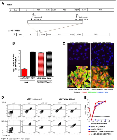
nucleotides in-frame (from 87 to 928) of the NS1 gene (Figure 1A and see Methods); this left a small fragment of NS1 consisting of the 86 N-terminal and 201 C-terminal nucleotides.
After in vitro transcription and electroporation of wild
type (WNV-WT) or Δ-NS1-WNV RNA into BHK21 or
BHK21 VEEV-WNV-NS1 cells (the latter express WNV NS1 in trans from an autonomously propagating VEEV replicon [24]), supernatants were harvested and evaluated for viral RNA by quantitative RT-PCR (qRT-PCR). WNV-WT RNA was recovered readily from supernatants of both BHK21 or BHK21 VEEV-WNV-NS1 cells. In comparison,
while similar amounts ofΔ-NS1-WNV RNA were present
in the supernatant of BHK21 VEEV-WNV-NS1 cells, much lower levels (>105-fold) were recovered from the su-pernatants of BHK21 cells (Figure 1B). To confirm that
Δ-NS1-WNV could be rescued only in cells expressing
NS1, supernatant from BHK21 VEEV-WNV-NS1 cells
transfected withΔ-NS1-WNV was used to infect a
sec-ond set of BHK21 or BHK21 VEEV-WNV-NS1 cells. At 30 hours after infection, cells were analyzed for WNV E and NS5 protein expression by confocal microscopy and flow cytometry. Only BHK21 VEEV-WNV-NS1 but
not BHK21 cells infected with Δ-NS1-WNV showed
evidence of productive infection and WNV antigen ex-pression (Figure 1C and D).
To address the relative efficiency of trans-complementation
ofΔ-NS1-WNV, growth kinetics of infectious WNV-WT
andΔ-NS1-WNV were compared on BHK21 and BHK21
VEEV-WNV-NS1 cells by focus-forming assay (Figure 1E).
Consistent with the immunofluorescence data,Δ-NS1-WNV
failed to produce infectious foci on BHK21 cells con-firming that it fails to replicate efficiently in the absence
of NS1 expression. However, Δ-NS1-WNV growth
kinet-ics on BHK21 VEEV-WNV-NS1 cells were comparable to WNV-WT infection on BHK21 cells, with no appreciable difference observed at any time point examined (P> 0.5). WNV-WT also replicated more efficiently on BHK21 VEEV-WNV-NS1 cells (10 to 15-fold higher levels com-pared to BHK21 cells, P< 0.01) at early time points (16 and 24 hours) after infection, suggesting a difference in in-fectivity of the cells was due to expression of the VEEV replicon or ectopic expression of WNV-NS1. Experiments were repeated with cells expressing a VEEV replicon that lacked NS1 (BHK21-VEEV empty). As these cells sus-tained similar levels of WNV-WT infection compared to the parent BHK21 cells (data not shown), ectopic expres-sion of NS1 improves infectivity of WNV-WT, at least during the early stages of replication.
NS1 protein expression is required for trans-complement ofΔ-NS1-WNV
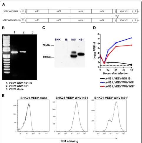
autonomously propagating replicon, it remains unclear whether this is due to expression of NS1 protein or the presence of RNA elements in the NS1 gene. Indeed, RNA elements in the flavivirus genome can bind host factors that are required for replication [26,27]. To evaluate this hypothesis, a stop codon was inserted into the codon of the eighth amino acid of NS1 after the signal peptide and cloned into the VEEV replicon (Figure 2A). BHK21 cells propagating the VEEV-WNV-NS1-internal stop (IS) repli-con transcribed NS1 RNA (Figure 2B) but did not express protein (Figure 2C): flow cytometric analysis showed that NS1 protein was produced in BHK21 VEEV-WNV-NS1 but not BHK21 VEEV-WNV-NS1-IS cells (data not shown). When BHK21 VEEV-WNV-NS1-IS cells were
in-fected withΔ-NS1-WNV, infectious virus was not
recov-ered from the supernatant (Figure 2D). Thus, the presence of an NS1 RNA transcript was not sufficient to trans-complement the infectivity defect associated with a dele-tion of NS1. Rather, NS1 protein is required for WNV replication.
NS1′trans-complementsΔ-NS1 viruses
The JEV serocomplex of flaviviruses produces a second NS1 species, NS1′, which is generated as a product of a
ribosomal frameshift [9,10,28]. As NS1′ reportedly
con-tributes to the pathogenesis of WNV [9], we tested whether its expression would complement the infectivity
of Δ-NS1. We ectopically expressed NS1′ (amino acids
768 to 1195 of the viral coding sequence) in a VEEV rep-licon in BHK21 cells (Figure 2C). Intracellular
expres-sion levels of NS1′ were comparable although slightly
lower than that achieved in cells ectopically expressing NS1 (Figure 2E). Viral growth analysis revealed that
transgenic expression of NS1′ also could complement
the replication defect associated with Δ-NS1-WNV
in-fection (Figure 2D).
A deletion of WNV NS1 is complemented by NS1 from different flaviviruses
A prior study showed that YFV with a deletion in NS1 was not trans-complemented by DENV-2 NS1 without a compensatory adaptive mutation in the NS4A gene [23].
To test whether Δ-NS1-WNV could be complemented
by NS1 from other flaviviruses, we expressed JEV, SLEV, DENV-2, and YFV NS1 using distinct VEEV replicons. Expression of each flavivirus NS1 was confirmed by flow cytometry using species-specific or cross-reactive (e.g.,
9NS1) anti-flavivirus NS1 MAbs (Figure 3A). Δ
-NS1-WNV that was prepared in BHK21 VEEV--NS1-WNV-NS1 cells was titered by plaque and focus-forming assays dir-ectly on BHK21 cells expressing JEV, SLEV, DENV-2, or YFV NS1; this approach limits the development of adaptive mutations. Each heterologous flavivirus NS1 expressed from VEEV replicons trans-complemented
Δ-NS1-WNV (Figure 3B). However, DENV-2, YFV, and
SLEV NS1 trans-complemented Δ-NS1-WNV with
smaller plaques size or lower number (5 to 240-fold) of infectious foci compared to cells expressing WNV NS1 (Figure 3C); these differences in relative infectivity could not be attributed to lower expression of heterologous NS1 (Figure 3A). Only cells expressing JEV NS1, which is the most closely related to WNV (~78% identity at the amino acid level), supported the large plaques and high viral yield that were seen with the complementing cell expressing WNV NS1. Given that adaptive mutations were required
for trans-complementation ofΔ-NS1 YFV with NS1 from
heterologous flaviviruses [23], we sequenced the complete
genomes of Δ-NS1 secreted WNV from cells expressing
WNV, DENV-2, or YFV NS1. No specific mutation was identified in viruses produced by trans-complementing cells expressing DENV-2 or YFV NS1 compared to WNV NS1 (data not shown). Thus, heterologous flavivirus NS1
can trans-complementΔ-NS1 WNV without further
pas-sage or adaptation.
Δ-NS1-WNV can enter cells and undergo input strand translation
To define the stage in the viral lifecycle that was affec-ted by a deletion in NS1 detailed cellular experiments were undertaken. Initially, we assessed whether
trans-complementedΔ-NS1-WNV efficiently attached and
en-tered cells, BHK21 cells were incubated with the same
MOI of WNV or Δ-NS1-WNV for one hour at 4°C or
37°C. After extensive washing, bound virus was quanti-fied by measuring genome copy number by qRT-PCR against the positive strand of WNV RNA. At the same
(See figure on previous page.)
Figure 1Trans-complementation ofΔ-NS1-WNV with ectopically expressed WNV NS1. A.Scheme for construction ofΔ-NS1-WNV. Nucleotides 87 to 928 of the NS1 gene were deleted after restriction digest with the BstEII enzyme.B.Recovery of WNV RNA in supernatant after transfection ofΔ-NS1-WNV or WNV-WT RNA into BHK21 or BHK21-VEEV-WNV NS1 cells. 12 hours post transfection, supernatant was harvested and assessed for levels of viral RNA by qRT-PCR of the E gene. The results are the average of two independent experiments performed in triplicate. C-D.Immunofluorescence(C)or flow cytometry(D)staining of WNV E and NS5 antigen after mock infection or infection ofΔ-NS1-WNV or WNV-WT in BHK21 or BHK21-VEEV-WNV NS1 cells. Cells were infected at an MOI of 5, and at 26 hr post infection, cells were fixed, permeabilized, and stained with anti-E (red), anti-NS5 (green), or a nuclear stain (blue). Results are representative of several independent experiments.
MOI, Δ-NS1-WNV showed equivalent levels of attach-ment to or entry of cells compared to WNV-WT (Figure 4A).
We next assessed steps prior to viral RNA synthesis and replication complex formation by evaluating the amount of viral antigen present in the cell at times that preceded
RNA replication. At the same MOI of 5, WNV orΔ
-NS1-WNV was added to BHK21 or BHK21 VEE--NS1-WNV-NS1 cells. Virus was incubated for one hour at 37°C, and un-bound virus was removed after extensive washing. Virus
internalization was tracked by confocal microscopy using an anti-WNV MAb against the E protein. At two hours
after WNV andΔ-NS1-WNV infection, punctate staining
of E protein from was observed in BHK21 and BHK21 VEE-WNV-NS1 cells (Figure 4B-D), consistent with fla-vivirus entry through endosomal pathways [29,30]. By 8 hours, BHK21 cells infected with WNV-WT or BHK21
VEE-WNV-NS1 cells infected with WNV-WT orΔ
[image:6.595.58.544.88.490.2]-NS1-WNV showed evidence of nascent E and NS5 protein syn-thesis, with a reticular pattern of staining consistent with
Figure 3Trans-complementation ofΔ-NS1-WNV by flavivirus NS1. A.Expression of homologous or heterologous flavivirus NS1 in BHK21 cells using VEEV replicons. WNV (wild type or internal stop (IS), DENV-2, JEV, SLEV, or YFV NS1 were cloned and expressed in VEEV replicons and stable cells propagating these were generated. Cells were stained for NS1 expression with an irrelevant control MAb (E16, anti-WNV E), an anti-DENV-2 NS1 MAb (2G6), anti-WNV NS1 MAb (4NS1), a cross-reactive NS1 MAb (9NS1), and anti-YFV NS1 MAb (4E3). The results are representative of several independent experiments. Red arrows indicate positive expression that was over levels of the negative control MAb. B.Growth kinetics ofΔ-NS1-WNV in BHK21-VEEV cells expressing WNV, DENV-2, SLEV, JEV, YFV NS1, or no insert (empty). Cells were infected at an MOI of 1, and supernatants were titrated on BHK VEEV WNV NS1 cells by focus-forming assay. The dashed line indicates the limit of detection of the assay. Results are the average of two independent experiments performed in triplicate. Error bards indicate standard deviations
its initial localization to the ER (Figure 4E-G, and data not shown). In contrast, at 8 hours, BHK21 cells infected with
Δ-NS1-WNV showed a loss of E protein staining and no
NS5 staining, as would be expected with a failure to repli-cate positive strand viral RNA.
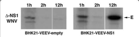
To further define defects in the viral lifecycle associ-ated with a deletion of NS1, we assessed input strand translation. To differentiate input strand protein during the initial translation period, cells were labeled biosyn-thetically and WNV E protein was immunoprecipitated from lysates at specified times. BHK21-VEEV-NS1 cells
or BHK21-VEEV-empty cells were infected withΔ
-NS1-WNV. One hour later, cells were placed in cysteine-methionine-free medium, pulsed for 30 minutes with 35
S-methionine-cysteine, and nascently generated E pro-tein was detected by immunoprecipitation (Figure 5). As
E protein translation was observed in Δ
-NS1-WNV-in-fected BHK21-VEEV-empty and BHK21-VEEV-NS1
cells, NS1 was not required incis for initial input strand translation. By two hours, however, translation of the in-put strand was no longer readily detected likely due to recruitment of the viral RNA into the replication com-plex. By later time points (e.g., 12 hours), E protein
translation again was observed in Δ-NS1-WNV-infected
BHK21-VEEV-NS1 cells but not in cells lacking trans-genic expression of NS1.
Δ-NS1-WNV shows a defect in viral RNA synthesis
NS1 is believed to be required for flavivirus infection be-cause of an essential function in viral RNA translation or
replication [22]. To address whetherΔ-NS1-WNV had a
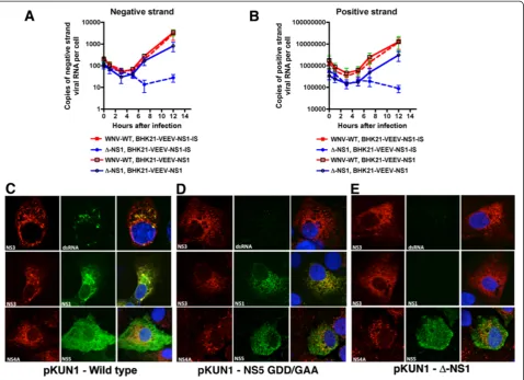
defect at or before RNA synthesis, we monitored viral RNA accumulation over time using asymmetric strand-specific qRT-PCR [31] (Figure 6A and B). Notably,
WNV-WT and Δ-NS1-WNV showed a similar trend in
BHK21 VEEV-WNV-NS1 cells through 5 hours after in-fection with decay in the levels of negative and positive strand RNA. However, by 7 hours after WNV-WT or
Δ-NS1-WNV infection, negative and positive strand
RNA increased in BHK21 VEE-WNV-NS1 cells,
al-though the levels of viral RNA inΔ-NS1-WNV-infected
[image:7.595.59.541.90.242.2]cells did not reach those seen with WNV-WT. In com-parison, in BHK21 VEE-WNV-NS1-IS (internal stop)
Figure 4Δ-NS1-WNV virus does not have an attachment defect. A.Direct binding to BHK21 cells. An MOI of 1 of WNV-WT orΔ-NS1-WNV was incubated at 4°C or 37°C with BHK21 cells for one hour. Unbound virus was removed by centrifugation and washing, and bound and/or internalized virus was quantified after cell lysis and RNA purification using qRT-PCR and a primer and probe set specific for positive strand specific WNV RNA. Values were normalized to 18S rRNA levels to account for possible differences in cell number. Results are the average of three independent experiments, and differences were not statistically significant.B-G.BHK21(B-C and E-F)or BHK21 VEEV NS1(D, G)were infected with WNV-WT(B,E)orΔ-NS1-WNV(C-D, and F-G)at an MOI of 5 for one hour and then unattached virus was removed by extensive washing. Two (top panels) or eight (bottom panels) hours later, cells were fixed, permeabilized and stained with anti-E (red) or anti-NS5 (green) antibodies, or a nuclear stain (blue). Yellow arrows indicate E protein in a punctate staining pattern prior to replication, consistent with virus that is entering cells through an endocytic pathway. Magenta arrows denote E and NS5 staining that occurs after viral replication has ensued. The results are representative of several independent experiments.
Figure 5Input strand translation ofΔ-NS1-WNV in
BHK21-VEEV-empty and BHK21-VEEV-WNV-NS1 cells.An MOI of 5 ofΔ-NS1-WNV was incubated at 37°C with (left) BHK21-VEEV-empty or (right) BHK21-WNV-VEEV-NS1 cells. At 1, 2, or 12 hours, cells were starved in cysteine-methionine deficient medium for 30 minutes, and then pulse-labeled with35S-cysteine-methionine for an additional 30 minutes. Cells were lysed (see Methods), immunoprecipitation was performed with an anti-E protein MAb, proteins were electrophoresed, and gels were subjected to autoradiography. The arrow indicates the mobility of the E protein band. The results are representative of three
[image:7.595.305.538.555.617.2]cells, WNV-WT showed increased negative and positive
strand RNA whereasΔ-NS1-WNV did not.
Δ-NS1-WNV does not form a replication complex
Given the absence of viral RNA accumulation in cells
in-fected with Δ-NS1-WNV, we hypothesized that NS1
might be essential for formation of the viral replication complex that forms on the ER membrane. To begin to assess this, we took advantage of an existing CMV launched infectious cDNA clone of the Kunjin strain of
WNV (pKUN1) [35], and generated Δ-NS1 (deletion of
amino acids 5 to 298 of NS1) and NS5 polymerase dead (GDD to GAA at amino acids 666 and 667) mutant plas-mids [36]. We used this approach so viral proteins could
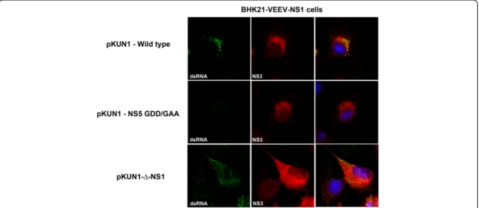
accumulate in the cell in the absence of a requirement for active RNA replication. Two days after transfection, Vero cells were analyzed by immunofluorescence mi-croscopy for co-localization of NS3 and NS5 in the rep-lication complexes. Transfection of the parent cDNA clone (pKUN1) resulted in the co-localization of NS3, NS5, and dsRNA in puncta at the ER (Figure 6C). In contrast, transfection of Vero cells with
pKUN1-NS5-GAA or pKUN1-Δ-NS1 resulted in accumulation of
NS3 and NS5 without recruitment of NS3 into puncta at the ER, the sites of the viral replication complex (Figure 6D and E). Moreover, labeling of double-stranded RNA was absent in NS5-GAA or
[image:8.595.60.539.89.436.2]pKUN1-Δ-NS1 transfected cells. In comparison, transfection of
Figure 6A deletion of WNV NS1 affects viral RNA synthesis and formation of the replication complex. A-B.Measurement of negative (A)or positive(B)strand viral RNA in BHK21-VEEV-NS1 or BHK21-VEEV-NS1-IS (premature stop codon) cells infected with WNV-WT orΔ-NS1-WNV. The indicated cells were infected with WNV-WT orΔ-NS1-WNV at an MOI of 5. At the indicated time points, viral RNA was harvested and strand-specific qRT-PCR [31,32] was performed. The results are the average of two independent performed in triplicate. The very low levels of negative strand viral RNA at input (t = 0) have been reported previously for DENV and WNV [33,34], and possibly reflect delivery of viral RNA in exosomes or defective viruses with inappropriate packing of RNA intermediates.C-E.Vero cells were transfected with CMV-launched infectious cDNA clones of(C)wild type KUNV (pKUN1-wild type),(D)KUNV with a mutation in NS5 that abolishes RNA polymerase activity (pKUN1-NS5 GDD/GAA), or(E)Δ-NS1-WNV KUNV (pKUN1-Δ-NS1). 48 hours later, cells were co-stained with antibodies against(top panels) NS3 and dsRNA, (middle panels) NS3 and NS1, or
BHK21-VEEV-NS1 complementing cells with
pKUN1-Δ-NS1 but not pKUN1-NS5-GAA resulted in the
forma-tion of replicaforma-tion complexes (identified by dsRNA) that co-localized with NS3 protein (Figure 7). Collectively, these data confirm that NS1 is required for viral RNA syn-thesis, and without this, replication complexes fail to form efficiently.
Discussion
Flavivirus NS1 is a non-structural glycoprotein that is expressed on the plasma membrane of infected cells and secreted into the extracellular space. Secreted NS1 antago-nizes complement function in solution and on the plasma membrane surface, and thus serves an immune evasion function [16-18,37]. Within the cell, even though it is lo-calized to the lumen of the ER and viral replication occurs on the cytosolic face of the ER, NS1 is an essential gene that regulates early viral RNA replication [22,38]. How this occurs has been poorly understood, although prior studies suggested NS1 is required for a step proximal to viral RNA synthesis [22], possibly through interactions with the viral transmembrane proteins NS4A [23] or NS4B [24], both of which have direct proximity to the replication complex on the cytosolic face of the ER membrane. In the current study, using two WNV strains that lack a func-tional NS1 gene and a trans-complementation system, we show that while WNV NS1 was not required for input strand translation of the infectious viral RNA, it was ne-cessary for negative and positive strand RNA synthesis and formation of the replication complex.
While the ability of ectopically-expressed NS1 to trans-complement flaviviruses (WNV or YFV) containing gene deletions in NS1 has been demonstrated previously [22,25], our studies show uniquely for WNV that heterol-ogous NS1 from DENV, JEV, SLEV, and YFV all can serve this function. Each heterologous flavivirus NS1 expressed
from VEEV replicons trans-complemented Δ-NS1-WNV
without further passage. Nonetheless, we observed differ-ences in efficiency, as trans-complementation with DENV, YFV, and SLEV NS1 resulted in smaller plaques sizes and/ or lower numbers of infectious foci compared to cells ec-topically expressing NS1 from homologous (WNV) or the most closely related (JEV) flaviviruses. In comparison, a
prior study showed thatΔ-NS1 YFV could not be
trans-complemented by DENV-2 NS1 without adaptation, pos-sibly due to an inability to interact with NS4A and the remainder of the YFV replication complex [23]. Thus, the requirements for regulating replication by NS1 appear conserved enough to allow for trans-complementation of
Δ-NS1-WNV by NS1 from other mosquito-transmitted
flaviviruses.
Our detailed kinetic experiments demonstrating that an absence of WNV NS1 affects viral RNA synthesis as judged by strand-specific qRT-PCR confirm and extend
earlier results with Δ-NS1 YFV that were obtained by
Northern blotting [22]. We show that trans-complemented
Δ-NS1 WNV bound to and entered cells, and the input
strand of genomic RNA was translated at early time points. Moreover, by using a CMV launched wild type and mutant
[image:9.595.58.540.90.299.2](Δ-NS1 or NS5 polymerase dead) WNV-KUNV, we
showed that NS1 is required for formation of the replica-tion complex and recruitment of other non-structural pro-teins (e.g., NS3) to the vesicle packets [39] associated with ER membranes; the strength of this experimental approach is that it allows for continued transcription and translation of viral non-structural genes in the absence of a require-ment for active replication. However, it remains unclear whether the absence of replication complex after
transfec-tion of Δ-NS1 WNV-KUNV was due to the lack of
pro-duction of dsRNA or the failure to recruit key viral and possibly, host proteins to the specialized ER membranes, which serve as the site of flavivirus replication.
Conclusions
Our experiments establish that WNV NS1 contributes to viral replication. Whereas soluble NS1 functions to antagonize innate immune responses through interac-tions with complement or pathogen recognition recep-tors [16,17,19,37], intracellular NS1 serves a discrete purpose during viral RNA synthesis, possibly to help form or stabilize the replication complex. Ongoing stud-ies are planned to define the precise molecular details by which NS1 executes functions at different stages of the flavivirus virus lifecycle.
Methods
Cells and viruses
BHK21 cells were grown in complete Dulbecco’s modified Eagle’s medium (DMEM) (supplemented with 10% fetal bovine serum (FBS), penicillin, streptomycin, 10 mM HEPES pH 7.3, and non-essential amino acids) in a 5% hu-midified CO2incubator at 37°C. BHK21 cells propagating VEEV replicons were maintained in complete DMEM
supplemented with 5 μg/ml of puromycin. Infection
ex-periments were performed with the New York 1999 strain (385–99) of WNV, which was derived from an infectious cDNA clone [40], or WNV-KUNV, as detailed below.
Generation of cells propagating VEEV replicons expressing flavivirus NS1
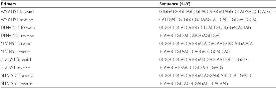
Full length NS1 including the N-terminal signal sequences from WNV (strain New York 1999, amino acids 768 to 1143), DENV-2 (strain D2S10, amino acids 776 to 1127), YFV (strain 17D, amino acids 760 to 1136), SLEV (strain GHA6, amino acids 766 to 1141), and JEV (strain 14-14-2, amino acids 771 to 1146) were amplified from viral RNA or infectious clone cDNA by RT-PCR and PCR using specific primers (Table 1), and cloned into pSTBlue TA
(EMD Millipore). To clone the NS1′form of WNV, one
additional nucleotide (T) was introduced at the site of the−1 frame shift by QuickChange mutagenesis using the following primers: forward: 5′-GATATGATTGACCCTT TTTCAGTTGGGCCTTCTG-3′, reverse: 5′-CAGAAGG CCCAACTGAAAAAGGGTCAATCATATC-3′, this re-sulted in the generation of an NS1′with an additional 52 amino acids at the C-terminus. After sequence verifica-tion, all NS1 genes were subcloned into a modified pSC-B W/VEEV shuttle vector and then into a VEEV replicon (pTC83new/Pac [41]; gift of I. Frolov) using an XbaI site. VEEV replicon plasmids encoding NS1 were linearized with MluI and used as templates forin vitrotranscription using an SP6 DNA-dependent RNA polymerase mMES-SAGE mRNA transcription kit (Ambion) according to the manufacturer’s instruction. RNA transcripts were intro-duced into cells using a GenePulser Xcell electroporator (Bio-Rad) at 850 V, 25 μF, and infinite Ω, and cells ex-pressing VEEV replicons were selected with puromycin
(5 μg/ml) over one week. Flavivirus NS1 expression was
confirmed by flow cytometry as described below.
Construction and production ofΔ-NS1 WNV-NY
[image:10.595.55.538.579.733.2]Using the pSTBlue vector containing WNV NS1 as a template, a second BstEII restriction enzyme site was in-troduced at nucleotide 87 of the WNV NS1 gene (from CATAGAC to GGTCACC) by site directed mutagenesis using the following primers: forward: 5′- GACAC
Table 1 Primers for cloning NS1 from different flaviviruses
Primers Sequence (5′-3′)
WNV NS1 forward GTGGATGGGCGGCCGCACCATGGATAGGTCCATAGCTCTCACGTTT
WNV NS1 reverse CATTGACTGCGGCCGCTAAGCATTCACTTGTGACTGCAC
DENV NS1 forward GCGGCCGCACCATGGTCTCACTGTCTGTGACACTAG
DENV NS1 reverse TCAAGCTGTGACCAAGGAGTTGAC
YFV NS1 forward GCGGCCGCACCATGGACATGACAATGTCCATGAGCA
YFV NS1 reverse TCAAGCTGTAACCCAGGAGCGCACCAG
JEV NS1 forward GCGGCCGCACCATGGACCGATCAATTGCTTTGGCC
JEV NS1 reverse TCAAGCATGAACCTGTGATCTGACG
SLEV NS1 forward GCGGCCGCACCATGGACAGGAGCATCTCGCTGACTC
production of any residual truncated protein using anti-NS1 MAbs that recognize different regions of the intact protein.
Trans-complementation plaque and focus-forming assays
The ability of different flavivirus NS1 to trans-complement
ΔNS1-WNV was determined by serially diluting Δ
-NS1-WNV (produced in cells ectopically expressing -NS1-WNV NS1) and then infecting BHK21 cells propagating VEEV replicons expressing WNV, JEV, SLEV, DENV-2, or YFV NS1 for one hour at 37°C. Subsequently, DMEM contain-ing final 4% FBS and 1% low meltcontain-ing agarose was overlaid and incubated an additional 3 days for plaque forming assay or DMEM containing final 4% FBS and 1% methyl-cellulose was overlaid and incubated for one day for the focus-forming assay. Plaques were stained with 1% crystal violet after fixation with 10% formaldehyde and then counted as described previously [42]. For the focus-forming assay, cells were fixed with 1% paraformalde-hyde for 20 minutes. After removal of the methylce-llulose, cells were washed three times with PBS, permeabilized with PBS supplemented with 1% saponin (w/v) and 1% BSA (w/v), and incubated with mouse E16 anti-WNV MAb [43] for three hours at room temp-erature or overnight at 4°C. Following three washes with PBS, cells were incubated for one hour with horseradish peroxidase-conjugated goat anti-mouse IgG (Sigma, 1/5,000 dilution), and after additional washes, foci were visualized with TrueBlue peroxidase substrate (KPL) and counted using a BioSpot Analyzer (C.T.L).
Western blotting
Supernatants from BHK21 VEEV-NS1-IS, BHK21
VEEV-WNV-NS1, or BHK21 VEEV NS1′cells were
sep-arated by 12% PAGE, transferred to nitrocellulose mem-brane using an iBlot dry blotting system (Invitrogen). Membranes were blocked for one hour at room temperature in 50 mM Tris, 150 mM NaCl, 0.05% Tween 20 (TBST) supplemented with 5% non-fat dry milk. Subsequently, WNV NS1 proteins were detected
using 16-NS1 MAb [44] (1 μg/ml) after incubating
membranes in TBST supplemented with 2.5% milk
DENV NS1, JEV NS1, SLEV NS1, or YFV NS1 were de-tached with HBSS containing 5 mM EDTA. Cells were washed twice with PBS on ice, fixed with 2% formalde-hyde in PBS for seven minutes, and washed again with HBSS. Cells were permeablized with 0.1% (w/v) saponin and 0.1% BSA in HBSS (permeabilization buffer) and then incubated with primary MAbs (WNV E16 as an isotype control, 9NS1 as a cross-reactive anti-NS1 MAb [44], 2G6 (DENV specific) [45], 4NS1 (WNV NS1-specific) [44], 10NS1 (reacts with WNV and JEV NS1) [44], and 4E3 (YFV NS1-specific) [46] for one hour on ice. After three washes with permeabilization buffer, cells were incubated with Alexa Fluor-647 conjugated goat anti-mouse IgG (Molecular Probe) for 30 minutes and then washed three additional times. Expression levels of NS1 were determined by flow cytometry on a BD FAC-SArray (Becton Dickinson) and data was processed with FlowJo software (Tree Star, Inc).
Asymmetric positive and negative strand qRT-PCR
Strand-specific real-time RT-PCR was performed as de-scribed using E gene primers [31,32]. Each transcript reaction was treated with DNAse (Ambion) thrice consecutively followed by RNA extraction with Trizol (Invitrogen) according to the manufacturer’s instruction. PCR was performed with and without reverse transcrip-tion to confirm that RNA was free of template DNA. Wild type BHK21 cells or BHK21 VEEV-WNV-NS1 cells (6 ×
105) were infected with WNV orΔ-NS1-WNV at an MOI
of 5. Total cellular RNA was harvested at specified time points using the RNEasy mini kit (Qiagen) following the manufacturer’s instructions, and viral RNA was quantified using fluorogenic quantitative RT-PCR (qRT-PCR). Strand specificity of the qRT-PCR was confirmed using individual asymmetric primer sets and positive or negative strand RNA that was generated fromin vitro transcription reac-tions after extensive DNAse treatment (data not shown).
Virus attachment assays
BHK21 cell monolayers were detached with HBSS and 5 mM EDTA, washed once with complete DMEM, and
aliquotted into eppendorf tubes and 106 FFU of WNV
or Δ-NS1-WNV was added in total volume of 100 μl.
After binding for one hour, unbound virus was removed by centrifugation (200 × g) and three washes with chilled PBS. Bound virus was quantified after cell lysis and RNA purification (RNEasy Kit, Qiagen) and quantification of genomic RNA using qRT-PCR and a primer and probe set specific for positive strand specific WNV RNA as de-scribed above. Values were normalized to 18S rRNA levels to account for possible differences in cell number.
Microscopic analysis of viral entry
BHK21 cells (6 × 10⁵) were infected with an MOI of 5 of
WNV or Δ-NS1-WNV for one hour at 37°C. Unbound
virus was removed after three rinses with PBS and complete DMEM was added for specified time points. Sub-sequently, cells were rinsed with PBS, fixed with 3% para-formaldehyde in PBS for 10 minutes, and permeabilized with PBS supplemented with 0.5% Triton-X 100. After additional washes with PBS and glycine (10 mM), cells were incubated with WNV E16 MAb or a negative control MAb (2G6, anti-DENV-2 NS1) for 20 minutes at room temperature in DMEM containing 10% BSA. After three washes with PBS and 10 mM glycine pH 7.4, cells were in-cubated for 15 minutes with AlexaFluor 555-conugated goat anti-mouse antibody (1/400 dilution), and nuclei were counterstained with To-PRO 3 (Molecular Probes). After several washes with PBS, cells were mounted on glass slides, and images were acquired with a laser scanning con-focal microscope (Zeiss LSM 510 META) and analyzed with LSM image browser software (Zeiss).
Radiolabeling and immunoprecipitation
To compare the initial translation rates, 6 × 10⁵ BHK21 cells or BHK21 VEEV-WNV-NS1 cells were infected at an
MOI of 5 of WNV or Δ-DNS1-WNV. At specific time
points after infection, cells were starved with methionine free DMEM for 15 minutes, and then labeled for 20 minutes with 50 mCi/ml of 35S methionine and cysteine
(EasyTag™EXPRES35S Protein Labeling Mix, PerkinElmer
Life Science). Labeled cells were solubilized in 800 μl of Lysis buffer (50 mM Tris-HCl, pH 8.0, 150 mM NaCl, 1% NP-40, 0.25% sodium deoxycholate, and 1 mM EDTA supplemented with a protease inhibitor cocktail (Sigma)) for 10 minutes on ice. Nuclei were removed by centrifuga-tion (13,800 × g) for five minutes at 4°C. Subsequently, ly-sates were incubated with WNV E16 MAb (10 mg per sample) for three hours at 4°C. WNV E protein-MAb complexes were immunoprecipitated with protein A-Sepharose beads (Invitrogen) after an additional one-hour incubation at 4°C. Beads were washed three times with 10 mM Tris pH 7.4, 150 mM NaCl, 1% sodium deoxycholate, and 1% NP-40 and proteins were eluted by boiling sam-ples at 95°C for five minutes in 4X SDS sample buffer
supplemented with 5% (v/v) β-mercaptoethanol. Eluates
were separated by 12% SDS-PAGE, gels were vacuum-dried on filter paper for one hour at 80°C, and proteins were visualized after exposure to Kodak Biomax light film.
Expression and analysis of CMV-launchedΔ-NS1-WNV-KUNV
Unique MluI restriction sites were generated within the
NS1 sequence in the full-length WNV expression vector pKUN1 [35] at residues 4 and 298 (described in [38]) using site-directed mutagenesis. Briefly, pKUN1 was
amplified with Pfu Ultra Hot-start polymerase
(Strata-gene) and primers incorporating MluI sequences (listed
in Table 2) as follows: 92°C for 2 min then 18 cycles of 92°C for 30 s, 55°C for 1 min and 68°C for 21 min. Fol-lowing restriction digestion and Sanger sequencing to verify mutations, plasmids were cleaved withMluIto move the majority of NS1-coding sequence, then
re-ligated and transformed into JM109
chemically-competent E.coli cells. Δ-NS1 plasmids were amplified
and extracted using a Hi-Speed Plasmid Midiprep kit (Qiagen). A mutation (NS5 GDD to GAA) that abolishes RNA polymerase activity has been described previously [38], and was made by site directed mutagenesis.
Vero or BHK21-VEEV-NS1 cells were transfected with
1 μg each of pKUN1, pKUN1-NS5-GAA or pKUN1-Δ
-NS1 cDNA using Lipofectamine 2000 (Life Technolo-gies) as described by the manufacturer. Two days hours after transfection, cells were fixed in either 1:1 acetone: methanol or 4% v/v paraformeldehyde and perme-abilized with 0.1% w/v Triton X-100. Cells were then in-cubated with MAbs to dsRNA (English & Scientific Consulting Bt. (Hungary)), NS1 and NS5 (kindly pro-vided by R. Hall, University of Queensland) or rabbit polyclonal antisera raised against NS3 [39] or NS4A [47]. Confocal images were collected on a Zeiss confocal microscope.
Data analysis
[image:12.595.306.537.643.732.2]All data were analyzed statistically using Prism software (GraphPad4, San Diego, CA). Differences in viral infec-tion or RNA levels were analyzed using a two tailed, un-pairedt-test.
Table 2 Primers for cloning CMV-launched pKUN-Δ-NS1
Primer Sequence (5′ –3′) (MluI)
final form. MSD and JMM supported the work with research grants from the National Institutes of Health (U54-AI057160) and the National Health and Medical Research Council of Australia (No. 100461). All authors read and approved the final manuscript.
Acknowlegements
We thank I. Frolov, S. Shresta, L. Kramer, A. Khromykh, R. Hall, P. Avirutnan and J. Schlesinger, and P. Y. Shi for the VEEV expression constructs, DENV-2 D2S10 and pKUN1 plasmids, and anti-NS1, anti-NS3, and anti-NS5 antibodies. We thank B. Lindenbach for experimental advice and critical comments on the manuscript.
Author details
1Department of Medicine, Washington University School of Medicine, Saint Louis,
MO 63110, USA.2Department of Molecular Microbiology, Washington University School of Medicine, Saint Louis, MO 63110, USA.3Department of Pathology and
Immunology, Washington University School of Medicine, Saint Louis, MO 63110, USA.4Department of Microbiology and Immunology, University of Melbourne,
Parkville, Melbourne 3010, Australia.
Received: 16 September 2013 Accepted: 14 November 2013 Published: 18 November 2013
References
1. Mackenzie JS, Gubler DJ, Petersen LR:Emerging flaviviruses: the spread and resurgence of Japanese encephalitis, West Nile and dengue viruses.
Nat Med2004,10:S98–109.
2. Muller DA, Young PR:The flavivirus NS1 protein: molecular and structural biology, immunology, role in pathogenesis and application as a diagnostic biomarker.Antiviral Res2013,98:192–208.
3. Flamand M, Megret F, Mathieu M, Lepault J, Rey FA, Deubel V:Dengue virus type 1 nonstructural glycoprotein NS1 is secreted from mammalian cells as a soluble hexamer in a glycosylation-dependent fashion.J Virol
1999,73:6104–6110.
4. Crooks AJ, Lee JM, Easterbrook LM, Timofeev AV, Stephenson JR:The NS1 protein of tick-borne encephalitis virus forms multimeric species upon secretion from the host cell.J Gen Virol1994,75(Pt 12):3453–3460. 5. Winkler G, Randolph VB, Cleaves GR, Ryan TE, Stollar V:Evidence that the
mature form of the flavivirus nonstructural protein NS1 is a dimer.
Virology1988,162:187–196.
6. Winkler G, Maxwell SE, Ruemmler C, Stollar V:Newly synthesized dengue-2 virus nonstructural protein NS1 is a soluble protein but becomes partially hydrophobic and membrane-associated after dimerization.
Virology1989,171:302–305.
7. Gutsche I, Coulibaly F, Voss JE, Salmon J, d’Alayer J, Ermonval M, Larquet E, Charneau P, Krey T, Megret F,et al:Secreted dengue virus nonstructural protein NS1 is an atypical barrel-shaped high-density lipoprotein.
Proc Natl Acad Sci USA2011,108:8003–8008.
8. Firth AE, Atkins JF:A conserved predicted pseudoknot in the NS2A-encoding sequence of West Nile and Japanese encephalitis flaviviruses suggests NS1′ may derive from ribosomal frameshifting.Virol J2009,6:14.
9. Melian EB, Hinzman E, Nagasaki T, Firth AE, Wills NM, Nouwens AS, Blitvich BJ, Leung J, Funk A, Atkins JF,et al:NS1′of flaviviruses in the Japanese encephalitis virus serogroup is a product of ribosomal frameshifting and plays a role in viral neuroinvasiveness.J Virol2010,84:1641–1647.
14. Noisakran S, Dechtawewat T, Rinkaewkan P, Puttikhunt C, Kanjanahaluethai A, Kasinrerk W, Sittisombut N, Malasit P:Characterization of dengue virus NS1 stably expressed in 293 T cell lines.J Virol Methods2007,142:67–80. 15. Noisakran S, Dechtawewat T, Avirutnan P, Kinoshita T, Siripanyaphinyo U,
Puttikhunt C, Kasinrerk W, Malasit P, Sittisombut N:Association of dengue virus NS1 protein with lipid rafts.J Gen Virol2008,89:2492–2500. 16. Chung KM, Liszewski MK, Nybakken G, Davis AE, Townsend RR, Fremont DH,
Atkinson JP, Diamond MS:West Nile virus non-structural protein NS1 inhibits complement activation by binding the regulatory protein factor H.Proc Natl Acad Sci USA2006,103:19111–19116.
17. Avirutnan P, Fuchs A, Hauhart RE, Somnuke P, Youn S, Diamond MS, Atkinson JP:Antagonism of the complement component C4 by flavivirus non-structural protein NS1.J Exp Med2010,207:793–806.
18. Avirutnan P, Hauhart RE, Somnuke P, Blom AM, Diamond MS, Atkinson JP: Binding of flavivirus nonstructural protein NS1 to C4b binding protein modulates complement activation.J Immunol2011,187:424–433. 19. Wilson JR, de Sessions PF, Leon MA, Scholle F:West Nile virus nonstructural
protein 1 inhibits TLR3 signal transduction.J Virol2008,82:8262–8271. 20. Mackenzie JM, Jones MK, Young PR:Immunolocalization of the dengue
virus nonstructural glycoprotein NS1 suggests a role in viral RNA replication.Virology1996,220:232–240.
21. Khromykh AA, Sedlak PL, Westaway EG:Trans-complementation analysis of the flavivirus Kunjin ns5 gene reveals an essential role for translation of its N-terminal half in RNA replication.J Virol1999,73:9247–9255. 22. Lindenbach BD, Rice CM:trans-Complementation of yellow fever virus
NS1 reveals a role in early RNA replication.J Virol1997,71:9608–9617. 23. Lindenbach BD, Rice CM:Genetic interaction of flavivirus nonstructural
proteins NS1 and NS4A as a determinant of replicase function.J Virol
1999,73:4611–4621.
24. Youn S, Li T, McCune BT, Edeling MA, Fremont DH, Cristea IM, Diamond MS: Evidence for a genetic and physical interaction between nonstructural proteins NS1 and NS4B that modulates replication of West Nile virus.
J Virol2012,86:7360–7371.
25. Khromykh AA, Sedlak PL, Guyatt KJ, Hall RA, Westaway EG:Efficient trans-complementation of the flavivirus kunjin NS5 protein but not of the NS1 protein requires its coexpression with other components of the viral replicase.J Virol1999,73:10272–10280.
26. Emara MM, Liu H, Davis WG, Brinton MA:Mutation of mapped TIA-1/TIAR binding sites in the 3′terminal stem-loop of West Nile virus minus-strand RNA in an infectious clone negatively affects genomic RNA amplification.
J Virol2008,82:10657–10670.
27. Davis WG, Blackwell JL, Shi PY, Brinton MA:Interaction between the cellular protein eEF1A and the 3′-terminal stem-loop of West Nile virus genomic RNA facilitates viral minus-strand RNA synthesis.J Virol2007, 81:10172–10187.
28. Sun J, Yu Y, Deubel V:Japanese encephalitis virus NS1′protein depends on pseudoknot secondary structure and is cleaved by caspase during virus infection and cell apoptosis.Microbes Infect2012,14:930–940. 29. van der Schaar HM, Rust MJ, Chen C, van der Ende-Metselaar H, Wilschut J,
Zhuang X, Smit JM:Dissecting the cell entry pathway of dengue virus by single-particle tracking in living cells.PLoS Pathog2008,4:e1000244. 30. van der Schaar HM, Rust MJ, Waarts BL, van der Ende-Metselaar H, Kuhn RJ,
Wilschut J, Zhuang X, Smit JM:Characterization of the early events in den-gue virus cell entry by biochemical assays and single-virus tracking.
31. Samuel MA, Whitby K, Keller BC, Marri A, Barchet W, Williams BRG, Silverman RH, Gale M, Diamond MS:PKR and RNAse L contribute to protection against lethal West Nile virus infection by controlling early viral spread in the periphery and replication in neurons.J Virol2006,80:7009–7019. 32. Samuel MA, Diamond MS:Type I IFN protects against lethal West Nile
Virus infection by restricting cellular tropism and enhancing neuronal survival.J Virol2005,79:13350–13361.
33. Diamond MS, Zachariah M, Harris E:Mycophenolic Acid inhibits dengue virus infection by preventing replication of viral RNA.Virology2002, 304:211–221.
34. Daffis S, Szretter KJ, Schriewer J, Li J, Youn S, Errett J, Lin TY, Schneller S, Zust R, Dong H,et al:2′-O methylation of the viral mRNA cap evades host restriction by IFIT family members.Nature2010,468:452–456.
35. Khromykh AA, Varnavski AN, Sedlak PL, Westaway EG:Coupling between replication and packaging of flavivirus RNA: evidence derived from the use of DNA-based full-length cDNA clones of Kunjin virus.J Virol2001, 75:4633–4640.
36. Khromykh AA, Kenney MT, Westaway EG:Trans-complementation of flavivirus RNA polymerase gene NS5 by using Kunjin virus replicon-expressing BHK cells.J Virol1998,72:7270–7279.
37. Krishna VD, Rangappa M, Satchidanandam V:Virus-specific cytolytic antibodies to nonstructural protein 1 of Japanese encephalitis virus effect reduction of virus output from infected cells.J Virol2009,83:4766–4777. 38. Khromykh AA, Sedlak PL, Westaway EG:Cis- and trans-acting elements in
flavivirus RNA replication.J Virol2000,74:3253–3263.
39. Westaway EG, Mackenzie JM, Kenney MT, Jones MK, Khromykh AA: Ultrastructure of Kunjin virus-infected cells: colocalization of NS1 and NS3 with double-stranded RNA, and of NS2B with NS3, in virus- induced membrane structures.J Virol1997,71:6650–6661.
40. Beasley DW, Whiteman MC, Zhang S, Huang CY, Schneider BS, Smith DR, Gromowski GD, Higgs S, Kinney RM, Barrett AD:Envelope protein glycosylation status influences mouse neuroinvasion phenotype of genetic lineage 1 West Nile virus strains.J Virol2005,79:8339–8347. 41. Petrakova O, Volkova E, Gorchakov R, Paessler S, Kinney RM, Frolov I:
Noncytopathic replication of Venezuelan equine encephalitis virus and eastern equine encephalitis virus replicons in Mammalian cells.J Virol
2005,79:7597–7608.
42. Diamond MS, Shrestha B, Marri A, Mahan D, Engle M:B cells and antibody play critical roles in the immediate defense of disseminated infection by West Nile encephalitis virus.J Virol2003,77:2578–2586.
43. Oliphant T, Engle M, Nybakken G, Doane C, Johnson S, Huang L, Gorlatov S, Mehlhop E, Marri A, Chung KM,et al:Development of a humanized monoclonal antibody with therapeutic potential against West Nile virus.
Nat Med2005,11:522–530.
44. Chung KM, Nybakken GE, Thompson BS, Engle MJ, Marri A, Fremont DH, Diamond MS:Antibodies against West Nile virus non-structural (NS)-1 protein prevent lethal infection through Fc gamma receptor-dependent and independent mechanisms.J Virol2006,80:1340–1351.
45. Puttikhunt C, Kasinrerk W, Srisa-ad S, Duangchinda T, Silakate W, Moonsom S, Sittisombut N, Malasit P:Production of anti-dengue NS1 monoclonal antibodies by DNA immunization.J Virol Methods2003,109:55–61. 46. Schlesinger JJ, Brandriss MW, Walsh EE:Protection against 17D yellow
fever encephalitis in mice by passive transfer of monoclonal antibodies to the nonstructural glycoprotein gp48 and by active immunization with gp48.J Immunol1985,135:2805–2809.
47. Mackenzie JM, Khromykh AA, Jones MK, Westaway EG:Subcellular localization and some biochemical properties of the flavivirus Kunjin nonstructural proteins NS2A and NS4A.Virology1998,245:203–215.
doi:10.1186/1743-422X-10-339
Cite this article as:Younet al.:Non-structural protein-1 is required for
West Nile virus replication complex formation and viral RNA synthesis.
Virology Journal201310:339.
Submit your next manuscript to BioMed Central and take full advantage of:
• Convenient online submission • Thorough peer review
• No space constraints or color figure charges • Immediate publication on acceptance
• Inclusion in PubMed, CAS, Scopus and Google Scholar
• Research which is freely available for redistribution