• No results found

Higher titers of some H5N1 and recent human H1N1 and H3N2 influenza viruses in Mv1 Lu vs MDCK cells

N/A
N/A
Protected

Academic year: 2020

Share "Higher titers of some H5N1 and recent human H1N1 and H3N2 influenza viruses in Mv1 Lu vs MDCK cells"

Copied!
9
0
0

Loading.... (view fulltext now)

Full text

Figure

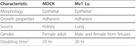
Table 1 Characteristics of MDCK and Mv1 Lu cells
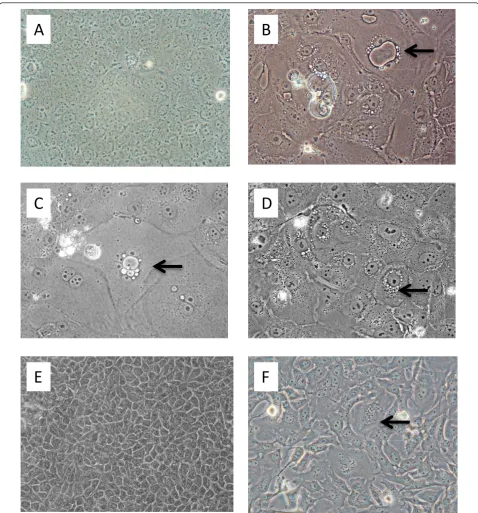
Figure 1 Microscopic analyses of cultured MDCK and Mv1 Lu cells. A. MDCK cells, normal (200× magnification); B
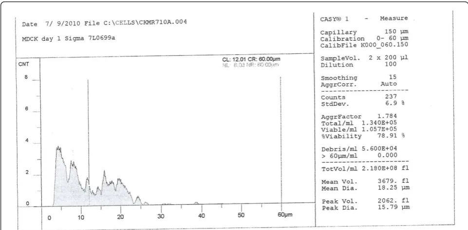
Figure 2 MDCK cell profile under optimal conditions and serum, one day post-seed. Ordinate, cell count (CNT); abscissa, diameter of cellor other particles (μm).
Figure 5 Appearance of MDCK and Mv1 Lu cells 48 hr after infection of the cells at a high MOI with a pandemic H1N1 2009 virusEnlarged nuclei and cellular granulation but no cellular detachment are evident in the MDCK cells (panel A), whereas most of the Mv1 Lu cellshave detached or are showing advanced CPE (panel B).
+4

References

Related documents