IDENTIFICATION AND CHARACTERISATION OF
BACTERIAL GENES ASSOCIATED WITH
RESISTANCE TO AND/OR DEGRADATION OF
ENVIRONMENTAL POLLUTANTS
A thesis submitted for the degree of
DOCTOR OF PHILOSOPHY
By
BELINDA L. DAVIS
B.Sc (Hons) (Applied Biology)
School of Engineering and Science
Victoria University
Melbourne, Victoria
I
DECLARATION
“I, Belinda Davis, declare that the PhD thesis entitled Identification and
Characterisation of Bacterial Genes Associated with Resistance to and/or Degradation
of Environmental Pollutants is no more than 100,000 words in length including quotes
and exclusive of tables, figures, appendices, bibliography, references and footnotes.
This thesis contains no material that has been submitted previously, in whole or in part,
for the award of any other academic degree or diploma. Except where otherwise
indicated, this thesis is my own work”.
II
ACKNOWLEDGEMENTS
I would sincerely like to thank my supervisor, Associate ProfessorMrinal Bhave, for
her patience and wealth of knowledge in the area of molecular genetics. She has bought
many an idea to this project and solved many an experimental problem. Countless
hours have been spent by Mrinal critically reviewing this thesis in its many stages, for
which I am grateful.
I would like to express my gratitude to my supervisor, Associate Professor Grant
Stanley, for his input and advice into the microbiology work performed in this thesis, in
particular, the growth curve studies. Thanks also to Grant for his ongoing emotional
support and encouragement.
I have a warm appreciation for Dr. Sarah Fraser for her help and support. Much valued
advice was given on the RNA aspects of this project at any time. Sarah has been a great
support, lending me her ear whenever required.
I would like to credit the late Dr. Nicholas McClure and Catherine Dandie for their
collaboration with the VUN 10010 isolate and their warm hospitality during my visit to
Flinders University, Adelaide.
I would like to acknowledge Shee Ping Ng from Swinburne University of Technology
for collaborative work regarding the Achromobacter sp. AO22 isolate.
Cheers to my fellow postgraduate students, in particular Joshua Johnson, Meredith
Chandler, Idris Mohammed, Karoline Tellbach, Kate Lauder, Matthew Knight and
Chutima Kongajaroon for their advice and exchange of ideas. Thanks also to Danny,
Mark, Bogdan, Camilla, Yadira, Nanna, Fabio and Kristian for the laughs and
friendship.
I would like to recognise the Technical staff of the School of Engineering and Science
at Victoria University, in particular, Dale Tomlinson, Charmaine DiQuattro, Michael
III Thanks to the Molecular Vaccines laboratory of the Department of Primary Industries
(Attwood) for the use of their Kinetic Microplate Reader.
I would like to express my gratitude to my employer of the last few years Schweppes, in
particular Graeme Kentish, Melissa Dawson and Natalie Tabone, for their
understanding whilst juggling my thesis and employment.
A big recognition goes to my family for their on-going support and assistance. You
may not have “understood” what I have been going through, but your encouragement
has been priceless.
Thanks to my friends for standing by me when “I can’t afford it” or “Am working on
my thesis”. Your understanding is much appreciated.
Last, but not least, to Socks and Ginge, my affection for you both made my time at uni
IV
PUBLICATIONS
Ng SP, Davis B, Palombo E and Bhave M (2009) A Tn5051-like mer-containing
transposon identified in a heavy metal tolerant strain Achromobacter sp. AO22 isolated
from an industrial site in Australia. BMC Research Notes. 2:38.
Davis BL and Bhave M (2004) An investigation of the formation of biofilms by heavy
metal resistant soil bacteria. The 12th Annual RACI Research and Development Topics
(Analytical and Environmental Division). The University of Melbourne, Melbourne,
Australia, December, 2004 (Oral Presentation).
Davis B, Stanley G and Bhave M (2003) An investigation of the presence and
expression of heavy metal resistance genes in a Gram-negative and a Gram-positive soil
bacterial isolate. XIX International Congress of Genetics. Melbourne, Australia, July,
2003 (Poster Presentation).
Davis B, Stanley G and Bhave M (2003) A study of the expression of mercury
resistance genes in a Gram-negative and a Gram-positive soil bacterial isolate. The 24th
Annual Conference on the Organisation and Expression of the Genome. Erskine House,
Lorne, Victoria, Australia, February, 2003 (Poster Presentation).
Davis B, Stanley G and Bhave M (2002) The investigation of bacterial isolates obtained
from heavy metal- and PAH-contaminated soil samples. AusBiotech 2002 National
Biotechnology and Investment Forum, August, 2002 (Oral Presentation).
Davis B and Bhave M (2002) Genes encoding mercury resistance and their expression
in soil bacteria. The 23rd Annual Conference on the Organisation and Expression of the
Genome. Erskine House, Lorne, Victoria, Australia, February, 2002 (Poster
Presentation).
Davis B, Gemmell C and Bhave M (2002) Genes encoding heavy metal resistance and
their dispersal in soil bacteria. The 22th Annual Conference on the Organisation and
Expression of the Genome. Erskine House, Lorne, Victoria, Australia, February, 2001
V
ABBREVIATIONS
< Less than
> Greater than
≥ Greater than or equal to
I One
II Two
+ Plus
- Minus
# Number
α Alpha
β Beta
λ Lambda
σ Sigma
oC Degrees Celsius
Registered trademark
µg Microgram
µg/kg Micrograms per kilogram
µg/L Micrograms per liter
µg/mL Micrograms per milliliter
µg/µL Micrograms per microliter
µL Microliter
µM Micromole
% Per cent
A Adenine
A Absorbance
Ag+ Silver
Ala Alanine
Amp Ampicillin
ANGIS Australian National Genomic Information Service
Asn Asparagine
AsO2- Arsenite
AsO43- Arsenate
Asp Aspartic acid
ATPase Adenosine triphosphatase
ATSDR Agency for Toxic Substances and Disease Registry
bp Base pairs
BSM Basal salt medium
C Cytosine
Cd(II) Cadmium
CdCl2 Cadmium chloride
cDNA Complementary DNA
C-Hg Carbon-Mercury
Co(II) Cobalt
Co(NO3)2 Cobalt nitrate
CO2 Carbon dioxide
CrO42- Chromium oxide
VI
Cu(II) Copper
Cys Cysteine
dCTP Deoxycytosine triphosphate
dH2O Distilled water
dNTP Deoxynucleotide triphosphate
DMSO Dimethyl Sulphoxide
DNA Deoxyribonucleic Acid
DTT Dithiothreitol
EDTA Ethylenediaminetetraacetic acid
EPS Extracellular polymeric substance
G Guanine
g/L Grams per liter
GI Gastrointestinal
Glu Glutamic acid
Gly Glycine
GSH Glutathione
Hg(0) Mercury
Hg(II) Mercury
HgCl2 Mercuric chloride
HgX2 Mercury compound
HMW High molecular weight
i.e. For example
IPTG Isopropyl β-D-1-thiogalactopyranoside
kb Kilobase
kDa Kilo daltons
L Liter
LB Luria broth
LMW Low molecular weight
M Molar
Met Methionine
MgCl2 Magnesium chloride
mg/L Milligrams per liter
mg/mL Milligrams per milliliter
MIC Minimum Inhibitory Concentration
mL Milliliter
mM Millimole
MMC Methylmercury chloride
NA Not applicable
NAT Nutrient agar plus 0.05 mM Tris pH 7.5
NaCl Sodium chloride
NADPH Nicotinamide adenine dinucleotide phosphate
NaOH Sodium hydroxide
NBT Nutrient broth plus 0.05 mM Tris pH 7.5
NCBI National Center for Biotechnology Information
ng Nanogram
Ni(II) Nickel
nm Nanometers
nM Nanomole
NMR Nuclear magnetic resonance
VII
OD Optical density
OP Operator/Promoter
P Phosphorous
PAH Polycyclic Aromatic Hydrocarbon
Pb(II) Lead
Pb(NO3)2 Lead nitrate
PCR Polymerase chain reaction
Phe Phenylalanine
PMA Phenylmercury acetate
pmol Picomole
ppm Parts per million
Pro Proline
PYR Pyrene
RNA Ribonucleic Acid
rpm Revolutions per minute
RT-PCR Reverse Transcription-Polymerase Chain Reaction
Sb(III) Antimony
SDS Sodium dodecyl sulfate
Ser Serine
sp. Species
SSC Sodium citrate buffer
T Thymidine
TAE Tris-Acetate-EDTA
TE Tris-EDTA
TeO32- Tellurite
Thr Threonine
TM Trademark
Tris Tris hydroxymethylaminoethane
Tyr Tyrosine
URF Untranslated reading frame
U/µL Units per microliter
U Units
UV Ultra violet
V Volts
VU Victoria University
W Watts
X Times
X-gal 5-bromo-4-chloro-3-indolyl- beta-D-galactopyranoside
Zn(II) Zinc
Zn(NO3)2 Zinc nitrate
VIII
SUMMARY
Bacteria were previously isolated from two separate sites, one contaminated with lead
and the other with Polycyclic Aromatic Hydrocarbons (PAHs). Alcaligenes sp. AO22
and Arthrobacter sp. E9 were identified from the lead contaminated site, while
Stenotrophomonas maltophilia was identified from the PAH contaminated site.
Minimum Inhibitory Concentration assays (MICs) were previously performed on
Alcaligenes sp. AO22 and Arthrobacter sp. E9 and they were found to be resistant to
varying levels of heavy metals and polymerase chain reactions suggesting the presence
of mercury (mer), copper (pco) and cadmium, zinc and cobalt (czc) resistance genes. S.
maltophilia VUN 10010, was previously investigated for its ability to degrade pyrene
and other HMW PAHs as a sole carbon and energy source. The purpose of the current
project was to further characterise the MICs of these isolates, along with biofilm
capabilities. The genetic basis of their heavy metal resistance was also investigated.
Alcaligenes sp. AO22 was further characterised as Achromobacter sp. AO22by analysis
of the gene encoding 16S RNA, while Arthrobacter sp. E9 was further characterised as
A. woluwensis. It was also determined that S. maltophilia VUN 10010 was not a pure
isolate, that a Mycobacterium species was also present. Attempts were made to isolate
the S. maltophilia from the Mycobacterium, however this was not achievable. For
further experiments, it was decided to work with VUN 10010 as a mixed culture.
This thesis confirmed the heavy metal MICs for Achromobacter sp. AO22 and A.
woluwensis E9 and determined the heavy metal MICs for Consortium VUN 10010.
Using a variety of methods, it was demonstrated that both Achromobacter sp.AO22 and
A. woluwensis E9 isolates retained similar levels of heavy metal resistance when
compared to the original study. Consortium VUN 10010 was determined to be resistant
to heavy metals at levels comparable to Achromobacter sp.AO22 and A. woluwensis
E9. All isolates were tested for their MIC in the presence of the organomercurial
compounds methylmercury chloride and phenylmercury acetate, with only VUN 10010
showing a slight resistance to these compounds.
All isolates were investigated for the presence of genes relating to mercury resistance.
IX divergently from the remainder of the operon. Each isolate also possessed the transport
genes, merT and merP, while partial merA genes were amplified. Attempts were made
to amplify the 3’ region of merA and beyond (i.e. merD, merE), however no
amplification was achieved. Internal sequences of the merB gene, encoding
organomercurial lyase, were achieved for all three isolates, while attempts were not
successful at amplifying the merC gene, encoding an additional transport protein.
Further analysis was performed to investigate the expression of the mer genes. In the
presence of HgCl2, Achromobacter sp.AO22 was found to rapidly adapt and grow at a
rate comparable to control cells, while it was found that A. woluwensis E9 was unable to
adapt as rapidly or match the tolerance levels of Achromobacter sp. AO22. RNA was
extracted from induced and control (no HgCl2) Achromobacter sp. AO22 cells and
Reverse Transcriptase PCR (RT-PCR) was performed. It was determined that merR
transcripts were induced in cells exposed to HgCl2, while merT and merP transcripts
were upregulated. Very little expression of merA was determined.
An investigation was performed to determine the abilities of all three isolates to form
biofilms in the presence of heavy metals. Pseudomonas aeruginosa, which is capable
of forming biofilms in a range of industrial and environmental settings, was used as a
comparative control in all biofilm experiments. It was determined that all three isolates
were capable of forming biofilms in the presence of mercuric chloride, lead nitrate, zinc
nitrate, cadmium chloride and cobalt nitrate.
MIC analysis of the three isolates also indicated the ability to grow in the presence of
lead nitrate and cadmium chloride. Primers were designed based on the pbr operon of
C. metallidurans CH34 and the cad operon of S. aureus pI258. No data was obtained to suggest sequences were present relating to these lead and cadmium resistance
mechanisms. To investigate the presence of PAH degradation genes in Consortium
VUN 10010, primers were designed based on six of reported genes found in PAH
degradation pathways. The presence of PAH-related genes could not be found in this
X
TABLE OF CONTENTS
DECLARATION I
ACKNOWLEDGEMENTS II
PUBLICATIONS IV
ABBREVIATIONS V
SUMMARY VIII
TABLE OF CONTENTS X
LIST OF FIGURES XVI
LIST OF TABLES XIX
CHAPTER 1 GENERAL INTRODUCTION AND LITERATURE
REVIEW
1
Background 3
1.1 Chemistry, uses and toxicity of heavy metals 3
1.2 Chemistry, uses and toxicity of Polycyclic Aromatic Hydrocarbons
5
1.3 Microbial resistance mechanisms to environmental contaminants
8
1.4 Mercury resistance 9
1.4.1 Regulation of the mer operon 9
1.4.1.1 MerR 9
1.4.1.2 The OP region of the mer operon 11
1.4.1.3 MerD 12
1.4.2 Transport of the Hg(II) ions 12
1.4.2.1 MerP 12
1.4.2.2 MerT 13
1.4.3 Mercuric reductase (MerA) 14
1.4.4 Additional mercury transport genes 15
1.4.4.1 MerC 15
1.4.4.2 MerE 16
1.4.4.3 MerF 16
1.4.5 Organomercurial resistance 17
1.4.5.1 MerB 17
1.4.5.2 MerG 18
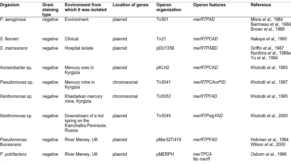
1.4.6 Diversity and organization of the mer operons 18
1.5 Lead resistance 26
1.6 Cadmium, zinc and cobalt resistance 29
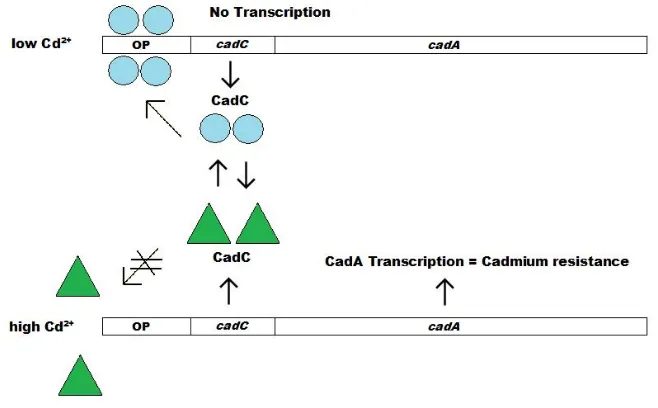
1.6.1 The cad operon 30
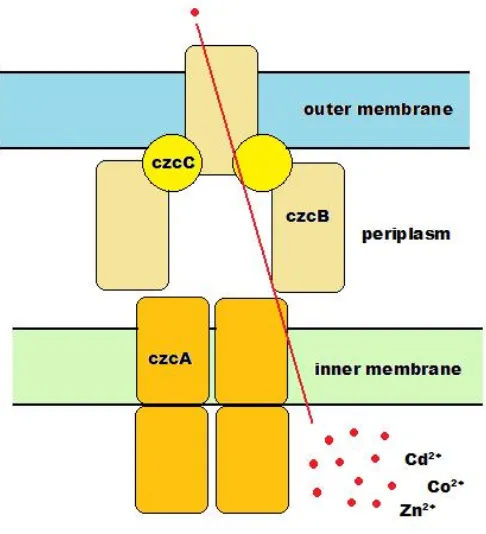
1.6.2 The czc operon 32
1.7 PAH degradation 35
1.7.1. The nah operon 35
1.7.1.1 Regulation of the nah operon 36
1.7.1.2 The upper nah operon (nah) 37
1.7.1.3 The lower nah operon (sal) 38
1.7.2 Diversity of genes relating to PAH degradation 39
1.8 The use of bacterial genes in environmental biotechnology 45
1.8.1 Bioremediation 45
XI
1.8.3 Biosorption 47
1.8.4 Biofilms 50
1.8.4.1 Biofilm structure, development and mechanisms of resistance
50
1.8.4.2 Environmental biofilms and heavy metals 52
1.9 Studies previously undertaken at Victoria University related to this project
52
1.9.1 Work based on heavy metal resistance 54
1.9.2 Work based on PAH degradation 55
1.9.3 Basis for investigating both heavy metal resistant and PAH degrading organisms
55
1.10 Aims of this project 56
1.10.1 Overall aim 56
1.10.2 Specific aims 56
1.11 Significance of this project 57
CHAPTER 2 MATERIALS AND METHODS 59
MATERIALS 61
2.1 Kits, reagents, enzymes and chemicals 61
2.1.1 Commercial kits and other reagents 61
2.1.2 Enzymes 61
2.1.3 Buffers and general stock solutions 61
2.1.4 Microbiological media and components 61
2.1.5 Sterilisation of microbiological media, reagents, glassware, consumables and antibiotic stocks
62
2.1.6 Disposal methods 62
BACTERIA 62
2.2 Bacteria used in this investigation 62
MICROBIOLOGICAL METHODS 64
2.3 Growth, storage and the investigation of the properties of AO22, E9 and VUN 10010 microorganisms in response to heavy metals and PAHs
64
2.3.1 Revival and growth of bacterial stocks 64
2.3.2 Gram-staining 64
2.3.3 Minimum inhibitory concentration assays (MICs) 65
2.3.4 Growth curve analysis 65
2.3.5 Growth curve analysis in the presence of mercuric chloride
66
2.3.6 Determination of viable cell counts by plating 67
2.3.7 Biofilm assays 67
2.3.8 Removal of Gram-negative bacteria from the Consortium VUN 10010
68
MOLECULAR TECHNIQUES 69
DNA TECHNIQUES 69
2.4 Isolation of genomic DNA from bacterial cells 69 2.4.1 Large scale isolation of genomic DNA from
Gram-negative bacteria
69
2.4.2 Large scale isolation of genomic DNA from Gram-positive bacteria
69
XII Gram-positive bacteria using the Wizard Genomic DNA Purification Kit (Promega)
2.5 Extraction of plasmid DNA from Gram-negative bacterial cells
71
2.6 Quantitation of DNA samples 71
2.7 Visualisation of DNA samples by agarose gel electrophoresis
71
2.8 PCR analysis 72
2.8.1 Criteria for design of oligonucleotide primers 72 2.8.1.1 Primer design for the amplification of mercury
resistance genes
73
2.8.1.2 Primer design for the amplification of lead resistance genes
73
2.8.1.3 Primer design for the amplification of cadmium resistance genes
73
2.8.1.4 Primer design for the amplification of genes relating to PAH degradation
74
2.8.2 PCR conditions 74
2.9 Purification of PCR products 77
2.9.1 Microspin Columns 77
2.9.2 ConcertTM Gel Extraction System 77
2.10 Cloning of PCR products 84
2.10.1 Ligation of purified PCR products into cloning vectors 85
2.10.2 Transformation of ligation reactions 85
2.10.3 Preparation of recombinant plasmids 85
2.10.4 Determination of the sizes of inserts by restriction digestion of clones of recombinant plasmids
86
2.11 Sequencing of DNA 87
2.11.1 Sequence data analysis 88
RNA TECHNIQUES 88
2.12 Isolation of RNA from bacterial cells 89
2.12.1 Determination of fixed cell numbers for RNA extractions
89
2.12.2 Isolation of RNA from Gram-negative bacteria 89
2.13 Quantitation of RNA samples 90
2.14 Reverse Transcription-PCR analysis 90
2.14.1 2.14.2
Removal of residual DNA from RNA samples cDNA synthesis
90 90
2.14.3 Reverse Transcription-PCR 91
CHAPTER 3 ESTABLISHING THE HEAVY METAL
RESISTANCE OF ACHROMOBACTER SP. AO22, A. WOLUWENSIS E9 AND CONSORTIUM VUN 10010
92
3.1 Introduction 93
3.2 Determination of heavy metal MICs by the spot plate method
94
3.3 Comparison of the spot plate and spread plate methods for MIC analysis
96
XIII
3.4.1 Gram-staining 97
3.4.2 Spray plates 99
3.4.3 Use of Lincomycin 99
3.4.4 Mycobacterium stab cultures: further attempts at the separation of the two organisms
100
3.5 MIC analysis of AO22, E9 and Consortium VUN 10010 using liquid broth
100
3.6 Determination of heavy metal MICs by the microtitre plate method
101
3.7 Organomercurial MICs 103
3.8 Growth curve analysis 104
3.9 16S sequencing 105
3.10 Discussion 108
CHAPTER 4 Molecular basis, growth and expression patterns of the mercury (mer) resistance operons of Achromobacter sp. AO22, A. woluwensis E9 and Consortium VUN 10010
114
4.1 Introduction 115
4.2 PCR analysis of the presence of mercury resistance genes based on the Tn501, Tn21 and pDU1358 operons
115
4.2.1 Amplification of the mer genes of Tn501 116 4.2.2 Amplification of the mer genes from the genomic DNA
of Achromobacter sp. AO22, A. woluwensis E9 and Consortium VUN 10010
116
4.2.3 Further amplification of the 3’ end of the merA gene and the merD gene from the genomic DNA of
Achromobacter sp. AO22, A. woluwensis E9 and Consortium VUN 10010
122
4.3 PCR analysis for detecting the presence of merC and merB genes
125
4.3.1 Amplification of merC from the genomic DNA of
Achromobacter sp. AO22, A. woluwensis E9 and Consortium VUN 10010
125
4.3.2 Amplification of merB from the genomic DNA of
Achromobacter sp. AO22, A. woluwensis E9 and Consortium VUN 10010
127
4.4 Sequence analysis of the genes associated with mer operons 128
4.4.1 merR and O/P sequence analysis 128
4.4.2 merT sequence analysis 129
4.2.3 merP sequence analysis 132
4.4.4 merA sequence analysis 133
4.4.5 merB sequence analysis 134
4.5 Mercuric chloride induction and growth curve analysis of Achromobacter sp. AO22
139
4.6 Growth profile of A. woluwensis E9 in the presence or absence of mercuric chloride
141
4.7 Expression of mer genes in Achromobacter sp. AO22 during HgCl2 stress
145
XIV
CHAPTER 5 THE FORMATION OF BIOFILMS BY
ACHROMOBACTER SP. AO22, A. WOLUWENSIS E9 AND CONSORTIUM VUN 10010
156
5.1 Introduction 157
5.2 Biofilm formation in the absence of heavy metal contaminants
158
5.3 Biofilm formation in the presence of mercuric chloride 159
5.4 Biofilm formation in the presence of lead nitrate 159
5.5 Biofilm formation in the presence of zinc nitrate 161
5.6 Biofilm formation in the presence of cadmium chloride 162
5.7 Biofilm formation in the presence of cobalt nitrate 163
5.8 Discussion 164
CHAPTER 6 PRELIMINARY INVESTIGATION INTO THE
PRESENCE OF GENES RELATING TO LEAD AND CADMIUM RESISTANCE IN ACHROMOBACTER SP. AO22, A. WOLUWENSIS E9 AND CONSORTIUM VUN 10010 AND GENES RELATING TO PAH DEGRADATION IN CONSORTIUM VUN 10010
169
6.1 Introduction 170
6.2 PCR detection of lead resistance genes in Achromobacter sp. AO22, A. woluwensis E9 and Consortium VUN 10010
171
6.3 Sequence analysis of a fragment obtained using pbr specific primers
179
6.4 PCR detection of cadmium resistance genes in
Achromobacter sp. AO22, A. woluwensis E9 and
Consortium VUN 10010
180
6.5 PCR detection of PAH degradation-encoding genes in Consortium VUN 10010
180
6.6 Sequence analysis of fragments obtained using pah specific primers
181
6.7 Discussion 182
CHAPTER 7 GENERAL CONCLUSIONS AND FUTURE
DIRECTIONS
186
7.1 Introduction 187
7.2 General conclusions 188
7.2.1 Further characterisation of Alcaligenes sp. AO22 and
Arthrobacter sp. E9 to the species level
188
7.2.2 Determination of Stenotrophomonas maltophilia VUN 10010 being a mixed culture
188
7.2.3 Confirmation of the heavy metal resistances of
Achromobacter sp. AO22 and A. woluwensis E9
188
7.2.4 Characterisation of the heavy metal resistances of VUN 10010
189
7.2.5 Determination of organomercurial resistance in
Achromobacter sp. AO22, A. woluwensis E9 and Consortium VUN 10010
XV 7.2.6 Determination of the formation of biofilms in the
presence of heavy metals by Achromobacter sp. AO22,
A. woluwensis E9 and Consortium VUN 10010
189
7.2.7 Determination of the presence of the mer operon in
Achromobacter sp. AO22, A. woluwensis E9 and Consortium VUN 10010
190
7.2.8 Growth profile of Achromobacter sp. AO22 in the presence of mercuric chloride
192
7.2.9 Growth profile of A. woluwensis E9 in the presence of mercuric chloride
192
7.2.10 mer gene expression in Achromobacter sp. AO22 192 7.2.11 Determination of the presence of the pbr operon in
Achromobacter sp. AO22, A. woluwensis E9 and Consortium VUN 10010
193
7.2.12 Determination of the presence of the cad operon in
Achromobacter sp. AO22, A. woluwensis E9 and Consortium VUN 10010
194
7.2.13 Determination of the presence of genes relating to PAH degradation in Consortium VUN 10010
194
7.3 Overall conclusion 194
7.4 Future directions 195
7.4.1 Further investigations of these isolates on a physiological level
196
7.4.2
7.2.3
Further investigations of these isolates on a molecular level
Further investigations of these isolates using practical applications
196
197
REFERENCES 198
APPENDIX 1 COMMERCIAL REAGENTS 221
APPENDIX 2 CHEMICALS AND REAGENTS 223
APPENDIX 3 MICROBIOLOGICAL MEDIA AND REAGENTS 228
APPENDIX 4 pGEM®-T EASY VECTOR 231
APPENDIX 5 MINIMUM INHIBITORY CONCENTRATION
ASSAY DATA
232
APPENDIX 6 RAW GROWTH CURVE DATA 234
APPENDIX 7 16S SEQUENCE COMPARISONS 235
APPENDIX 8 OD600 READINGS AND CELL COUNTS FROM MERCURIC CHLORIDE INDUCTION
EXPERIMENTS
239
APPENDIX 9 NEGATIVE CONTROL RT-PCR 241
XVI
LIST OF FIGURES
Figure 1.1 Examples of the chemical structures of some heavy metal compounds
5
Figure 1.2 Examples of the chemical structures of some PAHs 7 Figure 1.3 Diagrammatic representation of the protein products of the mer
operons
10
Figure 1.4 The two divergent promoters of the Tn501mer operon 12 Figure 1.5 Model for pbr Pb(II) resistance operon-encoded lead resistance
of C. metallidurans CH34
29
Figure 1.6 Mechanisms of transcription and expression of the cadCA
operon
32
Figure 1.7 Model for the function of the Czc efflux complex 34 Figure 1.8 The naphthalene degradation pathway in P. putida G7 40
Figure 2.1 Set up of biofilm assays in a microtitre plate 68 Figure 2.2 Location of primers designed to amplify the merRTPADEorf-2
genes
75
Figure 2.3 Location of primers designed to amplify the merC gene 76 Figure 2.4 Location of primers designed to amplify the merB gene 76 Figure 2.5 Location of primers designed to amplify the genes of the pbr
operon
80
Figure 2.6 Location of primers designed to amplify the cad operon 80 Figure 2.7 Location of primers designed to amplify the nahAa and nagAa
genes
81
Figure 2.8 Location of primers designed to amplify the nahAc, nagAc,
ndoAc and pahAc genes
81
Figure 2.9 Location of primers designed to amplify the nahAd, nagAd and
pahA4 genes
82
Figure 2.10 Location of primers designed to amplify the nahC, nagC
and pahC genes
82
Figure 2.11 Location of primers designed to amplify the nahE, nagE
and pahE genes
83
Figure 2.12 Location of primers designed to amplify the nahF, nagF and
pahF genes
83
Figure 3.1 Comparison of MIC results obtained in the current study with those obtained by Trajanovska et al., (1997)
95
Figure 3.2 Comparison of MIC results obtained using the spot plate and spread plate methods
98
Figure 3.3 Heavy metal MICs of VUN 10010, AO22 and E9 using microtitre plates
102
Figure 3.4 Growth curve analysis of AO22, E9 and VUN 10010 106 Figure 3.5 Comparison of the 16S RNA gene sequence of AO22 with the
16S RNA gene sequence of A. xylosoxidans (Accession #AJ50912)
109
Figure 3.6 Comparison of the 16S RNA gene sequence of E9 with the 16S RNA gene sequence of A. woluwensis (Accession# AY112986)
XVII Figure 4.1 PCR amplification of mer gene sections from the positive
control plasmid pACYC 184::Tn501
117
Figure 4.2 PCR amplification of mer genes from the genomic DNA of
Achromobacter sp. AO22
118
Figure 4.3 PCR amplification of mer genes from the genomic DNA of A. woluwensis E9
118
Figure 4.4 PCR amplification of the mer genes from the genomic DNA of Consortium VUN 10010
119
Figure 4.5 PCR amplification of Tn501 and genomic DNA of A. woluwensis E9 using additional mer primers
120
Figure 4.6 PCR amplification of Tn501 and the genomic DNA of Consortium VUN 10010 using additional mer primers
121
Figure 4.7 PCR amplification of Tn501 and the genomic DNA of
Achromobacter sp. AO22, A. woluwensis E9 and Consortium VUN 10010 using additional merA and merD primers
123
Figure 4.8 PCR amplification of Tn501 and the genomic DNA of
Achromobacter sp. AO22, A. woluwensis E9 and Consortium VUN 10010 using additional merA and merD primers and increased MgCl2 concentration
124
Figure 4.9 PCR amplification of merC using mer27-28 with genomic DNA of Achromobacter sp. AO22, A. woluwensis E9 and VUN 10010
126
Figure 4.10 PCR amplification of merB from genomic DNA of
Achromobacter sp.AO22, A. woluwensis E9 and VUN 10010 using internal primers
127
Figure 4.11 DNA sequence alignment of merR and OP regions from Tn501
(Z00027) with sequences of amplified fragments from
Achromobacter sp. AO22, A. woluwensis E9 and VUN 10010
130
Figure 4.12 Alignment of the MerR protein from Tn501 (Z00027) with the predicted MerR protein sequences from Achromobacter sp. AO22, A. woluwensis E9 and VUN 10010
130
Figure 4.13 DNA sequence alignment of merT from Tn501 (Z00027) with sequences obtained from the amplified fragments from
Achromobacter sp.AO22, A. woluwensis E9 and VUN 10010
131
Figure 4.14 Alignment of the MerT protein from Tn501 (Z00027) with the predicted MerT protein sequences from Achromobacter sp. AO22, A. woluwensis E9 and VUN 10010
131
Figure 4.15 DNA sequence alignment of merP from Tn501 (Z00027) with sequences of amplified fragments from Achromobacter sp. AO22, A. woluwensis E9 and VUN 10010
132
Figure 4.16 Alignment of the MerP protein from Tn501 (Z00027) with the predicted MerP protein sequences from Achromobacter sp. AO22, A. woluwensis E9 and VUN 10010
133
Figure 4.17 DNA sequence alignment of merA from Tn501 (Z00027) with sequences obtained from amplified fragments from
Achromobacter sp.AO22, A. woluwensis E9 and VUN 10010
135
Figure 4.18 Alignment of the MerA protein from Tn501 (Z00027) with the predicted MerA protein sequences from Achromobacter sp. AO22, A. woluwensis E9 and VUN 10010
XVIII Figure 4.19 DNA sequence alignment of merB from pDU1358(PDUMER)
with sequences obtained from amplified fragments from
Achromobacter sp.AO22, A. woluwensis E9 and VUN 10010
138
Figure 4.20 Alignment of the MerB protein from pDU1358 (PDUMER) with the predicted MerA protein sequences from
Achromobacter sp.AO22, A. woluwensis E9 and VUN 10010
138
Figure 4.21 OD600 readings of Achromobacter sp. AO22 cultures in the presence or absence of 0.075 mM HgCl2
140
Figure 4.22 Viable cell population of Achromobacter sp. AO22 cultures in the presence and absence of 0.075 mM HgCl2
142
Figure 4.23 Biomass profiles of A. woluwensis E9 cultures in the presence of 0.01 - 0.05 mM HgCl2 and in the absence of HgCl2
144
Figure 4.24 RNA prepared from Achromobacter sp.AO22 cells exposed to 0.075 mM HgCl2 and from control AO22 cells
146
Figure 4.25 RT-PCR amplification of Achromobacter sp. AO22 cDNA using the merR1-16 primer pair
147
Figure 4.26 RT-PCR amplification of Achromobacter sp. AO22 cDNA using the merT1-T2 (A) and merP1-P2 (B) primer pairs
148
Figure 4.27 RT-PCR amplification of Achromobacter sp. AO22 cDNA using the mer19-32 primer pair
149
Figure 5.1 Biofilm formation in the absence of heavy metals 158 Figure 5.2 Biofilm formation in the presence of mercuric chloride 160 Figure 5.3 Biofilm formation in the presence of lead nitrate 160 Figure 5.4 Biofilm formation in the presence of zinc nitrate 161 Figure 5.5 Biofilm formation in the presence of cadmium chloride 162 Figure 5.6 Biofilm formation in the presence of cobalt nitrate 163
Figure 6.1 PCR amplification of pbr genes using pbr specific primers with genomic DNA of Achromobacter sp. AO22
172
Figure 6.2 PCR amplification of pbr genes using pbr specific primers with genomic DNA of A. woluwensis
173
Figure 6.3 PCR amplification of pbr genes using pbr specific primers with genomic DNA of Consortium VUN 10010
173
Figure 6.4 PCR amplification of pbr genes using pbr specific primers and increased concentrations of MgCl2 with genomic DNA of Achromobacter sp. AO22
176
Figure 6.5 PCR amplification of pbr genes using pbr specific primers and increased concentrations of MgCl2 with genomic DNA of A. woluwensis E9
177
Figure 6.6 PCR amplification of pbr genes using pbr specific primers and increased concentrations of MgCl2 with genomic DNA of Consortium VUN 10010
178
Figure 6.7 PCR amplification of pah genes using pah specific primers with genomic DNA of Consortium VUN 10010
XIX
LIST OF TABLES
Table 1.1 Uses, contamination and toxicity of heavy metals 6
Table 1.2 Uses, contamination and toxicity of PAHs 7
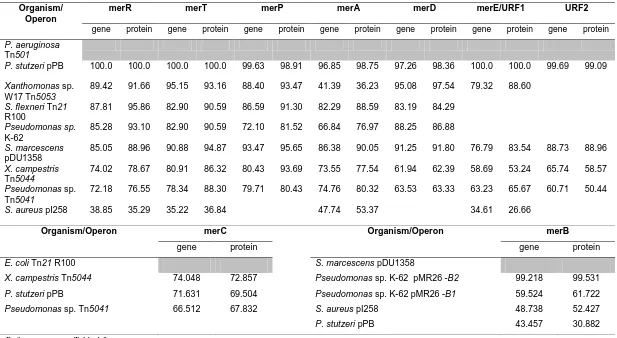
Table 1.3 Diversity and organization of the mer operons 20 Table 1.4 Homologies (%) of various mer genes and proteins 27 Table 1.5 Diversity and organization of genes relating to PAH
degradation
41
Table 1.6 Homologies (%) of the P. putida NAH7 PAH degradation genes compared to similar genes of other bacterial isolates
44
Table 1.7 Summary of biosensor constructs designed for the detection of heavy metals and PAHs
48
Table 1.8 Summary of biosorption constructs designed for the uptake of mercury using various applications
51
Table 1.9 Examples of the use of biofilms to reduce levels of heavy metals in contaminated samples
53
Table 2.1 Bacteria used in this investigation 63
Table 2.2 PCR primes used in this investigation 78
Table 3.1 Gram-stain results of various cultures and glycerol stocks of VUN 10010
99
Table 3.2 Heavy metal MICs of VUN 10010, AO22 and E9 using the liquid broth method
100
Table 3.3 Summary of heavy metal MICs of VUN 10010, AO22 and E9 using various methods
103
Table 3.4 Organomercurial MICs of AO22, E9 and VUN 10010 using the liquid broth method
104
Table 4.1 Results obtained from the PCR analysis of the positive control plasmid pACYC 184::Tn501 using the mer primers
117
Table 4.2 Results obtained from the PCR analysis of the genomic DNA from Achromobacter sp. AO22 using mer specific primers
119
Table 4.3 Results obtained using additional mer primers from Tn501 and the genomic DNA of A. woluwensis E9
120
Table 4.4 Results obtained using additional mer primers from Tn501 and the genomic DNA of Consortium VUN 10010
121
Table 4.5 Results obtained using additional merA and merD primers from Tn501 and the genomic DNA of Achromobacter sp. AO22, A. woluwensis E9 and Consortium VUN 10010
123
Table 4.6 Results obtained using additional merA and merD primers with increased concentrations of MgCl2, from Tn501 and the genomic DNA of Achromobacter sp. AO22, A. woluwensis E9 and Consortium VUN 10010
124
Table 4.7 Results obtained using merC primer pair mer27-28 from the genomic DNA of Achromobacter sp. AO22, A. woluwensis E9 and VUN 10010
XX Table 4.8 Results obtained using internal merB primer pairs to amplify
genomic DNA regions of Achromobacter sp. AO22, A. woluwensis E9 and VUN 10010
128
Table 4.9 Summary of mer genes detected in Arthrobacter sp. AO22, A. woluwensis E9 and VUN 10010
139
Table 4.10 OD600 readings from the A. woluwensis E9 cultures in the presence or absence of 0.03 mM HgCl2
143
Table 6.1 Results obtained from the PCR analysis of the genomic DNA from Achromobacter sp. AO22, A.woluwensis E9 and Consortium VUN 10010 using pbr specific primers
1
Chapter 1
General Introduction and Literature Review
Background 3
1.1 Chemistry, uses and toxicity of heavy metals 3
1.2 Chemistry, uses and toxicity of Polycyclic Aromatic Hydrocarbons
5
1.3 Microbial resistance mechanisms to environmental contaminants
8
1.4 Mercury resistance 9
1.4.1 Regulation of the mer operon 9
1.4.1.1 MerR 9
1.4.1.2 The OP region of the mer operon 11
1.4.1.3 MerD 12
1.4.2 Transport of the Hg(II) ions 12
1.4.2.1 MerP 12
1.4.2.2 MerT 13
1.4.3 Mercuric reductase (MerA) 14
1.4.4 Additional mercury transport genes 15
1.4.4.1 MerC 15
1.4.4.2 MerE 16
1.4.4.3 MerF 16
1.4.5 Organomercurial resistance 17
1.4.5.1 MerB 17
1.4.5.2 MerG 18
1.4.6 Diversity and organization of the mer operons 18
1.5 Lead resistance 26
1.6 Cadmium, zinc and cobalt resistance 29
1.6.1 The cad operon 30
1.6.2 The czc operon 32
1.7 PAH degradation 35
1.7.1. The nah operon 35
1.7.1.1 Regulation of the nah operon 36
1.7.1.2 The upper nah operon (nah) 37
1.7.1.3 The lower nah operon (sal) 38
1.7.2 Diversity of genes relating to PAH degradation 39
1.8 The use of bacterial genes in environmental biotechnology 45
1.8.1 Bioremediation 45
1.8.2 Biosensors 45
1.8.3 Biosorption 47
1.8.4 Biofilms 50
1.8.4.1 Biofilm structure, development and mechanisms of resistance
50
1.8.4.2 Environmental biofilms and heavy metals 52
1.9 Studies previously undertaken at Victoria University related to this project
52
1.9.1 Work based on heavy metal resistance 54
2 1.9.3 Basis for investigating both heavy metal resistant and
PAH degrading organisms
55
1.10 Aims of this project 56
1.10.1 Overall aim 56
1.10.2 Specific aims 56
3
BACKGROUND
Bacteria had previously been isolated from two sites, one contaminated with heavy
metals and the other site Polycyclic Aromatic Hydrocarbons (PAHs), and depending on
their origin was determined to grow in the presence of heavy metals and/or degrade
PAHs. This project focused on the genetic basis of these characteristics, searching for
the presence of genes relating to heavy metal resistances and PAH degradation. Such
information on the structure and function of these genes will facilitate decisions on the
suitability of these indigenous isolates for their use in a number of applications such as
the development of biosensors and biosorption systems and their use as biofilms. This
literature review will provide an overview on the chemistry, uses and toxicity of heavy
metals and PAHs and will discuss a number of genetic systems that encode resistances
to and/or degradation of these contaminants. Such systems will also be discussed with
respect to their role in the construction of biosensor and biosorption systems and how
bacteria capable of forming biofilms demonstrate higher resistances than planktonic
forms in the presence of these contaminants. The bacteria used in this project were
isolated by previous researchers. Background information on their studies will also be
provided in this chapter.
1.1 Chemistry, uses and toxicity of heavy metals
The term ‘heavy metals’ refers to metallic elements that have high atomic weights
(>100) and a relative density >5. Some heavy metals, such as cobalt, copper and zinc,
are essential micronutrients for biological systems, although they may be toxic in larger
amounts. Other metals, such as mercury, cadmium and lead, are biologically
non-essential and toxic in any quantity (Gadd, 1992). According to the Agency for Toxic
Substances and Disease Registry (ATSDR(a)), arsenic, lead and mercury comprise the
top 3 in the list of top 20 hazardous substances, while cadmium is ranked 7th. Major
sources of heavy metal contamination in the environment are the combustion of fossil
fuels, the operation of smelters and other industrial activities such as mineral mining
and processing, brewery and distillery wastes and the generation and use of agricultural
4 i. unreactive elemental mercury, which is a shiny, silver-white odorless liquid.
ii. inorganic mercurial salts and minerals (i.e. mercuric chloride, mercuric iodide,
mercuric oxide, mercuric sulphide, mercurous chloride).
iii. organic mercurials (i.e. methylmercury chloride, phenylmercury acetate, ethyl
mercury and merbromin) (Graeme and Pollack Jr, 1998).
Lead is a bluish-gray metal found in small amounts in the earth’s crust, although most
of the lead present in the environment is due to human activities (ATSDR(b)).
Cadmium is a natural element in the earth’s crust and is usually found as a mineral,
combined with other elements such as oxygen (cadmium oxide) or sulphur (cadmium
sulphate and cadmium sulphide) (ATSDR(c)).
Zinc, a bluish-white shiny metal, is one of the most common elements in the earth’s
crust and is found in air, soil, and water and is present in all foods (ATSDR(d)). It is an
essential co-factor for enzymes that control cell processes such as DNA synthesis,
growth, brain development, behavioral responses, reproduction, fetal development,
membrane stability, bone formation and wound healing. Zinc deficiencies may result in
growth retardation, anorexia, delayed sexual maturation, iron-deficiency anemia and
alterations in taste (Barceloux, 1999a). Cobalt is a naturally occurring element that may
be found in rocks, soil and water (ATSDR(e)). It is a relatively rare metal of gray
colour and is ductile, brittle, and magnetic. Cobalt is a necessary element in the
formation of vitamin B12 (hydroxocobalamin), which catalyses reactions such as the
synthesis of methionine, the metabolism of purines and folates and the formation of
methylmalonic acid in succinic acid (Barceloux, 1999b).
Heavy metals and their compounds are used widely in a number of industrial
applications, which often leads to environmental contamination, including of the air,
water and soil. Such contaminations may result in human exposure, which can often
lead to detrimental health problems. Figure 1.1 shows examples of heavy metal
structures, while the uses, sources of contamination and the target organs of the above
5
Figure 1.1 Examples of the chemical structures of some heavy metal
compounds
1.2 Chemistry, uses and toxicity of Polycyclic Aromatic Hydrocarbons (PAHs)
Polycyclic aromatic hydrocarbons (PAHs) comprise two or more fused benzene rings
(Figure 1.2), which may be in linear, angular or cluster arrangements. Generally PAHs
consist only of carbon and hydrogen atoms, although nitrogen, sulphur and oxygen
atoms may be substituted into the benzene rings to form heterocyclic aromatic
hydrocarbons. The stability of PAHs depends on the arrangement of the benzene rings,
angular PAHs such as pyrene, benzo[a]pyrene and coronene are the most stable, while
linear PAHs such as naphthalene and anthracene are the least stable. PAHs are
hydrophobic compounds, which makes them more persistent in the environment with
PAH solubility decreasing as the number of fused benzene rings increases (Cerniglia,
6
Table 1.1 Uses, contamination and toxicity of heavy metals Heavy
metal
Uses Contaminating sources Target organs
Mercury Production of chlorine gas and caustic soda. Used in dental fillings, batteries, skin lightening creams and antiseptic creams.
Contaminated fish and shellfish. Inhalation of vapors from spills, incinerators and the burning of mercury-containing fuels.
Release of mercury from dental work and medical treatments.
The practice of rituals that involve mercury.
Nervous system, brain, kidneys, developing fetus, lungs, GI tract, heart, skin and eyes.
Lead Production of batteries, ammunition, solder and pipes and X-ray shields.
Contaminated food and water. Exposure to lead-based paints. Working in industries that use lead. Use of health care products and folk remedies. Nervous system, kidneys, reproductive, system and blood.
Zinc Production of paint, rubber, wood
preservatives, ointments and alloys such as brass and bronze.
Used in coatings to prevent rust.
Breathing in zinc particles in the air of manufacturing sites.
Contaminated water near manufacturing or waste sites. Contaminated water or beverage that has been stored in containers or flows through pipes that have been coated with zinc to prevent rust.
Stomach, blood, pancreas and lungs.
Cadmium Production of batteries, pigments, metal coatings and plastics.
Breathing contaminated workplace air, and air near the burning of fossil fuels or municipal waste.
Contaminated water and foods (i.e. shellfish, liver, kidney).
Lungs, stomach and kidneys.
Cobalt Production of alloys. Used in aircraft engines, magnets grinding/cutting tools, artificial joints, medical sterilisation and research.
Working in industries that use cutting and grinding tools and those involving mining, smelting, refining or processing of cobalt or ores. Working at nuclear, irradiation or nuclear waste facilities.
Lungs, heart, skin and stomach.
7 They are formed naturally during thermal geologic reactions associated with fossil fuels
and mineral production, during the burning of vegetation in forest and bush fires and by
some bacterial and plant reactions (Cerniglia, 1992). The uses, sources of
contamination and the target organs of PAHs are summarised in Table 1.2.
Naphthalene Fluoranthene Phenanthrene
Pyrene Benzo[a]pyrene Coronene
Figure 1.2 Examples of the chemical structures of some PAHs
Table 1.2 Uses, contamination and toxicity of PAHs
Uses Contaminating sources Target organs
Production of coal tar, crude oil, creosate and roofing tar.
Used in dyes, plastics and pesticides.
Breathing air, containing PAHs in the workplace of coking, coal-tar and asphalt production plants, smokehouses and municipal waste facilities.
Breathing air, containing PAHs from cigarette smoke, wood smoke, vehicle exhausts.
Eating contaminated food and charred meats.
Drinking contaminated water or cow’s milk.
Carcinogenic -lungs -stomach Skin
In animals -skin
-immune system -reproductive system
8
1.3 Microbial resistance mechanisms to environmental contaminants
Bacterial cells resistant to the above environmental contaminants have previously been
isolated (Ji and Silver, 1995; Nies, 1999; Bruins et al., 2000). Analysis of their genetic
and physiological systems has revealed that they may possess one or more of the four
main types of mechanisms to protect against such contaminants. The four main types of
mechanisms are as follows:
Efflux systems: These types of mechanisms export toxic metal ions to the outside of the cell (Bruins et al., 2000). These may be non-ATPase or ATPase-linked and are
generally highly specific for a particular heavy metal ion (Nies and Silver, 1995).
P-type ATPases are common in heavy metal resistance mechanisms. These may be
described as a family of membrane proteins that perform active ion transport across
biological membranes (Apell, 2003).
Accumulation and complexation: These types of mechanisms serve to prevent the exposure of essential cellular components to the contaminant and may be a result of the
presence of metallothioneins or cysteine-rich proteins (Bruins et al., 2000).
Reduction: The contaminating ions are enzymatically reduced upon entry to the cell and the less toxic, reduced ions are exported from the cell into the environment (Nies,
1999).
Alteration of cellular components: Some microorganisms may adapt to the presence of heavy metals by altering the sensitivity of cellular components, which may be
achieved through mutations that decrease the sensitivity but do not alter basic function
or by increasing the production of the sensitive cellular component (Rouch et al., 1995).
A number of genes, located on bacterial plasmids and chromosomes, have been
identified that encode specific resistance to a number of heavy metal ions, including
Ag+, AsO2-, AsO43-, Cd(II), Co(II), CrO42-, Cu(II), Hg(II), Ni(II), Sb(III), TeO32- and
Zn2+ (Ji and Silver, 1995). The following sections will outline bacterial genetic systems
for resistances to mercury (mer), lead (pbr) and cadmium, zinc and cobalt (czc, cad)and
9
1.4 Mercury resistance
One of the most widely studied bacterial heavy metal resistance operons is the mer
operon, encoding mercury resistance, in a number of positive bacteria and
Gram-negative bacteria. Two of the most widely studied mer operons are Tn501 and Tn21.
The Tn501mer operon was originally isolated from plasmid pVS1 from a Pseudomonas
aeuruginosa strain isolated in Australia (Misra et al., 1984; Brown et al.,1986;
Barrineau et al., 1984). This archetypal mer operon contains five genes, merR, merD,
merT, merP and merA. Tn21 also carries an archetypal mer operon, with an additional
transport gene (merC) and was originally isolated on plasmid NR1 from Shigella
flexneri in Japan (Nakaya et al., 1960). Plasmid pDU1358 of Serratia marcescens
differs from the above two operons in that it carries an additional lyase gene (merB)
(Griffin et al., 1987). Figure 1.3 provides an overview of the functions of the various
genes/proteins of the mer operons, which will be discussed in the following sections.
1.4.1 Regulation of the mer operon
1.4.1.1 MerR
Extensive work has been carried out on the regulation of the mer operon by the merR
gene, which encodes a negative regulator of the remainder of the mer operon. In
Gram-negative bacteria, the merR gene is generally transcribed separately and in the opposite
direction compared to the other genes of the operon (Brown et al., 1986; Griffin et al.,
1987; Inoue, 1991; Kiyono, 1997; Schelert et al., 2004). An exception to this is the
marine bacterium Pseudoalteromonas haloplanktis, where the merR gene is
co-transcribed with the merTPCAD genes (Iohara et al., 2001). In the Gram-negative
bacteria Shewanella putrefacians plasmid pMERPH, the mer operon lacks merR and
merD genes (Osborn et al., 1996). In Gram-positive bacteria mer operons, the merR
genes are generally transcribed in the same direction as the rest of the operon (Laddaga
et al., 1987; Wang et al., 1987; Ravel et al., 1998; Huang et al., 1999a).
In the absence of Hg(II), the MerR protein binds as a homodimer to the promoter, which
is a region of dyad symmetry, located just upstream of the merT gene (Ross et al., 1989;
10 it binds with high specificity to MerR to provoke an allosteric change in the protein,
which is attached to the DNA of this operator region, leading to an unwinding of the
operator DNA (Heltzel et al., 1990; Ansari et al., 1992; Ansari et al., 1995; Parkhill et
al., 1998; Caguiut et al., 1999; Song et al., 2004). This leads to improved access of
RNA polymerase, which is bound simultaneously along with MerR to the promoter, to
the transcriptional start site (Lee et al., 1993; Livrelli et al., 1993). MerR can be cross
linked to the α, β and σ70 subunits of RNA polymerase, whether in the absence of the
DNA or when both MerR and Hg(II) are bound to the operator DNA (Kulkarni and
Summers, 1999).
Figure 1.3 Diagrammatic representation of the protein products of the mer operons.
The MerR proteins of Tn501 and Tn21 are 144 amino acids long and differ in nine
residues, three of which are conservative substitutions. Three cysteine residues are
conserved in all MerR proteins, which have been confirmed to be the site for Hg(II)
binding. In the Tn21 MerR, mutation of the three cysteines (Cys82, Cys117 and
Cys126) caused a loss of Hg(II)-inducible activation (Ross et al., 1989). The binding
site of MerR to Hg(II) lies at the interface of the homodimer and involves Cys82 from
one monomer and Cys117 and Cys126 from the other (Helmann et al., 1990; Caguiut et
al., 1999). In vitro metal binding studies have shown that MerR binds only one atom of
11 single Hg(II) ion to one site causes an allosteric change that renders the other site less
able to bind Hg(II) in competition with other thiols (O’Halloran and Walsh, 1987;
Shewchuk et al., 1989; Helmann et al., 1990; Zeng et al., 1998; Caguiut et al., 1999).
Mutants which affected the DNA binding (Glu22Lys and Arg25His) helped define the
DNA binding region (Parkhill et al., 1998), and indicated that the N-terminal
helix-turn-helix motif, rather than a similar motif more centrally in the protein, was responsible for
DNA binding (Ross et al., 1989). It has been shown that only residues 80-128 were
required for stable dimer formation and retained a high affinity for Hg(II) (Zeng et al.,
1998).
1.4.1.2 The OP region of the mer operon
The mer operator/promoter (OP), in the case of the mer operons of most Gram-negative
bacteria, is a 19 bp hyphenated sequence with 7 bp palindromes flanking a 4 bp AT-rich
center (Barrineau et al., 1984; Brown et al., 1986; Parkhill and Brown, 1990; Park et
al., 1992). An unusual feature of the mer OP region is that it lies within the spacer
region between the –10 and –35 regions of the PT promoter, slightly overlapping the –35
hexamer. The PT promoter has consensus –10 and –35 hexamers, but is unusual in that
it is 19 bp in length rather than the typical length of 17 bp found in most σ70 promoters
in bacteria (Figure 1.4). MerR binding in the absence of Hg(II)in this region does not
prelude, but rather fosters RNA polymerase occupancy of PT, albeit in a
transcriptionally inactive state until Hg(II) is present (O’Halloran et al., 1989; Frantz
and O’Halloran,1990; Heltzel et al., 1990; Kulkarni and Summers, 1999). Deletion
mutants have shown that the –35 and –10 sequences must be correctly separated by 19
bp for normal promoter activity (Lund and Brown, 1989; Parkhill and Brown, 1990).
When MerR, already bound to the OP region, binds to Hg(II), an increased reactivity of
bases occurs near the operator centre which leads to the unwinding of the operator DNA
(Ansari et al., 1992), making the –10 region available to RNA polymerase (Condee and
Summers, 1992). MerR requires a distinct operator contact for repression and
activation of PT (Park et al., 1992). Further studies show that another gene, merD, may
12
Figure 1.4 The two divergent promoters of the Tn501 mer operon. PT controls
the expression of the merTPAD genes and PR is the promoter for the regulatory merR
gene.
1.4.1.3 MerD
In Tn501, the merD gene is a small cysteine-rich open reading frame that lies just
downstream of merA (Brown et al., 1986). Its protein product (from Tn21) has been
observed to have an N-terminal region with a predicted helix-turn-helix motif similar to
that of MerR and has been shown to be translated in very small amounts (Lee et al.,
1989). Deletions of this gene, from pDU1358, have shown to have no effect on the
mercury resistant phenotype. (Nucifora et al., 1990; Mukhopadhyay et al., 1991). In
vitro, MerD from Cupriavidus metallidurans (formerly Ralstonia metallidurans)CH34
has been shown to form a ternary complex with MerOP and MerR (as described in
Section 1.4.1.2). It has been postulated that MerD displaces Hg-bound MerR from the
operator, allowing the synthesis of Hg(II)-free MerR, which switches off the induction
of mer genes in the absence of mercury (Champier et al., 2004).
1.4.2 Transport of the Hg(II) ions
1.4.2.1 MerP
The gene merP of Tn21 encodes a small periplasmic mercury binding protein, with the
72 (12 kDa) amino acid long mature MerP being processed from a 91 (13 kDa) amino
acid precursor (Jackson and Summers, 1982; Summers, 1986). This protein has been
shown to function as a monomer and binds a single Hg(II) ion via two cysteines, at
positions 14 and 17 (relating to Tn501 and Tn21) (Steele and Opella, 1997), which form
13 multiple repeats on the N-terminus of P-type ATPases, involved in influx/efflux of
transition metal cations in prokaryotes and eukaryotes (Bull and Cox, 1994). Loss of
either Cys14 or Cys17 has been shown to lead to an inability for MerP to bind Hg(II)
(Sahlman and Skarfstad, 1993). The Tn21 MerP protein has been shown to exist in an
oxidized (disulphide) or a reduced (dithiol) form, however, only in its reduced form,
with the Cys14 and Cys17 residues as free thiols, can the protein act as a receptor of
mercuric ions (Qian et al., 1998). NMR studies of MerP show that in the absence of
Hg(II), Cys14 of the reduced form is surface exposed and Cys17 is buried, however
when Hg(II) is bound, both cysteines are surface exposed (Steele and Opella, 1997). It
has been suggested that an electrostatic attraction between the buried Cys17 and Hg(II),
triggers a structural change upon Hg(II) binding (Powlowski and Sahlman, 1999).
Studies on the crystallized form of MerP from C. metallidurans CH34 propose that the
side-chain of Tyr66, which is a conserved residue in MerP proteins, and the main-chain
amide of Cys14 may play a role in the maintaining of Cys17 in an anionic form in the
reduced form of the protein. This study also suggests that Tyr66 and Phe38, also
conserved in MerP proteins, may be important in the mercury-binding reaction and
transfer of Hg(II) to MerT (Serre et al., 2004). MerP is believed to transfer Hg(II) to the
amino-terminal cysteines Cys24 and Cys25 (corresponding to Tn21 and Tn501) of
MerT (Hamlett et al., 1992; Morby et al., 1995). MerT is described below.
1.4.2.2 MerT
The gene merT encodes a product of 116 amino acids (12.4 kDa) and is an inner
(cytosolic) membrane protein strongly predicted to have three transmembrane helices,
the first of these having a cysteine pair which is thought to be accessible from the
periplasmic side (Sahlman et al., 1997; Liebert et al., 2000; Brown et al., 2002). In
Tn501, Cys24 and Cys25 in the first transmembrane domain have been found to be
essential for the transport of mercury ions through the cytoplasm (Morby et al., 1995;
Hobman and Brown, 1996). The second pair of cysteines is thought to lie on the
cytoplasmic face of the inner membrane between the second and third transmembrane
helices. Hg(II) may be transferred from the N-terminal-proximal cysteine pair to form a
di-coordinate protein complex with these cysteines, and then transferred to MerA
(Jackson and Summers, 1982; Schue et al., 2007). Mutations of Cys76Ser, Cys82Ser or
14 Mutations of Gly14Arg, Gly15Arg, Gly27Arg and Ala18Asp (also in Tn501) in the first
predicted transmembrane helix have been shown to cause a loss of mercury resistance
(Hobman and Brown, 1996).
1.4.3 Mercuric reductase (MerA)
The most widely observed mechanism of eubacterial mercury resistance is by the
reduction of the highly reactive cationic form of mercury, to volatile, relatively inert
monoatomic mercury vapor. This reduction is mediated by MerA (mercuric reductase),
encoded by the gene merA, which is a flavoprotein with a redox-active cysteine at the
active site (Fox and Walsh, 1982) which is a minimum of 1600 amino acids in length.
This cytoplasmic protein (Summers and Sugarman, 1974) is a homodimer (Fox and
Walsh, 1982) which catalyses the conversion of thiol-avid Hg(II) to volatile, uncharged
Hg(0), utilizing NADPH as a source of electrons (Furukawa and Tomomura, 1972).
When reduced by MerA, volatile Hg(0) diffuses through the cell membrane without the
need for a dedicated efflux system (Barkay et al., 2003).
MerA contains 8 cysteine residues, two of which (Cys135 and Cys140 in Tn501) are
located in the active site (Brown et al., 1983). In Tn501, the C-terminal cysteines
(Cys558 and Cys559) of one monomer lie near the redox-active cysteines of the other
monomer and could assist with Hg(II) binding at the active site (Brown et al., 1983).
Mutagenesis of two conserved pairs of cysteines in the N-terminus (Cys10 and Cys13)
and the C-terminus (Cys558 and Cys559) in Tn501 indicated no essential roles for
Cys10 and Cys13, but did identify a role in Hg(II) reduction for the latter two (Moore
and Walsh, 1989). Further, when compared to the wild-type enzyme, the Cys558Ala
and Cys559Ala mutants demonstrated 200-fold and 10-fold reductions in catalytic
activity respectively (Moore et al., 1992). It was also found in MerA from Bacillus
cereus RC607 that in the absence of the C-terminal cysteines, HgX2 substrates with
small ligands can access the redox-active cysteines, while those with large ligands could
not, indicating that the C-terminal cysteines play a crucial role in removing high-affinity
ligands before Hg(II) reaches the redox-active cysteines (Cys135 and Cys140) in the
inner active site (Engst and Miller, 1999). Approximately 77 amino acids in the
N-terminal domain of MerA are homologous to MerP (Misra et al., 1985; Schiering et al.,
15 site-directed mutagenesis of the N-terminal cysteines (Moore and Walsh, 1989) did not
have any influence on the catalytic properties in vitro.
In C. metallidurans CH34, the MerA protein contains an N-terminal sequence of 62
amino acids, referred to as MerAa, which contains a motif (Gly-Met-Thr-Cys-X-X-Cys)
homologous to part of MerP from the same organism. This MerAa N-terminal
sequence was expressed independently and two cysteine residues, found in the motif,
were found to be involved in the binding of one mercury atom, with an affinity
comparable to MerP, indicating that MerAa may play a role in mercury transport (Rossy
et al., 2004). Amino acid sequence analysis indicates that homologies within this
Gly-Met-Thr-Cys-X-X-Cys motif also exist between the N-terminal sequences of the MerA
proteins and the MerP proteins of Tn501 and Tn21 (Rossy et al., 2004). The MerAa
domain and the catalytic core of the Tn501 MerA were expressed as two separate
proteins. Results indicated that MerAa may be expressed as a soluble, monomeric
protein capable of binding Hg(II) and delivering it to the catalytic core of MerA.
However, in cells containing small molecular weight thiols (such as GSH), MerAa
appears to serve little function (Ledwidge et al., 2005).
1.4.4 Additional mercury transport genes
1.4.4.1 MerC
The merC gene of Tn21, pKLH2 of Acinetobacter sp. (Kholodii et al., 1993), Tn5041 of
Pseudomonas sp. (Kholodii et al., 1997) and pMERPH of Pseudomonas putrifaciens
(Osborn et al., 1996)for example, is located between the merP and merA genes. It
encodes the MerC protein, which ranges in size between 129 and 144 amino acids
(Peters et al., 1991; Kholodii et al., 1993; Yurieva et al., 1997; Liebert et al., 1999), has
four predicted transmembrane helices and is the largest of the mer operon-encoded
membrane proteins (Summers, 1986). Deletion analysis of the merC-encoding Tn21
operon indicated that the loss of MerP and MerT had some phenotypic effect on Hg(II)
resistance, while the lack of MerC did not change this or its Hg(II) volatilization
capabilities (Hamlett et al., 1992). Topological predictions suggest that the first
cysteine pair (Cys22 and Cys25) of the MerC of Tn21 lies just within the membrane on