Clinical and Experimental Gastroenterology
Dove
press
R E v i E w
open access to scientific and medical research
Open Access Full Text Article
Molecular and cellular pathways associated
with chromosome 1p deletions during colon
carcinogenesis
Claire M Payne
Cheray Crowley-Skillicorn
Carol Bernstein
Hana Holubec
Harris Bernstein
Department of Cell Biology and Anatomy, College of Medicine, University of Arizona Tucson, AZ, USACorrespondence: Claire M Payne Department of Cell Biology and Anatomy, College of Medicine, University of Arizona, Tucson, Arizona 85724-5044
Email [email protected]
Abstract: Chromosomal instability is a major pathway of sporadic colon carcinogenesis. Chromosome arm 1p appears to be one of the “hot spots” in the non-neoplastic mucosa that, when deleted, is associated with the initiation of carcinogenesis. Chromosome arm 1p contains genes associated with DNA repair, spindle checkpoint function, apoptosis, multiple microRNAs, the Wnt signaling pathway, tumor suppression, antioxidant activities, and defense against environmental toxins. Loss of 1p is dangerous since it would likely contribute to genomic instability leading to tumorigenesis. The 1p deletion-associated colon carcinogenesis pathways are reviewed at the molecular and cellular levels. Sporadic colon cancer is strongly linked to a high-fat/low-vegetable/low-micronutrient, Western-style diet. We also consider how selected dietary-related compounds (eg, excess hydrophobic bile acids, and low levels of folic acid, niacin, plant-derived antioxidants, and other modulatory compounds) might affect processes leading to chromosomal deletions, and to the molecular and cellular pathways specifically altered by chromosome 1p loss.
Keywords: chromosome 1p, colon carcinogenesis, molecular pathways, cellular pathways
Introduction
Chromosomal instability is a major feature of sporadic colon carcinogenesis.
1–11Eighty-five percent of colorectal cancers are aneuploid, the remaining 15% being
diploid.
5Chromosome 1p deletions in colon tumors have been reported by laboratories
from at least 15 countries around the world.
12–49Chromosome 1p deletions occur at
an early stage of colon carcinogenesis,
21,24,26–28,30,31,33,37,39,41–45and are strongly linked to
karyotypic evolution during colon cancer development.
43Many reports in the literature indicate that the macroscopically normal mucosa
proximal or distal to a colon cancer exhibit aneuploidy (loss or gain of chromosomes or
parts of chromosomes). Relevant to this review, Cianciulla et al
44reported that deletions
of chromosome 1p were simultaneously found in both the distant normal-appearing
mucosa of 76% of patients who also harbored 1p deletions in their cancer. These
findings indicate that the loss of chromosome 1p may be one of the “hot spots” among
the numerous defects in the non-neoplastic mucosa associated with the possible
initiation of colon carcinogenesis.
50–70The pioneering work of Paraskeva et al
71–75indicated the likely involvement
of chromosome 1p loss in in vitro immortalization
72,74and in the progression
of adenomas to carcinomas.
75The functional importance of loss of distal 1p in
colon tumorigenesis was demonstrated in 1993 by Tanaka et al
76who introduced
Clinical and Experimental Gastroenterology downloaded from https://www.dovepress.com/ by 118.70.13.36 on 20-Aug-2020
For personal use only.
Number of times this article has been viewed
This article was published in the following Dove Press journal: Clinical and Experimental Gastroenterology
Dovepress Payne et al
chromosomal band 1p36 into colon carcinoma cells and
found that their tumorigenicity was suppressed.
Chromosome 1p deletions can affect distinct pathways
of sporadic colon carcinogenesis, including both
chromo-somal instability and chromochromo-somal instability-negative
pathways. The underlying mechanisms associated with
the loss of chromosome 1p that may contribute to genomic
instabilty and drive colon carcinogenesis are loss of
genes associated with DNA repair, spindle checkpoint
function, apoptosis, multiple microRNAs (miRNAs), the
Wnt signaling pathway, tumor suppression, antioxidant
activities, and defense against environmental toxins.
77,78Since centromeric instability and resulting telomeric
fusions have been proposed as a mechanism for the loss
of chromosome 1p,
79the loss of genes located on
chromo-some 1p that function to ensure centromeric stability and
telomere integrity, in turn, can exacerbate chromosomal
instability throughout the genome. These 1p
deletion-associated pathways that may lead to colon carcinogenesis
will be reviewed at the molecular and cellular levels, and
dietary factors that affect these pathways (eg, excess
hydro-phobic bile acids, and low levels of folic acid, niacin,
plant-derived antioxidants, and other modulatory compounds)
will be explored. It is likely that certain dietary factors
prevent, initiate, or exacerbate genomic instability in
colon epithelial cells and thus have importance for colon
carcinogenesis.
Mechanisms of carcinogenesis
associated with the loss
of key genes on chromosome 1p
Chromosome 1, the longest human chromosome, is
gene-dense with 3141 genes.
80The genes located on chromosome 1
were identified with the assistance of the Weizmann Institute
of Science websites:
GeneLoc (www.genecards.weizmann.ac.il/geneloc/index.
shtml) and GeneCards – The Human Gene Compendium
(www.genecards.org). Genes located on the
p
arm of
chromosome 1 that are associated with protection against
oxidative stress, DNA damage, mitotic perturbations,
excessive cellular proliferation, development of apoptosis
resistance, aberrant colonic cell differentiation, and
environmental toxicity have been tabulated and the function
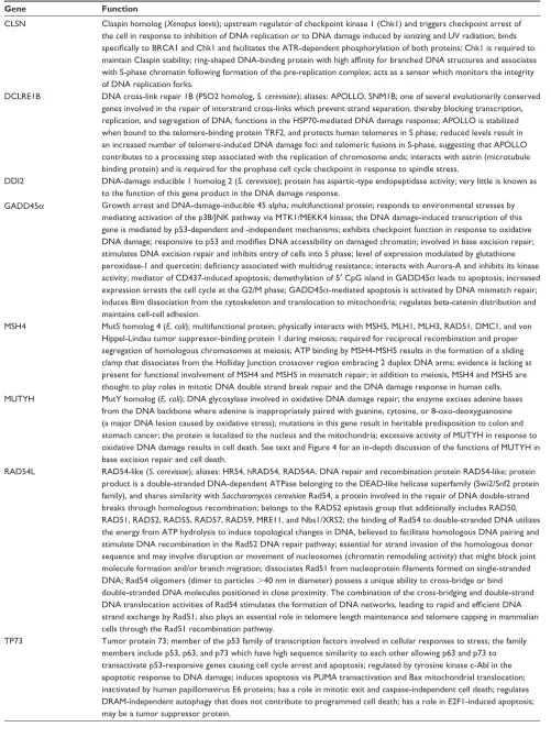
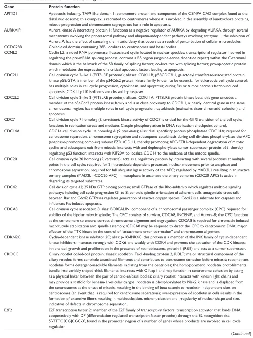
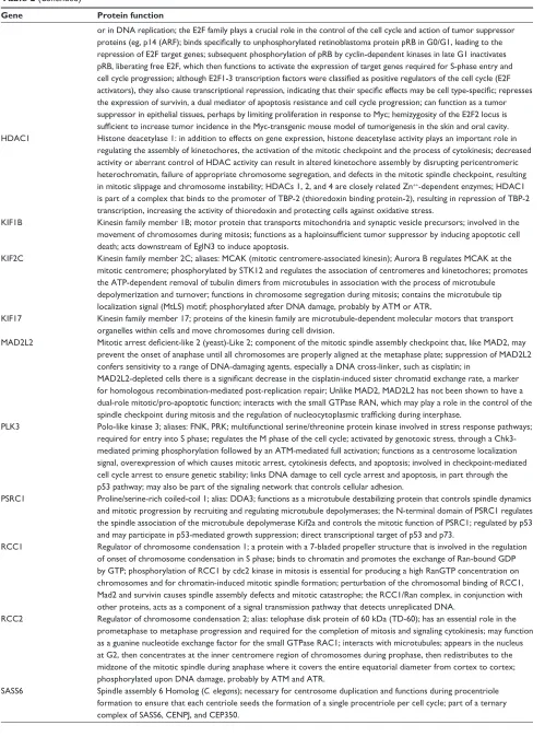
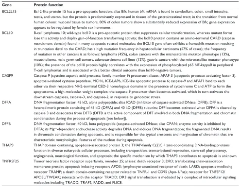
of the gene products described (Tables 1–8). Since many of
these genes have tumor suppressive capabilities, the
simulta-neous loss caused by a 1p deletion could initiate the
forma-tion of neoplastic clones and enhance tumorigenesis through
Darwinian selection.
8Mechanisms protective against genomic
instability
Cells with DNA damage, spindle damage, and dysfunctional
telomeres signal DNA damage responses.
81–84These DNA
damage responses include the activation of numerous
checkpoints that arrest the damaged cells in the G1, S, G2, or
M-phase of the cell cycle, depending upon the nature of the
damage or dysfunction and the stage of the cell cycle of the
target cell. DNA-damage checkpoints are activated following
direct damage to DNA.
85–91Spindle assembly checkpoints are
activated following damage to the mitotic machinery,
85,92–98or as a result of DNA damage during mitosis.
99Telomere
checkpoints are activated by defective telomeres.
100–106These
checkpoints prevent the damaged cell from completing
DNA replication and mitosis until all damage is repaired
(Figure 1), and thus prevent 1) mutations that could be formed
by replicating a damaged DNA template, 2) aneuploidy
that could result from chromosome mis-segregation, and
3) telomere fusions that result in anaphase bridges, broken
chromosomes, and translocations as a consequence of the
well-known breakage–fusion–bridge cycles.
107–114However, cells with excessive direct DNA damage,
115–122massive chromosome loss or chromosomal imbalances,
123prolonged activation or inhibition of the spindle checkpoint
pathways,
122–127or excessively shortened or dysfunctional
telomeres,
128–140initiate a cascade of molecular events that
ultimately leads to either caspase-dependent cell death,
141–143caspase-independent cell death,
144or a special form of
apoptosis referred to as mitotic catastrophe
145–148(Figure 2).
(Brightfield micrographs are shown in Figure 3 illustrating
the cellular alterations that accompany apoptosis [Figure 3A],
mitotic perturbation [Figure 3B], mitotic catastrophe
[Figure 3C], and micronuclei formation [associated with
aneuploidy] [Figure 3D]). The destructive and
cell-protective pathways are downstream of a common signal
transduction network that responds to DNA damage.
149The repair/survival and non-repair/cell death pathways are
probably activated simultaneously.
149The repair, checkpoint,
and cell death response to DNA damage are, however,
well co-ordinated,
150the interplay of positive and negative
regulatory loops resulting in a delayed death response to
DNA damage.
149DNA repair and the DNA damage
response (DDR) (Table 1)
The genes on chromosome 1p associated with DNA repair
or the DNA damage response (DDR) include CLSN,
DCL-RE1B (APOLLO), DDI2, GADD45
α
, MSH4, MUTYH,
Clinical and Experimental Gastroenterology downloaded from https://www.dovepress.com/ by 118.70.13.36 on 20-Aug-2020
Dovepress Chromosome 1p deletions during colon carcinogenesis
Table 1 DNA repair and DNA damage response genes
Gene Function
CLSN Claspin homolog (Xenopus laevis); upstream regulator of checkpoint kinase 1 (Chk1) and triggers checkpoint arrest of the cell in response to inhibition of DNA replication or to DNA damage induced by ionizing and Uv radiation; binds
specifically to BRCA1 and Chk1 and facilitates the ATR-dependent phosphorylation of both proteins; Chk1 is required to maintain Claspin stability; ring-shaped DNA-binding protein with high affinity for branched DNA structures and associates
with S-phase chromatin following formation of the pre-replication complex; acts as a sensor which monitors the integrity of DNA replication forks.
DCLRE1B DNA cross-link repair 1B (PSO2 homolog, S. cerevisiae); aliases: APOLLO, SNM1B; one of several evolutionarily conserved genes involved in the repair of interstrand cross-links which prevent strand separation, thereby blocking transcription, replication, and segregation of DNA; functions in the HSP70-mediated DNA damage response; APOLLO is stabilized when bound to the telomere-binding protein TRF2, and protects human telomeres in S phase; reduced levels result in an increased number of telomere-induced DNA damage foci and telomeric fusions in S-phase, suggesting that APOLLO contributes to a processing step associated with the replication of chromosome ends; interacts with astrin (microtubule
binding protein) and is required for the prophase cell cycle checkpoint in response to spindle stress.
DDi2 DNA-damage inducible 1 homolog 2 (S. cerevisiae); protein has aspartic-type endopeptidase activity; very little is known as to the function of this gene product in the DNA damage response.
GADD45α Growth arrest and DNA-damage-inducible 45 alpha; multifunctional protein; responds to environmental stresses by mediating activation of the p38/JNK pathway via MTK1/MEKK4 kinase; the DNA damage-induced transcription of this gene is mediated by p53-dependent and -independent mechanisms; exhibits checkpoint function in response to oxidative
DNA damage; responsive to p53 and modifies DNA accessibility on damaged chromatin; involved in base excision repair;
stimulates DNA excision repair and inhibits entry of cells into S phase; level of expression modulated by glutathione
peroxidase-1 and quercetin; deficiency associated with multidrug resistance; interacts with Aurora-A and inhibits its kinase
activity; mediator of CD437-induced apoptosis; demethylation of 5′ CpG island in GADD45α leads to apoptosis; increased expression arrests the cell cycle at the G2/M phase; GADD45α-mediated apoptosis is activated by DNA mismatch repair; induces Bim dissociation from the cytoskeleton and translocation to mitochondria; regulates beta-catenin distribution and maintains cell-cell adhesion.
MSH4 MutS homolog 4 (E. coli); multifunctional protein; physically interacts with MSH5, MLH1, MLH3, RAD51, DMC1, and von
Hippel-Lindau tumor suppressor-binding protein 1 during meiosis; required for reciprocal recombination and proper
segregation of homologous chromosomes at meiosis; ATP binding by MSH4-MSH5 results in the formation of a sliding clamp that dissociates from the Holliday Junction crossover region embracing 2 duplex DNA arms; evidence is lacking at present for functional involvement of MSH4 and MSH5 in mismatch repair; in addition to meiosis, MSH4 and MSH5 are thought to play roles in mitotic DNA double strand break repair and the DNA damage response in human cells. MUTYH MutY homolog (E. coli); DNA glycosylase involved in oxidative DNA damage repair; the enzyme excises adenine bases
from the DNA backbone where adenine is inappropriately paired with guanine, cytosine, or 8-oxo-deoxyguanosine (a major DNA lesion caused by oxidative stress); mutations in this gene result in heritable predisposition to colon and stomach cancer; the protein is localized to the nucleus and the mitochondria; excessive activity of MUTYH in response to oxidative DNA damage results in cell death. See text and Figure 4 for an in-depth discussion of the functions of MUTYH in base excision repair and cell death.
RAD54L RAD54-like (S. cerevisiae); aliases: HR54, hRAD54, RAD54A. DNA repair and recombination protein RAD54-like; protein product is a double-stranded DNA-dependent ATPase belonging to the DEAD-like helicase superfamily (Swi2/Snf2 protein family), and shares similarity with Saccharomyces cerevisiae Rad54, a protein involved in the repair of DNA double-strand breaks through homologous recombination; belongs to the RAD52 epistasis group that additionally includes RAD50, RAD51, RAD52, RAD55, RAD57, RAD59, MRE11, and Nbs1/XRS2; the binding of Rad54 to double-stranded DNA utilizes the energy from ATP hydrolysis to induce topological changes in DNA, believed to facilitate homologous DNA pairing and stimulate DNA recombination in the Rad52 DNA repair pathway; essential for strand invasion of the homologous donor
sequence and may involve disruption or movement of nucleosomes (chromatin remodeling activity) that might block joint molecule formation and/or branch migration; dissociates Rad51 from nucleoprotein filaments formed on single-stranded
DNA; Rad54 oligomers (dimer to particles .40 nm in diameter) possess a unique ability to cross-bridge or bind
double-stranded DNA molecules positioned in close proximity. The combination of the cross-bridging and double-strand
DNA translocation activities of Rad54 stimulates the formation of DNA networks, leading to rapid and efficient DNA
strand exchange by Rad51; also plays an essential role in telomere length maintenance and telomere capping in mammalian cells through the Rad51 recombination pathway.
TP73 Tumor protein 73; member of the p53 family of transcription factors involved in cellular responses to stress; the family
members include p53, p63, and p73 which have high sequence similarity to each other allowing p63 and p73 to
transactivate p53-responsive genes causing cell cycle arrest and apoptosis; regulated by tyrosine kinase c-Abl in the apoptotic response to DNA damage; induces apoptosis via PUMA transactivation and Bax mitochondrial translocation; inactivated by human papillomavirus E6 proteins; has a role in mitotic exit and caspase-independent cell death; regulates DRAM-independent autophagy that does not contribute to programmed cell death; has a role in E2F1-induced apoptosis; may be a tumor suppressor protein.
Clinical and Experimental Gastroenterology downloaded from https://www.dovepress.com/ by 118.70.13.36 on 20-Aug-2020
Dovepress Payne et al
Table 2 Mitosis-related and spindle checkpoint genes
Gene Protein function
APiTD1 Apoptosis-inducing, TAF9-like domain 1; centromere protein and component of the CENPA-CAD complex found at the distal nucleosome; this complex is recruited to centromeres where it is involved in the assembly of kinetochore proteins, mitotic progression and chromosome segregation; has a role in apoptosis.
AURKAiPi Aurora kinase A interacting protein 1; functions as a negative regulator of AURKA by degrading AURKA through several
mechanisms involving the proteasomal pathway and ubiquitin-independent pathways involving antizyme 1; the inhibition of
Aurora A has the effect of canceling the mitotic delay that occurs as a result of perturbation of cellular microtubules. CCDC28B Coiled-coil domain containing 28B; localizes to centrosomes and basal bodies.
CCNL2 Cyclin L2; a novel RNA polymerase ii-associated cyclin located in nuclear speckles; transcriptional regulator involved in regulating the pre-mRNA splicing process; contains a RS region (arginine-serine dipeptide repeat) within the C-terminal domain which is the hallmark of the SR family of splicing factors; co-localizes with splicing factors; pro-apoptotic protein which modulates the expression of a critical apoptotic factor, leading to apoptosis.
CDC2L1 Cell division cycle 2-like 1 (PiTSLRE proteins); aliases: CDK11B, p58CDC2L1, galactosyl transferase-associated protein kinase p58/GTA; a member of the p34Cdc2 protein kinase family known to be essential for eukaryotic cell cycle control; has multiple roles in cell cycle progression, cytokinesis, and apoptosis; during Fas or tumor necrosis factor-induced apoptosis, CDK11 p110 isoforms are cleaved by caspases.
CDC2L2 Cell division cycle 2-like 2 (PiTSLRE proteins); aliases: CDK11A, PiTSLRE protein kinase beta; this gene encodes a member of the p34Cdc2 protein kinase family and is in close proximity to CDC2L1, a nearly identical gene in the same chromosomal region; has multiple roles in cell cycle progression, cytokinesis (maintains sister chromatid cohesion) and apoptosis.
CDC7 Cell division cycle 7 homolog (S. cerevisiae); kinase activity of CDC7 is critical for the G1/S transition of the cell cycle; functions in replication stress and mediates Claspin phosphorylation in DNA replication checkpoint control.
CDC14A CDC14 cell division cycle 14 homolog A (S. cerevisiae); alias: dual specificity protein phosphatase CDC14A; required for centrosome separation, chromosome segregation and subsequent cytokinesis during cell division; phosphorylates the APC
(anaphase-promoting complex) subunit FZR1/CDH1, thereby promoting APC-FZR1-dependent degradation of mitotic
cyclins and subsequent exit from mitosis; interacts with and dephosphorylates tumor suppressor protein p53, thereby
regulating p53 function; interacts with KiF20A to localize CDC14 to the midzone of the mitotic spindle.
CDC20 Cell division cycle 20 homolog (S. cerevisiae); acts as a regulatory protein by interacting with several proteins at multiple
points in the cell cycle; required for 2 microtubule-dependent processes, nuclear movement prior to anaphase and chromosome separation; required for full ubiquitin ligase activity of the APC; regulated by MAD2L1 resulting in an inactive
ternary complex (MAD2L1-CDC20-APC) in metaphase; in anaphase the binary complex (CDC20-APC) is active in degrading its targeted substrates.
CDC42 Cell division cycle 42; 25 kDa GTP binding protein; small GTPase of the Rho-subfamily which regulates multiple signaling pathways including cell cycle progression G1 to S; controls spindle orientation of adherent cells; antagonistic cross-talk between Rac and Cdc42 GTPases regulates generation of reactive oxygen species; Cdc42 is a substrate for caspases and
influences Fas-induced apoptosis.
CDCA8 Cell division cycle associated 8; alias: BOREALIN; component of a chromosomal passenger complex (CPC) required for
stability of the bipolar mitotic spindle; The CPC consists of survivin, CDCA8, iNCENP, and Aurora-B; the CPC functions
at the centromere to ensure correct chromosome alignment and segregation; CDCA8 is required for chromatin-induced microtubule stabilization and spindle assembly; CDCA8 may be required to direct the CPC to centromeric DNA; major
effector of the TTK kinase in the control of “attachment-error-correction” and chromosome alignment.
CDKN2C Cyclin-dependent kinase inhibitor 2C; alias: p-18-iNK4C; this protein is a member of the iNK family of cyclin-dependent kinase inhibitors; interacts strongly with CDK6 and weakly with CDK4 and prevents the activation of the CDK kinases; inhibits cell growth and proliferation in the presence of retinoblastoma protein 1 (RB1) and acts as a tumor suppressor. CROCC Ciliary rootlet coiled-coil protein; aliases: rootletin, Tax1-binding protein 2, ROLT; major structural component of the
ciliary rootlet; forms centriole-associated filaments and contributes to centrosome cohesion before mitosis; recombinant rootletin forms detergent-insoluble filaments radiating from the centrioles; the homopolymeric rootletin protofilaments bundle into variably shaped thick filaments; interacts with C-Nap1 and may function in centrosome cohesion by acting
as a physical linker between the pair of centrioles/basal bodies; ciliary rootlet interacts with kinesin light chains and may provide a scaffold for kinesin-1 vesicular cargos; rootletin is phosphorylated by Nek2 kinase and is displaced from the centrosomes at the onset of mitosis, resulting in the binding of beta-catenin to rootletin-independent sites on
centrosomes (an event that is required for centrosome separation); overexpression of rootletin in cells results in the formation of extensive fibers resulting in multinucleation, micronucleation and irregularity of nuclear shape and size,
indicative of defects in chromosome separation.
E2F2 E2F transcription factor 2: member of the E2F family of transcription factors; transcription activator that binds DNA cooperatively with DP (differentiation regulated transcription factor proteins) through the E2 recognition site, 5′-TTTC[CG]CGC-3′, found in the promoter region of a number of genes whose products are involved in cell cycle regulation
(Continued )
Clinical and Experimental Gastroenterology downloaded from https://www.dovepress.com/ by 118.70.13.36 on 20-Aug-2020
Dovepress Chromosome 1p deletions during colon carcinogenesis
Table 2 (Continued)
Gene Protein function
or in DNA replication; the E2F family plays a crucial role in the control of the cell cycle and action of tumor suppressor
proteins (eg, p14 (ARF); binds specifically to unphosphorylated retinoblastoma protein pRB in G0/G1, leading to the repression of E2F target genes; subsequent phosphorylation of pRB by cyclin-dependent kinases in late G1 inactivates pRB, liberating free E2F, which then functions to activate the expression of target genes required for S-phase entry and cell cycle progression; although E2F1-3 transcription factors were classified as positive regulators of the cell cycle (E2F activators), they also cause transcriptional repression, indicating that their specific effects may be cell type-specific; represses
the expression of survivin, a dual mediator of apoptosis resistance and cell cycle progression; can function as a tumor suppressor in epithelial tissues, perhaps by limiting proliferation in response to Myc; hemizygosity of the E2F2 locus is
sufficient to increase tumor incidence in the Myc-transgenic mouse model of tumorigenesis in the skin and oral cavity.
HDAC1 Histone deacetylase 1: in addition to effects on gene expression, histone deacetylase activity plays an important role in regulating the assembly of kinetochores, the activation of the mitotic checkpoint and the process of cytokinesis; decreased activity or aberrant control of HDAC activity can result in altered kinetochore assembly by disrupting pericentromeric heterochromatin, failure of appropriate chromosome segregation, and defects in the mitotic spindle checkpoint, resulting in mitotic slippage and chromosome instability; HDACs 1, 2, and 4 are closely related Zn++-dependent enzymes; HDAC1
is part of a complex that binds to the promoter of TBP-2 (thioredoxin binding protein-2), resulting in repression of TBP-2 transcription, increasing the activity of thioredoxin and protecting cells against oxidative stress.
KiF1B Kinesin family member 1B; motor protein that transports mitochondria and synaptic vesicle precursors; involved in the
movement of chromosomes during mitosis; functions as a haploinsufficient tumor suppressor by inducing apoptotic cell
death; acts downstream of EglN3 to induce apoptosis.
KiF2C Kinesin family member 2C; aliases: MCAK (mitotic centromere-associated kinesin); Aurora B regulates MCAK at the mitotic centromere; phosphorylated by STK12 and regulates the association of centromeres and kinetochores; promotes the ATP-dependent removal of tubulin dimers from microtubules in association with the process of microtubule depolymerization and turnover; functions in chromosome segregation during mitosis; contains the microtubule tip localization signal (MtLS) motif; phosphorylated after DNA damage, probably by ATM or ATR.
KiF17 Kinesin family member 17; proteins of the kinesin family are microtubule-dependent molecular motors that transport organelles within cells and move chromosomes during cell division.
MAD2L2 Mitotic arrest deficient-like 2 (yeast)-Like 2; component of the mitotic spindle assembly checkpoint that, like MAD2, may
prevent the onset of anaphase until all chromosomes are properly aligned at the metaphase plate; suppression of MAD2L2 confers sensitivity to a range of DNA-damaging agents, especially a DNA cross-linker, such as cisplatin; in
MAD2L2-depleted cells there is a significant decrease in the cisplatin-induced sister chromatid exchange rate, a marker
for homologous recombination-mediated post-replication repair; Unlike MAD2, MAD2L2 has not been shown to have a dual-role mitotic/pro-apoptotic function; interacts with the small GTPase RAN, which may play a role in the control of the
spindle checkpoint during mitosis and the regulation of nucleocytoplasmic trafficking during interphase.
PLK3 Polo-like kinase 3; aliases: FNK, PRK; multifunctional serine/threonine protein kinase involved in stress response pathways;
required for entry into S phase; regulates the M phase of the cell cycle; activated by genotoxic stress, through a
Chk3-mediated priming phosphorylation followed by an ATM-Chk3-mediated full activation; functions as a centrosome localization signal, overexpression of which causes mitotic arrest, cytokinesis defects, and apoptosis; involved in checkpoint-mediated cell cycle arrest to ensure genetic stability; links DNA damage to cell cycle arrest and apoptosis, in part through the p53 pathway; may also be part of the signaling network that controls cellular adhesion.
PSRC1 Proline/serine-rich coiled-coil 1; alias: DDA3; functions as a microtubule destabilizing protein that controls spindle dynamics and mitotic progression by recruiting and regulating microtubule depolymerases; the N-terminal domain of PSRC1 regulates the spindle association of the microtubule depolymerase Kif2a and controls the mitotic function of PSRC1; regulated by p53 and may participate in p53-mediated growth suppression; direct transcriptional target of p53 and p73.
RCC1 Regulator of chromosome condensation 1; a protein with a 7-bladed propeller structure that is involved in the regulation of onset of chromosome condensation in S phase; binds to chromatin and promotes the exchange of Ran-bound GDP by GTP; phosphorylation of RCC1 by cdc2 kinase in mitosis is essential for producing a high RanGTP concentration on chromosomes and for chromatin-induced mitotic spindle formation; perturbation of the chromosomal binding of RCC1, Mad2 and survivin causes spindle assembly defects and mitotic catastrophe; the RCC1/Ran complex, in conjunction with other proteins, acts as a component of a signal transmission pathway that detects unreplicated DNA.
RCC2 Regulator of chromosome condensation 2; alias: telophase disk protein of 60 kDa (TD-60); has an essential role in the
prometaphase to metaphase progression and required for the completion of mitosis and signaling cytokinesis; may function
as a guanine nucleotide exchange factor for the small GTPase RAC1; interacts with microtubules; appears in the nucleus at G2, then concentrates at the inner centromere region of chromosomes during prophase, then redistributes to the
midzone of the mitotic spindle during anaphase where it covers the entire equatorial diameter from cortex to cortex;
phosphorylated upon DNA damage, probably by ATM and ATR.
SASS6 Spindle assembly 6 Homolog (C. elegans); necessary for centrosome duplication and functions during procentriole formation to ensure that each centriole seeds the formation of a single procentriole per cell cycle; part of a ternary complex of SASS6, CENPJ, and CEP350.
Clinical and Experimental Gastroenterology downloaded from https://www.dovepress.com/ by 118.70.13.36 on 20-Aug-2020
Dovepress Payne et al
RAD54L, and TP73. The functions of these gene products
are described in Table 1. The pathways that lead to the
prevention of genomic instability are diagrammatically
shown in Figure 4. DNA damage elicits a well orchestrated
and highly interactive series of events called the DDR,
which causes cells to undergo growth arrest so that DNA
damage can be adequately repaired. Although p53 mutation
or loss of heterozygosity (LOH) is a late event in colon
carcinogenesis,
151the loss of p73 (found on chromosome 1p)
through chromosomal deletion events may act early in colon
carcinogenesis. P73 is an important isoform of the p53 family,
since it performs many of the transcriptional functions of p53,
and may even target the same genes as p53 during the DDR.
In addition, TP73 has distinct transcriptional targets and
har-monizes with p53 and p63 to maintain genomic stability.
152–158In addition to its role in growth arrest after DNA damage to
allow DNA repair to take place, p73 plays an active role in
spindle dynamics, mitotic exit and chromosomal stability.
The PSRC1 (proline/serine-rich coiled-coil 1) gene found
on chromosome 1p (see Table 2) encodes a protein which is
a direct transcriptional target of both p53 and p73.
159PSRC1
functions as a microtubule destabilizing protein that controls
spindle dynamics and mitotic progression by recruiting
and regulating microtubule depolymerases.
160Through its
transcriptional activity, p73 is important for the M-to-G1
transition during mitosis.
161Functional knock-out of p73 gene
expression by small interfering RNAs alters mitotic
progres-sion, resulting in an increase of ana-telophase cells, the
accu-mulation of aberrant late mitotic figures, and the appearance
of abnormalities in the subsequent interphase.
161This novel
pathway involves the p73-mediated transcription of Kip2/
p57, a cyclin-dependent kinase inhibitor, and the coordination
of mitotic exit and transition to G1.
161,162Like p53, p73 has
been confirmed to be a tumor suppressor.
163–167Therefore, a
loss of p73 should have a major impact in the development
of genomic instability during carcinogenesis.
Table 3 Apoptosis-related genes
Gene Protein function
BCL2L15 Bcl-2-like protein 15 has a pro-apoptotic function; alias Bfk; human bfk mRNA is found in cerebellum, colon, small intestine, testis, and uterus, but the protein is predominantly expressed in tissues of the gastrointestinal tract; in the transition from normal human colonic mucosal tissue to tumors, 80% of colon tumors show a substantially reduced expression of Bfk; gene expression appears to be regulated by female sex hormones.
BCL10 B-cell lymphoma 10; wild-type bcl10 is a pro-apoptotic protein that suppresses cellular transformation, whereas mutant forms lose this activity and display gain-of-function transforming activity; the bcl10 protein contains an amino-terminal CARD (caspase recruitment domain) found in many apoptotic-related molecules; the BCL10 gene often exhibits a frameshift mutation resulting
in truncation distal to the CARD; has a high mutation frequency in hepatocellular carcinoma (57% of cases); the frequency
of mutation in other cancers is as follows: lymphoma (45%), colon cancers with the microsatellite mutator phenotype (13%), mesothelioma, male germ cell tumors, adenocarcinoma cell lines (12%), gastric cancers with the microsatellite mutator phenotype (10%); the presence of the bcl10 protein highly correlates with the expression of phosphorylated p65 NF-kappaB in peripheral T-cell lymphomas and is associated with a better clinical outcome than bcl10-negative tumors.
CASP9 Caspase-9 (cysteine-aspartic acid protease, family member 9) precursor; aliases: APAF-3 (apoptotic protease-activating factor 3), apoptosis-related cysteine peptidase, MCH6, iCE-LAP6, iCE-like apoptotic protease 6; caspase-9 and APAF1 bind to each other via their respective NH2-terminal CED-3 homologous domains in the presence of cytochrome C and ATP to form the apoptosome, a high-molecular-weight complex; the caspase-9 precursor then becomes activated, which in turn activates the downstream caspases, caspase-3, and caspase-2, in response to genotoxic stress.
DFFA DNA fragmentation factor, 45 kD, alpha polypeptide; alias iCAD (inhibitor of caspase-activated DNase, DFFB); DFF is a heterodimeric protein consisting of 45 kD (DFFA) and 40 kD (DFFB) subunits; DFF becomes activated when DFFA is cleaved by caspase 3 and dissociates from DFFB (DFFB is the active component of DFF involved in both DNA fragmentation and chromatin condensation during the process of apoptosis [see below]).
DFFB DNA fragmentation factor, 40 kD, beta polypeptide (caspase-activated DNase; alias CPAN; enzyme activity is inhibited by DFFA; its Mg++-dependent endnuclease activity degrades DNA and induces DNA fragmentation; the fragmented DNA results
in chromatin condensation during apoptosis, and is responsible for the typical crescents and margination of chromatin that are characteristic morphological features of the nucleus during apoptosis.
THAP3 THAP domain containing, apoptosis-associated protein 3; the THAP-family C(2)CH zinc-coordinating DNA-binding proteins function in diverse eukaryotic cellular processes, including transposition, transcriptional repression, stem-cell pluripotency,
angiogenesis, neurological function, and apoptosis; the specific mechanism by which THAP3 contributes to apoptosis is unknown.
TNFRSF25 Tumor necrosis factor receptor superfamily, member 25; aliases: death receptor 3; DR3; translocating chain-association membrane protein; apoptosis inducing receptor; APO3; lymphocyte-associated receptor of death; LARD; apoptosis-mediating receptor TRAMP; a death domain-containing receptor related to TNFR-1 and CD95 (Apo-1/Fas); receptor for TNFSF12/ APO3L/TwEAK; interacts with the adaptor TRADD; DR3 signal transduction is mediated by a complex of intracellular signaling molecules including TRADD, TRAF2, FADD, and FLiCE.
Clinical and Experimental Gastroenterology downloaded from https://www.dovepress.com/ by 118.70.13.36 on 20-Aug-2020
Dovepress Chromosome 1p deletions during colon carcinogenesis
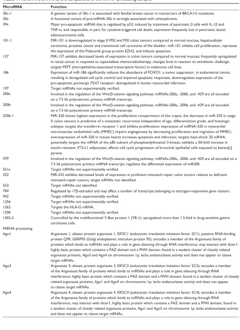
Table 4 MicroRNAs (miRNAs) and components of the miRNA processing complex
MicroRNA Function
30c-1 A genetic variant of 30c-1 is associated with familial breast cancer in noncarriers of BRCA1/2 mutations. 30e A functional variant of pre-miRNA-30e is strongly associated with schizophrenia.
34a Major pro-apoptotic miRNA that is regulated by p53; induced by treatment of pancreatic β cells with iL-1β and TNF-α, and responsible, in part, for cytokine-triggered cell death; expression frequently lost in pancreatic ductal
adenocarcinoma cells.
101-1 MiR-101 is downregulated in stage ii MSS and MSi colon cancers compared to normal mucosa, hepatocellular carcinoma, prostate cancer and transitional cell carcinoma of the bladder; miR-101 inhibits cell proliferation, represses the expression of the Polycomb group protein EZH2, and induces apoptosis.
137 MiR-137 exhibits decreased levels of expression in colon tumors compared to normal mucosa; frequently upregulated
in rectal cancer in response to capecitabine chemoradiotherapy; changes level in reaction to xenobiotic challenge; targets MiTF (micropthalmia-associated transcription factor) in melanoma cell lines.
186 Expression of miR-186 significantly reduces the abundance of FOXO1, a tumor suppressor, in endometrial cancer,
resulting in deregulated cell cycle control and impaired apoptotic responses; downregulates expression of the pro-apoptotic purinergic P2X7 receptor; dysregulated in human myocardial infarction.
197 Target miRNAs not experimentally verified.
200a involved in the regulation of the wnt/β-catenin signaling pathway; miRNAs-200a, -200b, and -429 are all encoded on a 7.5 kb polycistronic primary miRNA transcript.
200b involved in the regulation of the wnt/β-catenin signaling pathway; miRNAs-200a, -200b, and -429 are all encoded on a 7.5 kb polycistronic primary miRNA transcript.
320b-1 MiR-320 shows highest expression in the proliferative compartment of the crypts; the decrease in miR-320 in stage ii colon cancers is predictive of a metastatic recurrence independent of age, differentiation grade, and histologic subtype; targets the transferrin receptor 1 and inhibits proliferation; expression of miRNA-320 in myocardial microvascular endothelial cells (MMEC) impairs angiogenesis by decreasing proliferation and migration of MMEC; overexpression of miR-320 in mouse hearts increases apoptosis and infarction; targets heat-shock 20 mRNA; potentially targets the mRNA of the p85 subunit of phosphatidylinositol 3-kinase; exhibits a 50-fold increase in insulin-resistant 3T3-L1 adipocytes; affects cell cycle progression of bronchial epithelial cells exposed to benzo[a] pyrene.
429 involved in the regulation of the wnt/β-catenin signaling pathway; miRNAs-200a, -200b, and -429 are all encoded on a 7.5 kb polycistronic primary miRNA transcript; regulates the differential expression of miR200.
551a Target mRNAs not experimentally verified.
552 MiR-552 exhibits decreased levels of expression in proficient mismatch-repair colon tumors relative to deficient mismatch-repair tumors; target mRNAs not identified.
553 Target mRNAs not identified.
760 Regulated by 17β-estradiol and may affect a number of transcripts belonging to estrogen-responsive gene clusters. 942 Target mRNAs not experimentally verified.
1256 Target mRNAs not experimentally verified.
1262 Targets the HLA-G mRNA.
1290 Target mRNAs not experimentally verified.
1302-2 Controlled by the multifunctional Y-Box protein 1 (YB-1); upregulated more than 1.5-fold in drug-sensitive gastric carcinoma cells.
MiRNA processing
Ago1 Argonaute 1; aliases: protein argonaute 1, EiF2C1 (eukaryotic translation initiation factor 2C1), putative RNA-binding protein Q99, GERP95 (Golgi endoplasmic reticulum protein 95); encodes a member of the Argonaute family of proteins which binds to miRNAs and plays a role in gene silencing through RNA interference; may interact with dicer1; highly basic protein which contains a PAZ domain and a Piwi domain; found in a tandem cluster of closely related argonaute proteins, Ago3 and Ago4 on chromosome 1p; lacks endonuclease activity and does not appear to cleave target mRNAs.
Ago3 Argonaute 3; aliases: protein argonaute 3, EiF2C3 (eukaryotic translation initiation factor 2C3); encodes a member of the Argonaute family of proteins which binds to miRNAs and plays a role in gene silencing through RNA
interference; highly basic protein which contains a PAZ domain and a Piwi domain; found in a tandem cluster of closely related argonaute proteins, Ago1 and Ago4 on chromosome 1p; lacks endonuclease activity and does not appear to cleave target mRNAs.
Ago4 Argonaute 4; aliases: protein argonaute 4, EiF2C4 (eukaryotic translation initiation factor 2C4); encodes a member of the Argonaute family of proteins which binds to miRNAs and plays a role in gene silencing through RNA
interference; may interact with dicer1; highly basic protein which contains a PAZ domain and a Piwi domain; found in a tandem cluster of closely related argonaute proteins, Ago1 and Ago3 on chromosome 1p; lacks endonuclease activity and does not appear to cleave target mRNAs.
Clinical and Experimental Gastroenterology downloaded from https://www.dovepress.com/ by 118.70.13.36 on 20-Aug-2020
Dovepress Payne et al
Since base excision repair (BER) removes damage that
would otherwise be mutagenic in mammalian cells,
168–170BER is one of the most important DNA repair pathways in
the gastrointestinal tract. BER ameliorates environmentally
induced DNA damage in addition to the alkylation, oxidation,
and deamination events that occur during normal metabolic
processes.
171,172A critical enzyme in the base excision
repair pathway is MUTYH (MutY homolog or A/G-specific
adenine DNA glycosylase), whose germline mutation is
a known cause of MAP (MutYH-associated polyposis), a
recently described autosomal recessive colorectal adenoma
predisposition syndrome with a very high risk of colorectal
cancer.
173Myh
deficiency enhances intestinal tumorigenesis in
multiple intestinal neoplasia (
Apc
Min/+) mice.
174Interestingly,
Myh
deficiency in mice has a larger effect on tumor initiation
than on progression in the small bowel.
174Since 1p deletions
are observed in the human non-neoplastic mucosa of patients
with colon cancer,
44it is possible that Myh-deficient field
defects may initiate the process of colon carcinogenesis in
humans as it does in the mouse model. Since MUTYH-null
mouse embryonic stem cells exhibit a mutator phenotype,
175the loss of MUTYH can affect multiple pathways associated
with colon carcinogenesis. The role of MUTYH in the repair
of oxidative DNA damage begins with the formation of
8-oxo-guanine (8-oxoG) (see Figure 4), which then causes
a mispairing of the oxidized guanine base with adenine upon
DNA replication. Mismatch repair processes are activated
and MUTYH excises adenine leaving an apurinic (AP) site
resulting, after AP endonuclease action, in a DNA single
strand (ss) break.
176–180The activity of MUTYH, in
conjunc-tion with other glycosylases and the spontaneous generaconjunc-tion
of AP sites, may be quite extensive, since about 9000 AP
sites/cell occur daily.
168The AP site is then correctly repaired
by the sequential action of several enzymes which catalyze
template-directed insertion of one or a few nucleotides at the
previously damaged site.
172In addition to their role in DNA repair or the DDR,
MUTYH and p73 play important roles in the death of cells
that experience either excessive oxidative DNA damage or
chromosomal instability. The MUTYH-mediated cell death
pathway is described in the next section followed by a
sec-tion on the p73-mediated cell death pathway, which utilizes
part of the MUTYH pathway in its mediation of cell death
in response to excessive mitotic perturbation.
MUTYH/PARP/AiF pathway of cell death
MUTYH-mediated cell death has, as a central player, the
activa-tion of PARP-1 [poly(ADP-ribose) polymerase-1] (Figure 5).
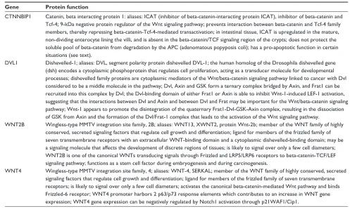
Table 5 Genes associated with the wnt signaling pathway
Gene Protein function
CTNNBiP1 Catenin, beta interacting protein 1: aliases: iCAT (inhibitor of beta-catenin-interacting protein iCAT), inhibitor of beta-catenin and Tcf-4; 9-kDa negative protein regulator of the wnt signaling pathway; prevents interaction between beta-catenin and Tcf-4 family members, thereby repressing beta-catenin-Tcf-4-mediated transactivation; in intestinal tissue, iCAT is upregulated in the mature, non-dividing enterocyte lining the villi, and is absent in the beta-catenin/TCF signaling region of the crypts; does not protect the soluble pool of beta-catenin from degradation by the APC (adenomatous popyposis coli); has a pro-apoptotic function in certain situations (see text).
DvL1 Dishevelled-1; aliases: DvL, segment polarity protein dishevelled DvL-1; the human homolog of the Drosophila dishevelled gene (dsh) encodes a cytoplasmic phosphoprotein that regulates cell proliferation, acting as a transducer molecule for developmental processes; dishevelled family proteins are cytoplasmic mediators of the wnt/beta-catenin signaling pathway linked to cancer with Dvl considered to be a middle molecule in the pathway; Dvl, Axin and GSK form a ternary complex bridged by Axin, and Frat1 can be recruited into this complex by Dvl; the Dvl-binding domain of either Frat1 or Axin is able to inhibit wnt-1-induced LEF-1 activation, suggesting that the interactions between Dvl and Axin and between Dvl and Frat may be important for the wnt/beta-catenin signaling
pathway; Wnt-1 appears to promote the disintegration of the quaternary Frat1-Dvl-GSK-Axin complex, resulting in the dissociation
of GSK from Axin and the formation of the Dvl/Frat-1 complex that leads to the activation of the wnt signaling pathway.
wNT2B wingless-type MMTv integration site family, 2B; aliases: wNT13, XwNT2, protein wnt-2b; member of the wNT family of highly conserved, secreted signaling factors that regulate cell growth and differentiation; ligand for members of the frizzled family of seven transmembrane receptors with an extracellular wNT-binding domain and a cytoplasmic dishevelled-binding domain; may be a signaling molecule that affects the development of discrete regions of tissues; is likely to signal over only a few cell diameters; wNT2B is one of the canonical wNTs transducing signals through Frizzled and LRP5/LRP6 receptors to beta-catenin-TCF/LEF signaling pathway; functions as a stem cell factor during embryogenesis and during carcinogenesis.
wNT4 wingless-type MMTv integration site family, 4; aliases: wNT-4, SERKAL; member of the wNT family of highly conserved, secreted signaling factors that regulate cell growth and differentiation; ligand for members of the frizzled family of seven transmembrane receptors; is likely to signal over only a few cell diameters; activates the canonical beta-catenin-mediated wnt pathway and binds Frizzled-6 receptor; wNT4 promoter harbors 2 p63/p73 response elements which contributes to an increase in wNT gene expression; wNT4 gene expression can be negatively regulated by Notch1 activation through p21wAF1/Cip1.
Clinical and Experimental Gastroenterology downloaded from https://www.dovepress.com/ by 118.70.13.36 on 20-Aug-2020
Dovepress Chromosome 1p deletions during colon carcinogenesis
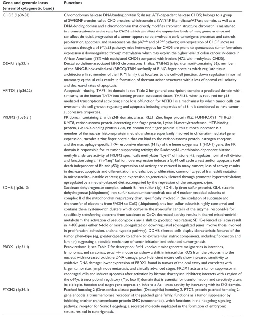
Table 6 Tumor suppressor genes
Gene and genomic locus (ensembl cytogenetic band)
Functions
CHD5 (1p36.31) Chromodomain helicase DNA binding protein 5; aliases: ATP-dependent helicase CHD5; belongs to a group of Swi/SNF proteins called CHD proteins, which contain a Swi/SNF-like helicase/ATPase domain, as well as a
DNA-binding domain and a chromodomain that directly modifies chromatin structure; chromatin is maintained
in a transcriptionally active state by CHD5 which can affect the expression levels of many genes at once and
can affect the quick progression of a tumor; appears to be involved in early tumorigenic processes and controls
proliferation, apoptosis, and senescence via the p16ink4a and p19Arf pathway; overexpression of CHD5 increases apoptosis through a p19Arf/p53 pathway; mice heterozygous for CHD5 are prone to spontaneous tumor formation; expression is downregulated through methylation, which may explain the higher level of colon cancer incidence in African Americans (78% with methylated CHD5) compared with iranians (47% with methylated CHD5). DEAR1 (1p35.1) Ductal epithelium-associated RiNG chromosome 1; alias: TRiM62 (tripartite motif-containing 62); member
of the RING-B-box-coiled-coil (RBCC)/TRIM subfamily of RING finger proteins which regulate tissue architecture; first member of the TRIM family that localizes to the cell–cell junction; down regulation in normal
mammary epithelial cells results in formation of aberrant acinar structures with a loss of normal cell polarity and decreased rates of apoptosis.
APiTD1 (1p36.22) Apoptosis-inducing, TAF9-like domain 1; see Table 2 for general description; contains a predicted domain with
similarity to the human TATA box-binding protein-associated factor, TAFII31, which is required for
p53-mediated transcriptional activation; since loss of function for APiTD1 is a mechanism by which tumor cells can overcome the cell growth-regulating and apoptosis-inducing properties of p53, it is considered to have tumor-suppressive properties.
PRDM2 (1p36.21) PR domain containing 2, with ZNF domain; aliases: RIZ1, Zinc finger protein RIZ, HUMHOXY1, MTB-ZF, KMT8, retinoblastoma protein-interacting zinc finger protein, Lysine N-methyltransferase, MTE-binding protein, GATA-3-binding protein G3B, PR domain zinc finger protein 2; this tumor suppressor is a
member of the nuclear histone/protein methyltransferase superfamily involved in chromatin-mediated gene
expression; encodes a zinc finger protein that can bind to the retinoblastoma protein, estrogen receptor, and the macrophage-specific TPA-responsive element (MTE) of the heme oxygenase 1 (HO-1) gene; the PR
domain is responsible for its tumor suppressing activity; the S-adenosyl-L-methionine-dependent histone
methyltransferase activity of PRDM2 specifically methylates “Lys-9” of histone H3; regulates normal cell division
and function using a “Yin-Yang” fashion; overexpression induces a G2-M cell cycle arrest and/or apoptosis (cell death independent of Rb and p53); expression and activity are reduced in many cancers; loss of activity results in decreased apoptosis and differentiation and enhanced proliferation; common target of frameshift mutation in microsatellite-unstable cancers; gene expression epigenetically silenced through promoter hypermethylation; upregulated by a methyl-balanced diet accompanied by the repression of the oncogene, c-jun.
SDHB (1p36.13) Succinate dehydrogenase complex, subunit B, iron sulfur (1p); SDH1, ip (iron-sulfur protein), GL4, succinic
dehydrogenase [ubiquinone] iron-sulfur subunit, mitochondrial; one of 4 nuclear-encoded subunits of complex II of the mitochondrial respiratory chain, specifically involved in the oxidation of succinate and the transfer of electrons from FADH to CoQ (ubiquinone); this iron-sulfur subunit is highly conserved and
contains three cysteine-rich clusters which comprise the iron-sulfur centers of the enzyme; responsible for
specifically transferring electrons from succinate to CoQ; decreased activity results in altered mitochondrial
metabolism, the activation of pseudohypoxia and a shift to glycolytic respiration; SDHB-silenced cells can result in .400 genes either 6-fold or more upregulated or downregulated (dysregulated genes involve those involved in proliferation, adhesion, and the hypoxia pathway); DDHB-silenced cells display characteristic features of the
tumor phenotype (eg, greater capacity to adhere to extracellular matrix components, including fibronectin and
laminin) suggesting a possible mechanism of tumor initiation and enhanced tumorigenesis.
PRDX1 (1p34.1) Peroxiredoxin 1; see Table 7 for description; Prdx1 knockout mice generate malignancies in intestines, lymphomas, and sarcomas; prdx1−/− mouse cells show a shift in intracellular ROS from the cytoplasm to the
nucleus with increased oxidative DNA damage; prdx1-deficient mouse cells show increased sensitivity to
oxidative DNA damage; lower expression of PRDX1 found in tumors of the oral cavity and correlates with larger tumor size, lymph node metastasis, and clinically advanced stages. PRDX1 acts as a tumor suppressor in esophageal cells and induces apoptosis after activation by histone deacetylase inhibitors; interacts with a region of the c-Myc transcriptional regulatory (Myc box ii) domain that is essential for transformation, and selectively alters its biological function and target gene expression; inhibits c-Abl kinase activity by interacting with its SH3 domain. PTCH2 (1p34.1) Patched homolog 2 (Drosophila); aliases: patched (Drosophila) homolog 2, PTC2, protein patched homolog 2;
gene encodes a transmembrane receptor of the patched gene family; functions as a tumor suppressor by inhibiting another transmembrane protein SMO (smoothened), which functions in the hedgehog signaling pathway; receptor for Sonic Hedgehog, a secreted molecule implicated in the formation of embryonic structures and in tunorigenesis.
(Continued)
Clinical and Experimental Gastroenterology downloaded from https://www.dovepress.com/ by 118.70.13.36 on 20-Aug-2020
Dovepress Payne et al
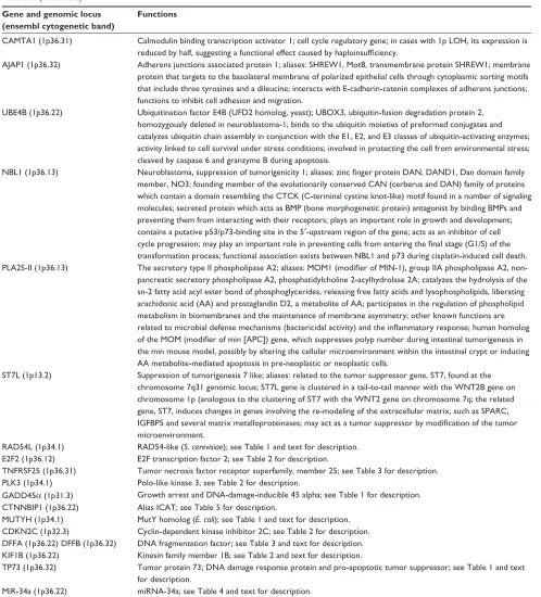
Table 6 (Continued)
Gene and genomic locus (ensembl cytogenetic band)
Functions
CAMTA1 (1p36.31) Calmodulin binding transcription activator 1; cell cycle regulatory gene; in cases with 1p LOH, its expression is
reduced by half, suggesting a functional effect caused by haploinsufficiency.
AJAP1 (1p36.32) Adherens junctions associated protein 1; aliases: SHREw1, Mot8, transmembrane protein SHREw1; membrane protein that targets to the basolateral membrane of polarized epithelial cells through cytoplasmic sorting motifs that include three tyrosines and a dileucine; interacts with E-cadherin-catenin complexes of adherens junctions; functions to inhibit cell adhesion and migration.
UBE4B (1p36.22) Ubiquitination factor E4B (UFD2 homolog, yeast); UBOX3, ubiquitin-fusion degradation protein 2, homozygously deleted in neuroblastoma-1; binds to the ubiquitin moieties of preformed conjugates and catalyzes ubiquitin chain assembly in conjunction with the E1, E2, and E3 classes of ubiquitin-activating enzymes;
activity linked to cell survival under stress conditions; involved in protecting the cell from environmental stress; cleaved by caspase 6 and granzyme B during apoptosis.
NBL1 (1p36.13) Neuroblastoma, suppression of tumorigenicity 1; aliases: zinc finger protein DAN, DAND1, Dan domain family
member, NO3; founding member of the evolutionarily conserved CAN (cerberus and DAN) family of proteins which contain a domain resembling the CTCK (C-terminal cystine knot-like) motif found in a number of signaling molecules; secreted protein which acts as BMP (bone morphogenetic protein) antagonist by binding BMPs and preventing them from interacting with their receptors; plays an important role in growth and development; contains a putative p53/p73-binding site in the 5′-upstream region of the gene; acts as an inhibitor of cell
cycle progression; may play an important role in preventing cells from entering the final stage (G1/S) of the
transformation process; functional association exists between NBL1 and p73 during cisplatin-induced cell death. PLA2S-ii (1p36.13) The secretory type II phospholipase A2; aliases: MOM1 (modifier of MIN-1), group IIA phospholipase A2,
non-pancreatic secretory phospholipase A2, phosphatidylcholine 2-acylhydrolase 2A; catalyzes the hydrolysis of the sn-2 fatty acid acyl ester bond of phosphoglycerides, releasing free fatty acids and lysophospholipids, liberating arachidonic acid (AA) and prostaglandin D2, a metabolite of AA; participates in the regulation of phospholipid metabolism in biomembranes and the maintenance of membrane asymmetry; other known functions are
related to microbial defense mechanisms (bactericidal activity) and the inflammatory response; human homolog of the MOM (modifier of min [APC]) gene, which suppresses polyp number during intestinal tumorigenesis in
the min mouse model, possibly by altering the cellular microenvironment within the intestinal crypt or inducing AA metabolite-mediated apoptosis in pre-neoplastic or neoplastic cells.
ST7L (1p13.2) Suppression of tumorigenesis 7 like; aliases: related to the tumor suppressor gene, ST7, found at the
chromosome 7q31 genomic locus; ST7L gene is clustered in a tail-to-tail manner with the WNT2B gene on chromosome 1p (analogous to the clustering of ST7 with the WNT2 gene on chromosome 7q; the related
gene, ST7, induces changes in genes involving the re-modeling of the extracellular matrix, such as SPARC,
IGFBP5 and several matrix metalloproteinases; may act as a tumor suppressor by modification of the tumor
microenvironment.
RAD54L (1p34.1) RAD54-like (S. cerevisiae); see Table 1 and text for description. E2F2 (1p36.12) E2F transcription factor 2; see Table 2 for description.
TNFRSF25 (1p36.31) Tumor necrosis factor receptor superfamily, member 25; see Table 3 for description. PLK3 (1p34.1) Polo-like kinase 3; see Table 2 for description.
GADD45α (1p31.3) Growth arrest and DNA-damage-inducible 45 alpha; see Table 1 for description. CTNNBiP1 (1p36.22) Alias iCAT; see Table 5 for description.
MUTYH (1p34.1) MutY homolog (E. coli); see Table 1 and text for description. CDKN2C (1p32.3) Cyclin-dependent kinase inhibitor 2C; see Table 2 for description. DFFA (1p36.22) DFFB (1p36.32) DNA fragmentation factor; see Table 3 and text for description. KiF1B (1p36.22) Kinesin family member 1B; see Table 2 and text for description.
TP73 (1p36.32) Tumor protein 73; DNA damage response protein and pro-apoptotic tumor suppressor; see Table 1 and text for description.
MiR-34a (1p36.22) miRNA-34a; see Table 4 and text for description. MiR-101-1 (1p31.3) miRNA-101-1; see Table 4 and text for description.
Excessive DNA ss breaks caused by the action of MUTYH
and AP endonuclease in the nucleus results in the activation of
PARP-1, which attaches polymers of ADP-ribose to proteins,
thereby opening up the chromatin to allow access of DNA
repair proteins.
181,182PARP initially serves as a survival protein
facilitating the rapid repair of DNA strand breaks, and also
prevents DNA degradation, in part, by inhibiting the activity
of deoxyribonucleases through the process of poly(ADP)
ribosylation.
183Since the synthesis of ADP-ribose polymers
consumes nicotinamide adenine dinucleotide (NAD
+),
184and
NAD
+is largely found in mitochondria where it participates in
the production of ATP (bottom right side of Figure 5), sustained
Clinical and Experimental Gastroenterology downloaded from https://www.dovepress.com/ by 118.70.13.36 on 20-Aug-2020
Dovepress Chromosome 1p deletions during colon carcinogenesis
PARP activation will consume energy reserves, resulting in cell
death, usually through the process of necrosis.
185–188A marked
deficiency in energy reserves may cause the ATP-dependent
Na
+/K
+transport proteins, which maintain ionic balance, to
fail, resulting in cell swelling and lysis of the cell,
189one of the
hallmarks of necrosis.
190In addition to the above energy catastrophe caused by
excessive PARP activity in the nucleus, persistent
single-stranded gaps in newly replicated DNA initiated by the action
of MUTYH
in mitochondria
can result in the fragmentation and
depletion of mitochondrial DNA (mtDNA)
191,192accompanied
by the loss of mitochondrial function culminating in cell
death
191,193(bottom right side of Figure 5). Dysfunctional
mitochondria can release Ca
++into the cytosol which can
activate calpains, causing Bax activation, lysosomal rupture,
and the release of cathepsins into the cytosol
191,194resulting
in a caspase-independent mode of cell death. Calpain
activation can also result in Bax activation, followed by Bax
oligomerization and mitochondrial damage, resulting in the
loss of the mitochondrial membrane potential.
There is another unique mechanism that can lead
to PARP-mediated cell death after excessive MUTYH
activity, in addition to the fragmentation of mtDNA,
energy catastrophe and calpain/lysosomal rupture/cathepsin
pathways of mitochondrial failure described above. The main
product of PARP-1 activity is the generation of polymers of
ADP-ribose (PAR). Although these polymers are usually
covalently bound to proteins, free PAR polymers are
themselves toxic
195–197and function as a death signal.
197–199The PAR polymers bind to mitochondria and induce the
release of tAIF (truncated apoptosis-inducing factor) from the
mitochondria into the cytosol
199(lower left side of Figure 5).
tAIF is then translocated to the nucleus where it binds to
DNA,
200–202causes DNA condensation
203and recruits DNA
degrading factors (eg, endogenous endo- and exo-nucleases)
resulting in DNA degradation
198,204(upper left side of
Figure 5). This series of events is part of an intricate program
of caspase-independent cell death,
203–213and is currently an
active area of research.
Several mechanisms have been proposed to explain
how tAIF is released from the mitochondria into the
cytosol.
210,214Prior to truncation, AIF is embedded in the inner
mitochondrial membrane,
215and the release of AIF requires
its cleavage
215,216from a 62 kDa AIF mitochondrial form to
a truncated 57 kDa soluble AIF form (tAIF).
217,218Calpain-I,
which is activated by Ca
++,
219and Ca
++-independent cathepsins
B, L, and S
218,220can cleave intramitochondrial AIF.
221–223The calpains and cathepsins can truncate AIF in the same
position at Gly102/Leu103.
218Calpain-I, however, appears
to be the critical enzyme regulating AIF processing in which
the AIF pathway is important for cell death.
219Oxidative
modifcation of AIF markedly increases the susceptibility
of AIF to calpain-I-mediated processing, most probably
through the exposure of a normally hidden calpain cleavage
site.
219Since the PAR polymer is a highly negatively charged
molecule, it could depolarize mitochondria leading to opening
of the mitochondrial membrane permeability transition pore
(MPTP) followed by the release of tAIF.
197,199PAR polymers
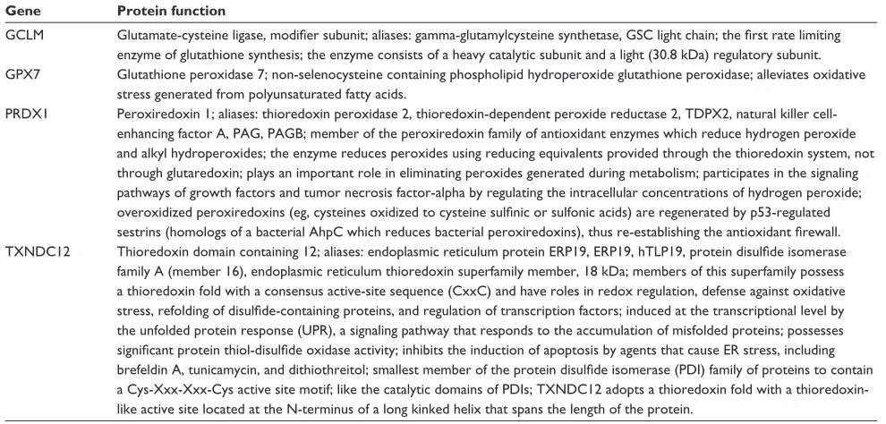
Table 7 Genes associated with antioxidant function
Gene Protein function
GCLM Glutamate-cysteine ligase, modifier subunit; aliases: gamma-glutamylcysteine synthetase, GSC light chain; the first rate limiting
enzyme of glutathione synthesis; the enzyme consists of a heavy catalytic subunit and a light (30.8 kDa) regulatory subunit. GPX7 Glutathione peroxidase 7; non-selenocysteine containing phospholipid hydroperoxide glutathione peroxidase; alleviates oxidative
stress generated from polyunsaturated fatty acids.
PRDX1 Peroxiredoxin 1; aliases: thioredoxin peroxidase 2, thioredoxin-dependent peroxide reductase 2, TDPX2, natural killer cell-enhancing factor A, PAG, PAGB; member of the peroxiredoxin family of antioxidant enzymes which reduce hydrogen peroxide
and alkyl hydroperoxides; the enzyme reduces peroxides using reducing equivalents provided through the thioredoxin system, not
through glutaredoxin; plays an important role in eliminating peroxides generated during metabolism; participates in the signaling pathways of growth factors and tumor necrosis factor-alpha by regulating the intracellular concentrations of hydrogen peroxide;
overoxidized peroxiredoxins (eg, cysteines oxidized to cysteine sulfinic or sulfonic acids) are regenerated by p53-regulated sestrins (homologs of a bacterial AhpC which reduces bacterial peroxiredoxins), thus re-establishing the antioxidant firewall.
TXNDC12 Thioredoxin domain containing 12; aliases: endoplasmic reticulum protein ERP19, ERP19, hTLP19, protein disulfide isomerase
family A (member 16), endoplasmic reticulum thioredoxin superfamily member, 18 kDa; members of this superfamily possess
a thioredoxin fold with a consensus active-site sequence (CxxC) and have roles in redox regulation, defense against oxidative stress, refolding of disulfide-containing proteins, and regulation of transcription factors; induced at the transcriptional level by
the unfolded protein response (UPR), a signaling pathway that responds to the accumulation of misfolded proteins; possesses
significant protein thiol-disulfide oxidase activity; inhibits the induction of apoptosis by agents that cause ER stress, including brefeldin A, tunicamycin, and dithiothreitol; smallest member of the protein disulfide isomerase (PDI) family of proteins to contain
a Cys-Xxx-Xxx-Cys active site motif; like the catalytic domains of PDis; TXNDC12 adopts a thioredoxin fold with a thioredoxin-like active site located at the N-terminus of a long kinked helix that spans the length of the protein.
Clinical and Experimental Gastroenterology downloaded from https://www.dovepress.com/ by 118.70.13.36 on 20-Aug-2020
Dovepress Payne et al
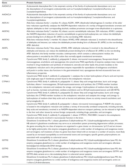
Table 8 Genes associated with protection against environmental and metabolic toxicity
Gene Protein function
AADACL3 Arylacetamide deacetylase-like 3; the enzymatic activity of the family of arylacetamide deacetylases carry out
the deacetylation of carcinogenic arylacetamides such as 4-acetylaminobiphenyl, 2-acetylaminofluorene, and
2-acetylaminaphthalene.
AADACL4 Arylacetamide deacetylase-like 4; the enzymatic activity of the family of arylacetamide deacetylases carry out
the deacetylation of carcinogenic arylacetamides such as 4-acetylaminobiphenyl, 2-acetylaminofluorene, and
2-acetylaminaphthalene.
AKR1A1 Aldo-keto reductase family 1, member A1; aliases ALDR1, ARM, dihydrodiol dehydrogenase 3; member of the aldo/ keto reductase superfamily; catalyzes the NADPH-dependent reduction of a variety of biogenic/xenobiotic aromatic and
aliphatic aldehydes to their corresponding alcohols; oxidizes proximate carcinogen trans-dihydrodiols to o-quinones.
AKR7A2 Aldo-keto reductase family 7, member A2; aliases: succinic semialdehyde reductase, SSA reductase, AFAR1; catalyzes the NADPH-dependent reduction of succinic semialdehyde to gamma-hydroxybutyrate; can reduce the dialdehyde
protein-binding form of aflatoxin B1 (AFB1) to the non-binding AFB1 dialcohol.
AKR7A3 Aldo-keto reductase family 7, member A3; aliases: AFAR2, AFB1 aldehyde reductase 2; involved in the detoxification of aldehydes and ketones; can reduce the dialdehyde protein-binding form of aflatoxin B1 (AFB1) to the non-binding
AFB1 dialcohol.
AKR7L Aldo-keto reductase family 7-like; aliases: AFAR3, AFB1 aldehyde reductase 3; involved in the detoxification of aldehydes and ketones; can reduce the dialdehyde protein-binding form of aflatoxin B1 (AFB1) to the non-binding
AFB1 dialcohol; this family member encodes a selenoprotein, which contains a selenocysteine residue; the selenocysteine is encoded by the UGA codon that normally signals translational termination.
CYP2J2 Cytochrome P450, family 2, subfamily J, polypeptide 2; aliases: microsomal monooxygenase, flavoprotein-linked
monooxygenase, arachidonic acid epoxygenase; the cytochrome P450 superfamily of enzymes catalyze many reactions involved in drug metabolism and synthesis of cholesterol, steroids and other lipids; this protein localizes to the endoplasmic reticulum and is the predominant enzyme responsible for epoxidation of endogenous arachidonic acid pools in cardiac tissue; also functions in the gastrointestinal tract; epoxygenase-derived eicosanoids have
anti-inflammatory properties.
CYP4Z1 Cytochrome P450, family 4, subfamily Z, polypeptide 1; catalyzes the in-chain hydroxylation of lauric acid and myristic acid; single-pass type ii membrane protein found in the endoplasmic reticulum.
CYP4A11 Cytochrome P450, family 4, subfamily A, polypeptide 11; aliases: fatty acid hydrolase, lauric acid omega-hydrolase, alkane-1 monooxygenase, 20-hydroxyeicosatetraenoic acid synthase; this CYP450 member localizes to the endoplasmic reticulum and catalyzes the omega- and omega-1-hydroxylation of medium-chain fatty acids such as laurate, myristate and palmitate; oxidizes arachidonic acid to 20-hydroxyeicosatetraenoic acid (20-HETE). CYP4A22 Cytochrome P450, family 4, subfamily A, polypeptide 22; aliases: fatty acid hydroxylase, lauric acid omega-hydrolase; this CYP450 member localizes to the endoplasmic reticulum and catalyzes the omega- and (omega-1)-hydroxylation of medium-chain fatty acids such as laurate and palmitate; shows no activity toward arachidonic acid and prostaglandin A1.
CYP4B1 Cytochrome P450, family 4, subfamily B, polypeptide 1; aliases: microsomal monooxygenase, P-450HP; this enzyme is located in the endoplasmic reticulum and oxidizes a variety of structurally unrelated compounds, including steroids, fatty acids and xenobiotics; involved in an NADPH-dependent electron transport pathway; can be induced to high levels in the liver and other tissues by various foreign compounds, including drugs, pesticides, and carcinogens.
CYP4X1 Cytochrome P450, family 4, subfamily X, polypeptide 1; aliases: CYPivX1, MGC40051; located in the endoplasmic reticulum and may be involved in neurovascular function in the brain.
GSTM1 Glutathione S-transferase Mu 1; aliases: glutathione S-alkyltransferase M1, S-(hydroxyalkyl)glutathione lyase M1, HB subunit 4; glutathione transferases may serve as an antioxidant system preventing degenerative cellular processes; the genes encoding the mu class of enzymes are organized in a gene cluster on chromosome 1p13.3 and are known to be highly polymorphic; this enzyme conjugates glutathione to a wide number of endogenous and exogenous toxins and carcinogens; null mutations of class mu genes have been linked with an increase in a number of cancers, most likely
caused by an increased susceptibility to environmental toxins and carcinogens; specific genetic polymorphisms are
associated with susceptibility to colorectal cancer.
GSTM2 Glutathione S-transferase Mu 2; aliases: glutathione S-alkyltransferase M2, S-(hydroxyalkyl)glutathione lyase M2; this enzyme conjugates glutathione to a wide number of endogenous and exogenous toxins and carcinogens; alleviates benzo[a]pyrene-diolepoxide-DNA damage.
GSTM3 Glutathione S-transferase Mu 3; aliases: glutathione S-alkyltransferase M3, S-(hydroxyalkyl)glutathione lyase M3; this enzyme conjugates glutathione to a wide number of endogenous and exogenous toxins and carcinogens; GSTM1 and GSTM3 allele variants are a risk-modulating factor in colorectal cancer patients.
GSTM4 Glutathione S-transferase Mu 4; aliases: glutathione S-alkyltransferase M4, S-(hydroxyalkyl)glutathione lyase M4; this enzyme conjugates glutathione to a wide number of endogenous and exogenous toxins and carcinogens; active on 1-chloro-2,4-dinitrobenzene.
(Continued)
Clinical and Experimental Gastroenterology downloaded from https://www.dovepress.com/ by 118.70.13.36 on 20-Aug-2020