Clinical and Experimental Gastroenterology
Risk factors of converting to laparotomy in
laparoscopic appendectomy for acute appendicitis
Tomoyuki Abe1 Takashi Nagaie1 Mitsuhiro Miyazaki1 Miho Ochi2
Tatsuro Fukuya2 Kiyoshi Kajiyama1
1Department of Surgery, Aso Iizuka
Hospital, Iizuka City, Fukuoka, Japan;
2Department of Radiology, Aso Iizuka
Hospital, Iizuka City, Fukuoka, Japan
Correspondence: Takashi Nagaie Department of Surgery, Aso Iizuka Hospital, 3–83 Yoshio-machi, Iizuka 820-8505, Japan Phone +81 948 22 3800 Fax +81 948 29 8747 Email [email protected]
Purpose: Laparoscopic appendectomy (LA) for acute appendicitis has several advantages over open appendectomy (OA). In cases of complicated appendicitis, LA is converted to OA at a constant rate, though converting appendectomy (CA) has several disadvantages. We retrospectively determined preoperative risk factors for failure of LA and subsequent conversion to OA.
Methods: Consecutive cases of preoperative computed tomography (CT) and attempted LA were retrieved from our hospital database and grouped by procedure (LA versus CA). Patients
with negative appendectomies (n = 28), opened appendectomy (n = 210), delayed interval
appendectomy (n = 3), or who were ,14 years of age were excluded.
Results: Average patient age, preoperative C-reactive protein (CRP) level, and diffuse peritonitis were significantly different between the groups. CT inflammation and occurrence of complicated appendicitis were significantly higher in CA than LA. Conversion to OA was mostly because of dense adhesions, diffuse peritonitis, and difficulties in excision of the appendix due to perforation or severe inflammation from surgical point of view. Postoperative complications were significantly lower in LA than CA, although the rate of intraoperative abscess was not different.
Conclusion: Most patients with acute appendicitis can be successfully treated with LA. We identified the following significant risk factors of CA: CT inflammation grade 4 or 5; complicated appendicitis; higher preoperative CRP level; and diffuse peritonitis.
Keywords: laparotomy, laparoscopic appendectomy, acute appendicitis
Introduction
For more than a century, open appendectomy (OA) has been the standard surgery
for acute appendicitis.1 Since it was first introduced by Semm in 1983,2 laparoscopic
appendectomy (LA) has become an increasingly prevalent intervention.
Laparoscopic surgery has several advantages, including the use of small incisions to obtain good quality visualization and access to the abdominal cavity and rapid
postoperative recovery.3 Meta-analyses of randomized, controlled trials suggest that LA
has several distinct benefits over OA, including less postoperative pain, shortened hospital stay, and lower superficial surgical site infection rates.4–8 In contrast, the rate of
intra-abdominal abscess (IAA), which is one of the most concerning intra-abdominal postoperative
complications, occurs almost three times more often in LA than after OA.9
Acute appendicitis is subdivided into two groups with respect to inflammatory grading: simple versus complicated appendicitis. Simple appendicitis, of which phlegmonous appendicitis is the most common type, is considered as a good indication
Dove
press
C A S E S E R I E S
open access to scientific and medical research
Open Access Full Text Article
Clinical and Experimental Gastroenterology downloaded from https://www.dovepress.com/ by 118.70.13.36 on 20-Aug-2020
For personal use only.
Number of times this article has been viewed
This article was published in the following Dove Press journal: Clinical and Experimental Gastroenterology
for LA. For complicated appendicitis, defined as acute gangrenous appendicitis and/or perforation of the appendix leading to localized or diffuse peritonitis, no clear consensus favoring LA has been established.
Converting appendectomy (CA), ie, converting from an LA to an OA procedure, occurs if intraoperative complications arise during LA or the severity of disease prohibits a safe laparoscopic intervention. It is well-known that CA increases medical costs and operative times; in addition, the benefits of the laparoscopic approach and outcomes, such as fewer surgical site infections and shortened hospital stays, are lost.10 Therefore, preoperative criteria that can be used to
decide the ideal operative approach for individual patients are required.
The present study was designed to evaluate the preoperative indicators of clinical symptoms and radiological inflammatory grading by computed tomography (CT) to define parameters that may prove useful in predicting the failure of LA. Furthermore, we evaluated the feasibility and efficacy of LA in selected patients with complicated appendicitis.
Materials and methods
Patients
From April 2001 to December 2008, appendectomy for acute appendicitis was performed on 532 patients in the Department of Surgery at Iizuka Hospital, Japan. Of these, 262 patients who underwent LA and 29 patients who underwent CA were enrolled into this study, retrospectively. The conversion rate was 10%. Patients with negative
appendectomies (n = 28), who were younger than 14 years
of age, or who underwent open appendectomy (n = 210),
and delayed interval appendectomy (n = 3) with acute
appendicitis, were excluded from this study. Patients with delay in operation for more than one week after abdominal symptoms happened underwent conservative treatment or delayed interval appendectomy.
Methods
In our department, diagnosis of acute appendicitis is made according to findings of clinical symptoms, laboratory data, contrast CT, and/or abdominal ultrasonography (US). The indications for either OA or LA are based on the attending surgeon’s opinion and the patient’s condition. The following data were collected for analysis: patient’s background; laboratory data; and perioperative outcomes. Findings of CT were evaluated, retrospectively, in detail: appendix location; appendicolith; cecal wall thickening involving the base of the
appendix; and lymphadenopathy. The extent of inflammation was graded by using imaging features seen anywhere along the course of the appendix and in the periappendiceal region. A 6-point scale was defined as follows: a grade of 0 indicates a normal appendix; grade 1, a possibly abnormal appendix, eg, one at least 6 mm in diameter without intraluminal fluid, or with wall enhancement, or containing an appendicolith; grade 2, an abnormal appendix, eg, diameter 6 mm with wall enhancement, without adjacent fat stranding; grade 3, an abnormal appendix surrounded by fat stranding; grade 4, abnormal appendix surrounded by fat stranding and fluid;
and grade 5, inflammatory mass or abscess.11
Terms and definitions
Complicated appendicitis was defined as acute gangrenous appendicitis and/or perforation of the appendix leading to localized peritonitis. It was defined, retrospectively, histologically.
Intra-abdominal abscess was confirmed when it was known that fluid collection diagnosed at US or CT contained pus at US- or CT-guided aspiration, or when clinical signs with positive laboratory findings, with or without pathology, were
demonstrated by CT or US.5 Since radiological imaging was
not always required, postoperatively IAA was confirmed with or without it.
Surgeons
Surgical procedures were performed by the attending surgeon or residents with at least 2 years of surgical training. Residents were always assisted by an attending surgeon who had more than 10 years of experience in laparoscopic and open surgical techniques. A total number of 19 surgeons participated in this study.
Operative techniques
Technique of laparoscopic retrograde
appendectomy
Under general anesthesia, LAs were performed using a standardized 3 - or 4- trocar approach (umbilical, 10–12 mm port; suprapubic, 10–12 mm port; lower-right quadrant, 5 mm port; and optional lower-left quadrant, 5 mm port). With the patient in the Trendelenburg position and right side up, the small bowel was retracted away from the lower right quadrant. An inflammatory mass or hard adhesions, if present, was dissected gently with blunt instruments. The appendix was divided using an intestinal stapler (Endo-GIA 30, US Surgical Corp, Norwalk, CT, USA) or two pretied loops (Endoloops, Ethicon, Johnson and Johnson, Arlington, TX, USA) and Dovepress
Abe et al
Clinical and Experimental Gastroenterology downloaded from https://www.dovepress.com/ by 118.70.13.36 on 20-Aug-2020
removed through one of the two 10–12 mm ports, in general with use of a specimen bag (Endo-Catch, US Surgical Corp, Mansfield, MA, USA). Generally, intraperitoneal irrigation was performed in all cases. Particularly in case of an abscess or perforated appendix, the lower-right quadrant, right paracolic gutter, and pelvis were irrigated with 2–3 L of physiological saline.
Open appendectomy
Open appendectomy was performed via a standard McBurney’s splitting incision of the lower-right quadrant muscle or by a lower middle abdominal incision (lower-right pararectal incision). After the appendix was removed, a stump ligature was performed with invagination. If an abscess or perforated appendix was found, a drainage tube was used for a few days as required. The tube type was selected by the attending surgeon.
Statistical methods
The SPSS software (Version 4.11; Abacus Concepts Inc, Berkeley, CA, USA) was used for multivariate adjustment of all covariates by means of stepwise regression analysis on a Windows computer. Statistical significance was defined by a P-value of less than 0.05 using the Student’s t-test and Fisher’s exact test. Data are presented as a proportion (eg,
percent of total) or as the mean ± standard deviation (SD).
All hazard ratios (HRs) are presented with 95% confidence intervals (CI).
Results
Of the 291 patients who underwent appendectomy, there were 262 LA procedures (90.0% of the total) and 29 CA procedures (10.0%) between April 2001 and December 2008. The patient demographics and characteristics are summarized
in Table 1. The overall average age was 38.7 ± 18.7 years
with a range from 15–86 years. The LA patients were
significantly younger than the CA patients (37.0 ± 18.0 years
for LA versus 54.1 ± 17.9 years for CA; P= 0.001). There
were no significant differences between the LA and CA groups in terms of sex distribution, body mass index (BMI)
(22.7 ± 3.6 years for LA versus 22.1 ± 4.8 years for CA),
symptoms such as vomiting and diarrhea (93 for LA versus 6 for CA), previous appendicitis history (46 for LA versus 5 for CA), or abdominal surgery (21 for LA versus 6 for CA).
However, there was a statistical difference in terms of the American Society of Anesthesiologists (ASA)
ratio, which was higher in CA than LA (an ASA ratio 3
was observed in 6 LA patients versus 4 CA patients;
P= 0.002). CA had lower white blood cell (WBC) counts
than LA (11,413 ± 4,112 mm3 versus 13,412 ± 4,609 mm3;
P = 0.019) and higher C-reactive protein (CRP) level
(10.3 ± 9.4 mg/dL versus 4.1 ± 6.1 mg/dL; P= 0.0019).
Diffuse peritonitis was more frequently seen in CA (12 patients, or 41.4%) than in LA (12 patients, or 4.6%) (P= 0.0005).
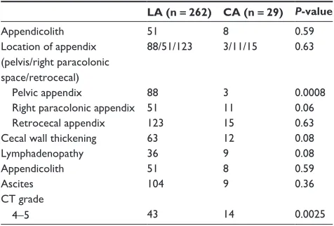
All patients had undergone preoperative abdominal contrast CT. One radiologist, who was blinded with respect to any clinical information or whether the operations were converted, performed the retrospective assessment of CT grading. CT findings about the appendix are summarized in Table 2, which clearly shows that there were no differences in the ratio of appendicolith, cecal wall thickening, lymph-adenopathy, or ascites between the two groups. In terms of
Table 1 Patients’ demographics
Variable LA CA P-value
Number of patients 262 29
Age (years) 37.0 ± 18.0 54.1 ± 17.9 0.001
Male/female ratio 164/101 22/7 0.11
BMI 22.7 ± 3.6 22.1 ± 4.8 0.52
ASA 1-2/3-4 ratio 256/6 25/4 0.0021 Symptoms
Vomiting Diarrhea
73 20
5 1
0.17 0.30 Previous abdominal
surgery
21 6 0.12
Previous appendicitis historya
46 5 0.97
WBC (mm3) 13412 ± 4609 11413 ± 4112 0.019
CRP (mg/dL) 4.1 ± 6.1 10.3 ± 9.4 0.0019
Diffuse peritonitis 12 12 0.0005
Notes: Data are mean ± SD. aPrevious history of appendicitis treated conservatively. Abbreviations: LA, laparoscopic appendectomy; CA, converted to the open appendectomy; BMI, body mass index; ASA, American Society of Anesthesiologists; WBC, white blood cell count; CRP, C-reactive protein level.
Table 2 CT findings and grading
LA (n =262) CA (n =29) P-value
Appendicolith 51 8 0.59
Location of appendix (pelvis/right paracolonic space/retrocecal)
88/51/123 3/11/15 0.63
Pelvic appendix 88 3 0.0008
Right paracolonic appendix 51 11 0.06
Retrocecal appendix 123 15 0.63
Cecal wall thickening 63 12 0.08
Lymphadenopathy 36 9 0.08
Appendicolith 51 8 0.59
Ascites 104 9 0.36
CT grade
4-5 43 14 0.0025
Abbreviations: CT, computed tomography; LA, laparoscopic appendectomy; CA, converted to open appendectomy.
Dovepress Risk factors of converting appendectomy
Clinical and Experimental Gastroenterology downloaded from https://www.dovepress.com/ by 118.70.13.36 on 20-Aug-2020
the location of the appendicitis, the pelvic appendix was a
good indicator of LA (P= 0.0008). A CT grade of greater
than 4 was significantly more often associated with CA compared to LA (14 patients versus 43 patients, respectively;
P= 0.0025).
Univariate analysis helped identify six factors associated with complicated appendicitis, namely older age, ASA ratio
3, lower WBC count, higher CRP, diffuse peritonitis, and
CT grade 4 or 5. Multiple stepwise regression analysis was performed to assess the potential preoperative risk factors of CA. Only the predictors with value in the range of 0.05 and 0.1 were included in the analysis. Table 3 shows that diffuse
peritonitis (OR = 9.75; 95% CI: 3.25–29.3); CT grade 4 or
5 (OR = 3.91; 95% CI: 1.46–10.5); CRP levels .10 mg/dL
(OR = 3.44; 95% CI: 1.22–9.71); and complicated
appen-dicitis (OR = 3.79; 95% CI: 1.33–10.8) are the risk factors
associated with CA.
Operative outcomes and postoperative complications are summarized in Table 4. The LA group was associated with
shorter operative time than CA (81.6 ± 32.1 minutes for LA
versus 148.8 ± 49.4 minutes for CA; P= 0.0001). CA had
more intraoperative bleeding than LA (3.4 ± 29.7 mL for
LA versus 127.8 ± 196.3 mL for CA; P= 0.002). Incidence
of SSI and IAA was not significantly different, but the overall perioperative complications rate was higher in CA
than in LA (8.8% for LA versus 34.5% for CA; P= 0.007),
including postoperative ileus, intra-abdominal abscess, and enteritis. There was no mortality in either group. Patients in the LA group were discharged earlier than in
CA (7.1 ± 6.3 days for LA versus 14.3 ± 8.6 days for CA;
P= 0.0001). Pathologically, final diagnosis of complicated
appendicitis was higher in CA (21 cases, or 72.4%) than
LA (73 cases, or 27.9%; P= 0.0001).
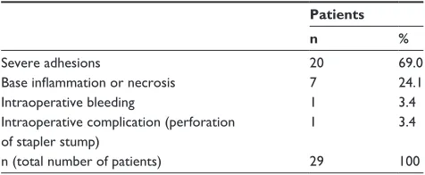
The reasons for converted appendectomy have been summarized in Table 5. The most frequent reasons were severe adhesions (69%), secondary base inflammation or necrosis (24.1%), and bleeding from the appendiceal artery (3.4%). Intraoperative complication arose in one case, and perforation of the stapler stump occurred during intraperitoneal irrigation.
Discussion
In recent years, there have been several advancements in laparoscopic surgery and intraoperative instruments. These improvements have contributed to several advantages of LA over the open technique, including reduced postoperative pain, fewer SSIs, and earlier discharge from the hospital. In the literature, LA has been reported to be associated with less analgesic use, early start of oral nutrient intake, shorter hospital stay, and lower incidence of SSI and IAA.4,10,12,13 The
disadvantages of LA are the use of disposable instruments, which adds to the cost and increases the operative time
compared to OA.14,15 Our study shows that LA has distinct
superiority over CA, owing to the shorter operative time (81.6 ± 32.1 minutes), less bleeding (3.4 ± 29.7 mL), reduced
hospital stay (7.1 ± 6.3 days), and lower frequency of overall
postoperative complications (8.0%). In the same period covered by the study, we performed open appendectomy in 210 patients for acute appendicitis and found that LA had distinct benefits over OA about surgical postoperative complications.
Table 4 Operative and postoperative outcomes
LA (n =262) CA (n =29) P-value
Operative time (minutes) 81.6 ± 32.1 148.8 ± 49.4 0.0001 Bleeding volume (mL) 3.4 ± 29.7 127.8 ± 196.3 0.0020 Complicated appendicitis
(pathologically gangrenous and/or perforation)
73 21 0.0001
Hospital stay (days) 7.1 ± 6.3 14.3 ± 8.6 0.0001
Overall complications 21 10 0.0070
Surgical site infection 9 5 0.066
Postoperative ileus 3 2 0.52
Intra-abdominal abscess 7 2 0.94
Enteritis 1 0 0.16
Intraoperative complications 2 1 0.45
Note: Data is represented as mean ± SD.
Abbreviations: LA, laparoscopic appendectomy; CA, converted to open appendectomy; SD, standard deviation.
Table 5 Reasons for CA
Patients
n %
Severe adhesions 20 69.0
Base inflammation or necrosis 7 24.1
Intraoperative bleeding 1 3.4
Intraoperative complication (perforation of stapler stump)
1 3.4
n (total number of patients) 29 100
Abbreviation: CA, converted appendectomy.
Table 3 Important risk factors predicting CA
Odds ratio 95% CI P-value
CT inflammation grade more than 4
3.91 1.46–10.5 0.007
Complicated appendicitis 3.79 1.33–10.8 0.012 High CRP level (.10 mg/dL) 3.44 1.22–9.71 0.019 Diffuse peritonitis 9.75 3.25–29.3 0.0001
Abbreviations: CA, converted appendectomy; CI, confidence interval; CRP, C-reactive protein level; CT, computed tomography.
Dovepress
Abe et al
Clinical and Experimental Gastroenterology downloaded from https://www.dovepress.com/ by 118.70.13.36 on 20-Aug-2020
The most controversial complication is the frequency and morbidity of postoperative IAA. In our study, there was no significant difference in the incidence of abscess formation
between LA and OA, and this is similar to another report.16
Tuggle et al provided evidence from a nationwide study showing that, in cases of complicated appendicitis, LA is superior in terms of superficial and deep wound infections; in contrast to our results, LA was associated with an increased
incidence of postoperative IAA.11 Markides et al reported that
LA has advantages in terms of less SSI compared to OA in complicated acute appendicitis, and (in agreement with our results), there was no significant additional risk of IAA.17 In
terms of incidence, LA patients have been reported to have
12% fewer cases of IAA as a postoperative complication.18
It is thought that the low frequency of SSI in LA may be due to the fact that the use of a specimen bag and a laparoscopic port prevent direct attachment of the resected appendix to the surgical site.
The rates of conversion reported in the literature are variable. Liu et al reported a conversion rate of 9.7% from
LA to OA,19 attributed to a variety reasons associated
with patients, surgeons, or technical factors. The 10.0% conversion rate in this study is in accordance with other
published studies,10 although lower conversion rates
(0%–3.3%) have been reported.20 The conclusion from
our study and others is that conversion itself lengthens the operative time, leads to a longer hospital stay, and causes a high incidence of postoperative complications. Higher postoperative complications required additional intervention, such as abdominal drainage, which could lengthen hospital stays. Understanding the factors associated with a higher chance of conversion may be useful, not only for surgeons to select patients for laparoscopic intervention appropriately, but also for patients to be able to make a better informed decision about their treatment. Our study shows that the significant preoperative risk factors in CA were older age, an ASA ratio of greater than 3, high CRP, diffuse peritonitis, a CT grade of 4 or 5, and a complicated type of appendicitis.
A clear consensus as to the superiority of LA versus OA for uncomplicated appendicitis has been established. On the other hand, the superiority of either intervention, especially in the case of patients with complicated appendicitis, is still uncertain. It is known that acute gangrenous and perforating appendicitis (defined as complicated acute appendicitis) is associated with a significant increased risk of postoperative complications, and such cases are regarded as contraindicated
for LA.4,9 Our study data revealed that 94 patients (32.3%)
with complicated appendicitis were identified with clinical and pathological findings, and the differences in LA distribution (72 patients, or 27.4%) versus CA (22 patients, or
75.9%) were significant (P= 0.0001). Garg et al also reported
that LA for complicated appendicitis is feasible and safe.3 It
is associated with less postoperative pain, lower incidence of infectious complications, and reduced length of hospital stay when compared to OA.
Reasons for CA in complicated appendicitis are perforation or necrosis of the appendix, and this friability often made us carry out surgical removal to the extent of ileocecal resection. The main reason of conversion was severe dense adhesions (n = 20), which also limit the amount of intra-abdominal space to perform a laparoscopic intervention. Conversion to open appendectomy would be inevitable in such cases. Nineteen patients (98.5%) with severe adhesions had previous appendicitis history. The location of the appendix is also an important factor with severe adhesions, 15 patients (75%) with retrocecal and five patients’ (25%) appendix could not be removed from the cecum or intestine which made us do CA. More attention to appendix locations and previous abdominal medical history must be paid to the patient with
a CT grade .3. We undertook abdominal drainage in two
patients with postoperative abdominal abscess in CA. Five patients with surgical site infection and ileus made hospital stays longer than LA. The discharge criteria is that the patient is fully recovered from complications; so a higher rate of
complications in CA compared with LA (P= 0.007) leads
to longer hospital stays. We attempted to complete LA even with severe adhesive or inflamed cases, so operation time in CA was twice as long as LA.
A limitation of our study was primary open appendectomy. The decision of whether primary LA or OA was made was determined by the attending surgeon, not by apparent criteria. By choosing only those patients in whom LA was attempted
lead to verification bias. OA had CT grade 1 (n = 5); grade
2 (n = 27); grade 3 (n = 83); grade 4 (n = 36); and grade
5 (n = 53). A CT grade .3 in OA was apparently higher
than LA (89 cases versus 58 cases); however, the share of complicated appendectomies in the same time frame was apparently lower in the OA group than the LA group (28 cases versus 73 cases). Retrospectively, our decision whether primary LA or OA had relationships with preoperative CT grade, discrepancy of preoperative CT grade, and histological data was confirmed.
In conclusion, the present study has identified four independent risk factors of conversion: diffuse peritonitis on physical examination, CT grade of 4 or 5, high CRP
Dovepress Risk factors of converting appendectomy
Clinical and Experimental Gastroenterology downloaded from https://www.dovepress.com/ by 118.70.13.36 on 20-Aug-2020
Clinical and Experimental Gastroenterology
Publish your work in this journal
Submit your manuscript here: http://www.dovepress.com/clinical-and-experimental-gastroenterology-journal
Clinical and Experimental Gastroenterology is an international, peer-reviewed, open access journal, publishing all aspects of gastroenterology in the clinic and laboratory, including: Pathology, pathophysiology of gastrointestinal disease; Investigation and treatment of gastointes-tinal disease; Pharmacology of drugs used in the alimentary tract;
Immunology/genetics/genomics related to gastrointestinal disease. This journal is indexed on CAS. The manuscript management system is completely online and includes a very quick and fair peer-review system. Visit http://www.dovepress.com/testimonials.php to read real quotes from published authors.
(.10 mg/dL), complicated appendicitis. We also found
that, even in cases of complicated appendicitis, LA could be successfully performed and was associated with important benefits over patients who had undergone CA.
Disclosure
The authors report no conflicts of interest in this work.
References
1. Berry J Jr, Malt RA. Appendicitis near its centenary. Ann Surg. 1984;200(5):567–575.
2. Semm K. Endoscopic appendectomy. Endoscopy. 1983;15(2):59–64. 3. Garg CP, Vaidya BB, Chengalath MM. Efficacy of laparoscopy in
com-plicated appendicitis. Int J Surg. 2009;7(3):250–252.
4. Wei B, Qi CL, Chen TF, et al. Laparoscopic versus open appendectomy for acute appendicitis: a metaanalysis. Surg Endosc. 2011;25(4): 1199–1208.
5. Hellberg A, Rudberg C, Kullman E, et al. Prospective randomized multicentre study of laparoscopic versus open appendicectomy. Br J Surg. 1999;86(1):48–53.
6. Williams MD, Collins JN, Wright TF, Fenoglio ME. Laparoscopic versus open appendectomy. South Med J. 1996;89(7):668–674.
7. Bennett J, Boddy A, Rhodes M. Choice of approach for appendicectomy: a meta-analysis of open versus laparoscopic appendicectomy. Surg Laparosc Endosc Percutan Tech. 2007;17(4):245–255.
8. Kim CB, Kim MS, Hong JH, Lee HY, Yu SH. Is laparoscopic appendectomy useful for the treatment of acute appendicitis in Korea? A meta-analysis. Yonsei Med J. 2004;45(1):7–16.
9. Siewert B, Raptopoulos V, Liu SI, Hodin RA, Davis RB, Rosen MP. CT predictors of failed laparoscopic appendectomy. Radiology. 2003; 229(2):415–420.
10. Nana AM, Ouandji CN, Simoens C, Smets D, Mendes da Costa P. Laparoscopic appendectomies: results of a monocentric prospective and non-randomized study. Hepatogastroenterology. 2007;54(76): 1146–1152.
11. Tuggle KR, Ortega G, Bolorunduro OB, et al. Laparoscopic versus open appendectomy in complicated appendicitis: a review of the NSQIP database. J Surg Res. 2010;163(2):225–228.
12. Yeh CC, Wu SC, Liao CC, Su LT, Hsieh CH, Li TC. Laparoscopic appendectomy for acute appendicitis is more favorable for patients with comorbidities, the elderly, and those with complicated appendicitis: a nationwide population-based study. Surg Endosc. 2011;25(9): 2932–2942.
13. Li X, Zhang J, Sang L, et al. Laparoscopic versus conventional appendectomy – a meta-analysis of randomized controlled trials. BMC Gastroenterol. 2010;10:129.
14. Chung RS, Rowland DY, Li P, Diaz J. A meta-analysis of randomized controlled trials of laparoscopic versus conventional appendectomy. Am J Surg. 1999;177(3):250–256.
15. Fallahzadeh H. Should a laparoscopic appendectomy be done? Am Surg. 1998;64(3):231–233.
16. Khalili TM, Hiatt JR, Savar A, Lau C, Margulies DR. Perforated appendicitis is not a contraindication to laparoscopy. Am Surg. 1999;65(10):965–967.
17. Markides G, Subar D, Riyad K. Laparoscopic versus open appendectomy in adults with complicated appendicitis: systematic review and meta-analysis. World J Surg. 2010;34(9):2026–2040.
18. So JB, Chiong EC, Chiong E, et al. Laparoscopic appendectomy for perforated appendicitis. World J Surg. 2002;26(12):1485–1488. 19. Liu SI, Siewert B, Raptopoulos V, Hodin RA. Factors associated
with conversion to laparotomy in patients undergoing laparoscopic appendectomy. J Am Coll Surg. 2002;194(3):298–305.
20. Martin LC, Puente I, Sosa JL, et al. Open versus laparoscopic appendectomy. A prospective randomized comparison. Ann Surg. 1995;222(3):256–261.
Dovepress
Dove
press
Abe et al
Clinical and Experimental Gastroenterology downloaded from https://www.dovepress.com/ by 118.70.13.36 on 20-Aug-2020