Aldo Quattrone, MD
Maurizio Morelli, MD
David R. Williams, PhD,
FRACP
Basilio Vescio, PhD
Gennarina Arabia, MD,
MSc
Salvatore Nigro, PhD
Giuseppe Nicoletti, MD
Maria Salsone, MD
Fabiana Novellino, MD
Rita Nisticò, MD
Franco Pucci, PhD
Carmelina Chiriaco, PhD
Pierfrancesco Pugliese,
MD
Domenico Bosco, MD
Manuela Caracciolo, MD
Correspondence to Prof. Quattrone: aldo.quattrone@gmail.com
MR parkinsonism index predicts vertical
supranuclear gaze palsy in patients with
PSP
–
parkinsonism
ABSTRACT
Objective:
To identify a biomarker for predicting the appearance of vertical supranuclear gaze
palsy (VSGP) in patients affected by progressive supranuclear palsy–parkinsonism (PSP-P).
Methods:
Twenty-four patients with PSP-P were enrolled in the current study. Patients were
clin-ically followed up every 6 months until the appearance of VSGP or the end of the follow-up (4
years). Participants underwent MRI at baseline and at the end of follow-up. Magnetic resonance
parkinsonism index (MRPI), an imaging measure useful for diagnosing PSP, was calculated.
Results:
Twenty-one patients with PSP-P completed follow-up, and 3 patients dropped out.
Eleven of 21 patients with PSP-P developed VSGP after a mean follow-up period of 28.5 months
(range 6–48 months), while the remaining 10 patients with PSP-P did not develop VSGP during
the 4-year follow-up period. At baseline, patients with PSP-P who later developed VSGP had
MRPI values significantly higher than those of patients not developing VSGP without overlapping
values between the 2 groups. MRPI showed a higher accuracy (100%) in predicting VSGP than
vertical ocular slowness (accuracy 33.3%) or postural instability with or without vertical ocular
slowness (accuracy 71.4% and 42.9%, respectively).
Conclusions:
Our study demonstrates that MRPI accurately predicted, on an individual basis, the
appearance of VSGP in patients with PSP-P, thus confirming clinical diagnosis in vivo.
Neurology® 2016;87:1266–1273GLOSSARY
H-Y5Hoehn-Yahr;MCP5middle cerebellar peduncle;MMSE5Mini-Mental State Examination;MRPI5magnetic reso-nance parkinsonism index;PD5Parkinson disease;PSP5progressive supranuclear palsy;PSP-P5progressive supra-nuclear palsy–parkinsonism; RS5Richardson syndrome; SCP 5superior cerebellar peduncle; UPDRS-ME 5Unified Parkinson’s Disease Rating Scale–Motor Examination;VSGP5vertical supranuclear gaze palsy.
There is emerging evidence that progressive supranuclear palsy (PSP) includes 2 main clinical
phenotypes: the classic Richardson syndrome (RS) and the PSP-parkinsonism (PSP-P).
1–10RS
is characterized by postural instability with falls and vertical supranuclear gaze palsy (VSGP) that
occur within 2 years from disease onset.
1,4,7–10By contrast, PSP-P is characterized mainly by
parkinsonism, resting tremor, and a moderate levodopa response with falls and VSGP that may
occur later in the course of the disease.
1,4,7,8A recent retrospective study in 19 patients with
definite PSP-P demonstrated that VSGP appeared from 7 to 19 years (mean 10.8 years) after
disease onset.
6Evidence indicates that VSGP is one of the more specific neurologic signs for
differentiating patients with PSP-P from those with Parkinson disease (PD).
8For these reasons,
in the absence of VSGP, diagnosing PSP-P may be challenging.
At present, no clinical or radiologic biomarker can predict the appearance of VSGP in
pa-tients with PSP-P. Magnetic resonance parkinsonism index (MRPI) has been proven to
accu-rately differentiate patients with PSP from those with PD on an individual basis.
11–19From the Institute of Neurology (A.Q., M.M., G.A., F.P.), Magna Graecia University, Catanzaro, Italy; Neuroimaging Research Unit, Institute of Molecular Bioimaging and Physiology (A.Q., B.V., S.N., G.N., M.S., F.N., R.N., C.C., M.C.), National Research Council, Catanzaro, Italy; Department of Medicine (Neuroscience) (D.R.W.), Monash University, Melbourne, Australia; Neurology Unit (P.P.), Annunziata Hospital, Cosenza, Italy; and Department of Neuroscience (D.B.), San Giovanni di Dio Hospital, Crotone, Italy.
Go to Neurology.org for full disclosures. Funding information and disclosures deemed relevant by the authors, if any, are provided at the end of the article. The Article Processing Charge was paid by the authors.
Moreover, the MRPI can predict the evolution
of undetermined parkinsonisms toward PSP
phenotypes, thus suggesting its usefulness for
identifying patients with PSP in very early
stages of the disease.
20,21In the current study,
we investigated the possible usefulness of
MRPI in predicting the appearance of VSGP
in patients with PSP-P.
METHODS Patients.Twenty-four patients with a clinical diagnosis of PSP-P were consecutively recruited from among those referred to the Institute of Neurology, University Magna Graecia, Catanzaro, Italy, between May 2009 and September 2014. One of the authors (M.M.) with .10 years of experience in movement disorders clinically evaluated all patients. We made the diagnosis of PSP-P according to internationally accepted clinical criteria and expert guidelines.1,4 Diagnostic criteria for PSP-P included
asymmetric bradykinesia or tremor associated with at least one of the following clinical features developed after 2 years from disease onset: isolated postural instability with backward falls, isolated slowness of vertical saccades, or both postural instability with backward falls and slowness of vertical saccades.1,4 For each patient, a complete medical
history, neurologic examination, and clinical assessment with the Unified Parkinson’s Disease Rating Scale–Motor Examination (UPDRS-ME)22and Hoehn-Yahr (H-Y) rating
scale23(off medications overnight) were available. We used
the Mini-Mental State Examination (MMSE)24 to assess
cognitive performance in all patients. Exclusion criteria were history of neuroleptic use within the past 6 months, presence of serum or urinary abnormalities (iron, ferritin, transferrin, calcium, parathormone, ceruloplasmin), evidence of normal striatal uptake in dopamine transporter123I-FP-CIT-SPECT,
or evidence on the MRI scan of vascular lesions in the basal ganglia.
Standard protocol approvals, registrations, and patient consents.All study procedures and ethics aspects were approved by the institutional review board. In addition, written informed consent was obtained from all participants who were examined as part of the study.
MRI acquisition and analysis.All patients had brain MRI with a 3T MR750 GE MRI scanner and an 8-channel head coil. All study participants underwent the same MRI protocol, including 3-dimensional T1-weighted volumetric spoiled gradient echo (sagittal section; repetition time/echo time 9.2/ 3.7 milliseconds; slice thickness 1.0 mm; frequency and phase-encoding matrix 256 3 256; flip angle 128; field of view 25.6 mm), T2-weighted fast spin echo (axial section; repetition time/echo time 5462/85 milliseconds; slice thickness 4.0 mm; frequency and phase-encoding matrix 5123256; field of view 24 mm), and T2-weighted fluid-attenuated inversion recovery (axial section; repetition time/echo time/inversion time 9500/ 100/2250 milliseconds; slice thickness 4.0 mm; frequency and phase-encoding matrix 5123256) sequences. Two independent raters with.10 years of experience in neuroradiology who were blinded to patient diagnoses analyzed the images. Midbrain area, pons area, middle cerebellar peduncle (MCP) width, and superior cerebellar peduncle (SCP) width were measured according to the methods described previously.11,25The MRPI was calculated by
multiplying the midsagittal area of the pons/midsagittal area of
midbrain (P/M) by the MCP width/SCP width: [(P/M) 3 (MCP/SCP)].11
Study design.In this cohort study, patients underwent neu-rologic examination and MRI at baseline and at the end of the follow-up. At baseline and follow-up, levodopa response was assessed both in the off state (off medication overnight) and 2 hours after drug administration as a clinical improvement of
$20% on the UPDRS-ME score. Patients were clinically followed up every 6 months by one of the authors (M.M.) who was blinded to the MRI measurements. For each patient with PSP-P, follow-up was calculated as the time interval between the date of baseline evaluation (clinical examination and MRI measurements) and the appearance of clinical evidence of VSGP with normal vestibular-ocular reflex or as the time period of 4 years in the absence of VSGP. Palsy of both voluntary downward and upward vertical gaze and palsy of downward and marked limitation of upward vertical gaze were the established ocular motor criteria for the diagnosis of VSGP.26
Intrarater and interrater evaluation.To assess the intrarater reliability, each measurement was repeated (images in a different order) 2 weeks later by the same rater who was blinded to the first MRI evaluation. Two independent raters performed interrater evaluation in both MRI evaluations (baseline and follow-up).
version 2.15.1, R Foundation for Statistical Computing, Vienna, Austria, 2012).
RESULTS
Demographic, clinical, and radiologic
data of patients with PSP-P are listed in table 1.
Twenty-one of 24 patients with PSP-P completed
the study; 3 patients (1 patient with slowness of
vertical saccades and 2 patients with postural
instability with backward falls and slowness of
vertical saccades) dropped out. In particular, 1
patient died and 2 patients were lost during the
follow-up.
Eleven of 21 patients (52.3%) with PSP-P
devel-oped VSGP during the follow-up. The mean
6
SD
duration of clinical follow-up period for these
pa-tients was 28.5
6
13.6 months (range 6
–
48
months). None of the remaining 10 patients with
PSP-P developed VSGP within 48 months of the
clinical observational period. Baseline comparisons
of clinicoradiologic data between patients with
PSP-P developing and not developing VSGP are
shown in table 1 and figure 1. More than 50% of
patients in both groups showed levodopa
respon-siveness. It is noteworthy that the patients with
PSP-P who developed VSGP showed baseline
MRPI values (mean 14.77; range 12.52
–
20.24)
that were significantly higher (
p
,
0.001) than
those detected in patients who did not develop
VSGP (mean 9.77; range 7.90
–
11.34) without
overlapping values between the 2 groups (table 1).
Follow-up comparisons of clinicoradiologic data
between these 2 patient groups are shown in table 2.
Of note, most patients in both groups lost levodopa
responsiveness, with this result being more evident
in those patients who developed VSGP.
Compar-isons between MRPI values in patients with PSP-P
Table 1 Baseline clinicoradiologic data in the whole group of patients with progressive supranuclear palsy–parkinsonism (PSP-P) and in patients developing and not developing vertical supranuclear gaze palsy (VSGP)
PSP-P (whole sample) PSP-P developing VSGP PSP-P not developing VSGP pValuea
n 24b 11 10 —
M/F, n 19/5 10/1 6/4 0.15c
Age at examination, yd 68.566.1 (57–78) 68.964.9 (58–75) 68.767.7 (57–78) 0.94e
Age at disease onset, yd 62.6
66.7 (51–75) 62.664.9 (55–68) 63.168.7 (51–75) 0.88e
Disease duration, yd 5.962.2 (3–9) 6.362.4 (3–9) 5.662.2 (3–9) 0.51e
MMSE scoref 24 (17
–29) 23.5 (17–25) 24.5 (22–27) 0.21g
UPDRS-ME scoref 31.5 (16–48) 34 (31–42) 27 (16–37) 0.001g
H-Y scoref 3.0 (2
–5) 3 (3–5) 2 (2–3) ,0.001g
Levodopa responsivenessh 14 (58.3) 6 (54.5) 6 (60) 1c
Clinical features
Isolated postural instability with backward falls after 2 y of disease onset
1 0 1 0.48c
Slowness of vertical saccades after 2 y of disease onset
14 5 8 0.18c
Postural instability with backward falls and slowness of vertical saccades after 2 y of disease onset
9 6 1 0.063c
Brain MRI measurementsd
Pons area, mm2 476.0657.1 (380–585) 478.5658.1 (398–562) 478.1658.1 (407–585) 0.99e
Midbrain area, mm2 98.6620.2 (58–136) 86.7614.2 (58–110) 110.7613.8 (88–136) 0.001e
MCP width, mm 8.560.8 (6.3–10.0) 8.561.0 (6.3–10.0) 8.660.6 (8.0–9.7) 0.60e
SCP width, mm 3.5160.6 (2.20–4.50) 3.2360.5 (2.20–3.90) 3.8560.3 (3.45–4.50) 0.005e
MRPI 12.4663.7 (7.85–21.71) 14.7762.4 (12.52–20.24) 9.7761.2 (7.90–11.34) ,0.001g
Abbreviations: H-Y5Hoehn-Yahr; MCP5middle cerebellar peduncles; MMSE5Mini-Mental State Examination; MRPI5magnetic resonance parkinsonism index; SCP5superior cerebellar peduncles; UPDRS-ME5Unified Parkinson’s Disease Rating Scale–Motor Examination.
aClinicoradiologic comparisons between patients with PSP-P developing and not developing VSGP.
bThree of 24 patients with PSP-P were lost during follow-up.
cFisher exact test.
dData are expressed as mean6SD (range).
eThettest.
fData are expressed as median (range).
gMann-WhitneyUtest.
developing and not developing VSGP at baseline
and follow-up are shown in figure 2. The
mixed-model analysis of variance showed a significant
effect of status (developing, not developing;
p
,
0.001) and no significant effect of time (baseline,
follow-up;
p
5
0.38), while the interaction of
sta-tus and time was shown to be slightly significant
(
p
5
0.045), thus suggesting that the change in
MRPI values from baseline to follow-up evaluation
was higher in patients with PSP-P developing
VSGP than in those not developing VSGP.
Sensitivity, specificity, positive predictive value,
negative predictive value, and diagnostic accuracy of
baseline clinical features and MRI measurements to
predict VSGP in patients with PSP-P are summarized
in table 3.
There was an excellent intrarater correlation
between repeated MRI measurements at baseline
(intraclass correlation coefficient: pons area 0.991;
midbrain area 0.992; MCP width 0.988; SCP
width 0.987) and at follow-up (intraclass
correla-tion coefficient: pons area 0.989; midbrain area
0.991; MCP width 0.992; SCP width 0.990).
Moreover, there was an excellent interrater
correla-tion in both measurements at baseline (intraclass
correlation coefficient: pons area 0.990; midbrain
area 0.987; MCP width 0.986; SCP width 0.988)
and follow-up (intraclass correlation coefficient:
pons area 0.986; midbrain area 0.993; MCP width
0.991; SCP width 0.989).
DISCUSSION
The results of our study show that
MRPI is a highly accurate magnetic resonance
mea-sure for predicting VSGP in patients with clinical
phenotypes that fulfilled the criteria for PSP-P. In
particular, MRPI is more powerful in predicting
VSGP than clinical features suggestive of PSP-P
such as slowness of vertical saccades and postural
instability with or without slowness of vertical
saccades.
In the last decade, a new nosology for PSP
1–9has
emerged that confirmed the original observations
10but also separated out several clinical subtypes that
would otherwise not satisfy the established consensus
criteria for PSP.
26According to the results of a large
clinicopathologic series of patients with PSP, some
researchers renamed the classic PSP form as RS and
proposed the term PSP-P for the clinical phenotype
similar to PD, which accounted for up to one-third of
their PSP cases.
1,4The major difference relates to the
regional distribution of pathology and the resulting
clinical phenomenology with greater tau burden in
RS than in PSP-P.
1–6In the early stage of the disease,
PSP-P phenotype shows a clinical picture similar to
that observed in patients with PD characterized by
asymmetrical onset, resting tremor, rigidity, moderate
initial response to levodopa, a later age at onset, and
a more favorable disease course compared with RS.
1,4Indeed, in a recent retrospective review,
8no clinical
feature distinguished PSP-P from PD in the early
stage of the disease. VSGP appeared to be the most
specific clinical sign of PSP-P compared to PD and
multiple system atrophy,
8but this clinical sign often
occurs later during the course of the disease. A recent
retrospective study in 19 patients with definite PSP-P
demonstrated that VSGP appeared from 7 to 19 years
(mean 10.8 years) after disease onset.
6Thus, at the
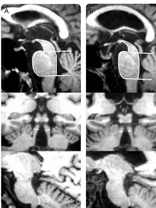
Figure 1 MRI measurements in patients with progressive supranuclear palsy–parkinsonism (PSP-P) developing or not developing vertical supranuclear gaze palsy (VSGP)
Midsagittal T1-weighted volumetric spoiled gradient echo (GE) MRIs of midbrain and pons areas (top row). Coronal T1-weighted volumetric spoiled GE MRIs of superior cerebellar peduncle (SCP) width (intermediate row). Midsagittal T1-weighted volumetric spoiled GE MRIs of middle cerebellar peduncle (MCP) width (bottom row). (A) A patient with PSP-P not developing VSGP; (B) a patient with PSP-P developing VSGP. (A) Midbrain area 108 mm2;
pons area 422 mm2; SCP width 3.65 mm; MCP width 8.95 mm; magnetic resonance
par-kinsonism index (MRPI) 9.58. (B) Midbrain area 88 mm2; pons area 434 mm2; SCP width
present time, diagnosing PSP-P may be challenging in
the early phase of the disease in the absence of VSGP.
To the best of our knowledge, no biomarker exists
that is capable of predicting the appearance of VSGP
in patients with clinical features fulfilling criteria for
PSP-P.
In the last few years, several MRI measurements
such as the ratio of midbrain to pons areas
27–32and
MRPI
11–19have been reported to accurately diagnose
patients with PSP. Both MRI measurements have
proven to be useful tools for diagnosing PSP,
11–19,27–32but the ratio of midbrain to pons areas was reported to
be influenced by aging
33and thus is less reliable than
MRPI for diagnosing this disease, which typically
occurs in older people. Moreover, increased MRPI
val-ues were reported to be capable of predicting PSP in
patients with undetermined parkinsonisms, a finding
of great interest for diagnosing these diseases at a very
early stage.
21In the current study, we found that 11 of 21
pa-tients with PSP-P developed VSGP after a mean
follow-up period of 28.5 months (range 6
–
48
months), while the remaining 10 patients with PSP-P
did not develop VSGP during the follow-up period of
up to 4 years. These 2 groups did not show significant
differences in baseline demographic or clinical variables
except for UPDRS-ME and H-Y scores, which were
Table 2 Clinicoradiologic comparisons in patients with progressive supranuclear palsy–parkinsonism (PSP-P)developing and not developing vertical supranuclear gaze palsy (VSGP) at the end of follow-up
PSP-P developing VSGP PSP-P not developing VSGP pValue
n 11 10 —
M/F, n 10/1 6/4 0.15a
Age at examination, yb 71.465.4 (60–79) 73.067.5 (61–82) 0.58c
Disease duration, yb 9.363.4 (5–15) 10.062.5 (7–14) 0.58c
MMSE scored 20.5 (19–21) 20.0 (19–24) 0.94e
UPDRS-ME scored 41 (36–46) 32 (21–39) 0.001e
H-Y scored 4 (3–5) 2.5 (2–3) 0.001e
Levodopa responsivenessf 2 (18.1) 3 (30) 0.64a
Brain MRI measurementsb,g
Pons area, mm2 480.7660.4 (375–545) 440.4641.8 (378–502) 0.14c
Midbrain area, mm2 82.4611.1 (65–96) 111.0611.2 (99–133) ,0.001c
MCP width, mm 8.660.8 (7.1–9.9) 8.760.7 (8.1–9.8) 0.87e
SCP width, mm 3.2660.5 (2.50–3.85) 3.6060.3 (3.20–4.20) 0.14c
MRPI 15.4861.3 (14.11–17.86) 9.7661.6 (7.37–11.57) ,0.001c
Abbreviations: H-Y5Hoehn-Yahr; MCP5middle cerebellar peduncles; MMSE5Mini-Mental State Examination; MRPI5 magnetic resonance parkinsonism index; SCP5superior cerebellar peduncles; UPDRS-ME5Unified Parkinson’s Disease Rating Scale–Motor Examination.
aFisher exact test.
bData are expressed as mean6SD (range).
cThettest.
dData are expressed as median (range).
eMann-WhitneyUtest.
fNumber (percentage) of patients who showed a clinical improvement of$20% in comparison with that detected in the off state.
gNine of 11 patients (81.8%) with PSP-P developing VSGP and 7 of 10 patients (70%) with PSP-P not developing VSGP
repeated the MRI examination at the end of follow-up period.
Figure 2 Box plots of baseline and follow-up magnetic resonance parkinsonism index measurements in patients with progressive supranuclear palsy–
parkinsonism (PSP-P) developing (A) or not developing (B) vertical supranuclear gaze palsy (VSGP)
slightly higher in patients developing VSGP with
a large overlap between the 2 groups. It is noteworthy
that patients with PSP-P who developed VSGP
showed at baseline significantly higher MRPI values
(mean 14.77; range 12.52
–
20.24) than those
de-tected in patients with PSP-P who did not develop
VSGP at the end of follow-up (mean 9.77; range
7.90
–
11.34) without overlapping values between
the 2 groups. This result is in accordance with a
pre-vious study showing that among patients with
unde-termined parkinsonism, baseline MRPI had higher
values in patients who later developed PSP compared
with those not developing this disease. At the end of
the follow-up period, the MRPI values of patients
who developed VSGP were significantly higher than
those detected at baseline (mean 15.48; range 14.11
–
17.86), whereas no differences between baseline and
follow-up MRPI values (mean 9.76; range 7.37
–
11.57) were observed in patients not developing
VSGP (figure 2). Our findings therefore indicate that
patients with PSP-P who developed VSGP had, at
the end of follow-up, a worsening atrophy of
infra-tentorial brain structures measured by MRPI, a
find-ing not observed in patients with PSP-P not
developing VSGP.
Of note, the patients with PSP-P not developing
VSGP showed baseline MRPI values very similar to
those reported in our recent study
33in patients with
PD with similar motor disability scores (MRPI mean
values 9.77 vs 9.85, respectively). Although baseline
MRPI was not different between these 2 groups, the
PSP-P midbrain area value was smaller than that
de-tected in PD, suggesting that this brain structure may
be involved early in patients with PSP-P not
develop-ing VSGP. Why patients with PSP-P not developdevelop-ing
VSGP had baseline and follow-up MRPI values lower
than those developing VSGP is still unknown.
Because the clinical development of VSGP in patients
with PSP-P may take up to 17 years,
6it is possible
that the patients not developing VSGP after 4 years of
follow-up may require a longer period of clinical
observation before developing VSGP.
Alternatively, because falls occur very frequently in
the elderly and ocular slowness is a clinically difficult
sign to evaluate, it is possible that some patients with
PD have been misdiagnosed as having PSP-P.
How-ever, because the large majority of these patients not
developing VSGP (7 of 10) showed no levodopa
responsiveness at the end of follow-up, the diagnosis
of PD seems unlikely. Whether these patients can be
considered to have PSP-P in a very early stage or are
affected by other forms of parkinsonism needs to be
elucidated. Pathologic studies in these patients not
developing VSGP are warranted.
Of note, in patients with PSP-P developing
VSGP, baseline MRPI showed a higher sensitivity
(100%) and specificity (100%) in predicting the
clin-ical development of VSGP than clinclin-ical features
typ-ically occurring in PSP-P. The combination of
postural instability with backward falls associated
with slowness of vertical saccades showed an accuracy
of 71.4%, while isolated postural instability with
backward falls or isolated slowness of vertical saccades
had an accuracy of 42.9% and 33.3%, respectively.
These results are in agreement with those of our
pre-vious study
21in which we demonstrated that clinical
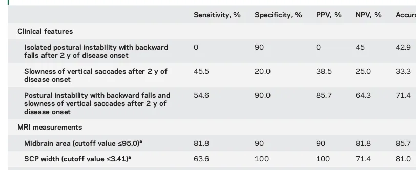
Table 3 Performances of clinical features and magnetic resonance parkinsonism index (MRPI) values forpredicting vertical supranuclear gaze palsy in patients with progressive supranuclear palsy–
parkinsonism
Sensitivity, % Specificity, % PPV, % NPV, % Accuracy, %
Clinical features
Isolated postural instability with backward falls after 2 y of disease onset
0 90 0 45 42.9
Slowness of vertical saccades after 2 y of disease onset
45.5 20.0 38.5 25.0 33.3
Postural instability with backward falls and slowness of vertical saccades after 2 y of disease onset
54.6 90.0 85.7 64.3 71.4
MRI measurements
Midbrain area (cutoff value£95.0)a 81.8 90 90 81.8 85.7
SCP width (cutoff value£3.41)a 63.6 100 100 71.4 81.0
MRPI (cutoff value‡12.52)a 100 100 100 100 100
Abbreviations: NPV5negative predictive value; PPV5positive predictive value; SCP5superior cerebellar peduncles. McNemar tests showed significant differences in comparing the diagnostic accuracies of MRPI vs isolated postural instability with backward falls after the second year of disease onset (p50.001), MRPI vs slowness of vertical saccades after 2 years of disease onset (p,0.001), and MRPI vs postural instability with backward falls after the second year of disease onset and slowness of vertical saccades (p50.04).
features suggestive of PSP such as slowness of vertical
saccades or first-year falls had a lower accuracy
(61.9% and 73.8%, respectively) than MRPI
(92.9%) in predicting the evolution of undefined
parkinsonisms toward clinical phenotypes that
ful-filled the diagnostic criteria for possible or probable
PSP.
26There were some limitations to this study. We
used clinical criteria for the diagnoses of disease,
and none of our patients underwent autopsy. Thus,
it is possible that in some patients, including those
with PSP-P with normal MRPI, the clinical diagnosis
may be in error. However, these clinical evaluations
had been performed according to operational
diag-nostic criteria and expert guidelines
1,4,26,34and were
carried out by a specialist in movement disorders with
extensive experience in diagnosis and management of
PSP. In the current study, MRI was performed with
a 3T scanner, which is not always available in
move-ment disorder clinics. However, we previously
per-formed several studies on MRPI using a 1.5T
scanner,
11,15,21supporting the reliability of 1.5T
scan-ners for MRPI measurements also in patients with
PSP-P.
21The small sample size of patients with PSP-P is
another limitation of our study; however, PSP-P is
a rare disease, and imaging data repeated over time
were difficult to obtain. The large majority of
previ-ous studies of PSP-P enrolled a number of
partici-pants similar to ours. However, larger studies with
longer follow-up times are needed to confirm our
results.
Our study demonstrates that MRPI was more
powerful than clinical features in predicting the
appearance of VSGP in patients with PSP-P on an
individual basis. VSPG remains one of more specific
signs for diagnosing PSP-P, but its appearance in
some patients may require many years after disease
onset. Therefore, the negative predictive value of
baseline MRPI is strictly for VSGP at 4 years
of follow-up and not necessarily for the diagnosis of
PSP-P. Our study showed that MRPI accurately
pre-dicts VSGP and may help distinguish PSP-P from
other parkinsonisms in early stages of the disease.
AUTHOR CONTRIBUTIONS
Prof. Quattrone: drafting/revising the manuscript, study concept or design, analysis or interpretation of data, study supervision. Dr. Morelli: drafting/revising the manuscript, analysis or interpretation of data, acqui-sition of data. Dr. Williams: drafting/revising the manuscript, analysis or interpretation of data. Dr. Vescio: analysis or interpretation of data, sta-tistical analysis. Dr. Arabia: drafting/revising the manuscript, analysis or interpretation of data. Dr. Nigro: analysis or interpretation of data. Dr. Nicoletti: analysis or interpretation of data. Dr. Salsone: analysis or interpretation of data. Dr. Novellino: analysis or interpretation of data. Dr. Nisticò: analysis or interpretation of data. Dr. Pucci: analysis or interpretation of data. Dr. Chiriaco: acquisition of data. Dr. Pugliese: acquisition of data. Dr. Bosco: acquisition of data. Dr. Caracciolo: acqui-sition of data.
STUDY FUNDING
This study was supported by the PON Neuromeasures (PON03PE_00009_1).
DISCLOSURE
The authors report no disclosures relevant to the manuscript. Go to Neurology.org for full disclosures.
Received February 16, 2016. Accepted in final form June 7, 2016.
REFERENCES
1. Williams DR, de Silva R, Paviour DC, et al. Character-istics of two distinct clinical phenotypes in pathologically proven progressive supranuclear palsy: Richardson’s syndrome and PSP-parkinsonism. Brain 2005;128:1247– 1258.
2. Williams DR, Holton JL, Strand C, et al. Pathological tau burden and distribution distinguishes progressive supranu-clear palsy-parkinsonism from Richardson’s syndrome. Brain 2007;130:1566–1576.
3. Dickson DW, Rademakers R, Hutton ML. Progressive supranuclear palsy: pathology and genetics. Brain Pathol 2007;17:74–82.
4. Williams DR, Lees AJ. Progressive supranuclear palsy: clinicopathological concepts and diagnostic challenges. Lancet Neurol 2009;8:270–279.
5. Lang AE. Clinical heterogeneity in progressive supranu-clear palsy: challenges to diagnosis, pathogenesis and future therapies. Mov Disord 2014;29:1707–1709. 6. Respondek G, Stamelou M, Kurz C, et al. The phenotypic
spectrum of progressive supranuclear palsy: a retrospective multicenter study of 100 definite cases. Mov Disord 2014; 29:1758–1766.
7. Williams DR, Lees AJ, Wherrett JR, Steele JC. J. Clifford Richardson and 50 years of progressive supranuclear palsy. Neurology 2008;70:566–573.
8. Williams DR, Lees AJ. What features improve the accuracy of the clinical diagnosis of progressive supra-nuclear palsy-parkinsonism (PSP-P)? Mov Disord 2010;25:357–362.
9. Liscic RM, Srulijes K, Gröger A, Maetzler W, Berg D. Differentiation of progressive supranuclear palsy: clinical, imaging and laboratory tolls. Acta Neurol Scand 2013; 127:362–370.
10. Richardson JC, Steele J, Olszewski J. Supranuclear oph-thalmoplegia, pseudobulbar palsy, nuchal dystonia and dementia: a clinical report on eight cases of“heterogenous system degeneration.”Trans Am Neurol Assoc 1963;88: 25–29.
11. Quattrone A, Nicoletti G, Messina D, et al. MR imag-ing index for differentiation of progressive supranu-clear palsy from Parkinson disease and the Parkinson variant of multiple system atrophy. Radiology 2008; 246:214–221.
12. Hotter A, Esterhammer R, Schocke MF, Seppi K. Potential of advanced MR imaging techniques in the differential diagnosis of parkinsonism. Mov Disord 2009;24:S711–S720.
13. Hussl A, Mahlknecht P, Scherfler C, et al. Diagnostic accuracy of the magnetic resonance parkinsonism index and the midbrain-to-pontine area ratio to differentiate pro-gressive supranuclear palsy from Parkinson’s disease and the Parkinson variant of multiple system atrophy. Mov Disord 2010;25:2444–2449.
parkinsonism is distinct from progressive supranuclear palsy. Brain 2010;133:2410–2425.
15. Morelli M, Arabia G, Salsone M, et al. Accuracy of mag-netic resonance parkinsonism index for differentiation of progressive supranuclear palsy from probable or possible Parkinson disease. Mov Disord 2011;26:527–533. 16. Jones N. Movement disorders: imaging differentiates
pro-gressive supranuclear palsy from Parkinson disease. Nat Rev Neurol 2011;7:186.
17. Stamelou M, Knake S, Oertel WH, Höglinger GU. Mag-netic resonance imaging in progressive supranuclear palsy. J Neurol 2011;258:549–558.
18. Massey LA, Micallef C, Paviour DC, et al. Conventional magnetic resonance imaging in confirmed progressive supra-nuclear palsy and multiple system atrophy. Mov Disord 2012;27:1754–1762.
19. Colosimo C, Bak TH, Bologna M, Berardelli A. Fifty years of progressive supranuclear palsy. J Neurol Neurosurg Psy-chiatry 2014;85:938–944.
20. Karimi M, Perlmutter JS. MRI measures predict progres-sive supranuclear palsy: clinically useful? Neurology 2011; 77:1028–1029.
21. Morelli M, Arabia G, Novellino F, et al. MRI measure-ments predict PSP in unclassifiable parkinsonisms: a cohort study. Neurology 2011;77:1042–1047.
22. Fahn S, Elton RL. Unified Parkinson’s Disease Rating Scale. In: Fahn S, Marsden CD, Calne D, Goldstein M, eds. Recent Developments in Parkinson’s Disease. Florham Park, NJ: MacMillan Healthcare Information; 1987: 153–163.
23. Hoehn MM, Yahr MD. Parkinsonism: onset, progression, and mortality. Neurology 1967;17:427–442.
24. Folstein MF, Folstein SE, McHugh PR.“Mini-mental state:” a practical method for grading the cognitive state of patients for the clinician. J Psychiatr Res 1975;12:189–198.
25. Nicoletti G, Fera F, Condino F, et al. MR imaging of middle cerebellar peduncle width: differentiation of mul-tiple system atrophy from Parkinson disease. Radiology 2006;239:825–830.
26. Litvan I, Agid Y, Calne D, et al. Clinical research criteria for the diagnosis of progressive supranuclear palsy (Steele-Richardson-Olszewski syndrome): report of the NINDS-SPSP international workshop. Neurology 1996;47:1–9.
27. Oba H, Yagishita A, Terada H, et al. New and reliable MRI diagnosis for progressive supranuclear palsy. Neurology 2005;64:2050–2055.
28. Gröschel K, Kastrup A, Litvan I, Schulz JB. Penguins and hummingbirds: midbrain atrophy in progressive supranu-clear palsy. Neurology 2006;66:949–950.
29. Wszolek ZK, Slowinski J, Imamura A, Tsuboi Y, Broderick DF. New and reliable MRI diagnosis for pro-gressive supranuclear palsy. Neurology 2006;66:781. 30. Borroni B, Malinverno M, Gardoni F, et al. A
combina-tion of CSF tau ratio and midsaggittal midbrain-to-pons area ratio atrophy for the early diagnosis of progressive supranuclear palsy. J Alzheimers Dis 2010;22:195–203. 31. Arabia G, Quattrone A. The midbrain to pons ratio: a
sim-ple and specific MRI sign of progressive supranuclear palsy. Neurology 2013;81:2147.
32. Massey LA, Jäger HR, Paviour DC, et al. The midbrain to pons ratio: a simple and specific MRI sign of progressive supranuclear palsy. Neurology 2013;80:1856–1861. 33. Morelli M, Arabia G, Messina D, et al. Effect of aging on
magnetic resonance measures differentiating progressive supranuclear palsy from Parkinson’s disease. Mov Disord 2014;29:488–495.
34. Litvan I, Bhatia KP, Burn DJ, et al. Movement disorders society scientific issues committee report: SIC task force appraisal of clinical diagnostic criteria for Parkinsonian disorders. Mov Disord 2003;18:467–486.