581 | P a g e
Review on
Study of Vibrational Spectroscopy through FTIR.
A.
G. Patil
1, A.P. Maharolkar
2, S. L. Patankar
3, A G Murugkar
41
Vivekananda college, Aurangabad
2
Marathwada Institute of technology, Aurangabad
3
Vasantrao Naik College Aurangabad
4
Dept. of Physics, Dr. B. A. M. University, Aurangabad.
ABSTRACT
The past few years have seen rapid and intense growth in the use of infrared spectroscopy for at-line, on-line,
and even in-line analysis. This progress has been made possible by developments in the design of both FTIR
instruments and equipment to interface these instruments to chemical processes. It has been driven by the need
for real-time monitoring of the chemistry and other fields underlyingdifferent processes and by infrared’s ability
to provide a wealth of information about chemical structure. The present paper reviews some developments,
with details of FTIR instrument. Finally, it reviews a number of representative applications areas in which
process FTIR is currently being used.
Keywords
.:
FTIR, beam splitter, Detector, IR source
I. INTRODUCTION
Fourier-transform infrared spectroscopy (FTIR)[1] is a technique immensely applied to achieve
an infrared spectrum of absorption and emission of three different states like solid, liquid and
gases.high-spectral-resolution data over a broad spectral range has been collected by FTIR spectrometer. This is the fact
that it confers a important advantage over a dispersive spectrometer, which measures intensity above a narrow
range of wavelengths at a time[1].
The fact that a Fourier transforms (a mathematical process) is requisite to convert the raw data into the actual
spectrumcreated from the term Fourier-transform infrared spectroscopy. For same or other applications of this
kind of technique
[1]
.The aim of absorption spectroscopy (FTIR, ultraviolet-visible ("UV-Vis") spectroscopy, etc.) is to measure how
fine a sample absorbs light at each wavelength. The t majority straightforward method to do this, the "dispersive
spectroscopy" technique, is to shine a monochromatic light beam at a sample, measure how much of the light is
absorbed, and repeat for each different wavelength[1]. (This is how some UV–vis spectrometers work, for
example.)
A less intuitive way to obtain the same information is Fourier-transform spectroscopy is. Slightly than shining
a monochromatic beam of light at the sample, this technique excels a beam containing a lot of frequencies of
582 | P a g e
hold a different combination of frequencies, giving a second data point[1]. This procedure is repetitive for many
times[1]. Later, a processor takes all this information and works toward the back to infer what the absorption is
at each wavelength[1].
A broadband light source—one containing the complete spectrum of wavelengths to be calculated is used to
produce beam which is already explained above. [1]. The light shines into a Michelson interferometer—a
definite configuration of mirrors, one of which is stimulated by a motor. Shifting of this mirror causes, each
wavelength of light in the beam is periodically blocked, transmitted, blocked, transmitted by the interferometer,
due to wave interference. Dissimilar wavelengths are modulated at dissimilar rates, so that at each moment the
beam approaching out of the interferometer has a unlikespectrum [1].
Torevolve the raw data(light absorption for each mirror position) into the preferred result (light absorption for
each wavelength)PC processing is necessary. The processing required turns out to be a general algorithm called
the Fourier transform (hence the name "Fourier-transform spectroscopy"). The raw information is sometimes
called an "interferogram"[1].
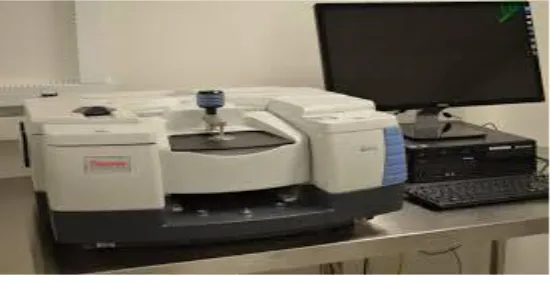
Fig 1: Actual Photograph of FTIR
II. ADVANTAGES OF FTIR
There are three majorreturn for an FT spectrometer compared to a scanning (dispersive) spectrometer
1. FTIR is having unique advance that is the multiplex or Fellgett's advantage. in order from all wavelengths is
togetherat the same time this occurs from this fact only [1]. It is outcome in a higher Signal-to-noise ratio for
a known scan-time for explanationpartial by a permanent detector noise donation (typically in the thermal
infrared spectral region where a photo-detector is limited by generation-recombination noise). For a
spectrum with m resolution elements, this amplify is equivalent to the square root of m. otherwise, it permit a
shorter scan-time for a known resolution. follow multiple scans are often averaged, rising the signal-to-noise
ratio by the square root of the number of scans[1].
2. FTIR is having another unique advance that is the throughput or Jacquinot'sbenefit. the detail that in a
dispersive gadget, the mono-chromator has entrance and way out slits which limit the amount of light that
passes through it gives this outcome from the same. The interferometer throughput is strong-minded only by
the diameter of the collimated beam pending coming from the source. Although no slits are wanted, FTIR
583 | P a g e
since convergent rays are modulated at dissimilar frequencies as the pathway difference is different. Such
anhole is called a Jacquinot stop.[1] For a known resolution and wavelength this circular holepermit more
light through than a slit, resulting in a superior signal-to-noise ratio[1].
3. Another important advantage of FTIR is the wavelength correctness or Connes' advantage. The wavelength
scale is standardize by a laser beam of recognized wavelength that overtake through the interferometer[1].
This is much more steady and precise than in dispersive instruments where the scale depends on the
mechanical group of diffraction gratings. In follow, the accuracy is partial by the divergence of the beam in
the interferometer which depens on the resolution[1].
Less sensitivity to wander off light, that is radiation of one wavelength appearing at another wavelength in the
spectrumis an additional small benefit is [1]. In dispersive gadget, this is the result of imperfections in the
diffraction gratings and accidental reflections. In FT instruments there is no direct equal as the apparent
wavelength is strong-minded by the modulation frequency in the interferometer[1].
III. COMPONENTS
IV. IR SOURCES
FTIR spectrometers are mostly used for capacity in the mid and near IR regions. For the mid-IR region,
2−25 µm (5000–400 cm−1), the most ordinaryfoundation is a silicon carbide element heated to about 1200 K.
The output is like to a blackbody[1]. Small wavelengths of the near-IR, 1−2.5 µm (10000–4000 cm−1), need a
higher temperature source, typically a tungsten-halogen lamp[8]. The long wavelength output of these is limited
to about 5 µm (2000 cm−1) by the absorption of the quartz envelope[1]. For the far-IR, particularly at
wavelengths further than 50 µm (200 cm−1) a mercury discharge lamp providesuperior output than a thermal
584 | P a g e
V. DETECTORSMid-IR spectrometers usually use pyroelectric detectors that react to modify in temperature as the intensity of
IR radiation lessening on them varies[1]. The responsive elements in these detectors are also deuterated
triglycine sulfate (DTGS) or lithium tantalate (LiTaO3). These detectors work at specific temperatures and
givesufficient sensitivity for most customapplications[1]. To attain the top sensitivity the time for a scan is
characteristically a few seconds. Cooled photoelectric detectors are in work for situations want higher sensitivity
or earlierresponse[1]. Liquid nitrogen cooled mercury cadmium telluride (MCT) detectors are the most
extensivelysecond-hand in the mid-IR. by means of the detectors an interferogram can be measured in as little as
10 milliseconds. Uncooled indium gallium arsenide photodiodes or DTGS are the commonoption in near-IR
systems[8]. extremelyresponsive liquid-helium-cooled silicon or germanium bolometers are applied in the
far-IR where both sources and beam splitters are inefficient[1].
VI. BEAM SPLITTER
The figure 3, shows Simple interferometer with a beam-splitter and compensator plate.
An ideal beam-splitter transmits and reflects 50% of the incident radiation. However, as any material has a
limited range of optical transmittance, several beam-splitters may be used interchangeably to cover a wide
spectral range[1]. For the mid-IR region the beam-splitter is usually made of KBr with a germanium-based
coating that makes it semi-reflective. KBr absorbs strongly at wavelengths beyond 25 μm (400 cm−1) so CsI is sometimes used to extend the range to about 50 μm (200 cm−1
)[8]. ZnSe is an option where water vapor can be
a problem but is partial to about 20μm (500 cm−1). CaF2 is the usual material for the near-IR, being both harder
and fewerresponsive to water vapor than KBr but cannot be used beyond about 8 μm (1200 cm−1). In a simple
585 | P a g e
only one time [1]. Theaccurate for this ansupplementary compensator plate of equal thickness is incorporated.
Far-IR beam-splitters are mostly based on polymer films and cover a limited wavelength range.[9]
VII. ATTENUATED TOTAL REFLECTANCE (ATR)
ATR is singleaccomplice of FTIR spectrophotometer to calculate surface possessions of solid or thin film
samples rather than their bulk properties[9]. Generally, ATR has a penetration depth of around 1 or 2
micrometers needy on your sample circumstances [1].
VIII. FOURIER TRANSFORM
The interferogram in carry out contains of a set of intensities calculated for discrete standards of retardation[8].
The dissimilarityamongconsecutive retardation principles is steady. Thus, a discrete Fourier transform is needed.
The fast Fourier transform (FFT) algorithm is used[1].
IX. FAR-INFRARED
The first FTIR spectrometers were urbanized for far-infrared range[1]. The cause for this has to do with the
mechanical lenience needed for high-quality optical presentation, which is connected to the wavelength of the
light being applied.. For the comparatively larger wavelengths of the far infrared, ~10 μm tolerances are
adequate, whereas for the rock-salt region tolerances have to be improved than 1 μm. A typical gadget was the
cube interferometer developed at the NPL[10] and marketed by Grubb Parsons. It applied a stepper motor to drive
the moving mirror, recording the detector response after each step was concluded [1].
X. MID-INFRARED FTIR
With the arrival of inexpensive microcomputers it turn out to be possible to have a computer devoted to
controlling the spectrometer, gather the data, doing the Fourier transform and presenting the spectrum[1]. This
suppled the impetus for the development of FTIR spectrometers for the rock-salt region. The problems of
manufacturing ultra-high precision optical and mechanical components had to be resolve. A broad range of
gadget are now obtainablecommercial[10]y. Although gadget design has turn out to be more complicated, the
basic main beliefs remain the similar. these days, the touching mirror of the interferometer moves at a steady
velocity, and sampling of the interferogram is activate by finding zero-crossings in the fringes of a minor
interferometer lit by a helium–neon laser[10]. In modern FTIR systems the steady mirror velocity is not
firmlyrequisite, as extended as the laser fringes and the original interferogram are recorded simultaneously with
higher sampling rate and then re-interpolated on a steady grid, as pioneered by James W. Brault[10]. This
awards very high wavenumber correctness on the ensuing infrared spectrum and avoids
wavenumber calibration errors[1].
XI. NEAR-INFRARED FTIR
The near-infrared region distances the wavelength range between the rock-salt section and the start of
586 | P a g e
applied mainly in industrial request such as process control and chemical imaging
[1]
.XII. APPLICATIONS
FTIR can beapplied in all function where a dispersive spectrometer was used in the past [10]In totaling, the
better sensitivity and speed have opened up novel areas of application. Spectra can be calculated in situations
where extremelysmall energy reaches the detector and scan rates can exceed 50 spectra a second. Fourier
transform infrared spectroscopy is appliedin geology, chemistry, materials and biology for and research
areas[1].
XIII. MICROSCOPY AND IMAGING
An infrared microscope permit samples to be observed and spectra calculated from regions as little as 5 microns
across. Images can be produce by mixing a microscope with linear or 2-D array detectors[1]. The spatial
resolution can approach 5 microns with tens of thousands of pixels. The images hold a spectrum for each pixel
and can be sight as maps presentation the intensity at any wavelength or grouping of wavelengths[10]. This
permits the distribution of different chemical species within the sample to be seen. Typical studies include
analyzing tissue sections as an alternative to conventional histopathology and examining the homogeneity of
pharmaceutical tablets[1].
XIV. NANOSCALE AND SPECTROSCOPY BELOW THE DIFFRACTION LIMIT
The spatial resolution of FTIR can be additionalenhanced below the micrometer scale by integrating it
into scanning near-field optical microscopy stage [1]. The equivalentmethod is called nano-FTIR and perm,
itsfor performing broadband spectroscopy on materials in ultra-small amount (single viruses and protein
complexes) and with 10 to 20 nm spatial resolution.[11]
XV. FTIR AS DETECTOR IN CHROMATOGRAPHY
The speed of FTIR allows spectra to be get from compounds as they are alienated by a gas chromatograph[1].
though this method is smallapplied compared to GC-MS (gas chromatography-mass spectrometry) which is
more responsive. The GC-IR method is mainlyhelpful for recognize isomers, which by their natural history have
identical masses[1]. Liquid chromatography fractions are supplementaryhardsince of the solvent there. One
notable exception is to measure chain branching as a function of molecular size in polyethylene using gel
permeation chromatography, which is likely using chlorinated solvents that have no absorption in the region in
question[1].
XVI. TG-IR (THERMOGRAVIMETRIC ANALYSIS-INFRARED SPECTROMETRY)
calculating the gas developed as a material is heated permitted qualitative identification of the species to set off
587 | P a g e
XV. CONCLUSION
FTIR is most effective method for qualitative and quantitative analysis[1]. This technique is safe, less time
consuming and works for solid, liquids systems in pristine as well as in mixed and complex form.Thisoffersus
the most perfect means and plan of identifying the surface species getting produced upon molecular adsorption
and the species being generated by surface reactions[1].
REFERENCES
[1.] https://en.wikipedia.org/wiki/Fourier-transform_infrared_spectroscopy
[2.] Griffiths, P.; de Hasseth, J. A. (18 May 2007). Fourier Transform Infrared Spectrometry (2nd ed.).
Wiley-Blackwell. ISBN 0-471-19404-2.
[3.] "The Infracord double-beam spectrophotometer". Clinical Science. 16 (2). 1957.
[4.] Peter R. Griffiths; James A. De Haseth (2007). Fourier Transform Infrared Spectrometry(2nd ed.). John
Wiley & Sons. ISBN 9780470106297.
[5.] Jump up^ [1][dead link]
[6.] Jump up^ "Agilent Technologies to Acquire Varian, Inc. for $1.5 Billion". Agilent. July 27, 2009.
[7.] Brault, James W. (1996). "New Approach to high-precision Fourier transform spectrometer
design". Applied Optics. 35 (16): 2891–2896. Bibcode:1996ApOpt..35.2891B. doi:10.1364/AO.35.002891.
[8.] Connes, J.; Connes, P. (1966). "Near-Infrared Planetary Spectra by Fourier Spectroscopy. I. Instruments
and Results". Journal of the Optical Society of America. 56 (7): 896–910. doi:10.1364/JOSA.56.000896.
[9.] Smith, D.R.; Morgan, R.L.; Loewenstein, E.V. (1968). "Comparison of the Radiance of Far-Infrared
Sources". J. Opt. Soc. Am. 58 (3): 433–434. doi:10.1364/JOSA.58.000433.
[10.]Griffiths, P.R.; Holmes, C (2002). Handbook of Vibrational Spectroscopy, Vol 1. Chichester: John Wiley
and Sons.
[11.] Chamberain, J.; Gibbs,J.E.; Gebbie, H.E. (1969). "The determination of refractive index spectra by fourier
spectrometry". Infrared Physics. 9 (4): 18209. Bibcode:1969InfPh...9..185C.
doi:10.1016/0020-0891(69)90023-2.
[12.]Amenabar, Iban; Poly, Simon; Nuansing, Wiwat; Hubrich, Elmar H.; Govyadinov, Alexander A.; Huth,
Florian; Krutokhvostov, Roman; Zhang, Lianbing; Knez, Mato (2013-12-04). "Structural analysis and
mapping of individual protein complexes by infrared nanospectroscopy". Nature