Western University Western University
Scholarship@Western
Scholarship@Western
Electronic Thesis and Dissertation Repository
7-28-2015 12:00 AM
Evaluation of Radiation Dose-Response in a Breast Cancer Brain
Evaluation of Radiation Dose-Response in a Breast Cancer Brain
Metastasis Model
Metastasis Model
Niloufar ZarghamiThe University of Western Ontario
Supervisor Dr. Eugene Wong
The University of Western Ontario
Graduate Program in Medical Biophysics
A thesis submitted in partial fulfillment of the requirements for the degree in Master of Science © Niloufar Zarghami 2015
Follow this and additional works at: https://ir.lib.uwo.ca/etd Part of the Medical Biophysics Commons
Recommended Citation Recommended Citation
Zarghami, Niloufar, "Evaluation of Radiation Dose-Response in a Breast Cancer Brain Metastasis Model" (2015). Electronic Thesis and Dissertation Repository. 3028.
https://ir.lib.uwo.ca/etd/3028
Evaluation of Radiation Dose-Response in a Breast Cancer Brain Metastasis Model
(Thesis format: Integrated Article)
by
Niloufar Zarghami
Graduate Program in Medical Biophysics
A thesis submitted in partial fulfillment of the requirements for the degree of
Master of Science
The School of Graduate and Postdoctoral Studies The University of Western Ontario
London, Ontario, Canada
Abstract
The second incidence of brain metastases is from breast cancer. Radiotherapy, a standard treatment for brain metastasis, limits cancer division by inducing DNA double-stranded breaks (DSBs). Currently, identical radiation doses are prescribed for all types of brain metastases but little is known about their histological responses. In this thesis, we initiated a radiation dose-response study in a triple-negative human breast cancer brain metastasis mouse model using a custom designed 3D-printed restrainer to assist half-brain irradiation. We quantified the amount of DSBs in tumors and mouse brain tissues using γ-H2AX marker at 30 minutes (acute) and 11 days (longitudinal) after treatment with doses of 8-24 Gy. We also evaluated tumors’ response using histology and MRI. In the longitudinal study we found significant differences in the amount of DSBs, tumor cell density, and nucleus size between irradiated surviving and non-irradiated tumors. These results gave insights to brain metastasis response after radiotherapy.
Keywords
Co
-
Authorship Statement
The following thesis contains one manuscript that has been submitted for publication and one in preparation to be submitted to the peer-reviewed scientific journal.
Chapter 2 encompasses the original manuscript submitted to the Journal of Medical Physics
entitled “Immunohistochemical evaluation of mouse brain irradiation targeting accuracy with 3D-printed immobilization device”. The manuscript was co-authored by Niloufar Zarghami, Michael Jensen, Srikanth Talluri, Paula Foster, Ann Chambers, Fred Dick and Eugene Wong. As the 1st author, I assisted with modeling and design of the head holder, conducting animal experiments, data acquisition, analysis and writing the manuscript. Michael Jensen provided advice regarding the design of the head holder and irradiation procedure. Srikanth Talluri helped with the histology and immunohistochemistry. Drs. Paula Foster, Ann Chambers and Fred Dick provided feedback for the manuscript. Dr. Eugene Wong helped determine research objectives, provided mentorship, supervision, and editorial assistance.
Chapter 3 contains basis of an original manuscript in preparation to be submitted to the
Acknowledgments
First and foremost, I would like to express my deepest appreciation to my supervisor Dr. Eugene Wong, who has inspired me to think beyond boundaries. I am very grateful to him for challenging me and pushing me much further than I thought possible. Through his leadership and guidance, I was motivated and excited to create and develop this thesis. I would like to thank Dr. Wong for the opportunity he has given me, the ample amount of support he provided and the doors he has opened for my future.
I would like to thank the members of my committee, Drs. Paula Foster and Ann Chambers for providing me with guidance and support. I am very grateful for their insightful comments and expertise. I would also like to thank Dr. Fred Dick for providing me the opportunity to pursue my research in the field of cancer biology; an area that I am passionate about and has provided me an unparalleled learning experience.
I would like to extend a massive thank you to members of Wong lab. I truly appreciated and enjoyed all of our group meetings and discussions. To Dr. Jeff Chen- I am most fortunate to have you in our meetings. You are an ultimate role model of medical physicist/scientist. To Michael Jensen- thank you for your leadership and support. You were always there to guide me. To Tom Hrinivich and Anthony Lausch- thank you for helping me with statistics and coding.
Most importantly, I would like to thank those closest to me. To my grandmother, mother, father and brother. Thank you for all your support, courage and unconditional love. You taught me the importance of perseverance, commitment, and academic inquisitiveness. My friends, Nastaran, Shahrzad, and Shamin - my life is filled with joy because of you! To Farhang-for your unconditional support and encouragement throughout my degree.
Table of Contents
Abstract ... ii
Co-‐Authorship Statement ... iii
Acknowledgments ... iv
List of Tables ... ix
List of Figures ... x
List of Abbreviations ... xii
List of Appendices ... xiv
Chapter 1 ... 1
1 General Introduction ... 1
1.1 Cancer ... 1
1.1.1 Breast Cancer ... 1
1.1.2 Triple-‐Negative Breast Cancer Subtype ... 3
1.1.3 Metastasis ... 4
1.1.4 Breast Cancer Metastasis ... 8
1.1.5 Breast Cancer Brain Metastases Preclinical Models ... 10
1.2 Radiation Therapy ... 11
1.2.1 Radiobiology Principles ... 11
1.2.2 DNA Double-‐Stranded Breaks (DSBs) ... 12
1.2.3 Radiation Therapy Dose-‐Response Relationship ... 15
1.2.4 Preclinical Radiation Therapy Devices ... 16
1.3 Magnetic Resonance Imaging (MRI) ... 17
1.3.1 Balanced Steady State Free Precession (bSSFP) MRI Sequence ... 18
1.4 Three-‐Dimensional (3D) Printing Technology ... 19
1.5 Thesis Objectives and Outline ... 20
1.6 References ... 22
Chapter 2 ... 32
2 Immunohistochemical Evaluation of Mouse Brain Irradiation Targeting Accuracy with 3D-‐Printed Immobilization Device ... 32
2.1 Introduction ... 32
2.2 Methods and Materials ... 33
2.2.1 Head Holder Design and 3D-‐Printing ... 33
2.2.2 Mouse Brain Irradiation ... 35
2.2.3 Immunohistochemistry ... 37
2.2.4 Analysis ... 38
2.3 Results ... 39
2.3.1 Mouse Set-‐up in 3D-‐printed Head Holder ... 39
2.3.2 Validating Beam Targeting Accuracy with γ-‐H2AX Staining ... 39
2.4 Discussion ... 41
2.5 Conclusions ... 42
2.6 Acknowledgment ... 42
2.7 References ... 43
Chapter 3 ... 45
3 Evaluation of Radiation-‐Induced DNA Double-‐Stranded Breaks and Tumor Response in a Breast Cancer Brain Metastasis Model ... 45
3.1 Introduction ... 45
3.2 Material and Methods ... 47
3.2.1 Cell Culture ... 47
3.2.2 Animal Model ... 48
3.2.3 In-‐Vivo MRI ... 49
3.2.4 Mouse Hemi-‐Brain Irradiation ... 49
3.2.5 Immunohistochemistry ... 51
3.2.6 Histological Quantification ... 53
3.2.7 MRI Analysis ... 56
3.3 Results ... 56
3.3.1 γ-‐H2AX Radiation Dose-‐response ... 56
3.3.2 Tumor Cell Nuclear Size ... 62
3.3.3 Tumor Cell Density ... 64
3.3.4 In-‐vivo Dose-‐Response ... 66
3.4 Discussion ... 69
3.5 Conclusions ... 72
3.6 Acknowledgment ... 72
3.7 References ... 73
Chapter 4 ... 80
4 Conclusions and Future Directions ... 80
4.1 Chapter 2 ... 80
4.1.1 3D-‐Printed Mouse Holder: Summary ... 80
4.1.2 Future Work ... 81
4.2 Chapter 3 ... 81
4.2.1 Acute Radiation Dose-‐Response Study: Summary ... 81
4.2.2 Acute Radiation Dose-‐Response Study: Future Work ... 82
4.2.3 Ex-‐Vivo Longitudinal Radiation Dose-‐Response Study: Summary ... 82
4.2.4 Ex-‐Vivo Longitudinal Radiation Dose-‐Response Study: Future Work ... 83
4.2.5 In-‐Vivo Longitudinal Radiation Dose-‐Response Study: Summary ... 83
4.2.6 In-‐Vivo Longitudinal Radiation Dose-‐Response Study: Future work ... 84
4.3 References ... 85
Appendices ... 87
Appendix A: Animal Use Protocol ... 87
Appendix B: Fluorescent γ-‐H2AX Immunohistochemistry Standard Operating Procedure .... 88
Appendix C: License and Permission Usage ... 89
List of Tables
Table 1-1: Breast cancer classification based on histopathological markers. ... 3
Table 2-1: Targeting accuracy measurement of γ-H2AX stained brain sections.. ... 40
List of Figures
Figure 1-1: Schematic of the metastasis process.. ... 5
Figure 1-2: Main sites of metastasis in breast cancer patients and their incidencep ... 9
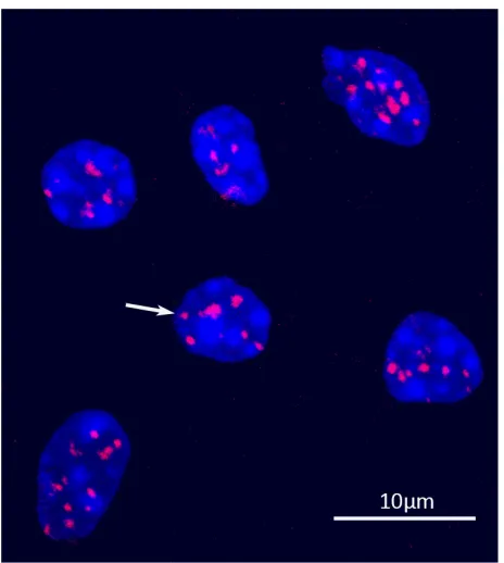
Figure 1-3: Image of mouse brain tissue 30 minutes after receiving ~1 Gy, stained with fluorescent anti-γ-H2AX antibody.. ... 13
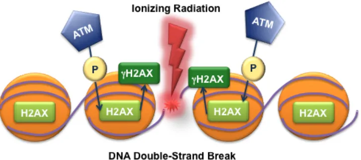
Figure 1-4: DNA is wrapped around core histone proteins. Upon DNA double-strand break, one of the histone proteins, H2AX, gets rapidly phosphorylated by ATM adjacent to the site of damage. The phosphorylated H2AX is termed γ-H2AX. ... 15
Figure 1-5: Processes involved in 3D-printing. ... 20
Figure 2-1: 3D-printed mouse head holder. ... 35
Figure 2-2: Pre-treatment CT images of the mouse brain positioned in 3D-printed head holder. ... 36
Figure 2-3: Beam’s eye view fluoroscopy image of the mouse from the top (dorsal-ventral view). ... 37
Figure 2-4: Fluorescence microscopy of γ-H2AX stained brain sections (red), imaged at 10X magnification. ... 39
Figure 3-1: Radiation dose-response experimental design. ... 50
Figure 3-2: Calculated dose distribution on coronal CT plane of the mouse brain. ... 51
Figure 3-4: Flow chart of the processes involved in the quantification of γ-H2AX intensity, tumor nucleus size and tumor cell density. ... 55
Figure 3-5: Whole brain image of γ-H2AX stained brain section (red), imaged at 10X. ... 57
Figure 3-7: Quantification of the intensity of γ-H2AX staining versus radiation dose 30
minutes after radiotherapy. ... 59
Figure 3-8: Residual DNA damage response 11 days post-irradiation. ... 60
Figure 3-9: Quantification of the intensity of γ-H2AX staining for the various radiation dose 11 days after radiotherapy. ... 61
Figure 3-10: DAPI staining of tumor nuclei 11 days after radiotherapy. ... 62
Figure 3-11: Average size of tumor nuclei 30 minutes after radiotherapy. ... 63
Figure 3-12: Average size of tumor nuclei 11 days after radiotherapy. ... 63
Figure 3-13: H&E stained sections of tumors 11 days after radiotherapy. ... 64
Figure 3-14: The tumor cell density 30 minutes after radiotherapy. ... 65
Figure 3-15: The tumor cell density 11 days after radiotherapy. ... 66
Figure 3-16: MR images (bSSFP) of the mouse brain at two-time points. ... 67
Figure 3-17: The mean fractional growth of brain metastases measured on MR images for various radiation doses. ... 68
List of
Abbreviations
3D Three-dimensional
53PB1 TP53 binding protein 1
ACVS Animal care and veterinary services ADC Apparent diffusion coefficient ANOVA Analysis of variance
ATM Ataxia telangiectasia mutated
ATR ATM-and Rad3-related
BED Biological effective dose
BB Ball bearing
BBB Blood-brain barrier
BCBM Breast cancer brain metastasis
BM Brain metastasis
bSSFP Balanced steady-state free precession
CAD Computer aided design
CBCT Cone beam computed tomography
CNS Central nervous system
CT Computed tomography
DAPI 4', 6-diamidino-2-phenylindole
DDR DNA damage response
DMEM Dulbecco’s modified eagle’s medium
DNA Deoxyribonucleic Acid
DNA-PKcs DNA-dependent protein kinase catalytic subunit
DSB Double strand break
DWI Diffusion weighted imaging
EGFP Enhanced green fluorescent protein
ER Estrogen receptor
HER2 Human growth factor receptor 2
IC Intra-cardiac
IgG Immunoglobulin G
IHC Immunohistochemistry
IR Ionizing radiation
IRIF Ionizing radiation-induced foci
KPS Karnofsky performance status
LF Longitudinal fissure
MRI Magnetic resonance imaging
PFA Paraformaldehyde
PFGE Pulsed field electrophoresis
PIKK Phosphoinositide 3-kinase-related-kinase
PR Progestron receptor
RT Radiation therapy
SRS Stereotactic radiosurgery
STL Stereolithography
T Tesla
TE Echo time
TNBC Triple-negative breast cancer
TR Repetition time
List of Appendices
Appendix A: Animal Use Protocol ... 87
Appendix B: Fluorescent γ-H2AX Immunohistochemistry Standard Operating Procedure on Mouse Frozen Sections ... 88
Chapter 1
1
General Introduction
Cancer is one of the most severe and potentially life-threating diseases. The high morbidity and mortality rate is primarily due to the progression to the metastatic stage. Radiation therapy has been, and continues to be, an important treatment modality for patients diagnosed with cancer. This is especially true in the treatment of brain metastases because of limited drug penetration through the blood brain barrier, or tumors become refractory after multiple lines of chemotherapies, or appear in locations that are inoperable, leaving radiation therapy the only management option. Mechanisms of tumor response/resistance to radiation therapy remain unclear. With the advent of new biological markers, tumor models and novel imaging techniques, we revisit these radiobiological challenges in the preclinical setting. This introductory chapter provides an overview of breast cancer and brain metastasis from this origin, treatment options, principles of radiobiology as well as techniques for quantifying radiation damage, preclinical radiation therapy and imaging technique used in this thesis.
1.1
Cancer
1.1.1
Breast Cancer
Breast cancer is the most common malignancy among both American and
Canadian women.[1,2] It is estimated in 2015, 231,840 women in the USA and 25,000 in
Canada will be diagnosed with breast cancer.[2,3] Breast cancer also has the second
highest mortality rate after lung and bronchus cancer in women. This cancer has a low
incidence in men and accounts for less than 1% of all breast cancer cases.[2,3]
within an individual tumor.[4] Breast cancer can be segregated into three groups based on its structure and origin:
1) Invasive ductal carcinoma, 75% of cases,
2) Invasive lobular carcinoma, ~15% of cases, and
3) Other types such as medullary, neuroendocrine, tubular, apocrine, metaplastic, adenoid
cystic and micropapillary, ~10% of cases.[4,5]
Another approach to classify breast cancer is by their histopathological characteristics. Based on the status of hormone receptors such as estrogen (ER) and progesterone (PR), overexpression of the human epidermal growth factor receptor 2 (HER2) oncogene and Ki-67 labeling index, a proliferation marker, breast cancer has
been classified to four major subtypes.[6,7] Although there are other classifiers for breast
cancer, histopathological markers are known to be widely adopted in the clinics.[4,8,9]
Table 1-1 shows breast cancer subtypes based on their molecular markers and their
prevalence. Other subtypes of breast cancer such as normal like, apocrine molecular type
and claudin-low type, are less frequent.
Table 1-1: Breast cancer classification based on histopathological markers [6,8]
1.1.2
Triple-Negative Breast Cancer Subtype
The triple-negative breast cancer (TNBC) subtype is defined by the absence of hormone receptors (ER, PR) and lack of HER2 expression on immunohistochemical (IHC) analysis in the clinical setting.[6] In this thesis, we employed a TNBC cell line for
our radiation dose-response study. Most of the TNBC cells have a ductal origin (90.7%) and are poorly differentiated.[11] This breast cancer subtype occurs more in young women, with African or Hispanic ancestry.[12] Triple-negative breast cancer usually associated with poor prognosis and aggressive clinicopathological features.[13] Less than 30% of triple-negative patients survive more than 5 years.[12] TNBC has also a distinct
Breast cancer subtype Approximate
prevalence
Marker expression status
Luminal-A 50-60% HER2 negative, ER and/or PR
positive, Ki-67 low
Luminal-B 15%-20%
HER2 negative, ER positive, either Ki-67 high or PR low HER2 positive, ER positive,
any Ki-67, any PR
HER2 overexpression 15-20% HER2 positive and amplified,
ER and PR negative
Triple - negative 15-25% HER2 negative, ER and PR
recurrence pattern. Patients diagnosed with triple-negative breast cancer are frequently young and recurrence occurs 3 to 5 years after initial diagnosis. The incidence of late recurrence (more than 5 years) in these patients is lower compared to other breast cancer subtypes.[11] The treatment of triple-negative cells remains challenging. The primary reason is, this subtype does not express ER/PR and HER2 receptors on the cell surface which limits the use of endocrinal therapy or available targeted drugs.[12] Breast-conserving surgery followed by radiation therapy and/or cytotoxic chemotherapy are the common treatment options for these patients.
1.1.3
Metastasis
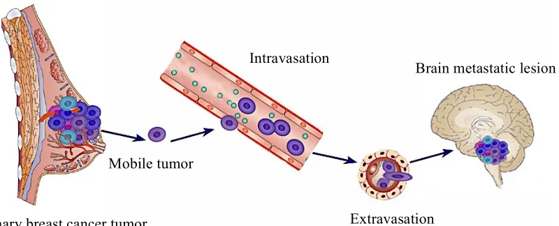
Frequently, it is not the primary cancer that a patient succumbs to, but what makes cancer a life-threating disease is when it gains the ability to escape from the primary site and spread to other organs. This process is known as metastasis.[14] Most of the tumors select either lymphatic or hematogenous (blood-stream) routes to leave the primary organ.[15] However, when these cells are in the circulatory system, the pattern and the micro-environmental conditions in the host organs play a crucial role in the ultimate fate of the primary tumor cells.[16–18] Metastasis is an inefficient process, and usually less than
0.01% circulating tumor cells can complete all the steps shown in Figure 1-1 and form metastasis [15,17,19]; thus this tiny subset is responsible for the majority of cancer deaths.
Figure 1-1: Schematic of the metastasis process. The metastatic cascade consists of multiple steps that the primary tumor cells have to take in order to travel and grow in the secondary site. Adapted from reference 19.
1.1.3.1
Brain Metastasis
Brain metastasis occurs when the primary tumor cells escape from their origin via arterial circulation, extravasate across the capillary membrane of brain parenchyma and finally seed and grow efficiently in the brain.[18] It is known that brain metastasis is the
primary reason for morbidity and mortality with 8.1% overall survival at two years.[20] Brain metastases occur approximately in 20-40% adult patients with systemic malignancies and the incidence is up to 10 times higher than primary malignant brain tumors.[18,21,22] In the USA, it is expected that 21,651 to 43,301 new cases of brain metastasis from any primary origin be diagnosed each year.[20] Nonetheless, the incidence and prevalence of brain metastasis is rising.[22] The primary reason is prolonged survival due to improved treatment outcomes of primary cancer. Some of monoclonal antibody drugs such as Trastuzumab (Herceptin), which are effective for treating systemic malignancies, cannot penetrate to the brain.[18,20] Moreover, advances in noninvasive screening tools and modalities such as magnetic resonance imaging (MRI), have contributed in detection of brain metastasis lesions.
Primary breast cancer tumor Mobile tumor
Intravasation
Extravasation
Breast cancer (20-30%) is the second most common source of brain metastases. Other primary malignancies responsible for brain metastasis include: Lung cancer (40-50%), melanoma (5-10%), lymphoma and other sites (4-6%).[18,23]
1.1.3.2
Brain Metastases Management
The treatment of patients diagnosed with brain metastasis remains clinically challenging due to unique features of the brain. Un-treated brain metastasis causes increased intracranial pressure, which results in median survival of only one month.[21,24] The main treatment options for brain metastasis are surgery, radiation therapy (RT), stereotactic radiosurgery (SRS) and corticosteroids (e.g. Dexamethasone). Chemotherapy is not routinely used as the primary treatment modality for patients diagnosed with brain metastasis.[21] Most of the treatments for these patients are considered to improve the quality of life and achieve neurological palliation. The optimal selection of treatment combination for brain metastasis relies on factors such as the anatomical location of the lesion, number and size of the brain metastases, patient’s age, performance status, extent of extracerebral tumor burden and histological features.[24]
Surgery is the treatment of choice if stringent conditions are satisfied. Surgery can be performed only on accessible sites and depends on the general condition of the patient, number and size of the metastasis.[20,21] Advances in neuro-oncology techniques have
enabled surgeons to resect both single and multiple lesions (2-4) with reduced risks of post-operative complications.[24]
uniform radiation dose while sparing the eyes. The radiation absorbed in the tissue is often represented as deposited energy per unit mass ([J/Kg]=Gy).[26] The most common
radiation dose/fractionation scheme for WBRT is 30 Gy delivered in 10 fractions over two weeks. There is no agreement on the optimum radiation dose schedule/fractionation, and the majority of clinical trials could not provide sufficient evidence of alternative dosing schedules.[25,27] Acute radiation side effects such as alopecia (hair loss), dry desquamation (skin peeling), excessive fatigue and transient neurological symptoms can be seen in patients that receive WBRT. However, using steroids during RT may help with these symptoms. Generally long-term and chronic RT complications in these patients are not seen [27,28] as the median survival of patients treated with WBRT alone is estimated to be 7 months,[20] but it has been reported that the response to the WBRT is related to the primary histology of brain metastasis.[25]
Stereotactic Radiosurgery (SRS) is another RT method, which employs collimated beams usually delivered in an arc around the tumor in a single or a small number of fractions. Unlike WBRT technique, SRS delivers the radiation toward a lesion identified on CT or MRI and spare the normal brain tissues by spreading low doses to a large volume. It is a minimally invasive procedure and associated with high local control rates (80-98% range).[24,28] Depending on the size of the tumor, radiation dose varies between 18-24 Gy in one fraction. SRS is a better treatment option for preserving neurocognitive function. However, SRS as the sole treatment has been advocated for patients with small lesions (maximum size < 3 cm) and with 3 or fewer metastases in their brain.[21,25,29] The overall survival of patients treated only with SRS has been
reported to be 7.5-12.5 months.[24]
WBRT is often used in combination with SRS. Retrospective studies have shown that the combination of these two modalities may improve the overall survival in patients diagnosed with brain metastases in the absence of extra-cranial progression.[24]
One of the unique hurdles of treating tumors in the brain is the blood-brain barrier (BBB). The tight junctions between brain endothelial cells control the passage of substrates from the blood into the brain parenchyma. Thus, the penetration of the chemotherapeutic drugs to the brain and their efficacy depend on their solubility, size and molecular structure.[18,21]
1.1.4
Breast Cancer Metastasis
The primary reason for high mortality rate in patients initially diagnosed with breast cancer is the metastasis. A substantial proportion of these patients (23.3%) develop distant metastasis.[30] The molecular subtype of breast cancer appears to affect the pattern of metastases. The probability of metastasis in the first five years after the breast cancer diagnosis in TNBC subtype is higher than other histological subtypes. The common sites for breast cancer metastases in order of prevalence are bone, liver, lung, brain and ovary.
[17,30,31] The triple-negative subtype has the tendency to metastasize to distal organs
Figure 1-2: Breast cancer patients most commonly die from metastases instead of primary tumor. The common distant sites for breast cancer metastasis are bones, liver, lungs and brain. Main sites of metastasis in breast cancer patients and their
incidence are shown.[31] Adapted from reference 28 and by permission from Macmillan Publishers Ltd: Nature Reviews Cancer, Nguyen et al. 9:274-284,
copyright© 2009 April Nature Publishing Group
1.1.4.1
Breast Cancer Brain Metastasis
Breast is the second common primary site associated with brain metastasis.[18]
With the advances in controlling primary breast tumor and systemic disease, the treatment of breast cancer brain metastasis (BCBM) has become a crucial component for improving the overall survival and quality of life in these patients.[33] It is estimated
10-Brain: ~10%
Bones: ~42% Liver:
30% of all breast cancer patients develop brain metastasis, which can either be symptomatic or asymptomatic.[34,35] An average of 1-year survival can be seen in less
than 20% of breast cancer patients from the time of diagnosis with brain metastasis, and approximately 50% of patients die because of the symptoms in their brain.[34,36,37] Breast cancer subtype has been known to be an independent prognostic factor.
Among different subtypes of breast cancer, HER2 and TNBC have higher risk of brain metastasis. Brain metastases occurs approximately in 10% of breast cancer patients with triple-negative subtype.[34] Additionally, TNBC has the poorest overall survival rate compared to the other subtypes, as this subtype does not benefit from systemic treatments. [34,35,38]
The treatment options for patients with BCBM is the same as other histological types of brain metastases.[34,39] Radiation therapy either as SRS or WBRT modality is an inevitable part of the treatment for BCBM patients. The lack of effective treatment due to the structure of the brain in addition to an increase in the incidence of brain metastasis requires advancing our knowledge about current therapies. So far, improvements in understanding breast cancer subtypes have led to the individualization of systemic treatments, but as these subtypes reach the brain and form metastasis the same radiation dose/fractionation is used. Not only the same radiation protocol is used for all subtypes of breast cancer but all types of brain metastasis regardless of their primary origin. Often, radiosensitivity of primary tumor is not taken into the account for radiation dose prescription; this is despite the fact that brain metastases from breast cancer are relatively more radiosetntisive than other primaries i.e. melanoma.[35,40] The question remains how
specific breast cancer subtype would respond to different doses of radiation.
1.1.5
Breast Cancer Brain Metastases Preclinical Models
arterial circulation to imitate the metastatic process. This method requires developing cell lines that grow preferentially in the brain when injected into the left ventricle of the beating animal heart.[41] In this thesis we employed MDA-MB-231–BR, the brain tropic
human triple-negative breast cancer cell line. These cells are injected directly into the left ventricle of the heart of female nude mice. The parental cell line (MDA-MB-231P) has been passaged six times in brain metastases and in culture to establish brain seeking cells (MDA-MB-231-BR).[42] This sub-line shares many characteristics of human brain craniotomy samples such as apoptosis, proliferation and neuro-inflammatory response.[41]
1.2
Radiation Therapy
Radiation therapy can control rapidly dividing tumor cells (benign or malignant) by creating breaks in their DNA (Deoxyribonucleic Acid). Ionizing radiation (IR) is often considered as a “double-edged sword”, where it can induce mutations in the normal healthy cells while eradicating tumor cell.[43]
1.2.1
Radiobiology Principles
1.2.2
DNA Double-Stranded Breaks (DSBs)
DNA double strand-breaks (DSBs) can be induced by an endogenous source, for example products of internal metabolism, or exogenously such as IR. DNA DSBs are the most biologically significant effect of ionizing radiation. It is known that exposure to 1 Gy of IR (photon) produces approximately 30-40 DNA DSBs in diploid mammalian cells.[45,46,48] DSBs are lesions formed when two opposite strands of the DNA duplex are broken in a distance less than ten base pairs.[45] The incidence of DSBs is relatively rare compared to other types of DNA damage, but the repair mechanism is more complex. In normal mammalian cells, it takes more than 50 minutes to repair 50% of DSBs after induction, whereas for other DNA damages such as base damage or single-strand break the repair will be achieved in less than 20 minutes.[47] Following DSBs a cell can take two different pathways to repair the break depending on its status in the cell cycle.[49]
1.2.2.1
DSBs Detection Techniques
demonstrates an example of our immunofluorescence staining of γ-H2AX to detect DNA DSB visualized under a confocal microscope. This assay has been employed for a number of applications in diagnosis and assessment of treatment efficacy of cancer.[57,58]
γ-H2AX has also been an attractive in-vivo biomarker for detection of malignant cells in blood samples and biopsies.[58,59] It has been shown that tumors and premalignant lesions demonstrate high amount of endogenous γ-H2AX due to replication stress, dysfunctional telomeres and genomic instability.[55,60–62] However, one of the caveats of this method is, it does not detect the actual physical break of the DNA, but the cellular activities following the damage. The timing of γ-H2AX foci loss does not precisely agree with the final rejoining of the DNA DSB leading to possible inconsistencies.[63,64]
1.2.2.2
Role of γ-H2AX
DNA damage happens in the context of chromatin.[47,58] Chromatin structure consists of DNA and proteins. The fundamental units of chromatin are called nucleosomes. Approximately 145-147 DNA base pairs wrap around four pairs (an octamer) of histone proteins (H2A, H2B, H3 and H4) to form a nucleosome.[65] Following DNA DSBs from IR, the cell senses the damage via ATM (Ataxia telangiectasia mutated), a member of the phosphoinositide 3-kinase-related-kinase (PIKK) family.[66] As a result, the histone variant H2A, H2AX (~10% of H2A), are rapidly phosphorylated in the vicinity of the DSB and the chromatin around the break.[63] The phosphorylated H2AX is termed γ-H2AX. Figure 1-4 demonstrates the cascade of activation of H2AX. Phosphorylation of H2AX is also controlled by other PIKK members such as ATR (ATM- and Rad3-related) and DNA-PKcs (DNA-dependent protein kinase catalytic subunit).[66] However, it is mostly ATM that governs the phosphorylation of H2AX following DNA DSB.[63] γ-H2AX serves as a beacon for the cell and triggers DNA damage response (DDR), recruiting detection, signaling and repair proteins to the DSB site.[49] For each DSB, hundreds to thousands of nearby H2AX molecules get phosphorylated to form a focus.[54,63] The number of foci is known to
correlate linearly with IR dose in most cell types.[61,67] However, at high radiation doses
such as 2 Gy, detecting individual focus shortly after IR (~30 minutes) is not possible due to overlapping foci and diffuse covering of γ-H2AX in tumor nuclei.[54,68,69] Counting
discrete foci either manually or automatically on 3D images acquired from confocal microscopy is the common technique employed for low doses of radiation.[61,70] At higher radiation doses where finding separate foci is challenging, intensity quantification techniques such as flow cytometry in cells or image intensity from fluorescence-stained tissue samples are commonly used.[68–71] It should be noted that γ-H2AX is a time sensitive marker. The phosphorylation of H2AX starts within 3 minutes after IR and gets to the maximum level approximately 30 minutes after IR exposure.[53–55] As DNA DSBs are repaired, γ-H2AX either gets dephosphorylated or removed from the chromatin.
[56,61,63] Resolving γ-H2AX over time allows us to assess DNA repair kinetics and
Figure 1-4: DNA is wrapped around core histone proteins. Upon DNA double-strand break, one of the histone proteins, H2AX, gets rapidly phosphorylated by ATM adjacent to the site of damage. The phosphorylated H2AX is termed γ-H2AX.
1.2.3
Radiation Therapy Dose-Response Relationship
One of the concerns in clinical radiobiology is the response of tumor and normal tissue to radiation doses. A sigmoid radiation dose-response curve has been considered for both tumor control and normal tissue complications.[26,46,72] In this relation, the response to radiation tends toward zero as dose approaches zero and tends toward 100% for large radiation doses.[26,45] The response of tumors to radiation can be measured by different end-points such as local tumor control and tumor regrowth delay.
and repair pattern and the microenvironment influenced by hypoxia and angiogenesis.[45] All of these factors play a crucial role in determining the response of the tumors to RT.
In-vivo dose-response studies in preclinical models have the advantage of both inherent tumor radiosensitivity and also the influence of the tumor’s microenvironment. To be able to design experiments using human cancer cell lines in an animal model that can recapitulate clinical dose-response challenges, simulating clinical scenarios in the lab plays a key role.[76] To deliver clinically relevant radiation doses, biological effective dose (BED) formula helps to convert the clinical dose/fractionation regimen to practical dose/fractionation on the models while having the same biological effect.[77,78] BED is a measure of the biological dose delivered by the specific total dose and fractionation:
𝑩𝑬𝑫=𝒏𝒅 [ 𝟏+ 𝒅 𝜶 𝜷
], (1-1)
Where n is the number of fractions, d is the dose per fraction and !! is a radiosensitivity parameter of the tissue.[45,79]
1.2.4
Preclinical Radiation Therapy Devices
To be able to investigate and validate some of the clinical and technological evolutions in the field of cancer and radiation therapy, radiation delivery units are needed for preclinical models. These devices enable us to mimic human radiotherapy conditions. For many decades, small animal models received radiation using Cs137 or Co60 irradiator boxes or clinical linear accelerators. Improvements in technology and the need for precise and accurate dose delivery motivated investigators to design and build imaging and radiation therapy devices dedicated to preclinical studies. Nowadays, preclinical machines capable of on-board imaging along with a range of beam energies (kV) appropriate for the size of small animals are available.[80]
(Princess Margaret Hospital) and modified micro-CT/RT scanner (Stanford University and University of Western Ontario) are the most commonly used devices in this field.[80,81]
Modified Micro-CT/RT scanner has the advantage of high-quality on-board imaging, computerized asymmetric collimator and live animal monitoring.[81] On-board imaging refers to the imaging unit that is installed on the treatment machine and allows imaging before treatment to confirm a match between treatment and planned setup. The combination of image guidance and asymmetric collimators allow irradiating small sub-regional fields in three-dimensions. This unit has been used to study the effect of radiation therapy on normal tissues and will be employed in this thesis for our tumor model.[82]
Precision and accuracy are significant factors in small animal radiation therapy. For instance, the width of the mouse brain is approximately 1 cm in its largest dimension, and sub-millimeter precision is required to irradiate sub-regions of the brain. Considering the scale of animal models, error margins should be less than 0.2 mm to deliver a precise conformal dose.[83] Accurate set-up and immobilization, help with precise radiation dose
delivery.[84] In the clinical setting, external stabilization devices such as head holder, bite blocks and thermoplastics are often used specifically for head and neck cases to minimize the patient’s movement and maximize the reproducibility of the treatment set-up.[85] In preclinical setting, restrainer devices are needed to position the animal in the desirable setting while maintaining animal’s body temperature and monitoring it during radiation dose delivery.
1.3
Magnetic Resonance Imaging (MRI)
high soft-tissue contrast. Gadolinium-DTPA (Gd) is the common contrast agent for detecting brain lesions.[86] In patients with brain metastases, this agent can penetrate leaky
and disrupted tumor-brain barriers, resulting in tumors with hyper-intensities.[87]
In brain metastases preclinical models, 3D MRI allows monitoring tumors temporally and spatially.[88,89] More specifically, this imaging modality is useful in assessing tumor volume and morphology before and after radiation to investigate the effect of treatment.[90] Sequential MR images followed by histological analysis are used in this thesis to investigate response of the tumors to the radiation at the small animal and cellular level.
1.3.1
Balanced Steady State Free Precession (bSSFP) MRI
Sequence
The balanced steady state free precession (bSSFP) MRI sequence was primarily used for cardiac imaging but recently has been employed to image other sites.[91,92] The
most important feature of this sequence is its ability to produce high-resolution images while keeping high signal-to-noise ratio in a reasonable scan time. The contrast in bSSFP is dependent on the ratio of tissue T2 relaxation rate to its T1 relaxation rate. This ratio means that in this sequence images of fat and fluid, which have different T1 and T2 relaxation time but have the same ratio of T2/T1, appear as bright regions.[91,92]
1.4
Three
-
Dimensional (
3D) Printing Technology
Three-dimensional (3D) printing is rapidly being developed and adopted. This
technology allows building tangible 3D solid objects from virtual designs. There are
three main steps to fabricate the desirable object: 1) Modeling 2) Printing 3) Processing.
Manual 3D modeling is usually done using computer aided design (CAD) (e.g.
AutoCad® and SolidWorks™). The drawing will be exported as Stereolithography
(.STL) file format to be readable by the printer. The printer software interprets the .STL
model to thin cross-sectional layers. The 3D-printer used in this thesis is Object30 Pro, an
acrylic based plastic printer. It lays down thin horizontal layers (28 microns) of liquid
resin, which would be cured by ultraviolet light. The layers will be fused together
automatically and create the final object. The processing step consists of smoothing the
surface of the object with sand paper and using a water jet to remove the residual
material. Figure 1-5 shows the processes involved in printing. We will be using
3D-printing technology in our small animal brain irradiation studies.
Three-dimensional printing technology brings benefits over the conventional
manufacturing method. It can be used to create customized objects with varying
complexities from virtual designs in an inexpensive and timely manner. This technology
has been used in various sectors such as industry, medicine, forensic science and
education and will have a great influence on many fields in the near future.[97]
Interestingly, this technology has found its way to preclinical laboratories to construct
objects needed for experimental designs.[98,99]
Furthermore, the nature of the digital file
offers new prospects for sharing and collaborating with researchers. This allows printing
Figure 1-5: Processes involved in 3D-printing. Modeling is done in AutoCAD and exported as .STL format. The 3D-printer converts the digital file to thin cross-sectional layers. The final 3D-printed object is shown on the 3D-printer tray. A final processing step is needed to remove residual material from the object.
1.5
Thesis Objectives and Outline
The main goal of this thesis is to quantify the response of breast cancer brain metastasis to different radiation doses. To achieve this, we had two specific objectives:
1. To improve and histologically evaluate radiation targeting accuracy for mouse hemi-brain radiation therapy on the micro-CT/RT unit.
2. To quantify the acute amount of induced DSBs in a breast cancer brain metastasis model for different radiation doses and evaluate dose-response in the brain metastasis model longitudinally after treatment.
Objective 1 is addressed in Chapter 2 where we utilized 3D-printing technology
for animal immobilization, enabling studies in objective 2. We designed a mouse
restrainer for the purpose of half-brain radiation and used 3D-printer for the fabrication.
The device was evaluated for targeting accuracy using immunohistochemistry. This
work has been submitted to the journal of Medical Physics.
Chapter 3 discusses the response of breast cancer brain metastases to different radiation doses utilizing our 3D-printed device. The amount of induced (acute) and residual (longitudinal) DNA double-strand breaks was quantified for both tumor and mouse normal brain cells. bSSFP MR imaging was used to determine the in-vivo
response of tumors in the mouse model following hemi-brain radiation. A manuscript based on this chapter is in preparation.
1.6
References
1. American Cancer Society. Cancer Facts & Figures 2015. 2015;
2. Canada S. Canadian Cancer Statistics Special topic : Predictions of the future burden of cancer in Canada. 2015;
3. Siegel RL, Miller KD, Jemal A. Cancer Statistics, 2015. CA Cancer J Clin 2015;65(1):5–29.
4. Bertos N, Park M. Breast cancer—one term, many entities? J Clin Invest 2011;121(10):3789–96.
5. Li CI, Uribe DJ, Daling JR. Clinical characteristics of different histologic types of breast cancer. Br J Cancer 2005;93(9):1046–52.
6. Yersal O, Barutca S. Biological subtypes of breast cancer: Prognostic and therapeutic implications. World J Clin Oncol 2014;5(3):412–24.
7. Schnitt SJ. Classification and prognosis of invasive breast cancer: from morphology to molecular taxonomy. Mod Pathol 2010;23 Suppl 2(S2):S60–4.
8. Harbeck N, Thomssen C, Gnant M. St. Gallen 2013: Brief preliminary summary of the consensus discussion. Breast Care 2013;8(2):102–9.
9. Inic Z, Zegarac M, Inic M, Markovic I, Kozomara Z, Djurisic I, et al. Difference between Luminal A and Luminal B Subtypes According to Ki-67, Tumor Size, and Progesterone Receptor Negativity Providing Prognostic Information. Clin Med Insights Oncol 2014;8:107–11.
10. Arteaga CL, Sliwkowski MX, Osborne CK, Perez E a., Puglisi F, Gianni L. Treatment of HER2-positive breast cancer: current status and future perspectives. Nat Rev Clin Oncol 2011;9(1):16–32.
12. Kim K, Lee E, Lee J, Bae J, Korea Breast Cancer Society . Clinicopathologic Signature of TNBC Patients with Good Prognosis. Cancer Res 2009;69(24):4065– 4065.
13. Anders C, Carey L. Understanding and treating triple-negative breast cancer. Oncol (willist Park NY) 2008;22(11):1233–43.
14. MacDonald IC, Groom AC, Chambers AF. Cancer spread and micrometastasis development: Quantitative approaches for in vivo models. BioEssays 2002;24(10):885–93.
15. Wong SY, Hynes RO. Lymphatic or hematogenous dissemination: How does a metastatic tumor cell decide? Cell Cycle 2006;5(8):812–7.
16. Joyce J a, Pollard JW. Microenvironmental regulation of metastasis. Nat Rev Cancer 2009;9(4):239–52.
17. Chambers AF, Groom AC, MacDonald IC. Dissemination and growth of cancer cells in metastatic sites. Nat Rev Cancer 2002;2(8):563–72.
18. Gavrilovic IT, Posner JB. Brain metastases: Epidemiology and pathophysiology. J Neurooncol 2005;75(1):5–14.
19. Smith S, Theodorescu D. Learning therapeutic lessons from metastasis supressor proteins. Nat Rev Cancer 2009;9:253–64.
20. Owonikoko TK, Arbiser J, Zelnak A, Shu H-KG, Shim H, Robin AM, et al. Current approaches to the treatment of metastatic brain tumours. Nat Rev Clin Oncol 2014;11(4):203–22.
21. Patchell R a. The management of brain metastases. Cancer Treat Rev 2003;29(6):533–40.
23. Lee JJ, Lotze MT. Molecular basis of metastasis. N Engl J Med 2009;360(16):1679; author reply 1679–80.
24. Zhang X, Zhang W, Cao W-D, Cheng G, Liu B, Cheng J. A Review of Current Management of Brain Metastases. Ann Surg Oncol 2012;19(3):1043–50.
25. Khuntia D. Review Article Contemporary Review of the Management of Brain Metastasis with Radiation. Adv Neurosci 2015;2015:13.
26. Steel GG. Basica Clinical Radiobiology. 4th ed. London: Hodder Education; 2009.
27. McTyre E, Scott J, Chinnaiyan P. Whole brain radiotherapy for brain metastasis. Surg. Neurol. Int.2013;4(Suppl 4):S236–44.
28. Kondziolka D, Niranjan A, Flickinger JC, Lunsford LD. Radiosurgery with or without whole-brain radiotherapy for brain metastases: the patients’ perspective regarding complications. Am J Clin Oncol 2005;28(2):173–9.
29. Ranjan T, Abrey LE. Current management of metastatic brain disease. Neurotherapeutics 2009;6(3):598–603.
30. Maxmen A. The hard facts. Nature 2012;485(7400):S50–1.
31. Vona-Davis L, Rose DP, Gadiyaram V, Ducatman B, Hobbs G, Hazard H, et al. Breast cancer pathology, receptor status, and patterns of metastasis in a rural appalachian population. J Cancer Epidemiol 2014;2014.
32. Andreopoulou E, Schweber SJ, Sparano JA. Therapies for triple negative breast cancer. Expert Opin Pharmacother 2015;16(7):983–98.
33. Kodack DP, Askoxylakis V, Ferraro GB, Fukumura D, Jain RK. Emerging Strategies for Treating Brain Metastases from Breast Cancer. Cancer Cell 2015;27(2):163–75.
current multidisciplinary management guideline. Clin Transl Oncol 2014;16(5):436–46.
35. De Ieso PB, Schick U, Rosenfelder N, Mohammed K, Ross GM. Breast cancer brain metastases – A 12 year review of treatment outcomes. The Breast 2015;3–10.
36. Cheng X, Hung M-C. Breast cancer brain metastases. Cancer Metastasis Rev 2007;26(3-4):635–43.
37. Niikura N, Hayashi N, Masuda N, Takashima S, Nakamura R, Watanabe KI, et al. Treatment outcomes and prognostic factors for patients with brain metastases from breast cancer of each subtype: A multicenter retrospective analysis. Breast Cancer Res Treat 2014;147(1):103–12.
38. Lin NU, Claus E, Sohl J, Razzak AR, Arnaout A, Winer EP. Sites of Distant Relapse and Clinical Outcomes in Patients with Metastatic Triple-Negative Breast Cancer: High Incidence of Central Nervous System Metastases. 2010;113(10):2638–45.
39. Wiggenraad R, Kanter AV De, Kal HB, Taphoorn M, Vissers T, Struikmans H. Dose-effect relation in stereotactic radiotherapy for brain metastases. A systematic review. Radiother Oncol 2011;98(3):292–7.
40. Nieder C, Walter K. Dose / effect relationships for brain metastases. Survival (Lond) 1998;346–50.
41. Lin NU, Amiri-Kordestani L, Palmieri D, Liewehr DJ, Steeg PS. CNS metastases in breast cancer: Old challenge, new frontiers. Clin Cancer Res 2013;19(23):6404– 18.
43. Brenner DJ. 2011 FAILLA AWARD LECTURE: Exploring Two Two-Edged Swords David. Radiat Res 2012;178(1):7–16.
44. Cohen-Jonathan E, Bernhard EJ, McKenna WG. How does radiation kill cells? Curr Opin Chem Biol 1999;3(1):77–83.
45. Hall EJ, Giaccia AJ. Radiobiology for the Radiologist. 7th ed. Philadelphia: Wolters Kluwer Health/Lippincott Williams & Wilkins; 2012.
46. RADIATION BIOLOGY: A HANDBOOK FOR TEACHERS AND STUDENTS. Vienna: INTERNATIONAL ATOMIC ENERGY AGENCY; 2010.
47. Lavelle C, Foray N. Chromatin structure and radiation-induced DNA damage: from structural biology to radiobiology. Int J Biochem Cell Biol 2014;49(how):84–97.
48. Thrall DE. Biologic basis of radiation therapy. Vet Clin North Am Small Anim Pract 1997;27(1):21–35.
49. Thompson LH. Recognition, signaling, and repair of DNA double-strand breaks produced by ionizing radiation in mammalian cells: The molecular choreography. Mutat Res - Rev Mutat Res 2012;751(2):158–246.
50. Motalleb G, Sanadgol N, Estakhr J, Shahraki E. Methods for DNA strand breaks detection. Res J Appl Sci Eng Technol 2012;4(13):1888–94.
51. Olive PL, Banáth JP. The comet assay: a method to measure DNA damage in individual cells. Nat Protoc 2006;1(1):23–9.
52. Kumari S, Rastogi RP, Singh KL, Singh SP. Dna Damage : Detection Strategies. EXCLI J 2008;7:44–62.
54. Pilch DR, Sedelnikova O a, Redon C, Celeste A, Nussenzweig A, Bonner WM. Characteristics of gamma-H2AX foci at DNA double-strand breaks sites. Biochem Cell Biol 2003;81:123–9.
55. Ivashkevich A, Redon CE, Nakamura AJ, Martin RF, Martin OA. Use of the γ -H2AX assay to monitor DNA damage and repair in translational cancer research. Cancer Lett 2013;487:109–13.
56. Kuo LJ, Yang LX. Gamma-H2AX - a novel biomarker for DNA double-strand breaks. In Vivo (Brooklyn) 2008;22(3):305–9.
57. Redon CE, Nakamura AJ, Martin OA, Parekh PR, Weyemi US, Bonner WM. Recent developments in the use of γ -H2AX as a quantitative DNA double-strand break biomarker. 3(2):168–74.
58. Bonner WM. γ-H2AX and other histone post-translational modifications in the clinic. 2013;1819(7):743–56.
59. Pouliliou S, Koukourakis MI. Gamma histone 2AX (γ-H2AX)as a predictive tool in radiation oncology. Biomarkers 2014;19:167–80.
60. Sedelnikova O a, Bonner WM. γH2AX in Cancer Cells ND ES SC RIB. 2006;(December):2909–13.
61. Bonner WM, Redon CE, Dickey JS, Nakamura AJ, Olga A, Solier S, et al. γH2AX and cancer. Cancer 2011;8(12):957–67.
62. Cornelissen B, Kersemans V, Darbar S, Thompson J, Shah K. Europe PMC Funders Group Imaging DNA damage in vivo using γ H2AX-targeted immunoconjugates. 2012;71(13):4539–49.
64. Löbrich M, Shibata A, Beucher A, Fisher A, Ensminger M, Goodarzi A a., et al. γH2AX foci analysis for monitoring DNA double-strand break repair: Strengths, limitations and optimization. Cell Cycle 2014;9(4):662–9.
65. Talbert PB, Henikoff S. Histone variants--ancient wrap artists of the epigenome. Nat Rev Mol Cell Biol 2010;11(4):264–75.
66. Rothkamm K, Horn S. γ-H2AX as protein biomarker for radiation exposure. Ann Ist Super Sanita 2009;45(3):265–71.
67. Barnard S, Bouffler S, Rothkamm K. The shape of the radiation dose response for DNA double-strand break induction and repair. Genome Integr 2013;4(1):1.
68. Hernández L, Terradas M, Martín M, Tusell L, Genescà A. Highly sensitive automated method for DNA damage assessment: Gamma-H2AX foci counting and cell cycle sorting. Int J Mol Sci 2013;14:15810–26.
69. Yu TY, Chu EHM, Lambur H, Olive PL. Expression of phosphorylated histone H2AX in cultured cell lines following exposure to X-rays. Int J Radiat Biol 2003;79(5):351–68.
70. Anderson D, Andrais B, Mirzayans R, Siegbahn E a, Fallone BG, Warkentin B. Comparison of two methods for measuring γ-H2AX nuclear fluorescence as a marker of DNA damage in cultured human cells: applications for microbeam radiation therapy. J Instrum 2013;8:6008–16.
71. Ismail IH, Wadhra TI, Hammarsten O. An optimized method for detecting H2AX in blood cells reveals a significant interindividual variation in the gamma-H2AX response among humans. Nucleic Acids Res 2007;35(5).
72. Bolus NE. Basic Review of Radiation Biology and Terminology. Nucl Med Technol 2001;29(2):67–74.
74. Nieder C, Berberich W, Schnabel K. Tumor-related prognostic factors for remission of brain metastases after radiotherapy. Int. J. Radiat. Oncol. Biol. Phys.1997;39(1):25–30.
75. Moding EJ, Kastan MB, Kirsch DG. Radiation, Strategies for optimizing the response of cancer and normal tissues to. Nat Rev Drug Discov 2012;29(6):997– 1003.
76. Butterworth KT, Prise KM, Verhaegen F. Small animal image-guided radiotherapy: status, considerations and potential for translational impact. Br J Radiol 2015;88(1045):20140634.
77. Fowler JF. 21 Years of biologically effective dose. Br J Radiol 2010;83(991):554– 68.
78. Marples B. The Management of Gynecologic Cancers:Radiobiology. 2013.
79. Woditschka S, Evans L, Duchnowska R, Reed LT, Palmieri D, Qian Y, et al. DNA double-strand break repair genes and oxidative damage in brain metastasis of breast cancer. J Natl Cancer Inst 2014;106:1–13.
80. Verhaegen F, Granton P, Tryggestad E. Small animal radiotherapy research platforms. Phys Med Biol 2011;56(12):R55–83.
81. Jensen MD, Hrinivich WT, Jung J a, Holdsworth DW, Drangova M, Chen J, et al. Implementation and commissioning of an integrated micro-CT⁄RT system with computerized independent jaw collimation. Med Phys 2013;40(8):0817061– 08170613.
83. Kiehl EL, Stojadinovic S, Malinowski KT, Limbrick D, Jost SC, Garbow JR, et al. Feasibility of small animal cranial irradiation with the microRT system. Med Phys 2008;35(10):4735–43.
84. Armour M, Ford E, Iordachita I, Wong J. CT Guidance is Needed to Achieve Reproducible Positioning of the Mouse Head for Repeat Precision Cranial Irradiation. Radaition Res 2010;173(1):119–23.
85. Perez CA, Brady LW. Perez and Brady’s principles and practice of radiation oncology. 6th ed. Philadelphia: Wolters Kluwer Health/Lippincott Williams & Wilkins; 2013.
86. Anzalone N, Gerevini S, Scotti R, Vezzulli P, Picozzi P. Detection of cerebral metastases on magnetic resonance imaging: intraindividual comparison of gadobutrol with gadopentetate dimeglumine. Acta radiol 2009;50(8):933–40.
87. Bellin MF, Van Der Molen AJ. Extracellular gadolinium-based contrast media: An overview. Eur J Radiol 2008;66(2):160–7.
88. Perera M, Ribot EJ, Percy DB, Mcfadden C, Simedrea C, Palmieri D, et al. In Vivo Magnetic Resonance Imaging for Investigating the Development and Distribution of Experimental Brain Metastases due to Breast Cancer. Transl Oncol 2012;5(3):217–25.
89. Zhou H, Chen M, Zhao D. Longitudinal MRI Evaluation of Intracranial Development and Vascular Characteristics of Breast Cancer Brain Metastases in a Mouse Model. PLoS One 2013;8(4).
90. Gazdzinski LM, Cormier K, Lu FG, Lerch JP, Wong CS, Nieman BJ. Radiation-induced alterations in mouse brain development characterized by magnetic resonance imaging. Int J Radiat Oncol Biol Phys 2012;84(5):e631–8.
92. Bhosale P, Ma J, Choi H. Utility of the FIESTA pulse sequence in body oncologic imaging: review. AJR Am J Roentgenol 2009;192(6 Suppl):83–93.
93. Bernas LM, Foster PJ, Rutt BK. Imaging iron-loaded mouse glioma tumors with bSSFP at 3 T. Magn Reson Med 2010;64(1):23–31.
94. Mallett CL, Mcfadden C, Chen Y, Foster PJ. Migration of iron-labeled KHYG-1 natural killer cells to subcutaneous tumors in nude mice, as detected by magnetic resonance imaging. Cytotherapy 2012;14(6):1–9.
95. Perera M, Ribot EJ, Percy DB, McFadden C, Simedrea C, Palmieri D, et al. In Vivo Magnetic Resonance Imaging for Investigating the Development and Distribution of Experimental Brain Metastases due to Breast Cancer. Transl Oncol 2012;5(3):217–25.
96. Percy DB, Ribot EJ, Chen Y, McFadden C, Simedrea C, Steeg PS, et al. In Vivo Characterization of Changing Blood-Tumor Barrier Permeability in a Mouse Model of Breast Cancer Metastasis. Invest Radiol 2011;46(11):718–25.
97. Chen C, Erkal JL, Gross BC, Lockwood SY, Spence DM. Evaluation of 3D Printing and Its Potential Impact on Biotechnology and the Chemical Sciences. Anal Chem 2014;86(7):3240–53.
98. Zhang L, Yuan H, Burk LM, Inscoe CR, Hadsell MJ, Chtcheprov P, et al. Image-guided microbeam irradiation to brain tumour bearing mice using a carbon nanotube x-ray source array. Phys Med Biol 2014;59(5):1283–303.
Chapter 2
2
Immunohistochemical Evaluation of Mouse Brain Irradiation
Targeting Accuracy with
3
D
-
Printed Immobilization Device
This chapter is adapted from the technical note, “Immunohistochemical evaluation of mouse brain irradiation targeting accuracy with 3D-printed immobilization device” under review in Journal of Medical Physics, by Zarghami N, Jensen MD, Talluri S, Foster PJ, Chambers AF, Dick FA and Wong E.
2.1
Introduction
More than 50 percent of cancer patients receive IR as a main part of their treatment.[1] Over the last few decades, sophisticated and computerized treatment planning, dose delivery and on-board imaging systems have been introduced and are used routinely. The biological responses of tumor and normal tissue to radiation therapy can be investigated using animal models. Accurate simulation of the patient’s treatment scenario on small animals in laboratories will facilitate translating experimental results to the clinical setting and help further our understanding of radiobiology.
Several groups have developed stereotactic holders to improve dose delivery.[7–9] Moreover, sophisticated commercial devices are available for different small-animal imaging machines.[10] While all these stereotactic holders have the same purpose of
improving animal positioning, they may lack some features for some small animal radiotherapy treatment situations. Such features may include physiological monitoring, animal warming, and the capability for fine position adjustments. Commercially available devices have tended to be designed for imaging rather than radiotherapy. More importantly, traditional fabrication methods may not be economical, especially if several customized variations of a stereotactic holder are required. To allow investigators to optimally position the animal for each treatment site and minimize trauma, it is desirable to economically fabricate multiple external holders for small animal radiation therapy.
In this technical note, we introduce a completely 3-dimensional (3D) printed mouse head holder for a micro-CT/RT system. We investigate the feasibility of using 3D printing technology to make a head holder and then evaluate the head holder’s capability for precise mouse brain irradiation. The targeting accuracy is verified with half brain irradiation, using fluorescent immunohistochemical staining for phosphorylated histone H2AX, γ-H2AX, a marker for DNA double-strand breaks[11][12], on frozen mouse brain sections. γ-H2AX is a sensitive bio-dosimeter and can detect radiation doses as low as 1.2mGy.[13] This novel device demonstrates the potential application of 3D-printing to
small animal experimental platforms.
2.2
Methods and Materials
2.2.1 Head Holder Design and 3D-Printing
To size the stereotactic holder, mouse gross anatomical measurements were done on 6-8 week old C57BL/6 and NU-Foxn1nu (Charles River Laboratories, Wilmington,
MA) mice. The mouse head holder was designed using AutoCADTM 2014 (Autodesk,
lnc. San Rafael, CA, USA) (Figure 2-1a). The head holder design has integrated anesthesia gas delivery and respiration pillow sensor. The nose cone position can be adjusted according to the size of the mouse snout and connects to a commercially available Mapleson-D (Patterson Scientific, USA) for anesthetic gas. The respiration pillow sensor is placed under the abdomen of the animal and its rate can be monitored during the procedure. The mouse incisors are placed in a bite bar inside of the nose cone. The bite bar and two length-adjustable ear bars immobilize and orient the head in the desired position. The 5° inclined bed allows the mouse to be in a neutral position while keeping the head level with the axis of gantry rotation. There are indents for all four paws and the tail of the animal to ensure reproducible set-up. A separate hot water circulation is wrapped around animal’s body to maintain its body temperature.
Figure 2-1: 3D-printed mouse head holder. a. Conceptual view of the head holder design in AutoCAD™ b. Photograph of 3D-printed holder with integrated respiratory monitoring pillow and adjustable nose cone, tooth bar and ear bars c. Mouse setup in head holder. A water blanket is used on top of the animal for thermal maintenance. A CT small ball bearing (BB) marker was placed on the right side of the head holder to help the user with the animal orientation on CT and fluoroscopy.
2.2.2 Mouse Brain Irradiation
3D-printed head holder (Figure 2-1c). Animals were placed in a feet first prone position inside the scanner. To validate targeting accuracy, the right half of brains from 10 adult mice (C57BL/6 or NU-Foxn1nu) received the minimum dose of 16 Gy in a single
fraction. Longitudinal fissure (LF) was determined as the anatomical target for the edge of the radiation field within the brain. Setup lasers were initially used to set the scanner landmark position relative to the head holder. CT images were used to verify the position of the ear bars and tooth bar (Figure 2-2a, b). Moreover, CT was used to check the mouse head alignment in 3D. Once the mouse was positioned for treatment, online dorsal-ventral fluoroscopy was acquired to identify the skull features and position the collimators. The collimators were moved so the animal body and left hemisphere of the brain were shielded. The right half of the brain was irradiated with a single field (14x20 mm2) from the animal’s ventral-dorsal direction (Figure 2-3).
Figure 2-3: Beam’s eye view fluoroscopy image of the mouse from the top (dorsal-ventral view). Animal is positioned prone in the 3D-printed head holder. The collimated radiation field (14x20 mm2) is fused on top of the open field.
2.2.3 Immunohistochemistry
Mice were perfused with 0.9% saline containing 4% paraformaldehyde (PFA) approximately 30 minutes after treatment. Brains were harvested and post-fixed in 4% PFA followed by placing them in successive sucrose solution (10-30%) until the specimen sank to the bottom.[14] Brain samples were embedded in Tissue-Tek OCT Compound (Sakura, Torrance, CA) and frozen. Cyrosectioning of coronal slices was performed with 10-µm slice thickness. Sections were stained for fluorescent γH2AX using a well-established protocol published by Ford et al.[14]. Sections were stained with
Millipore, Billerica, MA, USA). DNA counterstaining was achieved with incubation in DAPI (4', 6-diamidino-2-phenylindole, Vector Laboratories, Inc. Burlingame, CA). A motorized fluorescence-scanning microscope (Leica Inc.) was used to automatically acquire images with a 10X objective and stitch them together to form whole brain images on stained histology sections of 5 mice. To quantify targeting accuracy, another set of 10X images, focusing on the midbrain region was acquired with a fluorescence microscope (Carl Zeiss Canada Ltd) for all 10 mice. All images were acquired under the same microscope settings and exposure parameters.
2.2.4 Analysis
Visualization of the actual beam in tissue was possible using γ-H2AX staining on
Figure 2-4: Fluorescence microscopy of γ-H2AX stained brain sections (red), imaged at 10X magnification. DAPI counterstaining of DNA is shown in blue. Sections from four irradiated mice treated in 3D-printed head holder are shown. Intended target was the right half of the brain. (a-d) Example measurement of targeting error on zoomed 10X image. Distance between longitudinal fissure (white line) and γ-H2AX field edge is shown (e).
2.3
Results
2.3.1 Mouse Set-up in 3D-printed Head Holder
Mice were under anesthesia for approximately two hours and breathing rate was monitored during treatment. Mice recovered well from isoflurane after treatment without any signs of trauma to their ears or mouth.
![Table 1-1: Breast cancer classification based on histopathological markers [6,8]](https://thumb-us.123doks.com/thumbv2/123dok_us/7770104.1279026/18.612.97.530.89.484/table-breast-cancer-classification-based-histopathological-markers.webp)