The epidermal growth factor receptor/Erb-B/HER family
in normal and malignant breast biology
SUZANNE A. ECCLES*
The Institute of Cancer Research, Surrey, UK
ABSTRACT The EGFR/Erb-B receptor tyrosine kinases each play distinct and complementary roles in normal breast development. The four receptors form both homodimers and heterodimers in response to binding by ligands which show selectivity for one or more of the receptors (except Erb-B2). Together with the additional flexibility generated by the formation of different dimer pairs, these signalling networks play key roles in directing a variety of both autocrine and paracrine cellular responses. Complex two-way interactions between mammary epithelial cells and the surrounding stroma direct proliferation, duct formation, branching and terminal differentiation during puberty, pregnancy and lactation, with each receptor and ligand fulfilling distinct roles. Caricatures of the normal role of EGFR/Erb-B signalling resulting in aberrant cellular responses are seen in breast cancers, where over-expression and/or (less commonly) mutation of one or more of the receptors results in enhanced cell proliferation, motility, release of proteases and angiogenic factors. Given their importance in tumour progression and their links with resistance to chemotherapy and anti-endocrine therapy, Erb-B receptors (most notably Erb-B2) have been exploited as therapeutic targets. Monoclonal antibodies (e.g. trastuzumab, pertuzumab) and small molecule tyrosine kinase inhibitors (e.g. lapatinib, afatinib) have shown significant clinical responses in some breast cancer subtypes. Additional approaches include targeted toxins or drugs, peptide vaccines, immunRNase and chaperone inhibitors to deplete Erb-B2 protein levels. Greater understanding of the full spec-trum of Erb-B-mediated signalling pathways and their misregulation in breast cancer will provide additional strategies to control malignant progression.
KEY WORDS:
EGFR, c-Erb-B2, c-Erb-B3, c-Erb-B4, cancer
The EGFR/ERB-B/HER family
The epidermal growth factor receptor (EGFR) and its close relatives HER2/c-Erb-B2, Erb-B3 and Erb-B4 are type 1 transmem-brane receptor tyrosine kinases (RTK) with key roles in embryonic development, tissue renewal/repair and cancer. A great deal has been learned about their structure, signalling pathways and aber-rations linked to malignant transformation since the explosion of interest in this family in the 1980’s.
Early discoveries
(*)5DN'DJO\FRSURWHLQZDVWKHÀUVWPHPEHURIWKHIDPLO\ WREHLGHQWLÀHGDVWKHUHFHSWRUIRUDҊJURZWKIDFWRUҋ(*)ZKLFK regulated eyelid opening in mice, and as the binding partner for UDGLRODEHOOHG (*) RQ ÀEUREODVW FHOO PHPEUDQHV 6XEVHTXHQWO\ EGFR was found to have kinase activity when stimulated with ligands and to be capable of phosphorylating tyrosine residues on both itself and downstream targets. Even before EGFR was
identi-www.intjdevbiol.com
*Address correspondence to: Suzanne A. Eccles. Cancer Research UK Cancer Therapeutics Unit, The Institute of Cancer Research, McElwain Laboratories, Cotswold Rd, Belmont, Sutton, Surrey SM2 5NG, UK. Tel: +44(0)208-722-4210. Fax: +44(0)208-722-4134. e-mail: Sue.Eccles@icr.ac.uk - web: http://www.icr.ac.uk
Final, author-corrected PDF published online: 11 November 2011.
ISSN: Online 1696-3547, Print 0214-6282
© 2011 UBC Press Printed in Spain
Abbreviations used in this paper: ADAM, a disintegrin and metalloproteinase; AREG, am-phiregulin; BTC, betacellulin; EGF(R), epidermal growth factor (receptor); EREG, epiregulin; ER, oestrogen receptor; HB-EGF, heparin binding EGF; MEC, mam-mary epithelial cells; MMP, matrix metalloprotease; NRG, neuregulin(heregulin); RTK, receptor tyrosine kinase; (SM)TKI, (small molecule) tyrosine kinase inhibitor; TEB, terminal end bud; TGF_, transforming growth factor alpha; TN(BC), triple negative (breast cancer).
ÀHGWKHUHZHUHHDUO\LQGLFDWLRQVWKDW(*)WKHQWHUPHGHSLWKHOLDO growth factor, even though it is a mitogen for mesenchymal cells too) could be important in breast development as it stimulated growth of mouse mammary gland explants. The role of EGFR was later proved by showing impaired mammary gland development in mice harbouring EGFR mutations (Cohen, 1997).
Structure and function
OHYHODQGDQҊRXWSXWҋOD\HURIWUDQVFULSWLRQDOUHJXODWLRQDQGXOWLPDWHO\ cellular responses (Citri and Yarden, 2006).
All Erb-B receptors consist of an extracellular domain which binds ligands (except in the case of Erb-B2), a transmembrane region and a cytoplasmic domain with kinase activity. Although there are 10 possible combinations of Erb-B dimers, not all are fully biologically active. Erb-B2 has no known ligands, but is the preferred partner of all family members, due to an intrinsically extended interaction loop rendering it constitutively available for dimerisation. Erb-B2 can stabilise EGFR in a conformation that potentiates dimerisation and phosphorylation in the absence of ligand and alters endo-F\WRVLV DQG LQWUDFHOOXODU WUDIÀFNLQJ$OWHUQDWLYHO\ DW OHDVW SDUWLDO transactivation of EGFR can be achieved by ligand-independent intracellular mechanisms, such as G protein-coupled receptor (GPCR) stimulation of Src or elevated calcium levels (Prenzel et
al., 2000). Finally, the receptors interact with, and are modulated
by, steroid hormone receptors and co-receptors. Erb-B3 was generally accepted to be kinase dead due to the lack of several key functional residues including the catalytic base aspartate, but a recent paper suggests that it nevertheless retains the ability to transphosphorylate its own intracellular domain (Shi et al., 2010). In any event, it can certainly form a very active signalling complex with all other EGFR RTK, especially Erb-B2. There is a high degree of homology in the kinase domain of the four receptors (59-81%) but more divergence in the C-terminal domains (only 11-25% identity). In addition to cross-talk between members of the EGFR/Erb-B IDPLO\WKHUHLVHYLGHQFHIRUVLJQLÀFDQWLQWHUDFWLRQVZLWKRWKHU57. such as c-MET and IGF-1R, and it is possible that such alternative signalling pathways are linked to resistance to targeted therapies (Jin and Esteva, 2008). Erb-B receptors also integrate signals from the extracellular microenvironment by forming macromolecular clusters with integrins and tetraspanins in specialised membrane microdomains (Alexi et al., 2011)
Ligands
There are up to 13 recognised ligands of the EGFR family: EGF itself, heparin-binding (HB)-EGF, transforming growth factor (TGF)_, amphiregulin (AREG) epiregulin (EREG), epigen (EPG), betacellulin (BTC) and neuregulins (NRG) 1-6 (also known as heregulins), which have multiple splice variants. EGF and TGF_ are the key EGFR binding ligands, BTC can bind and activate all receptors, and the NRGs have a preference for Erb-B3 and Erb-B4. All EGF family ligands exist as membrane-anchored precursors and are cleaved by metalloproteases (mainly ADAMs) resulting in ectodomain shedding and the release of soluble factors. The cleaved products, particularly of HB-EGF, have been implicated in transactivation of adjacent Erb-B receptors, and the remaining intracellular carboxy-terminal fragments may have additional intracellular signalling functions (Higashiyama et al., 2008). The EGFR ligand shedding DQGVXEVHTXHQWUHFHSWRUDFWLYDWLRQFDQEHVWLPXODWHGE\PDQ\ factors, including cytokines which bind G-protein couple receptors, activating PKC and MAPK signalling pathways (in the so-called triple membrane-passing signal mechanism) or via Wnt ligands binding Fzd receptors. Uncleaved, membrane-bound ligands can also stimulate adjacent cells via a juxtacrine mechanism which may be particularly important in epithelial-stromal communication. There LVHYLGHQFHWKDWGLIIHUHQWOLJDQGVFDQSURPRWHVSHFLÀFSDWWHUQVRI EGFR phosphorylation and dictate the duration of signalling events and divergent cellular responses. For example, TGF_ and AREG
are more potent stimulators of motility and invasion than EGF. This is reportedly due to sustained activation of PLCa and MAPK by the IRUPHUOLJDQGVZKHUHDV(*)SURPRWHVPRUHUDSLGXELTXLWLQDWLRQ and degradation of EGFR.
Downstream signalling
Ligand binding induces conformational rearrangements of the receptors to expose the interaction loop, promoting association of both homodimers and heterodimers, followed by internalisation and/or phosphorylation events. The phosphorylated (activated) receptors act as docking points for a number of direct substrates DQGRUDGDSWRUSURWHLQV6\VWHPDWLFSURÀOLQJRISKRVSKRW\URVLQH LQWHUDFWLRQVLWHVKDVVKRZQWKDWWKHIRXUUHFHSWRUVKDYHVSHFLÀF patterns of binding partners, although each may be recruited to PRUHWKDQRQHVLWHDOEHLWZLWKGLIIHUHQWDIÀQLWLHVRUNLQHWLFV)RU H[DPSOHELQGLQJRI6KF*UERU3,.WR(UE%LVLQÁXHQFHGE\ the mode of activation and the dimerisation partner (Schulze et
al.7KLVGHJUHHRIÁH[LELOLW\WKHUHIRUHDOORZVGLIIHUHQWLDOUH7KLVGHJUHHRIÁH[LELOLW\WKHUHIRUHDOORZVGLIIHUHQWLDOUH -sponses to external stimuli in different microenvironmental contexts and the integration of stimuli into co-ordinated cellular functions.
67$7ZDVLGHQWLÀHGDVDGLUHFWELQGLQJSDUWQHURI(*)5DQG (UE%DQGWKLVLQWHUDFWLRQLVUHTXLUHGLQWKHEUHDVWGXULQJODFWD -tion (Schulze et al., 2005). Interestingly, EGFR and Erb-B4, the only fully functional receptors (in contrast to Erb-B2 and Erb-B3) KDYHWKHJUHDWHVWQXPEHURILQWHUDFWRUVDQGSUREDEO\IXOÀOVLPLODU functions in different cellular contexts in response to their preferred ligands (EGF family and NRGs respectively).
Erb-B3 is activated primarily by NRG-1 and -2 and is a strong activator of the PI3 kinase pathway, having six binding sites for the p85 regulatory subunit. The PI3 kinase pathway is a pivotal point in cell signalling (mainly via AKT and mTOR) regulating cell size, metabolism, survival and proliferation. Negative regulation of pro-apoptotic and growth inhibitory pathways is mediated via FOXO transcription factors and GSK3`. There are additional links to promotion of motility via Rac and Rho, and angiogenesis via activation of HIF-1_.
In summary, the major signalling pathways activated by EGFR-Erb-B receptors are mediated by PI3 kinase, Ras-Raf (MAPK), JNK, PLCa and result in a plethora of biological functions. Although initially termed “growth” factors, the ligands induce not only cell proliferation but also alter adhesion and motility and protect against apoptosis at the cellular level, and promote invasion and angiogenesis at the physiological level. Given that these signalling systems are critical in development, it is not surprising that their activation should result in multiple co-ordinated cell- and tissue-level responses in normal cells, but these are subverted by overexpression/misregulation in pathological processes such as cancer.
Roles in normal breast development
and their ligands are to some extent confounded by spatial and temporal complexities, a degree of redundancy, and many layers of regulation. Nevertheless, those functions that have been elu-cidated have proved informative for the better understanding of FRQVHTXHQFHVRIWKHLUPLVUHJXODWLRQLQWKHFDULFDWXUHVRIFDQFHU
The mammary gland is an unusual organ in that most of its development occurs not in embryonic development, but at puberty, stimulated by steroid hormones. Interestingly, the rudimentary em-EU\RQLFGXFWDOEUDQFKLQJDQGDOOVXEVHTXHQWSKDVHVRIHSLWKHOLDO differentiation are orchestrated by signals from the surrounding stroma; indeed mammary epithelial cells can be completely re-directed (e.g. toward salivary gland morphology and function) by placing them in association with mesenchymal cells from different tissues.
The earliest phases of breast development are oestrogen inde-pendent. In adolescence, oestrogen and oestrogen receptor (ER)_ induce the next stage of branching, and in the adult progesterone
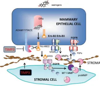
ADAM17 null mice show the same developmental defects as those lacking TGF-_, or HB-EGF, suggest-ing that this protease is primarily responsible for their processing and cleavage (Sahin et al., 2004). Further evidence of its physiological importance in mammary development was provided by its appropriate localisa-tion in the gland (although this was also the case for many other ADAMs). However, the reciprocal lack of its sole inhibitor (TIMP3) in TEBs suggests that ADAM17 would be active in areas of active ductal development. The means by which EGFR-activated stromal cells induce mammary epithelial development has also been intensively studied but it still not fully elucidated. *LYHQWKHPXOWLSOLFLW\RIFHOOXODUUHVSRQVHVUHTXLUHG FHOOSUROLIHUDWLRQVWURPDOҊLQYDVLRQҋRIGXFWVWHUPLQDO differentiation) it is likely that many simultaneous cel-lular responses are elicited. MT1-MMP (MMP14) is enriched in stroma surrounding TEBs and its activity, like that of ADAM17, would be enhanced by the lo-cal downregulation of TIMP3. MMP14 may stimulate ductal branching by activating MMP2 and degrading collagen 1. However it is membrane-bound and could RQO\ LQÁXHQFH HSLWKHOLDO FHOOV LQGLUHFWO\ 6WUXFWXUDO defects in TEBs in Erb-B2-/- mammary epithelium PD\FRQWULEXWHWRWKHREVHUYHGGHÀFLHQF\LQGXFWDO penetration into the fatpad, secondary to misregulation of matrix metalloproteinases, since branching mor-SKRJHQHVLVUHTXLUHV003DQG003SURPRWHV secondary and tertiary branching. Another potential contributory mechanism is signalling via FGFR2 on mammary epithelial cells stimulated by stromal FGF10 providing both mitogenic stimuli and guidance cues for ductal development (Sternlicht and Sunnarborg, 2008) (Fig. 2).
Other Erb-B family members: additional roles in later mammary differentiation
AREG becomes strongly repressed during preg-QDQF\ DQG ODFWDWLRQ DQG (*)5 LV QRW UHTXLUHG IRU alveolar development. Erb-B2 in the epithelium is UHTXLUHGIRUGXFWDORXWJURZWKDQG7(%GHYHORSPHQW and its functional partner is thought to be Erb-B3,
Puberty Signalling Biological responses
Stromal cells (EGFR+) stimulated by AREG induce MEC proliferation and ductal elongation
FGFs, by binding to FGFR on MEC, may serve as guidance cues for duct formation Fat pad ‘invasion’ by ducts is potentiated
by proteases, upregulated by EGFR/Erb-B2
MMPs activated by EGFR-MT1-MMP stimulate branching
Erb-B2 and Erb-B3 regulate ductal branching density and morphology
Erb-B2 controls TEB development via a role in cellular compartmentalisation
Erb-B3 also influences TEB development via different complementary mechanisms
AREG is strongly repressed
Erb-B3 is important in morphogenesis, signalling via PI3K
Erb B2 heterodimers respond to NRG1 in alveolar development
Erb-B4 is required for lobuloalveolar development (activated via NRG1Į?)
AREG is strongly repressed
Erb-B2/B3/B4 heterodimers are required for lactation
Erb-B4 sustains differentiation and milk production via Stat5 in luminal progenitor MEC Erb-B4 soluble intracellular fragment signals
are important
Fig. 1. Differential roles of EGFR/Erb-B receptors and ligands during normal mammary gland development and maturation. The Figure illustrates the time- and differentiation-dependent expression of the four Erb-B receptors and key ligands at puberty and during adult breast maturation, pregnancy and lactation. Their contrasting roles in ductal outgrowth, terminal end-bud (TEB) development, alveolar maturation and lactation are shown, and differential expression in epithelial and stromal cells is highlighted.
(PRG) plays a key role. The major contribution of Erb-B recep-tors is during puberty, pregnancy and lactation, when the steroid hormones upregulate production of many growth factors, including those of the EGF family.
EGFR signalling and the role of proteolytic regulation of ligands Both EGF and TGF_ in vitro and under certain circumstances
in vivo can stimulate growth of mammary epithelium and ductal
since knock-out of either gene results in similar abnormalities in these processes.
Erb-B3 appears when mammary glands mature and Erb-B4 is only expressed during pregnancy and lactation. In Erb-B3-/- mice, there was a decrease in the size of TEBs but increases in branch density and the number of TEBs. This was associated with an increase in apoptosis but no change in cell proliferation rates in TEBs. The major signalling pathway activated by Erb-B3 in this context seems to be PI3 kinase (Stern, 2008). Nrg3 polymorphisms can also result in abnormalities in rodent mammary gland develop-ment, also likely via activation of Erb-B3. Thus Erb-B3 plays a key role in regulating morphogenesis of mammary epithelium (Fig. 1). During pregnancy and lactation, Erb-B2, in association with its SDUWQHUVDQGLQUHVSRQVHWRQHXUHJXOLQVLVUHTXLUHGIRUDOYHRODU differentiation and milk protein production. Erb-B4 is essential for lobuloalveolar development and for maintaining lactation report-edly via Stat5a. The ligand(s) responsible for activating Erb-B4 in WKHVHSURFHVVHVKDYHQRWEHHQÀQDOO\HOXFLGDWHGDOWKRXJK15* HRG1_ has been implicated. Most of Erb-B4’s functions in the mammary gland seem to be mediated by a soluble intracellular fragment (4ICD). This fragment can localise in mitochondria and nuclei, eliciting different functional responses in cells. The 180 kDa membrane-bound Erb-B4 is cleaved by ADAM17, releasing a 120 kDa ligand-binding ectodomain and an 80kDa transmembrane peptide (m80) with kinase activity (Blobel et al., 2009). The latter
in many cancers, including breast, linked to a multiplicity of molecular mechanisms including RYHUH[SUHVVLRQ GXH WR JHQH DPSOLÀFDWLRQ RU epigenetic mechanisms, activating mutations of the receptors themselves or activation in-duced by autocrine/paracrine ligands. EGFR is IUHTXHQWO\DFWLYDWHGE\KHWHURGLPHULVDWLRQZLWK other TKR, and also heterologous receptors such as GPCR via Src. Recently a novel mechanism of EGFR signalling has been suggested: EGF-induced translocation to the nucleus associated with p-Tyr-1068 and indirect binding to DNA via STAT3 enabling EGFR to act as a transcriptional regulator of genes such as cyclin D1 and iNOS - discussed in (Burness et al., 2010).
Links to breast cancer
All of the four receptors are overexpressed to varying degrees in breast cancer, with their promi-nence being in rank order Erb-B2>EGFR>Erb-B3>Erb-B4. Many ligands, including NRG splice variants are also overexpressed, suggesting the possibility of autocrine signalling, although the combined measurement of EPG and NRG4 were the strongest predictors of relapse free interval and overall survival (McIntyre et al., 2010).
EGFR
Many early reports of expression/overexpres-sion of EGFR and its ligands in breast cancer and links to prognosis were contradictory as they were largely based on immunohistochemi-cal data and did not necessarily address the activation state of the signalling pathway. More
Fig. 2. EGFR/Erb-B signalling linked to epithelial cell-stromal cell interactions in ductal development in the mammary gland. The Figure illustrates the reciprocal interactions be-The Figure illustrates the reciprocal interactions be-tween stromal cells and mammary epithelial cells (MEC) during normal development. ADAM sheddases, regulated by oestrogens, release ligands such as AREG which stimulate EGFR on neighbouring stromal cells. Additional proteases (e.g MMP2 activated by MT1-MMP) release growth factors which reciprocally stimulate MECs to proliferate. MMPs are also required for ductal ‘invasion’ into the fat pad. Adapted from (Sternlicht and Sunnarborg, 2008).
fragment is released from the membrane by presenilin-dependent a-secretase cleavage. Cleavage can be stimulated by ligand bind-ing (generally HRG, HB-EGF or BTC) or simply in response to Erb-B4 overexpression.
Nuclear 4ICD in secretory mammary epithelium signalling via Stat5a is thought to be the major driver of lactation since Stat5a transcriptionally regulates `-casein and whey acidic protein (WAP) promoters. The proposed mechanism is as follows: Erb-B4 when activated becomes phosphorylated at Y964, providing a docking site for Stat5a SH2 domains. The following regulated intra-membrane proteolysis (RIP) previously described results in liberation of the 4ICD-Stat5a complex and its translocation to the nucleus. It has been suggested that the 4ICD simply acts as a Stat5a chaperone, but may also serve as a regulator of transcription (Jones, 2008) or indeed have intrinsic independent transactivation activity. The 4ICD fragment also functions as a selective ER_ co-activator since it regulates expression of PGR, SDF-1 and Erb-B4 itself. This involves ER_ recruitment not to canonical ERE sites but to AP-1 sites in a complex with c-Jun (DeNardo et al., 2007). In contrast, cytosolic 4ICD may have different functions, which may explain VRPHRIWKHDSSDUHQWO\FRQWUDGLFWRU\ÀQGLQJVHVSHFLDOO\LQUHODWLRQ to breast cancer biology.
Mutations and mechanisms of activation
recent gene expression analyses and also functional studies have FODULÀHGWKHNH\UROHWKDW(*)5PD\SOD\LQVSHFLÀFEUHDVWFDQFHU subsets (Foley et al., 2010).
0XWDWLRQVLQ(*)5DUHUDUHLQEUHDVWFDQFHUVEXWLWLVDPSOLÀHG in some cases (e.g. metaplastic subtype) (Burness et al., 2010) DQGLVDOVRKLJKO\H[SUHVVHGZLWKRUZLWKRXWDPSOLÀFDWLRQLQEDVDO breast cancers, a subset of triple negative breast cancers (TNBC; i.e. lacking ER, PGR or Erb-B2). TNBCs represent 10-17% of all breast cancers, are more common in certain non-Caucasian ethnic groups (e.g. those of African descent) and tend to occur at less than 50 years of age. These cancers are also generally of high grade and show distinct patterns of metastasis; notably visceral, liver and brain involvement leading to particularly poor prognosis (Dawson
et al., 2009). EGFR expression was found to be higher in patients
with nodal or distant metastases than in those without (Sutton et
al., 2010). Also, TGF_ and NRG2` and the proteases responsible IRUWKHLUFOHDYDJHDQGDFWLYDWLRQDUHIUHTXHQWO\RYHUH[SUHVVHGLQ ER- cancers (suggesting autocrine signalling) whereas AREG is expressed in ER+ cancers and may rather promote paracrine activation via the stroma (Foley et al., 2010).
TNBC is particularly prevalent in women carrying a BRCA1 mutation and EGFR overexpression is found in 67% of BRCA1 related cancers vs ~ 18% of sporadic cancers. Using human mam-mary epithelial cell (hMEC) cultures it was shown that even partial suppression of BRCA1 function (using RNAi) could induce EGFR expression and an increase in EGFR+ cancer stem-like cells, suggesting that this receptor could provide a growth advantage at early stages of transformation. Treating (MMTV-Cre) BRCA1ÁR[ÁR[ p53+/-WUDQVJHQLFPLFHZLWKWKH(*)5LQKLELWRUHUORWLQLEVLJQLÀFDQWO\ increased the latency period before mammary tumours developed, although this was limited to ER- and not ER+ subtypes (Burga et
al., 2011). These data suggest that early intervention with EGFR
LQKLELWRUV FRXOG EH EHQHÀFLDO WRBRCA1 mutation carriers in a
preventive setting. However, in established cancers, clinical trials to date have not shown major responses in unselected patient populations (Burness et al., 2010).
,QÁDPPDWRU\EUHDVWFDQFHULVSDUWLFXODUO\DJJUHVVLYHZLWKWKH majority of patients having disease in their lymph nodes and over one third with distant metastases at the time of diagnosis. It is FKDUDFWHULVHGE\ORVVRI(5DFWLYDWLRQRI1)ѣ%RYHUH[SUHVVLRQ of RhoC GTPase (resulting in a highly angiogenic phenotype) and a hyperactivated MAPK signalling path which has been linked to overexpression of EGFR and Erb-B2 (Van Laere et al., 2007).
EGFR has also been implicated as a key player in the mitogenic and motogenic effects mediated by the HGF-c-MET signalling axis in breast cancer. HGF and/or c-MET expression increase with tumour progression and each is independently associated with poor prognosis. Cross-talk between these RTK has been identi-ÀHGLQVHYHUDOWXPRXUW\SHVZLWK+*)DEOHWRWUDQVDFWLYDWH(*)5 and conversely EGFR ligands activating c-MET via intracellular signalling pathways. EGFR inhibitors have been shown to attenu-ate HGF-mediattenu-ated proliferation, motility and invasion of several breast cancer cell lines in vitro.
Erb-B2
Neu WKH UDW KRPRORJXH RI (UE% ZDV ÀUVW LGHQWLÀHG LQ D chemically-induced neuroblastoma and shown to be similar to a UHWURYLUDORQFRJHQHY(UE%UHODWHGWR(*)50XOWLSOHVXEVHTXHQW VWXGLHVLGHQWLÀHG(UE%+(5LQRIEUHDVWFDQFHUVZKHUH
it is associated with poor prognosis, although it is an early event occurring in over half of in situ carcinomas. Interestingly, an Erb-B2 subtype clearly emerged from a genetic analysis of breast cancer ZKLFKLGHQWLÀHGÀYHPROHFXODUVLJQDWXUHVZLWKGLVWLQFWELRORJLFDO properties (Sorlie, 2004). Breast cancers may express between 25-50 copies of the ERB-B gene resulting in up to 2 million recep-tors per cell. This differential provides a relatively tumour-selective therapeutic target, as levels are absent or low in most normal adult tissues. One exception is the heart, which may explain some of the cardiomyopathies seen with Erb-B targeted therapies, particularly when they were administered with anthracyclines, which is no longer a recommended combination (Procter et al., 2010).
(UE%SRVLWLYHFDQFHUVKDYHVRPHXQLTXHELRORJLFDOSURSHUWLHV including increased sensitivity to doxorubicin (perhaps because the HER2JHQHLVFRDPSOLÀHGZLWKWRSLVRPHUDVHWKHWDUJHWRI doxorubicin) and relative refractoriness to anti-endocrine agents (partly due to an inverse relationship between Erb-B2 and ER_ ex-pression levels). It has been suggested that Erb-B2+ breast cancers may be especially prone to post-surgical recurrences due to their SUROLIHUDWLYHUHVSRQVHVWRJURZWKIDFWRUVLQZRXQGÁXLGDQGWKDW this could be prevented by trastuzumab. However, only a subset of Erb-B2+ breast cancers responds to trastuzumab, suggesting ad-ditional levels of complexity. Recently, more detailed analyses have revealed not only genetic heterogeneity within Erb-B2+ tumours, EXWDOVRVLJQLÀFDQWHSLJHQHWLFLQÁXHQFHV7KRVHWKDWH[SUHVVKLJK levels of hypoxia-regulated genes show characteristics of basal cancers, and those without behaved more like luminal cancers, showing that even oncogenic drivers such as Erb-B2 are susceptible to modulation by the host microenvironment (Gatza et al., 2011).
Erb-B3
In mouse mammary carcinoma models induced by PyVMT or mutated or overexpressed neu, (UE%LVIUHTXHQWO\DFWLYDWHGDQG found in association with Erb-B2, again attesting to the effective DVVRFLDWLRQRIWKLVVSHFLÀFGLPHULVDWLRQSDUWQHUVKLS7KHVHWXPRXUV DUHLQKLELWHGE\WKH(*)5LQKLELWRUJHÀWLQLEDQGWKHGXDOLQKLELWRU lapatinib: response has been associated with inhibition of Erb-B3 and AKT phosphorylation. In contrast resistance has been linked with a de novo point mutation in Erb-B2. In a panel of six Erb-B2-overexpressing human tumour cell lines, Erb-B3 knockdown by RNAi was as effective as Erb-B2 knockdown at inhibiting prolifera-tion in vitro, and xenograft tumour growth in vivo, whereas EGFR expression was dispensable. Preferential phosphorylation of Erb-B3 was also seen in Erb-B2+ human breast cancers, suggesting a pivotal role for Erb-B3 in Erb-B2-driven tumours (Stern, 2008).
disparate results reported could also be related to the subcellular localisation of Erb-B3 and its activation status, which in turn are regulated by ligand availability.
Erb-B4
There are contradictory data on the role of Erb-B4 in breast cancer since both positive and negative associations with prognosis have been described; also the full-length and cleaved (4ICD) splice variants may have different functions (Sundvall et al., 2008). It is reportedly associated with luminal A breast cancer subtypes (which have a better prognosis than other groups) perhaps linked to its role in differentiation. It is also generally associated with positive ER status and hence has been predicted to be oestrogen regulated. In support of this is the fact that its promoter contains three possible oestrogen response element half-sites and oestrogen recruits ER_ to one of these sites.
Inhibition of Erb-B4 expression can inhibit the proliferation of ER+ breast carcinoma cell lines, suggesting a growth promoting effect that is ER_-dependent and reliant upon cross-talk between these two signalling pathways. Other Erb-B family members also show reciprocal interactions, with mechanisms that include phosphoryla-tion of ER_ or its co-activators, linked to ER_-induced upregula-tion of Erb-B ligands and a fostering of autocrine signalling loops. However, the Erb-B4 mechanism involving interactions between a FHOOVXUIDFHDQGDQXFOHDUUHFHSWRULVXQLTXH
Paradoxically, stimulation of ER+ breast cancer cells with Erb-B4 ligand, however, can result in cell death, even though it is also dependent on proteolytic release of 4ICD. It has been proposed that, since this activity is independent of nuclear localisation, it may be the result of 4ICD activity in the cytosol. 4ICD contains motifs similar to pro-apoptotic BH3 proteins and localises in mitochondria and the endoplasmic reticulum; also its anti-tumour activity is at-tenuated by the caspase inhibitor zVAD, suggesting that it is a bone
ÀGH apoptosis inducer. In support of this function, an association between cytosolic expression of 4ICD and apoptotic cells was noted in human breast cancers (Jones, 2008). A unifying hypothesis to explain these disparate functions has been proposed whereby in early stages of breast cancer development, ligand (mainly HRG_) activation of Erb-B4 generates nuclear 4ICD. ER+ cells have a growth advantage and a 4ICD-dependent autocrine loop develops, shifting from a Stat5 activator of differentiation to an ER_ co-activator driving proliferation. At later stages both ER_ and Erb-B4 may be lost, perhaps being redundant in the face of additional even stronger oncogenic drivers (Jones, 2008). These intriguing possibilities (and their therapeutic implications) are currently being actively investigated.
These varying associations between the Erb-B receptors and breast cancer biology are interesting as they may echo their contrasting roles in mammary gland development: promoting cell proliferation/’invasion’ of ductal epithelial cells into the mammary fat in the case of EGFR/Erb-B2/3 and a function in epithelial differentiation for Erb-B4 (Stern, 2008). It may be that subsets of precursor cells, UHSUHVHQWDWLYHRIDVSHFLÀFVWDJHRIEUHDVWGHYHORSPHQWEHFRPH trapped in that phenotype, unresponsive to normal regulatory cues.
Ligands and downstream signalling pathways in breast
cancer
Given the importance of the ADAM17-AREG/TGF_-EGFR axis in
normal mammary development, it is not surprising that all of these elements – and those that they regulate such as additional proteases and growth factor signalling pathways - are misregulated in breast cancers. ADAM17, AREG and TGF_DUHIUHTXHQWO\XSUHJXODWHG with co-expression of TGF_ and EGFR being associated with par-ticularly poor prognosis. Antisense suppression of AREG reduced the tumorigenicity of immortalised human mammary epithelial cells and prevented EGFR becoming activated in response to exogenous ligands (Ma et al., 2010).
In an interesting series of experiments with human mammary epithelial cells at different stages of transformation from immor-talised – premaligant - tumorigenic, it was shown that progression was associated with upregulation of AREG and TGF_, rendering them independent of exogenous EGF. In 3D cultures, reversion to a non-malignant phenotype was achieved by inhibitors of proteolytic activity or EGFR, suggesting an autocrine MMP/ADAM-dependent EGFR activation pathway. Similar effects were achieved by ADAM17 siRNA (Kenny and Bissell, 2007), suggesting that this protease may be a good therapeutic target in EGFR-dependent breast cancers.
Angiogenesis, invasion and metastasis
$OOVROLGWXPRXUVUHTXLUHWKHDELOLW\WRFRRSWKRVWYDVFXODWXUH and/or stimulate de novo angiogenesis in order to grow progres-sively. The newly formed vasculature is leaky and provides a ready conduit for haematogenous dissemination. One of the major angiogenic growth factors is VEGF, which is upregulated not only by hypoxia, but also via the Erb-B oncogenes, likely via the PI3K-AKT signalling pathway. Erb-B2 and EGFR overexpres-sion tends to correlate with increased levels of VEGF A (and also lymphangiogenic cytokines VEGF C and D) and in some cases with higher microvessel density.
Hypoxia itself is a known adverse prognostic indicator, and many HIF-responsive genes (such as MMPs, CXCR4, c-MET, LOX) are implicated in angiogenesis and metastasis. In the case of the G-protein-coupled chemokine receptors such as CXCR4, and its ligand CXCL12/SDF-1_, their expression and activation has also been linked to site-selective metastasis. Erb-B2 and CXCR4 levels tend to correlate in breast cancers, and the former is reported to enhance CXCR4 expression and to inhibit ligand-induced degra-dation. Interestingly, inhibition of CXCR4 expression suppressed Erb-B2-mediated malignant potential suggesting a mechanistic linkage between the two signalling axes.
In order to metastasise, cells must detach from underlying ex-WUDFHOOXODUPDWUL[(&0DFTXLUHWKHDELOLW\WRVXUYLYHXQGHUWKHVH anchorage independent conditions, and demonstrate the ability to invade surrounding tissues and basement membranes. Invasion UHTXLUHV D PRWLOH SKHQRW\SH DQG LQ PRVW FDVHV LV SRWHQWLDWHG E\SURWHRO\WLFDFWLYLW\DOWKRXJKSURWHDVHLQGHSHQGHQWҊDPRHERLGҋ motility has also been described. An early manifestation of breast cancer is the ability of transformed but premalignant cells to proliferate within the ductal lumen, away from their natural ECM niches. Detachment usually induces downregulation of EGFR (and cell death) but this can be overcome by Erb-B2, which stabilises EGFR and `1 integrin via Erk-Sprouty2 signalling and a lowered DIÀQLW\ RI WKH KHWHURGLPHUV IRU F&EO ZKLFK QRUPDOO\ SURPRWHV (*)5WUDIÀFNLQJWRO\VRVRPHV(Grassian et al., 2011).
signal-ling pathways promoting cell motility; indeed EGFR ligands are very potent chemotactic factors, stimulating rapid migration along concentration gradients. AIB1/SRC3 is associated with breast LQYDVLRQ DQG PHWDVWDVLV$ VSOLFH YDULDQW 65&у SURPRWHV NRG-Erb-B2 mediated motility, co-operating with Erb-B2 to in-duce progression of DCIS to invasive ductal carcinoma. Erb-B2 reportedly selectively promotes MDA-MB-468 migration mediated by EGF via phosphorylation of tyr1248 and a transient activation of PLCa. Others have implicated Tyr1227 and Shc-Memo signalling in response to HRG in T47D cells, although in both cases PLCa was an important contributor. HRG`(UE%VLJQDOOLQJDOVRVLJQLÀFDQWO\ enhances metastasis via PI3 kinase signalling, since mutation of its six YXXM PI3K p85-binding domains inhibited breast carci-noma cell motility, invasion, vascular intravasation and invasion (Smirnova et al., 2011).
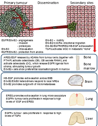
Overall, EGFR/Erb-B signalling has been linked to all aspects of metastasis: stimulation of angiogenesis, alterations in cell-cell and cell-matrix adhesion, upregulation of proteases and other key PROHFXOHV&;&5&'VSHFLÀFLQWHJULQVYHVVHOLQWUDYDVDWLRQ extravasation and organ-selective colonisation (Eccles, 2001) (Fig.
Organotropism and site selectivity of metastasis
The commonest site of breast cancer metastasis is the bone, DQGWKHUHLVDZHOOGHVFULEHGҊYLFLRXVF\FOHҋLQYROYLQJERWKWXPRXU and host cells that perpetuates tumour cell colonisation, metastasis development and bone destruction (Guise, 2010). Although there LVUHSRUWHGO\QRFOHDUDVVRFLDWLRQEHWZHHQKLJKOHYHOVRIVSHFLÀF Erb-B receptors (or their ligands) in bone metastases – and indeed Erb-B2 seems to be under-represented – there is good evidence of a role for ligand-activated proteases in the bone microenviron-ment (Foley et al., 2010).
Seminal work by Massague’s group deciphered the genetic determinants underlying key phases of metastasis in the MDA-MB-231 breast carcinoma model: escape from the primary tu-mour, extravasation and growth at secondary sites. Sublines with WURSLVP IRU VSHFLÀF VLWHV ERQH OXQJ EUDLQ ZHUH LVRODWHG DQG their key characteristics analysed. Osteoclast activity in the bone microenvironment is regulated by a balance between RANKL and osteoprotegerin (OPG). The presence of breast cancer cells upsets this balance, leading to a net increase in bone destruction via
os-Primary tumour Dissemina
Ɵ
on Secondary sites
AREG/EGF released by ADAMs from tumour cells (together with PTHrP) activate osteoblasts (OB). OB secrete RANKL and activate osteoclasts (OC), which release EGFR ligands from stroma, stimulating tumour growth
Erb-B2+ cells show preferential localisation/growth in marrow
Bone
Bone
marrow
Brain
Lung
Liver
EGFR/Erb-B2 - angiogenesis Erb-B2 – motility
- invasion Erb-B2/CXCR4- directional migration
- proteolysis Erb-B2/EGFR/EREG/HB-EGF extravasation
Erb-B3 - intravasation TGF activates MSC in metastatic “niche”Į
EGFR/Erb-B2 - protection from anoikis
HB-EGF promotes extravasation across BBB Erb-B2/ErbB3 heterodimers respond to local NRGs Erb-B2 promotes outgrowth of micrometastases
EREG promotes extravasation in lung microvasculature EGFR+ tumour cells proliferate in response to high levels of EGF and EREG
EGFR+ tumour cells proliferate in response to high levels of TGFĮ
Fig. 3. Role of EGFR/Erb-B receptors and ligands in metastasis and organotropism. Most if not all of the Erb-B family have been linked to an increased probability of breast cancer metastasis and, in some cases to site-selective growth. At the primary tumour site, EGFR and Erb-B2 in particular regulate factors that promote angiogenesis and invasion. Protection from anoikis and motility contribute to the ability of tumour cells to survive in the circulation, and specific ligands promote extravasation through the specialised microvasculature of different organs. There is limited involvement in the early generation of ‘pre-metastatic niches’, but finally, tumour cells expressing EGFR or Erb-B receptors respond to local growth factors to generate overt metastases. OB = osteoblasts, OC= osteoclasts.
3). There is now a growing appreciation that bone-marrow-derived mesenchymal stem cells (MSC) are recruited to the stroma of developing tumours and also contribute to formation of the ҊSUHPHWDVWDWLFQLFKHҋ6WLPXODWLRQRI06&ZLWK tumour cell-derived TGF_ simulated release of angiogenic factors and induced breast carci-noma cell migration; another example of mutual Erb-mediated tumour-host interactions involved in tumour progression (De Luca et al., 2011).
Dissemination to lymph nodes is a key factor in breast cancer staging, but increasingly iden-WLÀFDWLRQRIGLVVHPLQDWHGWXPRXUFHOOV'7&LQ the blood and in bone marrow aspirates is also being linked to poor prognosis. These cells IUHTXHQWO\RYHUH[SUHVV(*)5DQG(UE%HYHQ when the primary tumour is negative)(Braun et
al., 2001). Such observations have led to the
WHRFODVWVDQGUHOHDVHRIVHTXHVWHUHGJURZWKIDFWRUVZKLFKLQWXUQ stimulate growth and invasion of the tumour (Kang et al., 2003).
Ligands such as EGF or AREG shed by tumour cells activate EGFR-expressing osteoblasts to secrete less OPG; simultane-ously autocrine stimulation releases PTHrP from tumour cells to the same end, and the osteoblasts in turn release EGFR ligands and perpetuate the cycle of monocyte-derived osteoclast activation via RANKL or MCP and thus bone destruction (Foley et al., 2010). PTHrP has been recognised as one of the “metastasis virulence factors” within a bone metastasis gene signature. It transcription-ally regulates AREG and to a lesser extent TGF_ and HB-EGF, and may also increase ligand shedding via ADAM17. MMP1 and ADAMTS-1, additional members of the 11-gene signature, also increase AREG shedding and bone metastasis. Although MDA-MB-231 represents an ER_ negative breast cancer, since AREG is regulated by oestrogen, this pathway could be of more general VLJQLÀFDQFH LQ EUHDVW FDQFHU +RZHYHU LW UHPDLQV WR EH VHHQ whether EGFR inhibitors will have an impact, alone or in combina-tion, against bone metastasis.
A degree of organotypic metastasis selection is determined by the ability of tumour cells to extravasate through the phenotypically-GLVWLQFWYDVFXODWXUHZLWKLQVSHFLÀFRUJDQV7KHERQHPDUURZDQG OLYHUYDVFXODWXUHLVIHQHVWUDWHGDQGGRHVQRWSURYLGHDVLJQLÀFDQW barrier to tumour cell colonisation; however the lung endothelium has tight junctions and the blood-brain barrier (BBB) is even further specialised. EREG, together with COX-2, MMP-1 and ANGPTL4 has been linked to an enhanced capacity of breast tumour cells to extravasate in the lungs since together they can compromise the integrity of the pulmonary microvasculature. In contrast, HB-EGF,
&2;DQGVSHFLÀFDOO\WKH_2,6-sialyltransferase ST6GALNAC5 were strongly associated with breast cancer brain metastases (Bos et al., 2009) (Fig. 3).
The predilection of breast cancers expressing Erb-B oncogenes to metastasise to the brain may also be due to the fact that their cognate ligands (NRGs) are neural growth factors. It has been shown experimentally that Erb-B2 overexpression increases the outgrowth of breast cancer cells in the brain, rather than the initial VHHGLQJ HIÀFLHQF\ DQG IRUPDWLRQ RI PLFURPHWDVWDVHV(Palmieri
et al., 2007). The co-association of CXCR4 and Erb-B2 may be
linked to visceral metastases, and similarly cells overexpressing EGFR could respond to the high levels of ligands such as TGF_ and EREG in liver and lung (Eccles and Welch, 2007) (Fig. 3).
Therapy targeting the EGFR/Erb-B family
EGFR and Erb-B2 have been the main receptors considered as targets for immunotherapeutic approaches in breast cancer, mainly via antibody-based therapies, but also in active immunisation and gene therapy protocols, as well as ligand-targeted toxin and antisense/RNAi approaches and anti-Erb-B2 vaccines (Ladjemi
et al., 2010) (Table1; Fig. 4).
A novel means of inhibiting Erb-B expression and function is via HSP90 chaperone inhibitors such as 17-AAG and NVP-AUY922. HSP90 levels correlate with poor prognosis in breast cancer and (UE%LVDSDUWLFXODUO\VHQVLWLYHҊFOLHQWҋSURWHLQEHLQJKLJKO\GH -pendent on HSP90 for its correct folding and cellular localisation. HSP72, a related chaperone, has also been shown to be essential for Erb-B2-driven oncogenesis in transgenic mouse models by
Agent Type Target(s) Comments
Trastuzumab (Herceptin) and T-DM1 Humanised IgG1 monoclonal antibody Erb-B2 juxtamembrane region (domain IV) Approved for Erb-B2+ MBC and node+ early stage disease. Also conjugated to DM1, (maytansine toxin) for targeted delivery. Phase III. Active in trastuzumab-resistant cells
Pertuzumab (Omnitarg) Fully humanised IgG1 monoclonal antibody
Erb-B2 dimerisation domain (II) Inhibits dimerisation with EGFR and Erb-B3. Phase III breast cancer
Cetuximab (Erbitux) Chimaeric IgG1 monoclonal antibody EGFR ECD Little activity in EGFR+ breast cancer Panitumumab (Vectibix) Fully human IgG2 monoclonal antibody EGFR ECD Little activity in EGFR+ breast cancer
Ertumaxomab Bispecific monoclonal antibody Erb-B2 and FcȖRI/III Promotes ADCC via T cells. Phase II breast cancer Gefitinib (Iressa) Reversible TKI (quinazoline) EGFR kinase domain Limited activity in breast cancer
Erlotinib (Tarceva) Reversible TKI (quinazoline)
EGFR kinase domain Limited activity in breast cancer
Lapatinib (Tykerb) Reversible TKI 4-anilinoquinine
EGFR/Erb-B2 kinase domain Response linked primarily to Erb-B2 overexpression.
Neratinib (HKI-272)
Irreversible TKI Pan-Erb-B kinase domain
Active in cells with EGFR and Erb-B2 mutations. Phase I/II + temsirolimus in Erb-B2+ or TNBC
Afatinib (BIBW-2992) Irreversible TKI EGFR/Erb-B2 kinase domain
Active in trastuzumab-resistant breast cancer. Plans to trial in inflammatory BC and in several combinations
Canertinib (CI-1033)
Irreversible TKI Pan-Erb-B kinase domain
Phase II results poor in lung and ovarian cancer
AEE788 Reversible TKI EGFR, Erb-B2 VEGFR Added benefit with letrozole in preclinical breast cancer models. Phase I/II in other cancer types
BMS-599626 (AC480)
Reversible TKI EGFR/Erb-B2 kinase domain Inhibits EGFR-ErbB2 heterodimers. Phase I
Arry-334543 Reversible TKI EGFR/Erb-B2/B4 kinase domain
Phase II in breast cancer
MM-111 Bispecific fusion protein Blocks Erb-B3 ligand binding Targets ErbB2-B3 heterodimers
Tanespimycin (17-AAG) Ansamycin HSP90 chaperones Targets ErbB2, AKT, VEGFR, ERD. Phase III breast Retaspimycin
(IPI-504)
Ansamycin HSP90 chaperones Targets ErbB2, AKT, VEGFR ERD, Phase II breast
NVP-AUY922 Isoxazole resorcinol HSP90 chaperones Targets ErbB2, AKT, VEGFR, ERD. Phase I/II breast BIIB 021 Purine scaffold HSP90 chaperones Phase II in ER+ MBC + Exemestane
TABLE 1
regulating senescence signalling pathways (Meng et al., 2011). Other key client proteins are also important in breast tumour development (e.g. ER_) and progression, for example: AKT in cell survival and resistance to multiple agents; VEGF receptors in angiogenesis/lymphangiogenesis; FAK, Src and MET in inva-sion - to name but a few. HSP90 inhibitors induce depletion and proteasomal degradation of Erb-B2 and other client proteins in
vitro and in vivo, resulting in potent antitumour and antiangiogenic
activity in preclinical tumour models (Eccles et al., 2008), most notably in ER_+/Erb-B2+ BT474 xenografts. Recent clinical trial data in MBC where patients had progressed on trastuzumab are very promising (Modi et al., 2011). In preclinical and clinical stud-. In preclinical and clinical stud-ies, trastuzumab labelled with positron-emitting isotopes has also been used to monitor responses as described later in this review.
Inhibiting ligand binding/dimerisation
The anti-EGFR antibody cetuximab has been tested in combina-tion with a variety of standard chemotherapeutic agents in breast FDQFHU EXW ZLWK OLWWOH EHQHÀW DQG VRPHWLPHV XQDFFHSWDEOH VNLQ
fact that Erb-B2 positive tumours have a predilection for colonising the brain and because antibodies fail to cross the BBB effectively. Pertuzumab is another humanised antibody that inhibits Erb-B2 heterodimerisation with other family members by binding to the dimerisation loop of the former (i.e a different site from trastuzumab). It has shown some promise in Erb-B2+ breast and ovarian cancer patients and is also being evaluated in combination with trastuzumab and chemotherapy (CLEOPATRA trial) (Baselga and Swain, 2010). In general, Erb-B2 targeted therapies are only effective in cancers ZLWKJHQHDPSOLÀFDWLRQDQGVHQVLWLYHDVVD\VDUHQHHGHGWRGHWHU -mine those eligible (e.g. HercepTest or Oncotype Dx). Antibodies to Erb-B2 have also been employed to measure expression levels in tumours and also to monitor responses to therapy using positron emission tomography (PET) or Erb-B2-targeted nanoparticles in MRI approaches, since they report non-invasively on the level of membrane-exposed receptor (Capala and Bouchelouche, 2010). Similarly, ELISA or dot-blot assays can be performed on plasma samples to monitor the levels of Erb-B2 ECD and also reportedly correlate with tumour levels in several studies.
Advances in antibody-based therapies include the targeting of toxicity. Cetuximab is a chimeric IgG1,
whereas panitumumab is a fully human IgG2 anti-EGFR monoclonal antibody ZKLFKKDVEHHQOHVVIUHTXHQWO\HYDOXDWHG in breast cancer (Burness et al., 2010).
Trastuzumab, a humanised anti-Erb-B2 monoclonal antibody targeting the juxtamembrane region of the extracellular domain, has been more successful in clinical trials, particularly in combination with standard chemotherapy and in the adjuvant setting (Goel et al., 2011). This sensitisation to chemotherapy may involve downregulation of Mcl-1, an antiapoptotic protein and/or activation of PTEN which dephosphorylates AKT (a key survival signal) thereby promoting cell death. Trastuzumab is reportedly most active in tumours driven by Erb-B2 homodimers and is also effective in combination with antiendocrine therapies in ER+ tumours. Given the many key cellular functions activated downstream of Erb-B signalling, it is not surprising that trastuzumab (and other Erb-B-targeted therapies) also inhibit angiogenesis, which could contribute indi-rectly to tumour responses. Combinations of trastuzumab with bevacizumab, an antibody targeting the major angiogenic cytokine (VEGF) are also being trialled.
In addition to direct inhibition of Erb-B2 function, e.g. by promoting its internalisa-tion and degradainternalisa-tion, trastuzumab can pro-mote antibody-directed cellular cytotoxicity (ADCC) mediated by natural killer (NK) cells. Interestingly (but unfortunately) an increase in the incidence of brain metasta-ses has been observed in patients treated ZLWKWUDVWX]XPDE7KLVPD\UHÁHFWERWKWKH
prodrug activating enzymes or nanoparticles carrying cytotoxic payloads (Colombo et al., 2010). Recently, complexes of a com-. Recently, complexes of a com-pact human anti-Erb-B2 antibody and cytotoxic human pancreatic RNase have shown promise in vitro and in vivo in a rodent breast tumour model (Borriello et al., 2011). In experimental systems, it has been shown that using combinations of anti-Erb-B2 antibod-ies recognising different epitopes are more effective at inhibiting ligand-mediated invasion than single agents.
Inhibiting kinase activity
Several reversible and irreversible SMTKI with varying degrees of selectivity have been developed. These small molecule agents compete with ATP binding in the kinase domain of the receptor and inhibit downstream signalling. The most well known are the EGFR VHOHFWLYHLQKLELWRUVHUORWLQLEDQGJHÀWLQLEWKHGXDO(*)5(UE% inhibitor lapatinib and the more recently developed irreversible inhibitor afatinib.
*HÀWLQLEDQGHUORWLQLEKDYHVKRZQOLWWOHLIDQ\VLQJOHDJHQWDFWLYLW\ in breast cancer, even in triple negative cases which often have high levels of EGFR. Several trials are underway where EGFR TKI are used in combination with other cytotoxic or molecularly targeted agents.
Lapatinib binds the ATP-binding pocket of both EGFR and Erb-%SUHYHQWLQJDXWRSKRVSKRU\ODWLRQSRWHQWLDOO\OLPLWLQJҊHVFDSHҋ or rapid development of resistance to monotherapies. Unlike trastuzumab, activation of the PI3K pathway does not seem to LPSDLULWVHIÀFDF\,WKDVEHHQXVHGDORQHDQGLQFRPELQDWLRQZLWK both cytotoxic and molecularly targeted agents. Combination with trastuzumab improved median progression-free survival and re-duced the risk of disease progression in a phase III trial of heavily pre-treated patients and it has also been approved for use in those who have failed trastuzumab. Most responses have been seen in (UE%SRVLWLYHSDWLHQWVZLWKOLWWOHRUQREHQHÀWLQSDWLHQWVZLWK triple negative or EGFR-positive cancers (Burness et al., 2010). Neratinib is another orally active agent which irreversibly inhibits the kinase activity of EGFR, Erb-B2 and Erb-B4 which has shown promise in previously untreated and also trastuzumab-resistant breast cancers (Colombo et al., 2010). It is also being trialled in combination with paclitaxel, capecitebine and vinorelbine.
Links between Erb-B signalling and resistance to therapy
EGFR activation has been linked to resistance to the anti-endocrine agent tamoxifen, possibly via phosphorylation of AIB1, an ER_ co-activator which can result in mitogenic (rather than inhibitory) activity. Phosphorylation of ER_ (which can be achieved by several upstream kinases including Erb-B2) can also lead to oestrogen-independent transcription of ER_ targets, also resulting in resistance to tamoxifen. There is cross talk between ER_, c-Src, EGFR, and STAT5b in ER+ breast cancer cells and increased EGFR and c-Src signaling is associated with tamoxifen resist-ance. Constitutively active STAT5b increased DNA synthesis and conferred tamoxifen resistance in ER+ human breast carcinoma cell lines. Cells exposed to a pure anti-oestrogen (fulvestrant) ac-TXLUHGDUHVLVWDQWSKHQRW\SHZKLFKLQFOXGHGHOHYDWHGH[SUHVVLRQ and phosphorylation of EGFR and Erb-B2, together with TGF_ and AREG expression. More recently, fulvestrant has also been shown to upregulate Erb-B3 and/or Erb-B4 and to sensitise cells to
NRG`1 (Hutcheson et al., 2011). It is possible, therefore, that mito-. It is possible, therefore, that mito-genic autocrine signalling via any or all of the Erb-B receptors may subsume the role of oestrogen during breast cancer progression. EGFR activation stimulates signalling via AKT and ERK, and has been linked to multidrug resistance via upregulation of MRP-1,3,5 and 7. Erb-B3 (and also Erb-B4) has also been linked to resistance to anti-endocrine therapy (Sutherland, 2011) and to paclitaxel, reportedly by upregulating survivin via the PI3K-AKT-mTOR pathway. Amphiregulin expression has been associated with resistance to cisplatin in MCF7 cells and in patients via enhanced EGFR, AKT and ERK1 activation. EGFR and Erb-B2 activation may also be involved with resistance to radiotherapy, since both are upregulated/activated following exposure to ionising radiation. 6HYHUDOPHFKDQLVPVRIDFTXLUHGUHVLVWDQFHWR(*)5(UE% targeted therapies have been described, including subversion of trastuzumab binding to Erb-B2 by shed p95 ECD fragments (Ar- (Ar-ribas et al., 2011); a switch to alternative signaling via other Erb-B family members (notably Erb-B3), alternative RTK such as c-MET, ,*)5RU(SK$DQGÀQDOO\DFWLYDWHGGRZQVWUHDPVLJQDOOLQJHOH -ments (e.g. PI3 kinase-AKT-FOX1A) for example via loss of PTEN or by PIK3CA mutations. (Garrett and Arteaga, 2011). In the early days, the development of host antibodies against the therapeutic antibody could neutralise their activity upon repeat administration. However this has now largely been overcome by the development of humanised (or fully human) therapeutic antibodies.
In addition, there is evidence of primary resistance (or lack of sensitivity) in many patients, in spite of the fact that their tumours overexpress the target receptor, although the reasons for this – which are likely to be many and various – are less well understood. One mechanism may involve negative feedback loops in cases where trastuzumab fails to abolish Erb-B2 phosphorylation. This may occur due to trans-phosphorylation by other Erb-B receptors, following an AKT-dependent ADAM17-mediated release of Erb-B ligands (Gijsen et al., 2010)$OVRF65&KDVUHFHQWO\EHHQLGHQWLÀHGDV a common mediator of multiple trastuzumab resistance pathways LQERWKDFTXLUHGDQGde novo trastuzumab-resistant cells, involving
dephosphorylation by PTEN. Increased c-SRC activation conferred resistance in breast cancer cells and correlated with trastuzumab refractoriness in patients. Targeting c-SRC re-sensitized cells to trastuzumab and eliminated trastuzumab-resistant tumours in vivo, suggesting a potential clinical application of this strategy (Zhang
et al., 2011).
Molecular mechanisms of resistance to small molecule inhibi-tors generally differ from those linked to refractoriness to antibody therapy. In the former case, mutations in the kinase domain of (*)5HPHUJHZKLFKUHGXFHWKHELQGLQJDIÀQLW\607.,ZKHUHDV in the latter case, as described above, downregulation of the primary molecular target, a shift to non-inhibitable dimers (such as Erb-B2-IGF1R) or a short-circuit of the downstream signalling pathways is more common. Other reported resistance mechanisms include a mutation in exon 21 of the HER2 gene that encodes a SURWHLQZLWKUHGXFHGDIÀQLW\IRUWUDVWX]XPDEDQGXSUHJXODWLRQRI MUC4, a membrane-bound glycoprotein which binds Erb-B2 and can compete with trastuzumab binding.
cells are then subjected to microarray or to proteomic analysis to uncover the mechanisms of resistance. If this reveals a mutated target (as in the case of EGFR), then screens can be performed to identify new compounds which can bind the altered conformation; DOWHUQDWLYHO\ҊV\QWKHWLFOHWKDOҋVFUHHQVRIHLWKHUNQRZQWKHUDSHXWLF agents or siRNA libraries can be used to identify new vulnerabilities (Achilles’ heels) in the resistant cells which may be exploitable as targets. One good example is the multikinase inhibitor dasatinib, which shows synergistic anti-proliferative activity with trastuzumab in Erb-B2 expressing BT474 human breast carcinoma cells both
in vitro and in vivo (Seoane et al., 2010). Since one of the kinase
targets of dasatinib is c-SRC, this protocol may also help to prevent development of resistance as outlined in the previous section.
Cancer stem-like cells (CSC)
Cells which express one or more supposed markers of stem cells (e.g. CD44+/CD24- ALDH1+ and with their phenotypic char-acteristics (self-renewal capacity, anchorage-independent growth, pluripotency) have been isolated from mammary glands and breast cancers. It has been proposed that such cells may represent the true seeds of cancer, contributing to drug resistance (due to expres-VLRQRIGUXJHIÁX[SXPSVDQGORZSUROLIHUDWLYHUDWHDQGUHODSVH 1RUPDOEUHDVWVWHPFHOOVVKRZDҊEDVDOҋSKHQRW\SHKHQFHEUHDVW CSC may be endocrine resistant because they express low levels of ER_/LJDQGLQGXFHG(*)5VLJQDOOLQJLVUHTXLUHGWRVXSSRUWWKH clonogenicity of CSC derived from cancers and DCIS in soft agar, and overexpression of Erb-B2 in MEC increases the proportion of cells with CSC properties. Normal breast epithelial stem cells are highly dependent on EGFR and other RTK, raising the possibil-ity that their increased expression in endocrine-resistant breast FDQFHUVUHÁHFWVDQLQFUHDVHGSURSRUWLRQRI&6&VVHOHFWHGIRUE\ endocrine therapies. Several studies show that breast CSCs are ER_+/EGFR+/Erb-B2+, supporting this view (O’Brien et al., 2011). These observations suggest that Erb-B2 and/or EGFR inhibitors could target the CSC population, and indeed one small study with WKHGXDOLQKLELWRUODSDWLQLEVXJJHVWHGWKDWLWUHGXFHGWKHIUHTXHQF\ of CD44+/CD24- cells in breast cancer biopsies (Schmidt, 2008). )XUWKHUVWXGLHVDUHUHTXLUHGWRHVWDEOLVKZKHWKHUVWHPOLNHFHOOV perhaps shed early in cancer progression, may remain dormant and refractory to therapy until reactivated, when they may contribute to treatment relapse and metastasis. There is no doubt that we will need to pay more attention to the genotypic phenotypic properties of pre-existing micrometastases if we are ever to provide robust and sustained control of disseminated breast cancer.
Acknowledgements
SAE is funded by The Institute of Cancer Research and Cancer Re-search UK programme grant number C309/A8274 awarded to The Cancer Therapeutics Unit (Director, Professor Paul Workman). The ICR acknowl-edges NHS funding to the NIHR Biomedical Research Centre. I wish to acknowledge the many colleagues, past and present, with whom I have collaborated over the years on aspects of Erb-B biology and translational research, including the late Dr Christopher Dean, erstwhile Head of the Section of Immunology at ICR who pioneered our research on monoclonal antibodies to Erb-B2 and EGFR. I regret that due to space constraints it has not been possible to include all of the relevant references of scien-tists and clinicians all over the world who have contributed to our current understanding of this important receptor family and their exploitation for
SDWLHQWEHQHÀW
References
ALEXI, X., BERDITCHEVSKI, F. and ODINTSOVA, E. (2011). The effect of cell-ECM adhesion on signalling via the ErbB family of growth factor receptors. Biochem
Soc Trans 39: 568-573.
ARRIBAS, J., BASELGA, J., PEDERSEN, K. and PARRA-PALAU, J.L. (2011). p95HER2 and breast cancer. Cancer Res 71: 1515-1519.
BASELGA, J. and SWAIN, S.M. (2010). CLEOPATRA: a phase III evaluation of pertuzumab and trastuzumab for HER2-positive metastatic breast cancer. Clin
Breast Cancer 10: 489-491.
BLOBEL, C.P., CARPENTER, G. and FREEMAN, M. (2009). The role of protease activity in ErbB biology. Exp Cell Res 315: 671-682.
BORRIELLO, M., LACCETTI, P., TERRAZZANO, G., D’ALESSIO, G. and DE LORENZO, C. (2011). A novel fully human antitumour immunoRNase targeting ErbB2-positive tumours. Br J Cancer 104: 1716-1723.
BOS, P.D., ZHANG, X.H., NADAL, C., SHU, W., GOMIS, R.R., NGUYEN, D.X., MINN, A.J., VAN DE VIJVER, M.J., GERALD, W.L., FOEKENS, J.A. et al., (2009). Genes that mediate breast cancer metastasis to the brain. Nature 459: 1005-1009. BRAUN, S., SCHLIMOK, G., HEUMOS, I., SCHALLER, G., RIETHDORF, L.,
RIETHMULLER, G. and PANTEL, K. (2001). ErbB2 overexpression on occult metastatic cells in bone marrow predicts poor clinical outcome of stage I-III breast cancer patients. Cancer Res 61: 1890-1895.
BURGA, L.N., HU, H., JUVEKAR, A., TUNG, N.M., TROYAN, S.L., HOFSTATTER, E.W. and WULF, G.M. (2011). Loss of BRCA1 leads to an increase in epidermal growth factor receptor expression in mammary epithelial cells, and epidermal growth factor receptor inhibition prevents estrogen receptor-negative cancers in BRCA1-mutant mice. Breast Cancer Res 13: R30. doi:10.1186/bcr2850 BURNESS, M.L., GRUSHKO, T.A. and OLOPADE, O.I. (2010). Epidermal growth
factor receptor in triple-negative and basal-like breast cancer: promising clinical target or only a marker? Cancer J 16: 23-32.
CAPALA, J. and BOUCHELOUCHE, K. (2010). Molecular imaging of HER2-positive EUHDVWFDQFHUDVWHSWRZDUGDQLQGLYLGXDOL]HGҊLPDJHDQGWUHDWҋVWUDWHJ\Curr Opin Oncol 22: 559-566.
CITRI, A. and YARDEN, Y. (2006). EGF-ERBB signalling: towards the systems level.
Nat Rev Mol Cell Biol 7: 505-516.
COHEN, S. (1997). EGF and its receptor: historical perspective. Introduction. J
Mam-mary Gland Biol Neoplasia 2: 93-96.
COLOMBO, M., CORSI, F., FOSCHI, D., MAZZANTINI, E., MAZZUCCHELLI, S., MORASSO, C., OCCHIPINTI, E., POLITO, L., PROSPERI, D., RONCHI, S. et
al., (2010). HER2 targeting as a two-sided strategy for breast cancer diagnosis
and treatment: Outlook and recent implications in nanomedical approaches.
Pharmacol Res 62: 150-165.
DAWSON, S.J., PROVENZANO, E. and CALDAS, C. (2009). Triple negative breast cancers: clinical and prognostic implications. Eur J Cancer 45 Suppl 1: 27-40. DE LUCA, A., GALLO, M., ALDINUCCI, D., RIBATTI, D., LAMURA, L., D’ALESSIO,
A., DE FILIPPI, R., PINTO, A. and NORMANNO, N. (2011). Role of the EGFR ligand/receptor system in the secretion of angiogenic factors in mesenchymal stem cells. J Cell Physiol 226: 2131-2138.
DENARDO, D.G., CUBA, V.L., KIM, H., WU, K., LEE, A.V. and BROWN, P.H. (2007). (VWURJHQUHFHSWRU'1$ELQGLQJLVQRWUHTXLUHGIRUHVWURJHQLQGXFHGEUHDVWFHOO growth. Mol Cell Endocrinol 277: 13-25.
ECCLES, S.A. (2001). The role of c-erbB-2/HER2/neu in breast cancer progression and metastasis. J Mammary Gland Biol Neoplasia 6: 393-406.
ECCLES, S.A., MASSEY, A., RAYNAUD, F.I., SHARP, S.Y., BOX, G., VALENTI, M., PATTERSON, L., DE HAVEN BRANDON, A., GOWAN, S., BOXALL, F. et al., (2008). NVP-AUY922: a novel heat shock protein 90 inhibitor active against xe-nograft tumor growth, angiogenesis, and metastasis. Cancer Res 68: 2850-2860. ECCLES, S.A. and PAON, L. (2005). Breast cancer metastasis: when, where, how?
Lancet 365: 1006-1007.
ECCLES, S.A. and WELCH, D.R. (2007). Metastasis: recent discoveries and novel treatment strategies. Lancet 369: 1742-1757.
FOLEY, J., NICKERSON, N.K., NAM, S., ALLEN, K.T., GILMORE, J.L., NEPHEW, K.P. and RIESE, D.J., 2ND. (2010). EGFR signaling in breast cancer: bad to the bone. Semin Cell Dev Biol 21: 951-960.
antibod-ies and tyrosine kinase inhibitors: Mechanisms and clinical implications. Cancer
Biol Ther 11.
GATZA, M.L., KUNG, H.N., BLACKWELL, K.L., DEWHIRST, M.W., MARKS, J.R. and CHI, J.T. (2011). Analysis of tumor environmental response and oncogenic SDWKZD\DFWLYDWLRQLGHQWLÀHVGLVWLQFWEDVDODQGOXPLQDOIHDWXUHVLQ+(5UHODWHG breast tumor subtypes. Breast Cancer Res 13: R62. doi:10.1186/bcr2899 GIJSEN, M., KING, P., PERERA, T., PARKER, P.J., HARRIS, A.L., LARIJANI, B. and
KONG, A. (2010). HER2 phosphorylation is maintained by a PKB negative feedback loop in response to anti-HER2 herceptin in breast cancer. PLoS Biol 8: e1000563. GOEL, S., CHIRGWIN, J., FRANCIS, P., STUART-HARRIS, R., DEWAR, J., MILESH-KIN, L., SNYDER, R., MICHAEL, M. and KOCZWARA, B. (2011). Rational use of trastuzumab in metastatic and locally advanced breast cancer: Implications of recent research. Breast 20: 101-110.
GRASSIAN, A.R., SCHAFER, Z.T. and BRUGGE, J.S. (2011). ErbB2 stabilizes epidermal growth factor receptor (EGFR) expression via Erk and Sprouty2 in extracellular matrix-detached cells. J Biol Chem 286: 79-90.
GUISE, T. (2010). Examining the metastatic niche: targeting the microenvironment.
Semin Oncol 37 Suppl 2: S2-S14.
HIGASHIYAMA, S., IWABUKI, H., MORIMOTO, C., HIEDA, M., INOUE, H. and MAT-SUSHITA, N. (2008). Membrane-anchored growth factors, the epidermal growth factor family: beyond receptor ligands. Cancer Sci 99: 214-220.
HUTCHESON, I.R., GODDARD, L., BARROW, D., MCCLELLAND, R.A., FRANCIES, H.E., KNOWLDEN, J.M., NICHOLSON, R.I. and GEE, J.M. (2011). Fulvestrant-induced expression of ErbB3 and ErbB4 receptors sensitizes oestrogen receptor-positive breast cancer cells to heregulin beta1. Breast Cancer Res 13: R29. doi:10.1186/bcr2848
JIN, Q. and ESTEVA, F.J. (2008). Cross-talk between the ErbB/HER family and the type I insulin-like growth factor receptor signaling pathway in breast cancer. J
Mammary Gland Biol Neoplasia 13: 485-498.
JONES, F.E. (2008). HER4 intracellular domain (4ICD) activity in the developing mammary gland and breast cancer. J Mammary Gland Biol Neoplasia 13: 247-258. KANG, Y., SIEGEL, P.M., SHU, W., DROBNJAK, M., KAKONEN, S.M., CORDON-CARDO, C., GUISE, T.A. and MASSAGUE, J. (2003). A multigenic program mediating breast cancer metastasis to bone. Cancer Cell 3: 537-549.
KENNY, P.A. and BISSELL, M.J. (2007). Targeting TACE-dependent EGFR ligand shedding in breast cancer. J Clin Invest 117: 337-345.
LADJEMI, M.Z., JACOT, W., CHARDES, T., PELEGRIN, A. and NAVARRO-TEULON, I. (2010). Anti-HER2 vaccines: new prospects for breast cancer therapy. Cancer
Immunol Immunother 59: 1295-1312.
MA, L., HUET, E., SEROVA, M., BERTHOIS, Y., CALVO, F., MOURAH, S. and MENASHI, S. (2010). Antisense inhibition of amphiregulin expression reduces EGFR phosphorylation in transformed human breast epithelial cells. Anticancer
Res 30: 2101-2016.
MCINTYRE, E., BLACKBURN, E., BROWN, P.J., JOHNSON, C.G. and GULLICK, W.J. (2010). The complete family of epidermal growth factor receptors and their ligands are co-ordinately expressed in breast cancer. Breast Cancer Res Treat 122: 105-110.
MENG, L., HUNT, C., YAGLOM, J.A., GABAI, V.L. and SHERMAN, M.Y. (2011). Heat shock protein Hsp72 plays an essential role in Her2-induced mammary tumorigenesis. Oncogene 30: 2836-2845.
MODI, S., STOPECK, A., LINDEN, H., SOLIT, D., CHANDARLAPATY, S., ROSEN, N., D’ANDREA, G., DICKLER, M., MOYNAHAN, M.E., SUGARMAN, S. et al., (2011). HSP90 Inhibition Is Effective in Breast Cancer: A Phase II Trial of Tane-spimycin (17-AAG) Plus Trastuzumab in Patients with HER2-Positive Metastatic Breast Cancer Progressing on Trastuzumab. Clin Cancer Res 17:5132-5139. O’BRIEN, C.S., FARNIE, G., HOWELL, S.J. and CLARKE, R.B. (2011). Breast cancer
stem cells and their role in resistance to endocrine therapy. Horm Cancer 2: 91-103. PALMIERI, D., BRONDER, J.L., HERRING, J.M., YONEDA, T., WEIL, R.J., STARK, A.M., KUREK, R., VEGA-VALLE, E., FEIGENBAUM, L., HALVERSON, D. et al., (2007). Her-2 overexpression increases the metastatic outgrowth of breast cancer cells in the brain. Cancer Res 67: 4190-4198.
PRENZEL, N., ZWICK, E., LESERER, M. and ULLRICH, A. (2000). Tyrosine kinase signalling in breast cancer. Epidermal growth factor receptor: convergence point IRUVLJQDOLQWHJUDWLRQDQGGLYHUVLÀFDWLRQBreast Cancer Res 2: 184-190.
PROCTER, M., SUTER, T.M., DE AZAMBUJA, E., DAFNI, U., VAN DOOREN, V., MUEHLBAUER, S., CLIMENT, M.A., RECHBERGER, E., LIU, W.T., TOI, M. et al., (2010). Longer-term assessment of trastuzumab-related cardiac adverse events in the Herceptin Adjuvant (HERA) trial. J Clin Oncol 28: 3422-3428.
SAHIN, U., WESKAMP, G., KELLY, K., ZHOU, H.M., HIGASHIYAMA, S., PESCHON, J., HARTMANN, D., SAFTIG, P. and BLOBEL, C.P. (2004). Distinct roles for ADAM10 and ADAM17 in ectodomain shedding of six EGFR ligands. J Cell Biol 164: 769-779.
SCHMIDT, C. (2008). Lapatinib study supports cancer stem cell hypothesis, encour-ages industry research. J Natl Cancer Inst 100: 694-695.
SCHULZE, W.X., DENG, L. and MANN, M. (2005). Phosphotyrosine interactome of the ErbB-receptor kinase family. Mol Syst Biol 1: 2005 0008.
SEOANE, S., MONTERO, J.C., OCANA, A. and PANDIELLA, A. (2010). Effect of multikinase inhibitors on caspase-independent cell death and DNA damage in HER2-overexpressing breast cancer cells. J Natl Cancer Inst 102: 1432-1446. SHI, F., TELESCO, S.E., LIU, Y., RADHAKRISHNAN, R. and LEMMON, M.A. (2010).
ErbB3/HER3 intracellular domain is competent to bind ATP and catalyze auto-phosphorylation. Proc Natl Acad Sci U S A 107: 7692-7697.
SMIRNOVA, T., ZHOU, Z.N., FLINN, R.J., WYCKOFF, J., BOIMEL, P.J., POZZUTO, M., CONIGLIO, S.J., BACKER, J.M., BRESNICK, A.R., CONDEELIS, J.S. et al., (2011). Phosphoinositide 3-kinase signaling is critical for ErbB3-driven breast cancer cell motility and metastasis. Oncogene. doi:10.1038/onc.2011.275 SORLIE, T. (2004). Molecular portraits of breast cancer: tumour subtypes as distinct
disease entities. Eur J Cancer 40: 2667-2675.
STERN, D.F. (2008). ERBB3/HER3 and ERBB2/HER2 duet in mammary development and breast cancer. J Mammary Gland Biol Neoplasia 13: 215-223.
STERNLICHT, M.D. and SUNNARBORG, S.W. (2008). The ADAM17-amphiregulin-EGFR axis in mammary development and cancer. J Mammary Gland Biol
Neoplasia 13: 181-194.
SUNDVALL, M., ILJIN, K., KILPINEN, S., SARA, H., KALLIONIEMI, O.P. and ELENIUS, K. (2008). Role of ErbB4 in breast cancer. J Mammary Gland Biol
Neoplasia 13: 259-268.
SUTHERLAND, R.L. (2011). Endocrine resistance in breast cancer: new roles for ErbB3 and ErbB4. Breast Cancer Res 13: 106. doi:10.1186/bcr2878
SUTTON, L.M., HAN, J.S., MOLBERG, K.H., SARODE, V.R., CAO, D., RAKHEJA, D., SAILORS, J. and PENG, Y. (2010). Intratumoral expression level of epidermal JURZWKIDFWRUUHFHSWRUDQGF\WRNHUDWLQLVVLJQLÀFDQWO\DVVRFLDWHGZLWKQRGDO and distant metastases in patients with basal-like triple-negative breast carcinoma.
Am J Clin Pathol 134: 782-787.
VAN LAERE, S.J., VAN DER AUWERA, I., VAN DEN EYNDEN, G.G., VAN DAM, P., VAN MARCK, E.A., VERMEULEN, P.B. and DIRIX, L.Y. (2007). NF-kappaB DFWLYDWLRQLQLQÁDPPDWRU\EUHDVWFDQFHULVDVVRFLDWHGZLWKRHVWURJHQUHFHSWRU downregulation, secondary to EGFR and/or ErbB2 overexpression and MAPK hyperactivation. Br J Cancer 97: 659-669.
ZHANG, S., HUANG, W.C., LI, P., GUO, H., POH, S.B., BRADY, S.W., XIONG, Y., TSENG, L.M., LI, S.H., DING, Z. et al., (2011). Combating trastuzumab resistance by targeting SRC, a common node downstream of multiple resistance pathways.
Imke Albrecht and Gerhard Christofori Int. J. Dev. Biol. (2011) 55: 483-494
Evolution of angiogenesis
Ramón Muñoz-Chápuli
Int. J. Dev. Biol. (2011) 55: 345-351
Epithelial-Mesenchymal Transitions in development and disease: old views and new perspectives
M. Angela Nieto
Int. J. Dev. Biol. (2009) 53: 1541-1547
Cadherin-mediated cell-cell adhesion and tissue segregation in relation to malignancy
Ramsey A. Foty and Malcolm S. Steinberg Int. J. Dev. Biol. (2004) 48: 397-409
Expression of c-ets-1 and uPA genes is associated with mammary epithelial cell tubulogenesis or neoplastic scattering
$'HODQQR\&RXUGHQW:)DXTXHWWH;)'RQJ/H%RXUKLV%%RLOO\%9DQGHQEXQGHUDQG;'HVELHQV Int. J. Dev. Biol. (1996) 40: 1097-1108