Temporal and spatial expression patterns of Cdc25
phosphatase isoforms during early Xenopus development
NOBUSHIGE NAKAJO*
,1, YU-KI DENO
1, HIROYUKI UENO
1, CHIHIRO KENMOCHI
1,
KEN SHIMUTA
2and NORIYUKI SAGATA
11Department of Biology, Graduate School of Sciences, Kyushu University, Fukuoka, Japan and 2Department of Bacteriology I, National Institute of Infectious Diseases, Tokyo, Japan
ABSTRACT In early animal development, cell proliferation and differentiation are tightly linked and coordinated. It is important, therefore, to know how the cell cycle is controlled during early development. Cdc25 phosphatases activate cyclin-dependent kinases (Cdks) and thereby promote cell-cycle progression. In Xenopus laevis, three isoforms of cdc25 have been identified, viz. cdc25A,
cdc25B and cdc25C. In this study, we isolated a cDNA encoding a novel Xenopus Cdc25 phosphatase
(named cdc25D). We investigated the temporal and spatial expression patterns of the four cdc25
isoforms during early Xenopus development, using RT-PCR and whole-mount in situ hybridization.
cdc25A and cdc25C were expressed both maternally and zygotically, whereas cdc25B and cdc25D
were expressed zygotically. Both cdc25A and cdc25C were expressed mainly in prospective neural regions, whereas cdc25B was expressed preferentially in the central nervous system (CNS), such as the spinal cord and the brain. Interestingly, cdc25D was expressed in the epidermal ectoderm of the late-neurula embryo, and in the liver diverticulum endoderm of the mid-tailbud embryo. In agreement with the spatial expression patterns in whole embryos, inhibition of bone morphoge-netic protein (BMP), a crucial step for neural induction, induced an upregulation of cdc25B, but a downregulation of cdc25D in animal cap assays. These results indicate that different cdc25 isoforms are differently expressed and play different roles during early Xenopus development.
KEY WORDS:
cell cycle, Xenopus, cdc25, cdc25D, cell proliferation
In early animal development, cell-cycle progression and exit from the cell cycle must be precisely controlled, since aberrant cell proliferation results in malformation or hyperplasia. It is important, therefore, to know how the cell cycle is controlled during develop-ment. Despite the importance of cell-cycle control in embryogenesis, however, relatively less attention has been paid to the expression patterns of cell-cycle regulators than to those of other factors that are involved in patterning and differentiation. Since amphibian Xenopus laevis is one of the most intensely investigated model animals in both developmental biology and cell-cycle control at the molecular level, this animal is highly suitable for studies on the cell-cycle control during embryogenesis.
Over the last three decades, numerous studies have contributed to the understanding of the core mechanisms underlying cell-cycle control. Especially, extensive studies in yeast and mammalian cultured cells have identified many essential regulators that govern
www.intjdevbiol.com
*Address correspondence to: Nobushige Nakajo. Department of Biology, Graduate School of Sciences, Kyushu University, Hakozaki 6-10-1, Fukuoka 812-8581, Japan. Tel/Fax: +81-92-642-2617. e-mail: [email protected] - web: http://www.biology.kyushu-u.ac.jp/~hassei/sagata/sagata-top.html
Supplementary Material (two figures) for this paper is available at: http://dx.doi.org/10.1387/ijdb.113287nn
Accepted: 24 June 2011. Final, author-corrected PDF published online: 5 August 2011. Edited by: Makoto Asashima
ISSN: Online 1696-3547, Print 0214-6282 © 2011 UBC Press
Printed in Spain
Abbreviations used in this paper: BMP, bone morphogenetic protein; Cdk, cyclin-dependent kinase.
cdc25 genes have not been described yet.
There are three cdc25 isoforms, cdc25A, cdc25B and cdc25C, in mammalians and Xenopus laevis (Boutros et al., 2006). During early Xenopus development, Cdc25C proteins are constantly expressed in both oocytes and early embryos (Hartley et al., 1996), whereas Cdc25A proteins are abundant in cleavage-stage embryos and degraded rapidly at the midblastula transition (MBT) (Kim et al., 1999; Shimuta et al., 2002). While both cdc25A and cdc25C are expressed in early cleavage-stage embryos, cdc25B is not expressed until the MBT (Ueno et al., 2008). After the MBT, however, cdc25B is expressed in the neuroectoderm of early neu-rula embryos, and is essential for primary neurogenesis (Ueno et al., 2008). Although Cdc25 phosphatases are expressed in early Xenopus development, their roles in morphogenesis are poorly understood.
In this study, we have isolated a novel Xenopus cdc25 isoform (termed Xcdc25D; GeneBank Accession No. AB601986), and have examined the expression patterns of cdc25 isoforms (including Xcdc25D) during early embryogenesis. The spatial expression patterns of cdc25A, cdc25B, and cdc25C partially overlapped with
each other around the time of neurogenesis, but that of cdc25D exhibited a quite distinct pattern from those of the other cdc25 isoforms. Our results provide a framework for future studies on the role of the Cdc25 isoforms in early development.
Results
cDNA cloning of a novel Xenopus cdc25 isoform
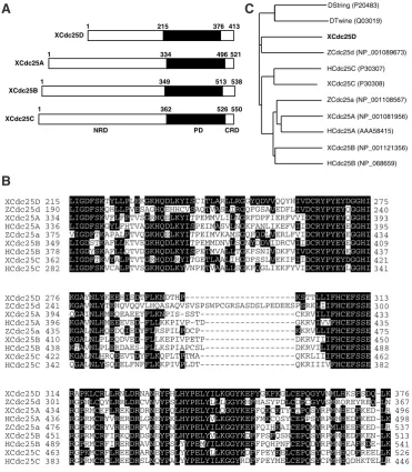
We performed BLAST search analysis in order to identify a possible novel cdc25 isoform(s) of Xenopus laevis, and found that some EST clones seem to encode a novel isoform. We then obtained a corresponding plasmid from NIBB (GeneBank Accession No. BJ037611), resequenced it, and found that this plasmid indeed contains a cDNA encoding a novel cdc25 isoform. Sequence analy-sis revealed that the predicted translation product of this cDNA has 413 amino acid residues and, like other cdc25 isoforms, consists of an N-terminal regulatory domain (NRD), a C-terminal phosphatase domain (PD), and a short C-terminal regulatory domain (CRD) (Fig. 1A). Neither the NRD nor the CRD of this Cdc25 isoform shares sequence homology with those of conventional Xenopus Cdc25 isoforms. However, the PD of this Cdc25 isoform shares 52%, 53%, and 56% identities with the PDs of Cdc25A, Cdc25B, and Cdc25C, respectively (Fig. 1B). Apparently, the PD of this isoform, excluding its inserted sequen-ce, is more similar to that of Zebrafish Cdc25d (Nogare et al., 2007) (Fig. 1B), and indeed, phylogenetic analy-sis showed that this isoform is most closely related to Zebrafish Cdc25d (Fig. 1C). Thus, hereafter, we call this Xenopus cdc25 isoform (X)cdc25D. A clear orthologue of cdc25D was found easily by BLAST in Xenopus tropicalis (GenBank Accession No. CR575588), but, interestingly, not in mammalians or chickens.
Temporal expression patterns of cdc25 isoforms during early Xeno-pus development
By RT-PCR, we first investigated the temporal expression patterns of cdc25 isoforms during early Xe-nopus development. In agreement
Fig. 1. Sequence analysis of the Xcdc25D product.(A) A schematic representation of
Xenopus Cdc25 isoforms. Three structural domains, i.e. the NRD, the PD, and the CRD, were defined according to Fauman
et al., (1998). (B) Alignment of the PD of Cdc25 proteins. Accession numbers are as in (C). D, Drosophila Melanogaster; X,
Xenopus laevis; H, Homo sapiens; Z, Zebra rerio. (C)Phylogenetic tree of cdc25 genes from various species, constructed by using the neighbor-joining method. Accession numbers used are shown.
LIGDFSKTYLLPLEKGKHQDLKYISCITLARLLRGGYQDVVQQYHIVDCRYPYEYDGGHI LIGDFSKQHLLPVESAGHQEHHCVSAQTVASLIRGQFGSAVEDFLIVDCRYPYEYQGGHI LIGDFSKVFLFPTVSGRHQELKYITPEMMVLILNGRFDPFIERFVVIDCRYPYEYQGGHI LIGDFSKGYLFHTVAGKHQDLKYISPEIMASVLNGKFANLIKEFVIIDCRYPYEYEGGHI VIGDFTKAPALPTVQGKHQELKYITPEIMVKAMSGQFQDLVERLFVIDCRYPYEYEGGHI LIGDSTKAFLLKTVEGKHQDLKYITPEMMDNVLSGNYDDVIDRCVIIDCRYPYEYEGGHI LIGDYSKAFLLQTVDGKHQDLKYISPETMVALLTGKFSNIVDKFVIVDCRYPYEYEGGHI LIGDFTKVYALPTVTGRHQDLRYITGETLAALIHGDFSSLVEKIFIIDCRYPYEYDGGHI LIGDFSKVCALPTVSGKHQDLKYVNPETVAALLSGKFQGLIEKFYVIDCRYPYEYLGGHI
KGAYNLYKEEHISDTFLKNDTHP---KSTTLLIFHCEFSSE KGAVNLYTEHQVQQVLHQASAQVSVSPSWPCGRSASDSLPEDEESPSRKLIIFHCEFSSE QGAINLHMEQEAEEYFLKNPIS-SST---CKRVILIFHCEFSSE KGAVNLHMEEEVED-FLLKKPIVP-TD---GKRVIVVFHCEFSSE KGALNLHQEDQIEDYFLRSPILPDCP---KKRVLLIFHCEFSSE KGALNLPLEQDVED-FLLKEPIVPETP---DKRVIIIFHCEFSSE KTAVNLPLERDAES-FLLKSPIAPCSL---DKRVILIFHCEFSSE KGALNLHRQEEVTDYFLKQPLTPTMA---QKRLIIIFHCEFSSE QGALNLYSQEELFNFFLKKPIVPLDT---QKRIIIVFHCEFSSE
RAPKLCRLLRNLDRNANRYPHLHYPELYILKGGYKEFYGKFKGLCEPQGYVNMLHKSFSDQ-LK
LIGDFSKTYLLPLEKGKHQDLKYISCITLARLLRGGYQDVVQQYHIVDCRYPYEYDGGHI LIGDFSKQHLLPVESAGHQEHHCVSAQTVASLIRGQFGSAVEDFLIVDCRYPYEYQGGHI LIGDFSKVFLFPTVSGRHQELKYITPEMMVLILNGRFDPFIERFVVIDCRYPYEYQGGHI LIGDFSKGYLFHTVAGKHQDLKYISPEIMASVLNGKFANLIKEFVIIDCRYPYEYEGGHI
VIGDFTKAPALPTVQGKHQELKYITPEIMVKAMSGQFQDLVERLFVIDCRYPYEYEGGHI LIGDSTKAFLLKTVEGKHQDLKYITPEMMDNVLSGNYDDVIDRCVIIDCRYPYEYEGGHI LIGDYSKAFLLQTVDGKHQDLKYISPETMVALLTGKFSNIVDKFVIVDCRYPYEYEGGHI LIGDFTKVYALPTVTGRHQDLRYITGETLAALIHGDFSSLVEKIFIIDCRYPYEYDGGHI LIGDFSKVCALPTVSGKHQDLKYVNPETVAALLSGKFQGLIEKFYVIDCRYPYEYLGGHI
KGAYNLYKEEHISDTFLKNDTHP---KSTTLLIFHCEFSSE KGAVNLYTEHQVQQVLHQASAQVSVSPSWPCGRSASDSLPEDEESPSRKLIIFHCEFSSE
QGAINLHMEQEAEEYFLKNPIS-SST---CKRVILIFHCEFSSE KGAVNLHMEEEVED-FLLKKPIVP-TD---GKRVIVVFHCEFSSE KGALNLHQEDQIEDYFLRSPILPDCP---KKRVLLIFHCEFSSE KGALNLPLEQDVED-FLLKEPIVPETP---DKRVIIIFHCEFSSE KTAVNLPLERDAES-FLLKSPIAPCSL---DKRVILIFHCEFSSE KGALNLHRQEEVTDYFLKQPLTPTMA---QKRLIIIFHCEFSSE
QGALNLYSQEELFNFFLKKPIVPLDT---QKRIIIVFHCEFSSE
with previous reports (Kim et al., 1999; Ueno et al., 2008; Hartley et al., 1996), cdc25A transcripts were constantly present at the cleavage stages with a slight decrease in its levels at later stages, cdc25B transcripts were expressed after the midblastula stage, and cdc25C transcripts were constantly present throughout the stages of early embryogenesis (Fig. 2). In contrast, cdc25D transcripts were scarcely detected at the preneurula stages, but became detectable during neurulation and persisted at least up to the late tailbud stage (Fig. 2). Thus, cdc25A and cdc25C are expressed both maternally and zygotically, whereas cdc25B and cdc25D are expressed zygotically in early Xenopus development.
Spatial expression patterns of cdc25 isoforms during early Xenopus development
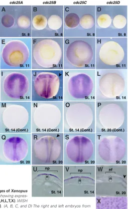
Although the spatial expression patterns of several cell-cycle regulators, such as cdks, cyclins, wee1 and myt1 isoforms, during early Xenopus development have been shown in previous studies (Vernon and Philpott, 2003; Leise and Muller, 2002), those of cdc25 isoforms have not been reported. We therefore examined the spatial expression patterns of Xenopus cdc25 isoforms using whole-mount in situ hybridization. In these experiments, no specific signals were detected with any of the sense probes (Fig. 3 M,N,O,P). Using an antisense probe, however, cdc25A transcripts were detected principally in the animal hemisphere from the blastula to initial gastrula stages (Fig. 3 A,E), and in the anterior neural plate at the early neurula stage (Fig. 3I). At the late neurula stage, cdc25A was expressed in the neural fold and the neural crest (Fig. 3Q), and, at the tailbud stage, it was weakly expressed in prospective retinal layers in eye vesicles (Fig 4 A,E,I).
Consistent with the results of RT-PCR (Fig. 2), cdc25B trans-cripts were not detectable in blastula embryos (Fig. 3B). During gastrulation, cdc25B transcripts were expressed in both the ecto-dermal and mesoecto-dermal regions (Fig. 3F). As previously reported (Ueno et al., 2008), at the early neurula stage, cdc25B transcripts exhibited bilateral expressions in the neural plate and trigeminal ganglions (Fig. 3 J,U), and, at the late neurula stage, they were expressed in the regions undergoing neurogenesis, such as the neural fold and eye anlagen (Fig. 3R). Furthermore, at the tailbud stage, cdc25B transcripts were expressed in the central nervous system, such as the spinal cord and the brain, including
prosen-cephalon, mesencephalon and rombencephalon (Fig. 4 B,F); they were also strongly expressed in the prospective retinal layers of eye vesicles (Fig. 4 B,F,J).
cdc25C, like cdc25A, was expressed exclusively in the animal hemisphere from the blastula to gastrula stages (Fig. 3 C,G). At the early neurula stage, cdc25C transcripts were expressed in the
cdc25B cdc25A
cdc25C
cdc25D
UFE 2 4 7 8 9 10 11 12 13 14 17 20 25 27 35
Blastula Gastrula Neurula Tailbud
ODC
Fig. 2. Nakajo et al.
(N/F)
Fig. 2. Temporal expression patterns of cdc25 isoforms during early Xenopus development. The transcripts of Xenopuscdc25 isoforms were analyzed by RT-PCR. ODC (ornithine decarboxylase) is a loading control. Nieuwkoop-Faber (N/F) stages are shown at the top.
Fig. 3. Spatial expression patterns of cdc25 isoforms at the indicated stages of Xenopus embryos. Whole-mount in situ hybridization (WISH) with antisense probes showing expres-sions of cdc25A(A,E,I,Q), cdc25B(B,F,J,R), cdc25C(C,G,K,S) and cdc25D(D,H,L,T,X). WISH
St. 8
St. 11
St. 20
n np
n nf np
n
St. 8 St. 8 St. 8
St. 11 St. 11 St. 11
St. 20 St. 20 St. 20
St. 14 St. 14
St. 20 St. 20
cdc25A cdc25B cdc25C cdc25D
St. 20 (Cont.)
St. 14 St. 14 St. 14 St. 14
St. 14 (Cont.) St. 14 (Cont.)
St. 14 (Cont.)
G
O
B
C
D
E
F
H
I
J
K
L
P
Q
R
S
T
A
M
N
U
V
W
X
neural plate (Fig. 3 K,V), and, at the late neurula stage, they were detected in the neural fold and neural crest regions (Fig. 3S). At the tailbud stage, cdc25C transcripts were broadly expressed in the head region (Fig. 4 C,G), particularly in the prospective retinal layers of eye vesicles (Fig. 4K).
Unlike cdc25A, B and C transcripts, cdc25D transcripts were undetectable up to the early neurula stage (Fig. 3 D,H,L), consis-tent with the results of RT-PCR (Fig. 2). Interestingly, at the late neurula stage, however, cdc25D transcripts were expressed in the epidermal ectoderm with a punctuate pattern, but not in the neural region (Fig. 3 T,W,X). At the tailbud stage, cdc25D transcripts were barely detected in the epidermal ectoderm, but, interestingly, their expression became apparent in the liver diverticulum endoderm (Fig. 4 D,H,L,M).
The effect of BMP inhibition on transcription of cdc25 isoforms
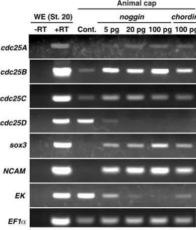
Given their spatially (as well as temporally) restricted expres-sion patterns, expresexpres-sions of different cdc25 isoforms would be regulated in different ways during development. In Xenopus, the ectodermal cell mass of the mid-blastula embryo (animal cap) is pluripotent, and is useful for addressing the developmental signal(s) (Lamb et al., 1993; Henry et al., 1996). Indeed, cdc25B expression in animal caps has been shown to be upregulated by inhibition of bone morphogenetic protein (BMP) (Ueno et al., 2008), a crucial event required for neural induction (Stern, 2005). We therefore investigated whether expression (or transcription) of other cdc25 isoforms would be affected by BMP inhibition, using animal cap assays and RT-PCR. Consistent with our previous report (Ueno et al., 2008), cdc25B transcription in animal caps was readily up-regulated by ectopic expression of noggin, a BMP antagonist (Munoz-Sanjuan and Brivanlou, 2002) (Fig. 5). Transcriptions of cdc25A and C were also up-regulated by noggin expression, albeit
to slightly lesser extents than cdc25B transcription; notably, howe-ver, cdc25D transcription was down-regulated dose-dependently by noggin expression (Fig. 5). Moreover, ectopic expression of
Fig. 4. Nakajo et al.
cdc25A
cdc25B
cdc25C
cdc25D
Fig. 4. Spatial expression patterns of cdc25 isoforms in tailbud-stage embryos (tailbud-stage 26). Whole-mount in situ hybridization showing expressions of cdc25A(A,E), cdc25B(B,F), cdc25C(C,G) and cdc25D(D,H). (A, B, C and D) Lateral views with anterior left. (E, F and G) Dorsal views with anterior left. (H) Ventral view with
anterior left. (I) Transversal section of the head region of (A). (J) Transversal section of the head region of (B); the arrow indicates mesencephalon. (K) Transversal section of the head region of (C). (I, J and K) The arrowhead indicates the eye vesicle. (L) Transversal section of (H). (M) Magnified view of the liver diverticulum endoderm of (L). The arrowhead indicates the liver diverticulum. No staining was detected with the sense probe (data not shown).
Animal cap
-RT WE (St. 20)
+RT Cont. 5 pg 20 pg 100 pg chordin
100 pg noggin
EF1α
EK cdc25A
cdc25C
cdc25D
NCAM sox3 cdc25B
Fig. 5. The effects of BMP inhibition on the expression of cdc25 isoforms in animal caps. Animal caps were isolated from late blastula Xenopus
embryos (st. 9) pre-injected with the indicated amounts of noggin or chordin
mRNA at the one-cell stage. The animal caps were cultured until sibling control embryos reached st. 20 and then analyzed by RT-PCR. sox3 and
NCAM are downstream markers of noggin and chordin. Epidermal keratin
(EK) is a marker for epidermis. EF1a is a loading control.
G
B
C
D
E
F
H
I
J
K
L
A
M
chordin, another BMP antagonist (Munoz-Sanjuan and Brivanlou, 2002), similarly affected the expression of cdc25 isoforms (Fig. 5). These results were consistent with the spatial expression patterns of the cdc25 iso-forms (Fig. 4). Thus, transcriptions of different cdc25 isoforms are likely to be differently regulated during the process of neural induction.
Discussion
In this study, we identified a novel isoform of Xeno-pus cdc25, termed cdc25D. In addition, we investigated the temporal and spatial expression patterns of all isoforms of cdc25 phosphatase during early Xenopus development, and found that they are different from each other during early development.
and Dunphy,W.G., 1992., Nakajo et al., 2000), whereas Cdc25A protein does not (Kim et al., 1999; Shimuta et al., 2002). Thus, Cdc25C protein seems to be the sole cdc25 isoform expressed in Xenopus oocytes, consistent with it being essential for Cdk1 activation during oocyte maturation in Xenopus (Izumi et al., 1992; Kumagai and Dunphy,W.G., 1992).
It has been shown that cells in the neural region actively proli-ferate during the neurula stage (Saka and Smith, 2001). Around the neurula stage, cdc25A, B, and C were expressed in the neural region, including the neural plate and the neural fold, and, in animal caps, their expressions were up-regulated by BMP inhibition, which is central to neural induction. Thus, cdc25A-C isoforms could pro-mote cell-cycle progression in the neural region. Notably, however, expression of cdc25B was more prominent in the neural region, and more readily induced by BMP inhibition in animal caps, than those of cdc25A and C. These results are consistent with our previous results that Cdc25B contributes significantly to cell proliferation in the neural region (Ueno et al., 2008). Previous studies showed that cdk1 and cyclin B transcripts are also preferentially expressed in the neural region (Vernon and Philpott, 2003), whereas wee1 and myt1 transcripts, encoding cdk1-inhibitory kinases, are barely expressed in the same region (Leise and Mueller, 2002). Thus, transcriptional regulations of these cell-cycle regulators would also contribute to cell-cycle progression in the neural region.
In contrast to other cdc25 isoforms, cdc25D transcripts were not expressed in the neural region, but were expressed in the epidermal ectoderm of the late-neurula embryos. Moreover, its transcription was suppressed by BMP inhibitors in animal caps. These results suggest that Cdc25D contributes to cell proliferation in non-neural ectoderms. Particularly, the punctuate expression pattern of cdc25D in the epidermal ectoderm of late-neurula embr-yos resembles that of a-tubulin, which is a marker of ciliated cells
(Deblandre et al., 1999). Thus, Cdc25D may have some role(s) in the formation of ciliated cell. Furthermore, and interestingly, at the tailbud stage, cdc25D transcripts were expressed in the liver diverticulum endoderm, which is the origin of embryonic liver in Xenopus (Nieuwkoop and Faber, 1994). Thus, Cdc25D might also be involved in the formation of embryonic liver.
Xenopus cdc25D has significantly closer homology with zebrafish cdc25d than with other conventional cdc25 isoforms. In zebrafish, however, cdc25d is expressed throughout early development and in the restricted ventral mesoderm and nasal placodes of the 24 hpf and 32 hpf embryos (Nogare et al., 2007). Therefore, despite their sequence homology, zebrafish cdc25d and Xenopus cdc25D seem to have different roles in early development. Furthermore, and surprisingly, neither mammalians nor chickens seem to pos-sess the cdc25D gene, suggesting that amniotes might have lost it during evolutionary processes. This might be related, however, to the potentially divergent roles of cdc25D even between zebrafish and Xenopus development.
The existence of distinct cdc25 isoforms in vertebrates may represent their different roles during developmental processes. This seems to be true for Xenopus development but not necessarily for mouse development. Recent studies in mice revealed the functional redundancy of Cdc25 isoforms during development; mice lacking both cdc25B and cdc25C developed normally, suggesting that cd-c25A compensates for the loss of cdc25B and cdc25C (Ferguson et al., 2005). In Xenopus, however, Cdc25A protein, unlike Cdc25B or C proteins, is largely degraded by Chk1 kinase just after the MBT
(Kim et al., 1999; Shimuta et al., 2002), and thereafter, cdc25A is expressed to very limited regions of the late-neurula and tailbud stage embryos. Thus, it appears that, in Xenopus, Cdc25A cannot compensate for the function of Cdc25B and Cdc25C.
To summarize, the temporal and spatial expression patterns of different cdc25 isoforms are different from each other during early Xenopus development. In addition, each orthologue of Cdc25 isoforms seems to have at least partially different roles in early development of different species.
Materials and Methods
Cloning of a novel isoform of Xenopus cdc25
The Xcdc25D EST was obtained from National Institute for Basic Biology (NIBB) with an identifier XL041a15 (GenBank accession number AB601986). The plasmid was resequenced with ABI PRISM 3100 Genetic Analyzer (applied biosystems, USA). Phylogenetic analysis was performed using the ClustalW program.
Xenopus embryos, in vitro transcription and animal cap assays
Xenopus eggs were artificially fertilized using Xenopus testis homo-genates and dejellied with 0.1 x Modified Barth’s saline (MBS) containing 2% cysteine for 20 minutes. Staging of embryos was done according to Nieuwkoop and Faber (1994). Capped RNAs were synthesized in vitro using a MEGAscript SP6 kit (Ambion) and injected into one-cell stage embryos. Injected embryos were cultured in 0.1 x MBS containing 3% Ficoll at 20˚C. Animal cap cells were obtained from stage 8.5 – 9 embryos, and then cultured in 1 x MBS containing 50 mg/ml gentamicin.
RNA extraction and RT-PCR
Total RNAs were extracted from five whole embryos at various stages and from twenty animal cap explants, using TRIzol reagent (invitrogen). The extracted total RNAs corresponding to one embryo or four animal
cap explants were used to synthesize cDNAs using oligo-dT20 primers
and SuperScript III reverse transcriptase (invitrogen). One-fortieth of the reaction products were subjected to PCR (94˚C for 30 sec, 52˚C for 30 sec, 72˚C for 1 min) with TAKARA Ex Taq (TAKARA). The PCR products were confirmed for their identities by sequencing and southern blotting (data not
shown). The primer sets (5′ to 3′; U, upstream and D, downstream) and
cycles used for PCR were: cdc25A (27 cycles)
U: GGCCACATACAGGGAGCCATTAACC D: TAGTTTCTTCAGCCGGCTGTACAGTTC cdc25B (30 cycles)
U: ACGTGGAAGACTTTCTGCTGAAGGAGC D: TCTCGCTTGCTCTTGTCTCCGGCCC cdc25C (27 cycles)
U: GGACACATAAAGGGTGCATTAAACC D: GCTTCATTATGCGGGCAATCTGTTC cdc25D (27 cycles)
U: GACGGAGGGCACATTAAGGGAGCCTAC D: GCTGGATGTTTTTTCAAGACAGTGC sox3 (27 cycles)
U: GCGCACATGAACGGCTGGACTA D: GTGTGGGAGGTGATGGCTGGAG
Primer sets for NCAM (30 cycles), epidermal keratin (20 cycles), and EF1-a (20 cycles) are described in Xenbase (http://www.xenbase.org/common). Under the present RT-PCR conditions, the amounts of the amplified target products were reflecting the amounts of template RNAs (Fig. S1), and the targets were amplified within a linear range of conditions (Fig. S2).
Whole-mount in situ hybridization
described by Takahashi et al., (2006). Open reading frame sequences of Xenopus cdc25A (Okazaki et al., 1996), cdc25B (Ueno et al., 2008), cdc25C (Kumagai and Dunphy, 1992) and cdc25D were subcloned into the pBlueScript KS+ plasmid (Agilent technologies). These plasmids were linealized, and digoxigenin-labeled RNA probes in sense and antisense orientations were transcribed using MEGAscript T7 and T3 kits (Ambion), respectively. For sectioning, stained embryos were embedded in gelatin/ sucrose (15% cold fish gelatin and 30% sucrose in PBS) and mounted with OCT compound (Tissue-Tek OCT compound, Sakura Finetek, USA), and 40 mm sections were prepared with a cryostat (Jung FrigoCut, Leica, Germany).
Acknowledgment
We thank M. Watanabe and S. Takahashi for helpful technical infor-mations, I. Ito and H. Udo for advice of preparing the cryosections, K. Ota for preparing the manuscript, and M. Ye for technical supports. This work was supported by a scientific grant from the Ministryof Education, Culture, Sports, Science, and Technology of Japanto N.S.
References
BOUTROS, R., DOZIER, C., DUCOMMUN, B (2006). The when and wheres of CDC25 phosphatases. Curr. Opin. Cell Biol. 18: 185-191.
BRIVANLOU, A.H., DARNELL, J.E., JR. (2002). Signal transduction and the control of gene expression. Science 295: 813-818.
CHEN, M.S., HUROV, J., WHITE, L.S., WOODFORD-THOMAS, T., PIWNICA-WORMS, H (2001). Absence of apparent phenotype in mice lacking Cdc25C protein phosphatase. Mol. Cell Biol. 21: 3853-3861.
COLEMAN, T. R., DUNPHY, W. G. (1994). Cdc2 regulatory factors. Curr. Opin. Cell. Biol. 6: 877-882.
DEBLANDRE, G.A., WETTSTEIN, D.A., KOYANO-NAKAGAWA, N., KINTNER, C. (1999). A two-step mechanism generates the spacing pattern of the ciliated cells in the skin of Xenopus embryos. Development 126: 4715-4728.
FERGUSON, A.M., WHITE, L.S., DONOVAN, P.J., PIWNICA-WORMS, H. (2005). Normal cell cycle and checkpoint responses in mice and cells lacking Cdc25B and Cdc25C protein phosphatases. Mol. Cell Biol. 25: 2853-2860.
HARTLEY, R.S., REMPEL, R.E., MALLER, J.L. (1996). In vivo regulation of the early embryonic cell cycle in Xenopus. Dev. Biol. 173: 408-419.
HENRY, G.L., BRIVANLOU, I.H., KESSLER, D.S., HEMMATI-BRIVANLOU, A.,
MEL-TON, D.A. (1996). TGF-b signals and a pattern in Xenopus laevis endodermal
development. Development. 122: 1007-1015.
IZUMI, T, WALKER, D.H., MALLER, J.L. (1992). Periodic changes in phosphorylation of the Xenopus cdc25 phosphatase regulate its activity. Mol. Biol. Cell. 3: 927-939. KIM, S.H., LI, C., MALLER, J.L., (1999). A maternal form of the phosphatase Cdc25A
regulates early embryonic cell cycles in Xenopus laevis. Dev. Biol. 212: 381-391. KUMAGAI, A. DUNPHY, W.G. (1992). Regulation of the cdc25 protein during the cell
cycle in Xenopus extracts. Cell 70: 139-151.
LAMB, T.M., KNECHT, A.K., SMITH, W.C., STACHEL, S.E., ECONOMIDES, A.N., STAHL, N., YANCOPOLOUS, G.D., HARLAND, R.M. (1993). Neural induction by the secreted polypeptide noggin. Science 262: 713-718.
LEISE, W.L., MUELLER, P.R. (2002). Multiple Cdk1 inhibitory kinases regulate the cell cycle during development. Dev. Biol. 249: 156-173.
MORGAN, D. O. (1995). Principles of CDK regulation. Nature 374: 131-134. MUNOZ-SANJUAN, I., BRIVANLOU, A.H. (2002). Neural induction, the default model
and embryonic stem cells. Nat. Rev. Neurosci. 3: 271-280.
NAKAJO, N., YOSHITOME, S., IWASHITA, J., IIDA, M., UTO, K., UENO, S., OKA-MOTO, K. AND SAGATA, N. (2000). Absence of Wee1 ensures the meiotic cell cycle in Xenopus oocytes. Genes Dev. 14: 328-338.
NIEUWKOOP, P.D., FABER, J. (1994). Normal Table of Xenopus laevis (Daudin). Garland Publishing Inc., New York.
NOGARE, D.E., ARGUELLO, A., SAZER, S., LANE, M.E. (2007). Zebrafish cdc25a is expressed during early development and limiting for post-blastoderm cell cycle progression. Dev. Dyn. 236: 3427-3435.
SAKA, Y, SMITH, J.C. (2001). Spatial and temporal patterns of cell division during early Xenopus embryogenesis. Dev. Biol. 229: 307-318.
SHIMUTA, K., NAKAJO, N., UTO, K., HAYANO, Y., OKAZAKI, K., SAGATA, N. (2002). Chk1 is activated transiently and targets Cdc25A for degradation at the Xenopus midblastula transition. EMBO J. 21: 3694-3703.
STERN, C.D. (2005). Neural induction: old problem, new findings, yet more questions. Development 132: 2007-2021.
TAKAHASHI, S., ONUMA, Y., YOKOTA, C., WESTMORELAND, J.J., ASASHIMA, M., WRIGHT, C.V. (2006). Nodal-related gene Xnr5 is amplified in the Xenopus genome. Genesis 44: 309-321.
UENO, H., NAKAJO, N., WATANABE, M., ISODA, M., SAGATA, N. (2008). FoxM1-driven cell division is required for neuronal differentiation in early Xenopus embryos. Development 135: 2023-2030.
Cyclin B2/cyclin-dependent kinase1 dissociation precedes CDK1 Thr-161 dephos-phorylation upon M-phase promoting factor inactivation in Xenopus laevis cell-free extract
Franck Chesnel, Franck Bazile, Aude Pascal and Jacek Z. Kubiak Int. J. Dev. Biol. (2007) 51: 297-305
Expression of the E2F family of transcription factors during murine development
Judith C Kusek, Robert M. Greene, Paul Nugent and M. Micheke Pisano Int. J. Dev. Biol. (2000) 44: 267-277 (2000)
p34(cdc2) and mitotic cyclin expression in the developing quail neuroretina.
Xavier Espanel, Anne Kastener, Oliver Stettler, Bertrand Tavitian, Gilbert Brun and German Gillet
Int. J. Dev. Biol. (1997) 41: 469-476
Cell reproduction: induction of M-phase events by cyclin-dependent cdc2
Takeo Kishimoto
Int. J. Dev. Biol. (1994) 38: 185-191
Meiosis reinitiation as a model system for the study of cell division and cell diffe-rentiation
Pierre Guerrier, Pierre Colas and Isabella Neant Int. J. Dev. Biol. (1990) 34: 93-109