Receptor-mediated uptake and transport
of macromolecules in the human placenta
HENNING SCHNEIDER
1and RICHARD K. MILLER*
,21Department of Obstetrics and Gynecology, Insel Spital, University of Berne, Berne, Switzerland and 2Departments of Obstetrics and Gynecology, Environmental Medicine and Pathology, University of
Rochester School of Medicine and Dentistry, Rochester, New York, USA
ABSTRACT The human placenta is required to be the anchor, the conduit and the controller during pregnancy. The survival of the baby and its associated placenta is dependent upon the placenta shielding the embryo/fetus from harm, e.g., autoimmune disease - thrombophilia, antiphospholipid syndrome or infections, while simultaneously providing for the passage of critical nutrients (e.g., amino acids, vitamins) and beneficial immunoglobulins. In a number of instances, the movements of macromolecules into and through the placenta can result in the passage of the intact molecules into the fetal circulation or in the case of proteins - catabolism to amino acids which are utilized by the placenta and the fetus for continued growth and development. The transfer of two such macromolecules, immunoglobulin G (IgG) and vitamin B12 (cyanocobalamin or B12), are exam-ined as to the unique receptor-mediated transfer capability of the human placenta, its transfer specificity as related to specific receptors and the role of endogeneous placental proteins (trancobalamins) in facilitating the recognition and transport of specifically B12. Brief compari-sons will be made to other animal species and the differences in specific organ transfer capabilities.
KEY WORDS: immunoglobulin IgG, transcobalamin, vitamin B12, placental transport, human
Overview of macromolecule transport and catabolism
by the placenta and extraembryonic membranes in
different species
The development of the embryo and fetus is dependent upon the function of the placenta and its extraembryonic membranes in every mammalian species. Of significance is the interdepen-dence of theses extraembryonic tissues in maintaining the con-ceptus and allowing for normal development.
Across species, macromolecules play multiple roles in provid-ing for this development. In particular, the transport physiologists and membrane biochemist focus on the transporters and the transcytosis that occurs for immuno-regulators and essential vitamins for normal development. Yet even the basic building blocks created by the degradation of these macromolecules provide for the growth of both fetus and placenta. The large contribution of amino acids from the metabolism of proteins, e.g., maternal albumin and other circulating proteins, provide the large
BIOLOGY
www.intjdevbiol.com*Address correspondence to: Henning Schneider. CH3122 Kehrsatz, Ahorn Weg 4, Switzerland. e-mail: Henning.Schneider@hispeed.ch or Richard K. Miller. Department of Obstetrics and Gynecology, University of Rochester Medical Center, Room 5-7550, 601 Elmwood Avenue, Rochester, New York 14642-8668 USA. e-mail: richardk_miller@urmc.rochester.edu
Final author-corrected PDF published online: 21 September 2009.
ISSN: Online 1696-3547, Print 0214-6282
© 2009 UBC Press Printed in Spain
Abbreviations used in this paper: Ag, antigen; Ab, antibody; Fab, antigen binding fragment of immunoglobulin; Fc, Fc receptor binding fragment of immunoglobulin; FcR, Fc receptor; FcγR I,II,II, subclasses of Fc receptors; FcRn, Fc receptor of the neonate; IC, immuno-complex; IgGs, class G immunoglobulins; IgG 1-4, subclasses 1-4 of immunoglobulins class G; LAMP2, lysosome-associated membrane protein; MSP1-42, merozoit surface protein 1-42 malaria protein; MHC, major histocompatibility complex; mRNA, messenger ribonucleic acid; STB, syncytiotrophoblast; TC I, II, III, Transcobalamin I,II,III.
continuous source of amino acids, which could not be sufficiently supplied by either passive diffusion or active transport.
does (cf Miller et al., 1976; Beck, 1981; Beckman et al., 1991a; Polliotti et al., 1997, Kim et al., 2009; Pentsuk and van der Laan, 2009). For the human and non-human primate, the chorioplacenta is the principal organ for catabolism and transport of the macro-molecules and important immunoglobulins.
This review will examine the current mechanism for transit and catabolism of immunoglobulin G (IgG), and transcobalamin II-vitamin B12 (TCII-B12 or holoTC) in the human placenta. For additional details concerning interspecies differences of the ma-ternal-fetal barrier and their relevance for the transfer of macro-molecule, IgG, the reader is referred to Pentsuk and van der Laan (2009).
Immunoglobulins
Introduction
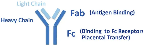
Immunoglobulins are Y-shaped molecules made of 2 antigen binding fragments (Fab) and the stem of the Y (Fc fragment) (Fig. 1). Specific binding of antigens at the Fab fragments results in the formation of immuno-complexes, whereas the Fc fragment inter-acts with effector systems of the immune response like comple-ment or Fc receptors on the surface of certain subpopulations of white blood cells. This starts a reaction cascade ultimately leading to the elimination of the antigen. In the human, class G immuno-globulins are the predominant antibodies (ab), which in the serum are present in four subclasses differing in their Fc regions leading to different affinities to Fc receptors (FcR).
FcRs apart from eliciting an immune response against the invasion of the body by antigens are also involved in the transport of free antibodies across various tissue barriers. A full comple-ment of maternal antibodies crosses intestine or placenta provid-ing the fetus or the neonate with a protective shield against infections during the first months of postnatal life. In addition, the interaction between placental tissue and immunoglobulins as well as immuno-complexes (IC) serves the purpose of protecting the fetus against infectious diseases and the maternal immune re-sponse to foreign and auto antigens in the sense of immune surveillance. To reconcile these conflicting objectives is one of the major challenges of evolution of reproduction.
Evolution of maternofetal transport of immunoglobulins dur-ing human pregnancy
Maternal antibodies providing immuno protection of the infant during the first months after birth in different species reach the fetus or newborn via different routes (Brambell, 1958). In the human, the placenta is the predominant route (Dancis et al., 1961; Linnet-Jepsen and Galatius-Jensen, 1958). Immunoglobulins in the fetal circulation almost exclusively consist of maternal IgG and a wide spectrum of different antibodies like specific IgG anti-tetanous toxoid, anti-group-A streptococcal carbohydrate and anti-herpes simplex virus have been described (Eichhorn et al., 1987; Osuga et al., 1992). Throughout evolution the placenta developed a complex transport system to allow a large variety of highly specific antibodies to cross the different tissue layers without interfering with the protective function of the barrier between maternal and fetal organism. In a recent study sugges-tive evidence is provided, that maternal antibodies reaching the fetus via the placenta or the infant via breast milk, do not only provide passive immunity against postnatal infection but may act
as immuno-modulatory agents helping to develop specific and long lasting immune responses in the infant (Gros et al., 2006).
In the human, in vivo data on transfer were originally derived from measurements of endogenous antibodies in paired samples of maternal and fetal sera, obtained at delivery or termination of pregnancy. Until the end of the first trimester the level of IgG in the fetal as compared with the maternal compartment is quite low (Dancis et al., 1961; Gitlin et al., 1969; Gusdon, 1969; Morell et al., 1972). With the introduction of cordocentesis performed for vari-ous diagnostic purposes a systematic study of the evolution of transmission of antibodies from the mother to the fetus at different gestational ages became possible (Malek et al., 1996). Between 17-22 and 28-32 weeks of gestation total IgG concentration increased from 1.44 + 0.67 g/l to 5.57 + 1.10 g/l, which is 10% and 50% of the maternal concentration respectively. Fetal levels of IgG continued to increase and at term the concentration with 11.98 + 2.18 g/L exceeded the maternal level, which was consis-tent with previous findings from cord blood sera taken at delivery at term (Malek et al., 1994; Longsworth et al., 1945; Kohler and Farr, 1966). The ratio between IgG1:IgG2 in fetal sera already at 17-22 weeks of gestation was higher than that in maternal samples (Malek et al., 1996). The curve of the changes in fetal:maternal ratio for IgG1, IgG3 and IgG4 in the second half of pregnancy demonstrated an exponential rise; whereas for IgG2, the increase of this ratio was linear.
The concentrations of the four subclasses of IgG in fetal sera showed, that levels of IgG1 exceeded the other subclasses at all stages of pregnancy (Malek et al., 1994 and 1996; Morell et al., 1971; Chandra, 1976; Catty et al., 1979). Comparing the slopes of these curves the following ranking of preferential transport was shown: IgG1> IgG3> IgG4> IgG2. Interestingly, small-for-gesta-tional age newborns have lower total IgG levels than their appro-priate-for-gestational-age peers (Yeung and Hobbs, 1968). This is largely due to lower levels of IgG1 (Chandra 1976; Catty et al., 1979).
At term, placental transmission of maternal IgG into the fetal circulation involves transfer across two cell layers, the villous syncytiotrophoblast (STB) and the endothelial cells lining fetal capillaries inside the villi. A number of different in vitro models have been applied to study the mechanisms involved. In addition to the explosive development of molecular biology, more recent technological advances in the field of histochemistry allowed for a more precise allocation of different antibodies and their recep-tors to defined subcellular structures (Takizawa et al., 2007). The
introduction of ultrathin cryosections for immunocytochemistry in the electron microscopy and for high-resolution immunofluores-cence as well as a combination of two or more imaging methods has opened a new dimension for our understanding of the complexities of these mechanisms.
Pathway for IgG passage across different tissue barriers in the placenta
Syncytiotrophoblast (STB)
The various subtypes of FcRs and their respective isoforms are differently expressed in the various tissue components of the human placenta and play different roles in the transport of IgG from the mother to the fetus (Saji et al., 1999).
The MHC class I-related FcRn or Fc receptor of the neonate had originally been described in the intestinal brush border of the neonatal rat (Jones and Waldmann, 1972; Rodewald, 1973; Wallace and Rees, 1980) and was found in the murine fetal yolk sac (Roberts et al., 1990). A human orthologue of FcRn was identfied as mRNA and protein in the STB of the human placenta (Story et al., 1994; Simister et al., 1996; Kristofferson and Matre, 1996); it is generally accepted, that FcRn mediates uptake of IgG at the syncytial surface of the placenta.
However, IgG binding to both the human and rodent variant of FcRn is different from other FcRs in that FcRns are pH dependant with a high affinity at pH 6.0 and no significant binding at the maternal blood pH of 7.4 (Rodewald, 1976; Martin et al.,2001). Furthermore, FcRn could not be detected at the apical plasma membrane but rather in a subapical endosomal compartment of STB (Kristoffersen and Matre, 1996). In view of these two find-ings, direct binding of IgG at the microvillous surface to FcRn can hardly explain uptake of IgG by the STB (Story et al., 1994). Since no direct evidence for binding of IgG to other proteins expressed in the microvillous surface like annexin II or placental alkaline phosphatase as intermediary carriers could be demonstrated (Stefaner et al., 1997; Simister and Story, 1997), it was postulated (Israel et al., 1995), that similar to the rodent yolk sac endoderm IgG in the syncytial layer internalizes nonspecifically by fluid-phase endocytosis and after reaching an acidified endosomal compartment binds to FcRn (Roberts et al., 1990). As shown recently by analyzing mice fetuses resulting from matings of FcRN +/- parents, it was found that FcRN -/- fetuses contained negligible amounts of IgG. Immunofluorescence together with immunoblotting showed that FcRN were expressed in the endo-derm of the yolk sac placenta, but not in other cells of the yolk sac placenta or in the chorioallantoic placenta of these fetuses. IgG was found in the endoderm of both FcRN -/- and FcRN+/- yolksac placentas and in the mesenchyme of the FcRN +/+, but was missing from the FcRN -/- yolksac placentas. It was concluded, that IgG may enter the endoderm constitutively but is moved out by FcRN receptors (Kim et al., 2009).
Functional studies using freshly isolated trophoblast cells (Sooranna and Contractor, 1991; Estermann et al., 1995) pro-vided details of the mechanisms involved in overcoming the trophoblast as part of the placental barrier. In choriocarcinoma-like cells expressing FcRn internalization of IgG by fluid-phase endocytosis with subsequent binding to the receptor and selec-tive sorting into a transcytotic pathway could be demonstrated (Ellinger et al., 1999). Immunocytochemical and labelled tracer approaches in human placental tissue provided evidence, that
tubulovesicular structures serve the transport of IgG to endosomes inside the STB and it was proposed, that binding to FcRn provides protection against lysosomal digestion (Leach et al., 1991; King, 1982). After transcytosis of the STB the contact with neutral pH outside of the basolateral plasma membrane favours uncoupling of IgG from FcRn with release into the interstitium.
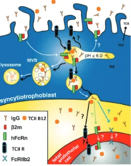
In situ, when villous syncytiotrophoblast is continuously ex-posed to high concentrations of IgG in maternal blood, saturation of the binding capacity for endosomal FcRn is reached, and excess unbound IgG is diverted to the lysosomal degradation route (Fuchs et al., 2006). This mechanism was demonstrated by confocal immunfluorescence microscopy, when the majority of
IgG was seen in LAMP2-positive (lysosome-associated mem-brane protein), late endosomes. Differently, in trophoblast de-rived BeWo cells the FcRn/IgG complex was detected in apical early endosomes, in recycling compartments and in vesicles close to the basolateral plasma membrane; the latter was prob-ably part of the transcytotic route. No FcRn or IgG could be detected in multivesicular, LAMP2-positive late endosomes and lysosomes (Leitner et al., 2006). From studies on co-localisation of IgG, FcRn and subcellular organelles identified by specific markers the following concept for the processing of IgG inside the syncytium in situ had been proposed (Fig. 2). After uptake in early endosomes IgG bound to FcRn can be routed to the basolateral plasma membrane or back to the apical plasma membrane with recycling to the maternal circulation. A large fraction of endog-enous IgG, which remains unbound to FcRn appears in multivesicular, LAMP2-positive, late endosomes and undergoes lysosomal degradation (Fuchs and Ellinger, 2004). The molecular mechanisms involved in the sorting of IgG to these different routes continue to be largely unknown.
As a model, the dual ex vivo perfusion of an isolated cotyledon of human placenta has proven to be particularly suited to study the actions of the different mechanisms involved in transmission of IgG from the mother to the fetus. By using a commercial prepara-tion of a mix of unlabelled IgG it had been demonstrated, that transfer of the four IgG subclasses was consistent with transfer rates derived indirectly from paired blood samples from the mother and the fetus as discussed above (Malek et al., 1995). As shown by Western immunoblot, samples from the maternal and fetal circuit had only one band in the range of 160 000 daltons, and there was no release of significant amounts of smaller IgG fragments into the fetal circulation. In another study with a similar model using radioactive iodine labelled IgG, 70% of the radiolabel recovered from the fetal circuit was attached to small molecules, which were not precipitable with trichloroacetic acid (Contractor et al., 1983). Whether the substantial catabolism, which can only be detected by using radiolabelled IgG, is real or a consequence of a denaturing effect of radiolabelling, cannot be decided on the basis of presently available data.
Electron microscopy had been used to trace the pathway of horseradish peroxidase conjugate of IgG through the different tissue layers of the human placenta (Leach et al., 1990). The delay of 2 hours between the start of perfusion of the intervillous space with the conjugated IgG and its detection in endothelial cells of the villous capillaries was consistent with the lag time after addition of IgG to the maternal perfusate until it was detected on the fetal side as observed in the study of Malek et al. (1995).
The current knowledge of the interaction between the FcRn receptor and IgG or its Fc fragment and its relevance for different functions has recently been reviewed (Ghetie et al., 2000). Mutated IgG Fc fragments have been used for functional studies on the role of the receptor for maternofetal transfer as well as for the half-life of IgG in the circulation. Ultimate proof of the central role of FcRn in the crossing of intact IgG from the maternal to the fetal side came from experiments studying mutated IgG in the dual ex vivo perfusion model. Maternofetal transfer of IgG1 as a wild-type antibody was compared to a recombinant, humanized (IgG1) antibody, where histidine had been replaced by alanine at the H435 position (His435 to H435A mutation) (Firan et al., 2001). This mutation interferes with binding of the antibody to
recombi-nant mouse and human FcRn, whereas binding to FcγRI, FcγRII and FcγRIII, which had also been postulated as mediators for the transport of maternal IgG, remains intact (Medesan et al., 1997). Comparing differently labelled wild-type antibody with the H435A mutant in the same experiment demonstrated a marked suppression of transfer for the mutated variant. This study also showed the value of the challenging method of ex vivo perfusion of whole placental tissue as a confirmation of the physiological relevance of data obtained from isolated trophoblast or endothe-lial cells alone.
Understanding the specifics of IgG transfer and the importance of the FcRn was further advanced by testing in the perfused human placenta the transit of a chimeric mouse/ human IgG Fab fragment used for anti coagulant therapy (Abciximab). No intact transmission of Fab fragments without the associated Fc portion could be demonstrated but much like albumin, the Fab fragment was catabolised by the human placenta (Miller et al., 2003). In contrast to Abciximab, Rituximab, another chimeric mouse/hu-man intact IgG and monoclonal anti-CD20 antibody was docu-mented to cross into the human fetus and reduce B-lymphocytes, both in the mother and in the newborn (Klink et al, 2008). Thus, the importance of the Fc end of the intact IgG is critical for the transfer of intact IgG whether human or mouse/human chimeric. The different routing of IgG internalized and bound to FcRn resulting in transcytosis or recycling has already been mentioned. Recycling with release of intact IgG into the maternal circulation is not restricted to the trophoblast but is related to binding to FcRn in general. Such release is also seen with endothelial cells (Ghetie et al., 2000). Consistent with this concept is the finding, that differences in binding affinity of the different subclasses of IgG to FcRn correlates with both, rates of maternofetal transfer and serum half-life (Ghetie et al., 1996; Israel et al., 1996; Medesan et al., 1996).
Endothelium of villous vasculature
FcγRI, RII, RIII are predominantly expressed in Hofbauer cells, the stromal macrophages of the placenta. Their role in transplacental passage of IgG is not entirely clear. It appears that isoforms of the three FcγRs may bind immune complexes and act as a protective barrier between the mother and the fetus. (Simister and Story, 1997; Simister, 1998; Simister, 2003).
The endothelial lining of the fetal capillaries inside the placental villi appears differently from other components of the placental barrier and is quite tight. The transcellular route is the only way for larger molecules to get from the maternal to the fetal circuit (Firth and Leach, 1996; Leach et al., 1991; King, 1982; Bright et al., 1994). Whereas the role of FcRn as trans-porter for IgG in helping to overcome the trophoblast part of the placental barrier is fairly-well established, the data on mecha-nisms for transport across the endothelium remain contradic-tory. Using immune staining, the presence of FcRn in the endothelium of capillaries in terminal villi have been described as absent or only occasionally seen (Simister et al., 1996; Lyden et al., 2001; Kristoffersen and Matre, 1996).
preferential from the basolateral to the apical surface, and IgG and FcRn colocalized in an intracellular endocytic compart-ment. In a more recent study the same group demonstrated, that FcRn binding of these cells discriminated between native IgG and IgG treated with diethylpyrocarbonate (DEPC) (Radulescu et al., 2004). The latter treatment by blocking histidine at the interaction site of IgG impaired the binding to FcRn. Whereas both types of ligands were internalized by the cells, the further processing differed dramatically. Intact FcRn binding protected native IgG from lysosomal digestion allowing active recycling as well as transcytosis of the native form with little accumulation inside the cells. The DEPC treated version displayed considerably higher intracellular accumulation and less recycling or transcytosis.
While these findings would fit very nicely into the general picture, it has been questioned, whether these cells indeed represent endothelium from terminal villi. It is generally agreed, that the expression of FcRn in endothelial cells from capillaries inside terminal villi is absent or very low and only increases along the vascular tree with clear expression in endothelium from cord vessels (Lyden et al., 2001). Furthermore these cells apparently do not express FcγRII or any other of the “classical” FcRs (Gafencu et al., 2003). FcγRII appears to be the only Fc receptor clearly expressed in terminal villous endothelium (Lyden et al., 2001). The expression of the FcγRIIb2 isotype is highest in the capillaries inside the terminal villi and decreases along the villous vascular tree toward the vessels in the cord (Lyden et al., 2001).”In the region of terminal villi, capillaries demon-strate sinusoidal dilatation with formation of vasculosyncytial zones, where the tissue layers separating maternal and fetal blood have been reduced to a thin syncytial covering, two basal laminas and the endothelium of the capillaries. This portion of the villous tree, therefore, would be particularly suited for transport of intact maternal IgGs into the fetal circulation.
Recently, an abundant expression of the FcγRIIb2 isoform in endothelial cells in sections from terminal villi was described.(Takizawa et al., 2005). Using a combination of high-resolution immunofluorescence and correlative electron mi-croscopy attempts to colocalize this receptor with a number of markers of different organelles in the endothelial cells were unsuccessful. Of particular note is the lack of colocalization with caveolae, which in analogy with endothelial cells from other organs until recently have been assumed to provide the cellular route for passive transcytosis of IgG (Tuma and Hubbard, 2003). Using double-label immunofluorescence stainings with anti-human IgG and anti-FcγRIIb2 a considerable overlap as a sign of co-localisation was found. Half of the FcγRIIb positive but so far unidentified vesicles contain eighty percent of endot-helial IgG.
Fluorescence intensity measurements, which were performed after labelling IgG with a fluorochrome-labelled IgG-antibody, displayed intense diffuse brightness in the extracellular matrix of the interstitium compared to much less intense fluorescent granulae inside the syncytiotrophoblast and the endothelium (Takizawa et al., 2005). This pattern of distribution of IgG in the different compartments of the placental barrier would be con-sistent with release of IgG from the syncytiotrophoblast with concentrative uptake by the extracellular matrix of the intersti-tium. The mechanism of the concentrative accumulation in the
interstitial layer remains unclear. A concentration gradient between the interstitium and the endothelium would support downward movement of IgG in the direction of the fetal circula-tion possibly supported by a FcγRIIb dependant transport system. The expression of FcγRIIb in endothelial cells for most of the vascular tree in the human placenta was recently con-firmed by Mishima and associates (2007).
Unanswered Questions
Apart from the uncertainties related to the mechanisms allowing transcytosis of IgG across the endothelium in the villous vasculature of the placenta there are a number of unanswered questions.
There is some indirect evidence, that IgG through binding may also act as a vehicle carrying proteins across the placental barrier from the maternal to the fetal side. Whereas human insulin does not cross the placenta, older investigations from pregnant diabetic women treated with animal insulin noted that placental transfer of insulin was related to the maternal level of specific insulin antibodies, which suggests a role for a complex of insulin and its antibody in transport (Bauman and Yalow, 1981; Menon et al., 1990). Secondly, correlation between the concentrations of tetanus Ag with anti-tetanus Ab found in cord blood as well as in the fetal compartment in the ex vivo model of the placenta perfused with serum containing tetanus Ag and Ab is consistent with the possibility, that the formation of an immuncomplex (IC) may be involved in the transplacental transport of the Ag (Malek et al., 1997 and 1998).
trans-ferred through the placenta and extraembryonic membranes to the fetus much like the Trojan horse of the past.
Transcobalamin proteins and the transport of vitamin
B12 (cyanocobalamin)
Vitamin B12 and transcobalamin proteins
Vitamin B12 (cyanocobalamin) is essential to normal develop-ment and is closely linked to folic acid. The balance of B12 and folic acid is critical to the prevention of birth defects and DNA synthesis (Watanabe, 2007). Normally in cells, B12 is transported by transcobalamin II, which circulates in the maternal blood along with transcobalamins I and III, which also bind B12 in the serum. Normally, the transcobalamin proteins are produced by the liver; however, from the earliest stages of pregnancy post implantation to term, the transcobalamin proteins are also produced by the human placenta (Ng et al. 1981; 1983). In addition, the human placenta has specific receptors for binding transcobalamin (Fried-man et al. 1982; Nexo and Hollenberg, 1980; Selig(Fried-man and Allen, 1978; Quadros et al., 1994). Under ex vivo perfusion conditions in the human placenta, B12 is transferred from mother to fetus with the appearance of the transcobalamin-bound B12 appearing in the fetal circulation (Perez-D’Gregorio et al. 1998) (Fig. 3).
The role of transcobalamin II in normal cell function is critical (Sereglhanoglu et al., 2008). During pregnancy, B12 has been noted to decline but not TCII-B12 (Morkbak et al., 2007). TCII is a 38 kD protein with a plasma half-life of 5 -90 minutes because of rapid absorption by cells (Gilbert, 1977). A rare genetic disease, transcobalamin II deficiency, has been reported (Qian et al., 2002). TC-II deficiency is associated with severe anemia and highly elevated methyl melonic acid levels in the blood. Only one women with a TCII deficiency is known to have become pregnant. Of interest has been the question, whether during pregnancy will she return to normal based upon the placenta producing and releasing TCII. This assumes that the baby in utero has at least one normal allele. Interestingly in three pregnancies, the mother with the TC-II deficiency has returned to normal methyl melonic acid levels early in the first trimester and maintained those normal levels throughout her pregnancy. The babies had normal B12 metabolism (RK Miller, J Mills and L Brody, unpublished observa-tions).
Such an observation of the placenta producing the carrier protein for the transport of a critical nutrient for fetal growth and development when the mother is deficient is another revelation of how important the placenta is in maintaining the development of the conceptus.
Across species, macromolecule transport is not always con-ducted by the chorioplacenta as it is in the primate. In the rabbit and rodent, the visceral yolk sac performs the duties for macromo-lecular transport. Both immunoglobulins and TCII – B12 are preferentially transferred across the visceral yolk sac (Brambell, 1958; Miller et al., 1976; Polliotti et al., 1998). These differences across species not only define the transport of macromolecules but also the catabolism of proteins and large molecules as sources of basic nutrients for the conceptus, e.g., amino acids. The combination of receptor medicated endocytosis – transcytosis and catabolism represent sites for disrupting normal supplies and the production of adverse embryonic and fetal development. In the rodent, birth defects have been produced by trypan blue and antisera against the yolk sac via mechanism disrupting these metabolic processes with direct actions on the embryo or fetus (Brent, 1964; Brent et al., 1970, 1983, 1990, Beck and Lloyd, 1967; Beckman et, 1991b; New and Brent, 1972).
Unanswered questions
As with IgG, the role of the fetal endothelial membrane and cells in transporting Vitamin B12 free or bound to transcobalamins remains to be examined. Does the endoethelial cell only transport TCII or does it have other mechansims for TC I and TCIII. It is known that all three proteins do appear in the fetal circuit binding B12 (Perez-D’Gregorio et al., 1998). We also know that the human placenta can produce and release these proteins. We do not know if TC I and TC III only bind free B12 in the fetal circulation or play a more active role in the transit of vitamin B12 once the B12 has entered the placenta. Future investigations will hopefully elucidate the control of this bulky vitamin and how it transits and binds to human placental proteins.
Summary
Thus, through examination of these important placental func-tions in multiple species, one can identify the critical roles played
in normal development of both embryo and fetus. Substantial work continues in understanding the specific cellular mechanisms controlling the transport and catabolism of these proteins and vitamins. This review hopefully will inspire the reader to further examine the cellular processes underlying the control of macro-molecule transport across the placenta.
References
ANTOHE, F., RADULESCU, L., GAFENCU, A., GHETIE, V. and SIMIONESCU, M. (2001) Expression of functionally active FcRn and the bidirectional transport of IgG in human placental endothelial cells. Hum. Immunol. 62: 93-105.
BAUMAN, W.A, and YALOW, R.S. (1981) Transplacental passage of insulin complexed to antibody. Proc. Natl. Acad. Sci. USA. 78: 4588-4590.
BRAMBELL, F.W.R. (1958). The passive immunity of the young mammal. Biol. Rev.
33: 485-531.
BECK F. (1981). Comparative placental morphology and function. In: Developmen-tal Toxicity, eds. Kimmel CA, Buelke-Sam J. New York: Raven Press, pp. 35-54.
BECK F, LLOYD JB. (1967). Griffiths A. Lysomal enzyme inhibition by trypan blue: A theory of teratogenesis. Science 157: 1180-1182.
BECKMAN DA, PUGARELLI J, KOSZALKA T, BRENT R, LLOYD J. (1991a). Sources of Amino Acids for protein synthesis during early organogenesis in the rat. Placenta 12: 37-46.
BECKMAN DA, ORNOY A, JENSEN M, ARNON J, BRENT RL. (1991b). Ultrastruc-ture and function of the rat yolk sac: damage caused by teratogenic anti-VYS serum and recovery. Teratology 44: 181-192.
BRENT RL. (1964). The production of congenital malformations using tissue antisera. II. The spectrum and incidence of malformations following the admin-istration of kidney antiserum to pregnant rats. Amer. J. Anat. 115: 525-542.
BRENT RL, AVERICH E, DRAPIEWSKI VA. (1970). Production of congenital malfromations using tissue antisera. Teratology 3: 198.
BRENT RL, BECKMAN DA, JENSEN M, KOSZALKA TR, DAMJANOV I. (1983). The embryopathologic effects of teratogenic yolk sac antiserum. Trophoblast Res 1: 335-346.
BRENT RL, BECKMAN DA, JENSEN M, KOSZALKA TR. (1990). Experimental yolk sac dysfunction as a model for studying nutritional disturbances in the embryo during early organogenesis. Teratology 41: 405-413.
BRIGHT, N.A., OCKLEFORD, C.D. and ANWAR, M. (1994). Ontogeny and distri-bution of Fc gamma receptors in the human placenta: transport or immune surveillance? J. Anat. 184: 297-308.
CATTY, D., DREW R. and SEGER R. (1979). Transmission of IgG subclasses to the human fetus. In: Hemmings, W.A. editor. Protein transmission through living membranes. Amsterdam: Elsevier/North-Holland, p. 37-43.
CHANDRA R.K. (1976). Levels of IgG subclasses, IgA, IgM, and tetanus antitoxin in paired maternal and fetal sera: findings in healthy pregnancies and placental insufficiency. In: Hemmings, W.A. editor. Maternofetal transmission of immuno-globulins. Cambridge: Cambridge Universiy Press; p. 77-87.
CONTRACTOR, S.F., EATON, B.M. and STANNARD P.J. (1983). Uptake and fate of exogenous immunoglobulin G in the perfused human placenta. J. Reprod. Immunol. 5 (5): 265-273.
DANCIS, J., LIND, J., ORATZ, M., SMOLENS, J. and VARA P. (1961). Placental transfer of proteins in human gestation. Am.J.Obstet.Gynecol. 82: 167-171.
Eichhorn, M.S., Granoff, D.M., Hahm, M.H. and Quinn, A., Shackleford, P.G. (1987). Concentrations of antibodies in paired maternal and infant sera: Rela-tionship to IgG subclasses. J.Pediatr. 111: 783-788.
ELLINGER, A.L., SCHWAB, M., STEFANESCU, A., HUNZIKER, W. and FUCHS, R. (1999). Immunoglobulin G transport across trophoblast-derived BeWo cells: a model system to study immunoglobulin G transport in the placenta. Eur. J. Immunol. 29: 733-744.
ESTERMANN, A.L., DANCIS, J., LEE, J.D., RINDLER, M.J. (1995): Two mecha-nisms for IgG uptake in cultured human trophoblast: evidence for a novel high affinity receptor. Pediatr. Res. 38: 1-6.
FIRAN, F., BAWDON, R., RADU, C., OBER, R.J., EAKEN, D., ANTOHE, F. GHETIE,V. and WARD, E.S. (2001). The MHC class I-related receptor, FcRn,
plays an essential role in the maternofetal transfer of γ-globulin in humans. Int. Immunol.13: 993-1002.
FIRTH, J.A. and LEACH, L. (1996). Not trophoblast alone: a review of the contribution of the fetal microvasculature to transplacental exchange. Placenta
17: 89-96.
FRIEDMAN, PA.,SHIA, MA., WALLACE, KJ. (1977) A saturable high affinity binding site for transcobalaminII-vitamin B12 complexes in human placental membrane preparations. J. Clin. Invest. 89: 13-24.
FUCHS, R. and ELLINGER, I. (2004). Endocytic and transcytotic processes in villous syncytiotrtophoblast: role in nutrient transport to the human fetus. Traffic
5: 725-738.
FUCHS,R., LEITNER,K., ELLINGER I., BUSCH B., EXNER B. and ZIMMER, K.P. (2006). IgG transport in placental trophoblasts. Placenta 27: Abstracts, W15,
A.11.
GAFENCU, A., HELTIANU, C., BURLAKU, A., HUNZIKER, W. and SIMIONESCU, M. (2003). Investigation of IgG receptors expressed on the surface of human placental endothelial cells. Placenta 24: 664-676.
GHETIE, V., HUBBARD, J.G., KIM, J.K., TSEN, M.F., LEE, Y. and WARD, E.S. (1996). Abnormally short serum half-lives of IgG in beta-2-microglobulin defi-cient mice. Eur. J. Immunol. 26: 690-696.
GHETIE, V. and WARD, E.S. (2000). Multiple roles for the major histocompatibility complex class I-related receptor FcRn. Annu. Rev. Immunol. 18: 739-766.
GILBERT, HS. (1977) Inhibition of vitamin B12 binding to transcobalamin II at low pH: basis of a procedure for quantification of circulating TCII and R binders. J. Lab. Clin. Med. 89: 13-24.
GITLIN, D. and BIASUCCI, A. (1969). Development of γg, γa, γm, βic/βia, c’1 esterase inhibitor, ceruloplasmin, transferrin, hemopexin, haptoglobin, fibrino-gen, plasminofibrino-gen, α1-antitrypsin, orosomucoid, β-lipoprotein, α 2-macroglobu-lin and prealbumin in the human conceptus. J. Clin. Invest. 48: 1433-1446.
GROS, L., PELEGRIN, M., PLAYS, M., PIECHAZCYK, M. (2006). Efficient mother to child transfer of antiretroviral immunity in the context of preclinical monoclonal antibody-based immunotherapy. J. Virol. 80: 10191-10200.
GUSDON, J.P. (1969). Fetal and maternal immunoglobulin levels during preg-nancy. Am. J. Obstet. Gynecol. 103: 895-900.
ISRAEL, E.J., PATEL, V.K., TAYLOR S.F., MARSHAK-ROTHSTEIN, A. and SIMISTER, N.E. (1995). Requirement for a β2-microglobulin-associated Fc receptor for acquisition of maternal IgG by fetal and neonatel mice. J. Immunol.
154: 6246-651.
ISRAEL, E.J., WISKER, D.F., HAYES, K.C., SCHOENFELD, D. and SIMISTER, N.E. (1996). Increased clearance of IgG in mice that lack beta-2-microglobulin: possible protective role of FcRn. Immunol. 89: 573-578.
JONES, A.E. and WALDMANN, T.A. (1972). The mechanism of intestinal uptake and transcellular transport of IgG in the neronatal rat. J. Clin. Invest. 51:
2916-2927.
KIM, J., MOHANTY, S., GANESAN, L.P., HUA, K., JARJOURA, D., HAYTON, W.L., ROBINSON, J.M. and ANDERSON, C.L. (2009). FcRn in the yolk sac endoderm of mouse is required for IgG transport to fetus. J. Immunol. 182: 2583-2589.
KING, B.F. (1982). Absorption of peroxidase-conjugated immunoglobulin G by human placenta: an in vitro study. Placenta 3: 395-406.
KING, C.L., MAY, K., GRUBE, M., MALHOTRA, I., LONG, C., MANDALIYA, K., FUSCH, C. and SCHNEIDER, H. (2008). Placental malaria and antibody-dependent transplacental transfer of malaria antigens. Placenta 29: A.4 – S13.
KLINK, D.T., VAN ELBURG, R.M., SCHREURS, M.W.J. and VAN WELL, G.T.J. (2008). Rituximab administration in third trimester of pregnancy suppresses neonatal B-cell development. Clin. Dev. Immunol. 2008:271363.
KOHLER, P.F. and FARR, R.S. (1966). Elevation of cord over maternal IgG immunoglobulin: evidence for an active placental IgG transport. Nature 210:
1070-1071.
KRISTOFFERSEN, E.K. and MATRE, R. (1996). Co-localisation of beta 2-microglobulin and IgG in human placental syncytiotrophoblasts. Eur. J. Immunol.
26: 505-507.
LEACH, L., EATON, B.M., FIRTH, J.A. and CONTRACTOR, S.F. (1990). Uptake and intracellular routing of peroxidase-conjugated immunoglobulin G by the perfused human placenta. Cell Tissue Res. 261: 383-388.
Immuno-cytochemical and labelled tracer approaches to uptake and intracellular routing of immunoglobulin-G (IgG) in human placenta. Histochem. J. 23: 444-449.
LEITNER, K., ELLINGER, I., GRILL, M., BRABEC, M. and FUCHS, R. (2006). Efficient apical IgG recycling and apical-to-basolateral transcytosis in polarized BeWo cells overexpressing hFcRn. Placenta 27: 799-811.
LINNET-JEPSEN, P. and GALATIUS-JENSEN, F. (1958). On the inheritance of the gm serum group. Acta Genet. 8: 164-196.
LONGSWORTH, L.G., CURTIS, R.M. and PEMBROKE, J.R. (1945). The electro-phoretic analysis of maternal and fetal plasma and sera. J. Clin. Invest. 24:
46-53.
LYDEN, T.W., ROBINSON, J.M., TRIANDAPANI, S., TEILLAUD, J.L., GARBER, S.A., OSBORNE, J.M., FREY, J., BUDDE, P. and ANDERSON, C.L. (2001). The Fc receptor for IgG expressed in the villous endothelium of the human placenta is Fc gamma RIIb2. J. Immunol. 166: 3882-3889.
MALEK, A., SAGER, R. and SCHNEIDER H. (1994). Materno-fetal transport of immunoglobulin G and its subclasses during the third trimester of human pregnancy. Am. J. Reprod. Immunol. 32: 8-14.
MALEK, A., SAGER, R., ZHAKER, A. and SCHNEIDER H. (1995). Transport of immunoglobulin G and its subclasses across the in vitro-perfused human
placenta. Am. J. Obstet. Gynecol. 173: 760-767.
MALEK, A., SAGER, R., KUHN, P., NICOLAIDES, K.H. and SCHNEIDER H. (1996). Evolution of maternofetal transport of immunoglobulins during human pregnancy. Am. J. Reprod.Immunol.36: 248-255.
MALEK, A., SAGER, R., LANG, A.B.,AND SCHNEIDER H. (1997). Protein trans-port across the in vitro perfused placenta. Am.J.Reprod.Immunol.38: 263-271.
MALEK, A., SAGER, R., and SCHNEIDER H. (1998). Transport of proteins across the human placenta. Am.J.Reprod.Immunol.40: 347-351.
MARTIN, W.L., WEST, AP, GRAN L. and BJORKMAN P.J. (2001) Crystal structure at 2.8 A of an FcRn/heterodimeric Fc complex: mechanism of pH dependant binding. Mol. Cell 7: 867-877.
MEDESAN, C., RADU, C., KIM, J.K., GHETIE, V. and WARD, E.S. (1996). Localisation of the site of the IgG molecule that regulates maternofetal transmis-sion in mice. Eur. J. Immunol. 26: 2533-2536.
MEDESAN, C., RADU, C., KIM, J.K., GHETIE, V. and WARD, E.S.‘(1997). Delin-eation of the amono acid residues involved in transcytosis and catabolism of mouse IgG1. J. Immunol. 158: 2211-2217.
MENON, R.K., COHEN, R.M., SPERLING, M.A., CUTFIELD, W.S., MIMOUNI, F., KHOURY, J.C. (1990). Transplacental passage of insulin in pregnant women with insulin-dependent diabetes mellitus. Its role in fetal macrosomia. N. Engl. J. Med. 323: 309-315.
MILLER, R.K., KOSZALKA, T.R. and BRENT, R.L. (1976) Transport Mechanisms for Molecules Across Placental Membranes, In: Cell Surface Reviews - The Cell Surface in Animal Development, Volume 1, eds., G. Poste and G.L. Nicolson,
pp. 145 223.
MILLER, R.K., MACE, K., POLLIOTTI, B., DERITA, R., HALL, W., ANDTREACY, G. (2003) Marginal Transfer of ReoPro™ (Abciximab) Compared with Immuno-globulin G (F105), Inulin and Water in the Perfused Human Placenta In Vitro. Placenta 24: 727-738.
MISHIMA, T., KURASAWA, G., ISHIKAWA, G., MORI, M., KAWAHIGASHT,Y., ISHIKAWA T., LUO S.-S., TAKIZAWA, T., GOTO, T., MATSUBARA S., TAKESHITA, T., ROBINSON, JM., and TAKIZAWA,T. (2007). Endothelial expression of Fc Gamma Receptor IIb in the human full-term placenta. Placenta
28: 170-174.
MORELL, A., SKVARIL, F., VAN LOGHEM E., KLEEMOLA, M. (1971). Human IgG subclasses in maternal and fetal serum. Vox Sang 21: 481-492.
MORELL, A., SKVARIL, F., STEINBERG, A.G., VAN, L.E. and TERRY W.D. (1972). Correlations between the concentrations of the four subclasses of IgG and gm allotypes in normal human sera. J. Immunol. 108: 195-206.
MORKBAK, AL, HVAS, A-M, MILMAN, N, NEXO E (2007) Holotranscobalamin remains unchanged during pregnancy. Longitudinal changes of cobalamins and their binding proteins during pregnancy and postpartum. Haematologica 92:
1711-1712.
NEW, D.A, BRENT RL., (1972) Effect of yolk sac antibody on rat embryos grown in culture. J. Embryol. Expt. Morphol. 27: 543-53.
NEXO, E, HOLLENBERG, MD. (1980). Characterization of the particulate and soluble acceptor for transcobalman II from human placenta and rabbit liver.
Biochem. Biophys. Acta 628: 190-200.
NG, WW., MILLER, RK., and CATUS, R. (1981) Macromolecule Transfer in the Human Trophoblast: Transcobalamin II Vitamin B12. Placenta 3: 145-169.
NG, WW., and MILLER, R.K. (1983) Transport of Nutrients in the Early Human Placenta: Amino Acid, Creatine, Vitamin B12, Trophoblast Research 1: 123
132.
OSUGA, T., MORISHIMA T., HANADA N., NISHIKAWA K., ISOBE, K. and WANTANABE K. (1992). Transfer of specific IgG and IgG sub-classes to herpes simplex virus across the blood-brain barrier and placenta in preterm and term newborns. Acta Paediatr. 81: 792-796.
PENTSUK, N. and VAN DER LAAN, J.W. (2009). An interspecies comparison of placental antibody transfer: new insights into developmental toxicity testing of monoclonal antibodies. Birth Def. Res. (Part B) 86: 328-344.
POLLIOTTI, B.M. PANIGEL, M. and MILLER, R.K. (1997) Free Vitamin B12 and Transcobalamin II- vitamin B12 complex uptake by the rat visceral yolk sac: effects of inhibitors, Reprod.Toxicol. 11: 616-626.
PEREZ D’GREGORIO, R. and MILLER, R.K. (1998) Transport and Endogenous Release of Vitamin B12 in the Dually Perfused Human Placenta, J Pediatrics
132: S35-42.
QIAN, L, QUADROS, EV, REGEC, A., ZITOUN, J ROTHENBERG, SP. (2002) Congenital transcobalamin II deficiency due to errors in RNA editing. Blood Cells Mol. Dis. 28: 134-142.
QUADROS, EV, SAI, P., ROTHENBERG, SP. (1994) Characterization of the human placental membrane receptor for transcobalamin II-cobalamin. Arch. Biochem. Biophys. 308: 192-199.
RADULESCU, L., ANTOHE, F., JINGA, V., GHETIE, V. and SIMIONESCU, M. (2004). Neonatal Fc receptors discriminates and monitors the pathway of native and modified immunoglobulin G in placental endothelial cells. Hum. Immunol.
65: 578-585.
ROBERTS, M.R., GUENTHERT, M. and RODEWALD, R.J. (1990). Isolation and characterization of the Fc receptor from the fetal yolk sac of the rat. J.Cell Biol.
111: 1867-1876.
RODEWALD,R.J. (1973). Intestinal transport of antibodies in the newborn rat.
J.Cell Biol. 58: 189-211.
RODEWALD,R.J. (1976). pH dependant binding of immunoglobulins to intestinal cells of the neonatal rat. J.Cell Biol. 71: 666-696.
SAJI, F., SAMEJIMA, Y., KAMIURA, S., KOYAMA, M. (1999). Dynamics of immunoglobulins at the materno-fetal interface. Rev. Reprod. 4: 81-89.
SELIGMAN, PA, ALLEN, RH. (1978) Characterization of the receptor for transcobalamin II isolated from human placenta. J. Biol. Chem. 253: 1766-72
SEREFHANOGLU, S, AYDOGDU, I, KEKILLI, E, IIHAN, A, KUKU I (2008) Measur-ing Holotranscobalamin II, an early indicator of negative vitamin B12 balance, by radioimmunoassay in patients with ischemic cerebrovascular disease. Ann. Hematol. 87: 391-395.
SIMISTER, N.E., STORY, C,M., CHEN, H.L., HUNT, J.S. (1996). An IgG transport-ing Fc receptor expressed in the syncytiotrophoblast of human placenta. Eur. J. Immunol. 26: 1527-1531.
SIMISTER N.E. and STORY, C,M. (1997). Human placental Fc receptors and the transmission of antibodies from mother to fetus. J.Reprod.Immunol. 37: 1-23.
SIMISTER, N.E. (1998). Human placental Fc receptors and the trapping of immune complexes. Vaccine 16: 1451-1452.
SIMISTER, N.E. (2003). Placental transport of immunoglobulin G. Vaccine 21:
3365-3369.
SOORANNA, S.R. and CONTRACTOR, S.F. (1991) Vectorial transcytosis of immunoglobulin G by human trophoblst cells in culture. Exp. Cell Res. 192:
41-45.
STEFANER, I., STEFANSCU, A., HUNZIKER, W. and FUCHS, R. (1997) Expres-sion of alkaline phosphatase does not correlate with IgG binding, internalization and transcytosis. Biochem. J. 327: 585-592.
STORY, C,M., MIKULSKA, J.E. and SIMISTER, N.E. (1994). A major histocompat-ibility complex class I-like Fc receptor cloned from human placenta: possible role in transfer of immunoglobulin G from mother to fetus. J.Exp.Med. 180:
2377-2381.
Application. J. Nippon Med. Sch.74: 268-273.
TAKIZAWA, T., ANDERSON, C.L. and ROBINSON, J. M. (2005). A novel FcγR defined, IgG containing organelle in placental endothelium. J. Immunol. 175:
2331-2339.
TUMA, T.L. and HUBBARD, A.L. (2003). Transcytosis: crossing cellular barriers.
Physiol. Rev. 83: 871-932.
WALLACE, K.H. and REES A.R. (1980). Studies on the immunoglobulin-G Fc-fragment receptor from neonatal rat small intestine. Biochem. J. 188: 9-16.
WATANABE, F (2007). Vitamin B12 Sources and Bioavailability. Exp. Biol. Med.
232: 1266-1274.
YEUNG, C.Y. and HOBBS, J.R. (1968). Serum gamma-globulin levels in normal, premature, post-mature and “small for dates” newborn babies. Lancet 1:
1167-1170.
Further Related Reading, published previously in the Int. J. Dev. Biol.
See our recent Special Issue Epigenetics & Development edited by Saadi Khochbin and Stefan Nonchev at: http://www.ijdb.ehu.es/web/contents.php?vol=53&issue=2-3
See Special Issue Pattern Formation edited by Michael K. Richardson and Cheng-Ming Chuong at: http://www.ijdb.ehu.es/web/contents.php?vol=53&issue=5-6
Trophoblast phagocytic program: roles in different placental systems
Estela Bevilacqua, Mara-Sandra Hoshida, Andrea Amarante-Paffaro, Andrea Albieri-Borges and Sara Zago-Gomes.
Int. J. Dev. Biol. (2010) 54: 495-505 (doi: 10.1387/ijdb.082761eb)
Immunoregulatory molecules in human placentas: potential for diverse roles in pregnancy
Joan S. Hunt, Judith L. Pace and Ryan M. Gill
Int. J. Dev. Biol. (2010) 54: 457-467 (doi: 10.1387/ijdb.082831jh)
Spatiotemporal expression of the selenoprotein P genein postimplantational mouse embryos
Se-Ra Lee, Jung-Min Yon, In-Jeoung Baek, Mi-Ra Kim, Chun-Gui Park, Beom-Jun Lee, Young-Won Yun and Sang-Yoon Nam
Int. J. Dev. Biol. (2008) 52: 1005-1011
Puzzles of mammalian fertilization - and beyond
J. Michael Bedford
Int. J. Dev. Biol. (2008) 52: 415-426
An activating mutation in the PDGF receptor-beta causes abnormal morphology in the mouse placenta
Camilla Looman, Tong Sun, Yang Yu, Agata Zieba, Aive Ahgren, Ricardo Feinstein, Henrik Forsberg, Carina Hellberg, Carl-Henrik Heldin, Xiao-Qun Zhang, Karin Forsberg-Nilsson, Nelson Khoo, Reinald Fundele and Rainer Heuchel
Int. J. Dev. Biol. (2007) 51: 361-370
A simple in vivo approach to investigate invasive trophoblast cells
Juan A. Arroyo, Toshihiro Konno, Darya C. Khalili and Michael J. Soares Int. J. Dev. Biol. (2005) 49: 977-980
Control of reproduction by Polycomb Group complexes in animals and plants
Anne-Elisabeth Guitton and Frederic Berger Int. J. Dev. Biol. (2005) 49: 707-716
Commitment of hematopoietic stem cells in avian and mammalian embryos: an ongoing story
Françoise Dieterlen-Lièvre Int. J. Dev. Biol. (2005) 49: 125-130
Melanoma Cell Adhesion Molecule (MCAM) expression in the myogenic lineage during early chick embryonic development.
Cristina Pujades, Borhane Guez-Guez and Dominique Dunon Int. J. Dev. Biol. (2002) 46: 263-266
The molecular organization of endothelial junctions and their functional role in vascular morphogenesis and permeability.
E Dejana, M G Lampugnani, O Martinez-Estrada and G Bazzoni Int. J. Dev. Biol. (2000) 44: 743-748
Changes in the placenta and in the rat embryo caused by the demethylating agent 5-azacytidine.
M Vlahovic, F Bulic-Jakus, G Juric-Lekic, A Fucic, S Maric and D Serman Int. J. Dev. Biol. (1999) 43: 843-846