MP-03.01
The interaction of urinary components with biomaterials in the urinary tract: Ureteral stent discolouration
Chew, Ben H.1; Chan, Justin1; Hirayama, Takahiro2; Iwamura, Masatsugu2;
Branda, Neil3; Lange, Dirk1
1Urologic Sciences, University of British Columbia, Vancouver, BC, Canada;
2Urology, Kitasato University, Kitasato, Minami, Sagamihara, Kanagawa,
Japan; 3Chemistry, Simon Fraser University, Burnaby, BC, Canada
Introduction and Objectives: Ureteral stents are fraught with complications,
including infection, encrustation, and significant patient discomfort. Recent studies from Japan suggested that the discolouration of indwelling stents changes their surface characteristics in a way that promotes stent-associated complications. Occasional stent discolouration has been observed, how-ever, not much is known about how it happens and whether it changes the surface characteristic of stents. This work studies mechanisms that lead to stent discoloration and characterizes changes in stent surface charac-teristics.
Methods: 20 indwelling Polaris Ultra and Percuflex Plus stents (made of
the same polymer material) with varying degrees of discolouration were collected from patients in both Japan and Canada. Surface characterization was conducted using scanning electron microscopy, Fourier transmission infrared spectroscopy, and secondary ion mass spectrometry. Conditioning film components from each stent were analyzed using quadrupole time of flight mass spectrometry. Degrees of encrustation of stents removed from patients and artificially discoloured stent pieces were investigated using an established stent encrustation model.
Results: Stent discolouration was caused by a reaction between
sulfur-containing compounds in urine and the bismuth subcarbonate in the stents used to make them radiopaque. It was determined that a reducing agent is also required, making this a reaction involving multiple urinary compounds. Furthermore, no significant changes in the bulk material of discoloured stents were observed compared to non-discoloured stents. No evidence of unusual crystal deposition on the surface of discoloured stents was found.
Conclusions: Ureteral stent discolouration is triggered by a reaction between
bismuth subcarbonate within the stent material and sulfur containing uri-nary components and does not result in significant changes in the bulk material of indwelling stents.
MP-03.02
Pre-clinical rationale for combination PI3K and epigenetic path-way inhibition in advanced prostate cancer
Toren, Paul J.1,2; Allman, Jared1; Kim, Soojin1; Zoubeidi, Amina1
1Department of Urologic Sciences, University of British Columbia,
Vancouver, BC, Canada; 2Department of Surgery, Université Laval, Quebec,
QC, Canada
Introduction and Objectives: The PI3K/Akt pathway is frequently activated
in aggressive and resistant prostate cancer. Here, we detail our pre-clinical evaluation of AZD8186, a novel β and δ selective PI3K small-molecule inhibitor. Further, we investigate how increased transcription of the Myc oncogene may represent a targetable mechanism of resistance to mono-therapy PI3K/Akt inhibition through targeting of the epigenetic reader BET proteins with JQ1.
Methods: Human prostate cancer cell lines LNCaP and 22RV1 were tested
for sensitivity to AZD8186 in vitro. Caspase-3 activity and flow cytometry were used to assess apoptosis. Western blotting and RT-qPCR were used to measure AR and myc pathway genes and protein expression. Castrate
resistant LNCaP xenografts were treated with increasing doses of AZD8186.
Results: In vitro, LNCaP cells were sensitive to AZD8186, with decreases
in cell proliferation and increases in apoptosis. In vivo, there was a dose-dependent decrease in tumour growth velocity with AZD8186 with on-target decreases in pAkt observed in xenograft tumours. Further, increases in Myc protein and mRNA levels were seen in AZD8186-treated xeno-grafts compared to control. Downstream increases in EGFR and IGF-IR mRNA transcripts induced by AZD8186 were also seen in vitro and in vivo. Addition of the BET inhibitor JQ1 decreased the increase of Myc induced by AZD8186 and also partially abrogated the increases in EGFR and IGFR. Greater suppression of PSA expression was seen with the combination of AZD8186 and JQ1 compared to AZD8186 and enzalutamide. Similar results were seen in the 22RV1 cell line.
Conclusions: Inhibition of PI3K with AZD8186 inhibits growth of
PTEN-negative LNCaP cells. However, feedback activation of Myc and AR path-ways occur. Our results suggest that combined BET inhibition and PI3K inhibition is a rational combination strategy and further studies are war-ranted for prostate cancers with an activated PI3K/Akt pathway.
MP-03.03
Targeting Gli1 and Gli2 in the sonic hedgehog pathway inhibits bladder cancer progression
Raven, Peter A.1,2; Frees, Sebastian2; Zhou, Betty2; Chavez-Munoz, Claudia2;
Cox, Michael2; So, Alan I.1,2
1Urology, University of British Columbia, Vancouver, BC, Canada;
2Vancouver Prostate Centre, Vancouver, BC, Canada
Introduction and Objectives: The sonic hedgehog (SHH) signaling pathway
regulates a variety of embryonic developmental processes and abnormal activation of the SHH pathway may lead to an increase in cell survival and metastasis in different cancers, including bladder cancer (BC). Glioma-associated proteins (Gli), a family of transcription factors, have been inter-related with the expression levels of the SHH pathway, suggesting Gli activity is a direct indicator of SHH pathway activity. Here we hypothesize that blockage of SHH pathway may be an efficacious way of stopping BC progression.
Methods: SHH pathway protein expression was assessed by
immunohisto-chemical staining in our human BC tissue array of superficial, invasive, and lymph node-metastasized transitional cell carcinoma. In addition, a panel of BC cell lines was assessed for SHH pathway activation, function, and responsiveness. Proliferation and cell death were measured in response to small-molecule pathway inhibitors and antisense oligonucleotides (ASO) targeted to Gli 1 and 2. These results were confirmed in a novel in-vivo murine non-muscle invasive BC model.
Results: An array of human BC tissues showed SHH pathway activation
through a significant upregulation of the SHH pathway proteins PTCH, SMO, and Gli1, and Gli2 in invasive BC when compared to non-invasive cancer and normal bladder tissue. Similarly, in a panel of urothelial car-cinoma cells lines, Gli1 and Gli2 proteins were highly expressed in more aggressive cell lines (UM-UC3, T24) compared to less aggressive ones (RT4, SV-HUC) . Inhibition of Gli1 and Gli2 signalling with ASO reduced tumour cell proliferation, migration, and invasion in vitro in the more aggressive cell lines. In vivo, GLI2 ASO significantly abrogated tumour growth when compared to control tumours.
Conclusions: Gli 1 and Gli2 ASO may be a potential new therapy to stop
progression and invasion in bladder cancer improving the survival of blad-der cancer patients.
MP-03.04
STAT1 inhibition restored chemotherapy sensitivity in chemo-resistant bladder cancer
Hayashi, Tetsutaro1,2; Seiler, Roland1; Bell, Robert H.1; Ettinger, Susan1;
Wang, Kendric1; Goriki, Akihiro1,2; Oo, Htoo1; Awrey, Shannon1;
Altamirano-Dimas, Manuel1; Matsubara, Akio2; Collins, Colin1; Black,
Peter C.1
1The Vancouver Prostate Centre and Department of Urologic Sciences,
University of British Columbia, Vancouver, BC, Canada; 2Department of
Urology, Hiroshima University, Hiroshima, Japan
Introduction and Objectives: Cisplatin-based combination chemotherapy
is the standard treatment for patients with advanced bladder cancer (BC). Although responses are observed in up to 60% of patients, these are rarely durable. Therefore, novel targets are essential for designing new treatments. Here, we studied to identify a genomic signature associated with resistance to cisplatin (CDDP) and gemcitabine (GEM), and explore novel therapeutic targets in BC.
Methods: Three chemosensitive BC cell lines (RT112, UM-UC3,
UM-UC13) were treated serially with increasing concentrations of CDDP up to 10 µM, 3.3 µM, 3.3 µM, or GEM up to 10 µM, 10 µM, 0.1µM, respectively in order to establish resistance (CDDP-R and GEM-R). Gene expression microarray analysis of the resistant cell lines compared to the sensitive parental lines was conducted.
Results: We ranked the top differentially expressed genes between the
parental chemosensitive cell lines and the derived chemoresistant cell lines. Genes related to interferon (IFN) signaling made up 10 of the top 15 up-regulated genes. Pathway analysis revealed that IFN/STAT1 signal-ing is activated in chemoresistant cell lines. Knockdown of STAT1 in these chemoresistant cell lines decreased expression of IFITM1, IFI27, IFI44L, IFI6, IFITM1, and IFIT1, which are downstream target genes of IFN/STAT1 signaling. Next, we studied IFN/STAT1 effect on proliferation in chemoresistant cells. STAT1 knockdown without CDDP/GEM treatment increased cell growth by cell cycle progression. This is accompanied with increased SKP2 and decreased p27. However, STAT1 knockdown with CDDP/GEM treatment decreased cell growth and increased apoptosis, suggesting that STAT1 silencing combined with chemotherapy restored chemotherapy sensitivity.
Conclusions: IFN/STAT1 signaling is activated in chemoresistant BC cell
lines and its inhibition can resensitize to chemotherapy. These genes war-rant further study for their potential for rational cotargeting in the context of chemoresistant BC.
MP-03.05
Development of a bio-layer interferometry-based assay for studying insulin-like growth factor (IGF)/IGF-binding protein interactions
Alsabban, Abdulrahman1,2; Cox, Michael E.1; Ghaffari, Mazyar1
1The Vancouver Prostate Centre, University of British Columbia, Vancouver,
BC, Canada; 2Department of Urology, King Abdulaziz University, Jeddah,
Saudi Arabia
Introduction and Objectives: The IGF axis is comprised of two hormone
growth factors (IGF-I and IGF-II), two cell surface receptors (IGF-IR and IGF-IIR), six high-affinity binding proteins (IGFBP-1 to -6), and several other factors that regulate IGF actions in essentially all tissues. Activation of IGF-IR promotes cell cycle progression and inhibits apoptosis, and has angiogenic and metastatic activities in various cancers. These effects depend on the bioavailability of the IGFs, which is regulated by IGFBPs. Increased IGFBP-2 and IGFBP-5 expression is observed in castration-resistant prostate cancer. We hypothesize that blocking the IGF binding site of IGFBPs can affect the bioavailability of IGFs to target tissues, and thus can be used for treatment of various IGF-responsive diseases, includ-ing prostate cancer.
Methods: We have developed a bio-layer interferometry (BLI)-based in
vitro assay to study IGFBP-5/IGF-I binding kinetics. This will allow for high throughput screening of factors that can affect this intermolecular interaction for use as a platform to screen for compounds targeting this and other IGFBPs.
Results: The assay uses biotinylated IGF-I bound to strepavidin-coated
biosensors, which in optimized buffer conditions, bind recombinant
IGFBP-5 with high affinity (dissociation rate constant (Kdis) of 1.2 x 10⁻⁴
(1/s)). We found that recombinant IGF-I can efficiently disrupted this
interaction (Kdis 1.5 x 10⁻³ 1/s), while the amino-terminal IGF-I mutant,
E3R, exhibits an intermediate competitive activity (Kdis 5.51 x 10⁻⁴ 1/s)
and insulin exhibits a lower competitive activity (Kdis 5.3 x 10⁻⁴ 1/s).
Conclusions: Results from the development of this BLI-based IGFBP-5/
IGF-I interaction assay demonstrates the capacity to differentiate relative competitive activity of compounds that target the high affinity IGF-I bind-ing site of IGFBPs. This assay will serve as a platform to screen for factors that might be developed as rationale therapeutics to disrupt sequestration of IGF-I by IGFBPs.
MP-03.06
Targeting heat-shock protein 27 inhibits tumour growth and enhances sensitivity to sorafenib treatment in human renal cancer cells
Frees, Sebastian1; Chavez-Munoz, Claudia1; Zhou, Betty1; Raven, Peter
A.1; Fazli, Ladan1; Chi, Kim1; Lawson, Keith2; Finelli, Antonio2; Gleave,
Martin E.1; So, Alan I.1
1Urologic Sciences, University of British Columbia, Vancouver, BC,
Canada; 2Department of Surgical Oncology, University of Toronto,
Toronto, ON, Canada
Introduction and Objectives: Novel targeted therapies, tyrosine kinase
inhibitors (TKI), have led to significant improvement in outcome of patients with metastatic renal cell carcinoma (mCCRCC). However, eventual drug resistance to TKI universally occurs due to upregulation of non-VEGF genes that allows for tumour progression. Heat shock protein 27 (Hsp27), a chaperone molecule involved in cell stress response, has been shown to be involved in treatment resistance in many malignancies. In this study, we assess the expression of Hsp27 in CCRCC and the effects of knocking down Hsp27 in cCCRCC with or without TKIs.
Methods: Immunhistochemistry of a tissue microarray from 15 patients
treated with sorafenib was stained for expression of Hsp27. Several renal cell lines (CAKI-1, CAKI-2, 7680 and ACHN) were treated with sorafenib and analysed for Hsp27 expression. After specific knockdown of Hsp27 with siRNA and antisense oligonucleotide (OGX-427) cell viability was measured using Presto Blue.
Results: Staining of the tissue microarray revealed a significant
upregula-tion in the protein expression of Hsp27 after treatment compared to a historical untreated cohort. In human renal cancer cell lines, we were able to observe similar results. Targeting Hsp27 with siRNA and antisense oligonucleotide revealed a decrease in cell viability in combination with sorafenib treatment.
Conclusion: Treatment of renal cell carcinoma with sorafenib leads to
an upregulation of the Hsp27 in-vivo and in vitro. Targeting Hsp27 in combination with sorafenib treatment might lead to better response rates in the treatment of advanced renal cancer.
MP-03.07
Orthotopic sunitinib-resistant renal cell carcinoma xenograft mouse model
Frees, Sebastian1; Moskalev, Igor1; Raven, Peter A.1; Zhou, Betty1;
Chavez-Munoz, Claudia1; Black, Peter C.1; So, Alan I.1
1Urologic Sciences, University of British Columbia, Vancouver, BC,
Canada
Introduction and Objectives: Current first-line treatments of renal cell
Methods: Luciferin expressing-CAKI-1 human renal cancer cells were
injected under the renal capsule of athymic nude mice using ultrasound guidance. After initial tumour growth, mice were treated with SU at 40 mg/kg, which was increased sequentially (60, 80, and 100 mg/kg) at each tumour passage. Tumours were monitored twice weekly by ultrasound
and bioluminescence (IVIS). Tumours exceeding 200 mm3 by ultrasound
or 1010 photons/sec by IVIS were considered endpoints. For each
pas-sage, tumours were harvested, dissociated, and injected in single-cell
suspension (5 x 10⁵ cells) into the following batch of mice, considering
this a cycle.
Results: Initial treatment of SU at 40 mg/kg showed decrease in tumour
size of about 30% using both methods. After 14 days of treatment, tumours started to regrow, despite continuous treatment. In the second cycle, tumours did respond to a subsequent treatment of 40 mg/kg, proving resistance. Even after increasing the dose of SU in the following cycles to 60 and 80 mg/kg, respectively, there was no response observed. 100 mg/kg had to be terminated due to severe side effects in the animals.
Conclusions: This study describes the first orthotopic xenograft RCC
SU-resistant mouse model created in angiogenic in vivo environment, opening the possibility to study not only the mechanism of SU resistance, but also to find alternative therapies to overcome SU resistance.
MP-03.08
Towards understanding the interplay between androgen recep-tors, androgens, and IKKε in the tumour growth control of pros-tate cancer cells
Huon, Yannick1,2,3; Péant, Benjamin1,2,3; Leclerc-Desaulniers, Kim1,2,3;
Delvoye, Nathalie1,2,3; Mes-Masson, Anne-Marie2,3,4; Saad, Fred2,3,5
1Université de Montréal, Montreal, QC, Canada; 2L’Institut de Cardiologie
de Montréal, Montreal, QC, Canada; 3Centre de recherche, Centre
hospit-alier de l’Université de Montréal, Montreal, QC, Canada; 4Department of
Medicine, Université de Montréal, Montreal, QC, Canada; 5Department
of Surgery, Université de Montréal, Montreal, QC, Canada
Introduction and Objectives: Following androgen-depletion therapy,
pros-tate cancer (PCa) can progress from hormone-sensitive (HS) to a castrate-resistant state. We have demonstrated the specific high expression of IKKε in advanced stages of PCa. We are performing experiments to determine whether androgen and hormone dependence influence IKKε function.
Methods: We used HS 22Rv1 PCa cells that overexpress IKKε in an induc-ible manner. We injected these cells into SCID mice and compared 72 castrated and 72 uncastrated animals with or without IKKε induction. In order to study the implication of the androgen receptor (AR) itself, we studied the effect of AR knockdown in relation to differential IKKε expression in 22Rv1 cells. Cell proliferation and clonogenic capability were performed.
Results: Growth of xenografts is decreased when IKKε is expressed, and thus mice survival is increased (p=0.0002). Interestingly, this effect is weakened upon castration, with a reduce effect on mice survival (p=0.0017). In vitro experiments don’t reveal any effect of concomitant IKKε expression and AR knockdown on proliferation and clonogenic abil-ity. In order to more faithfully mimic in vivo castration, we are repeating these results through the chemical inhibition of the AR.
Conclusions: Our results demonstrate that the presence of androgen
concomitant with IKKε expression had a positive effect on the survival of mice. Unexpectedly, this benefit is reduced upon castration, suggest-ing that the positive tumour growth activity of IKKε is contextual and influenced by androgen. On the other hand, in vitro experiments fail to reveal any differences in oncogenic capability of cells upon IKKε and AR knockdown, although AR signalling inhibition may have a different effect. Alternatively, immune regulatory factors, such as NK cells and macro-phages, may contribute to the results seen in xenografts. To address this, we are studying the presence of infiltrating immune cells in our xenografts by immunohistochemistry.
MP-03.09
CCN3 is a prognostic biomarker and functional mediator of prostate cancer bone metastasis
Danker, Matthew R.1; Ouellet, Veronique2; Perkins, Dru1; Annis, Matthew
G.1; Schmitt, Estelle2; Dong, Zhifeng1; Mes-Masson, Anne-Marie2; Saad,
Fred2; Siegel, Peter1
1Goodman Cancer Research Centre, McGill University, Montreal,
QC, Canada; 2Centre de recherche and Urology, Centre hospitalier de
l’Université de Montréal, Montreal, QC, Canada
Introduction and Objectives: CCN3 has been implicated in promoting
the formation of osteolytic prostate cancer (PCa) bone metastases. The C-terminal domain (CT) of CCN3 binds growth factors, heparin sulfate proteoglycans, activates Notch signaling, and promotes dimerization of CCN family members. We hypothesize that the CT domain is required to promote osteolytic PCa bone metastasis and that CCN3 represents a prog-nostic biomarker in primary PCa tumours to predict recurrence to bone.
Methods: LNCaP C4-2 cells were first tagged with a ZSGreen-Luciferase
reporter construct. Tagged LNCaP C4-2 cells were then transduced with control, CCN3 wild-type (WT) or CCN3 CT domain mutant (∆CT) ret-rovirus. The cell populations were subjected to intra-cardiac injection in immune-compromised mice. Animals were imaged bi-weekly for bioluminescence (IVIS 1000) and at endpoint by live animal micro-computed tomography scan. To evaluate the expression of CCN3 in human prostate cancer tissue, we accessed the Canadian Prostate Cancer Biomarker Network tissue microarray test series composed of adjacent benign and tumour cores from 250 radical prostatectomy specimens. Immunohistofluorescence was performed followed by a semi-automated image analysis using VisiomorphDP software.
Results: Intra-cardiac injection revealed bone metastases exclusively in
the mandibles, with incidence of 60% in mice injected with vector con-trol cells, 90% with CCN3WT-expressing cells, and 60% with CCN3∆CT mutant-expressing cells. Ex vivo micro-CT scans have revealed a signifi-cant decrease in bone mineral density in the mandibles of mice injected with CCN3WT expressing cells compared to vector control or CCN3∆CT mutant cells. Meanwhile, immunofluorescence staining with anti-CCN3 antibody of primary prostate tumours was significantly associated with recurrence to bone.
Conclusions: We conclude that CCN3 promotes the formation of bone
metastases in a CT-domain fashion and is a valid biomarker for predicting PCa recurrence to bone.
MP-03.10
Biomarker validation by immunofluorescence: A novel approach to follow prostate cancer progression
Clairefond, Sylvie1; Ouellet, Véronique1; Péant, Benjamin1; Barrès,
Véronique1; Fragoso, Gabriela1; Mes-Masson, Anne-Marie1,2; Saad, Fred1,3
1Centre de recherche, Centre hospitalier de l’Université de Montréal
(CRCHUM), Montreal, QC, Canada; 2Département de médecine,
Université de Montréal, Montreal, QC, Canada; 3Département de
chirur-gie, Université de Montréal, Montreal, QC, Canada
Introduction and Objectives: Prostate cancer (PCa) is the most frequently
diagnosed form of cancer and the third leading cause of cancer-related mortality in Canada. As PCa patient management varies widely, there is a need to find putative biomarkers for clinical use to identify patients with poor prognosis, where treatment options would be impacted. In this context, it is necessary to validate biomarkers for stratification of PCa. Our aim is to evaluate several biomarkers by immunofluorescence (IF) and correlate their expression with patient clinical data in order to identify the best prognostic biomarkers, either alone or in combination.
Methods: Biomarker antibodies (PUMA, NOXA, EGFR, and ErbB3) were
Results: Analysis has been completed for PUMA. As expected from
previ-ous results, we found no significant association with the prognosis of PCa patients. The predictive PUMA/NOXA combination that has previously been shown to be prognostic remains under investigation. Two com-mercial NOXA antibodies were non-specific and a third is in study. The EGFR antibody has been validated and the staining with epithelial mark-ers (CK8 and CK18) and the basal cell marker (p63) is being optimized. The biomarker ErbB3 has been validated and the analyses of expression levels are ongoing.
Conclusions: Our multimarker approach allows us to quantitatively
compare the expression of individual markers within the tumour micro-environment, as well as at the sub-cellular level, providing rich datasets for biomarker selection and validation.
MP-03.11
Development of a urine-based inflammatory test for prostate cancer
Gevariya, Nikunj1,2; Robitaille, Karine1,2; Bergeron, Alain1,2; Ben-Zvi, Tal1,2;
Allard, Marc-Andre1,2; Fradet, Yves1,2; Bisson, Nicolas1; Fradet, Vincent1,2
1Cancer Research Centre, Université Laval, Quebec, QC, Canada;
2Laboratoire d’uro-oncologie, CHU de Québec Research Centre, Quebec,
QC, Canada
Introduction and Objectives: Chronic inflammation is a potential causal
factor of prostate cancer (PCa). However, a non-invasive test assessing prostatic inflammation is inexistent. The aim of this project is to develop a non-invasive urine-based test to measure prostate inflammation and stratify risk of PCa. Specifically, here, we sought to identify proteins, inflammatory-related in particular, only present in post-digital rectal exam (DRE) urine.
Methods: We collected urine samples before and after DRE in 10
con-senting men precon-senting for prostate needle biopsy. A total of 22 urine samples were passed through shotgun mass spectrometry protocols. First, we optimized conditions in order to specifically analyze secreted pro-teins. Then, we compared global secreted protein expression of pre- and post-DRE urine samples (total 18 samples) of nine patients (three of each: Gleason (G)8, G6, and without cancer groups) to discover cancer-specific biomarkers of inflammation. Protein abundance were compared using the Welch t-test with p values <0.1 declared significant.
Results: The condition optimization process defined our protocol to use
of 5 µg of protein for a two-hour run duration from undepleted urine samples. We identified 69 proteins greater than two-fold more abun-dant in post-DRE urine as compared with pre-DRE urine. We identified 50 proteins found only in PCa versus patients without cancer. Of these, four proteins were involved in inflammatory pathways, including known cancer identifiers CD-74 and CD-276, the latter also being an immune-checkpoint. We identified 24 proteins differentially found in G8 versus G6 patients.
Conclusions: We identified candidate protein signatures of PCa and
spe-cifically of high-grade PCa. These must be validated in larger cohorts. Only few of these proteins are implicated in inflammatory pathways mak-ing shotgun proteomics not relevant to identify them. Targeted proteomics will be needed to measure inflammatory-related cytokines.
MP-03.12
A comparative analysis of uropathogen prevalence and suscep-tibility profiles in a prospectively managed HIV-positive cohort in Dublin, Ireland
O’Kelly, Fardod1; Lundon, Dara1; Tinago, Willard2,3; Macken, Alan2; Daly,
Padraig1; O’Malley, Kiaran1,2; Mallon, Patrick2
1Department of Urological and Reconstructive Surgery, Mater Misericordiae
University Hospital, Dublin, Ireland; 2Division of Infectious Diseases,
Mater Misericordiae University Hospital, Dublin, Ireland; 3Department
of Biostatistics, University of Zimbabwe, Harare, Zimbabwe
Introduction and Objectives: Multiple factors impact resistance profiles
of uropathogens. The rates vary greatly between countries, where resis-tance rates to certain antibiotics have reached critical levels. There is no data on the prevalence of urinary tract infections (UTIs) or resistance in HIV-positive populations within the U.K. or Ireland.
Methods: The institutional prospective database was analyzed, in addition
to clinical data for 539 HIV-positive patients. Prevalence, pathogenicity, and clinicodemographic characteristics were collated. Statistical analysis was carried out with SPSS (IBM).
Results: The cohort comprised of 539 patients, 314 (58.26%) of whom
were male, with females occupying significantly more of the culture-positive MSU (+MSU) group (76.7%) (p<0.001). The median age (at the time of anti-retroviral treatment (ART) initiation) was 40 years, and there was no significant age difference between groups. Controlling for other factors, a positive MSU was statistically more likely in Caucasians (adjusted odds ratio (OR) 3.54, 95% CI 1.31; 9.57), females (OR 11.01, 95% CI 4.67; 25.94), with advancing age (OR 1.75/10-year increment, 95% CI 1.10; 2.77), and with intravenous /injecting drug use (IVDU) (OR 5.96, 95% CI1.54; 23.14). There was no significant difference observed in the +MSU rate, and the incidence in the general population over this
period was 7.037%. E. coli was the most common causative organism
present in all culture specimens and Klebsiella pneumoniae co-infection
in 0.23%. Repeat infection was seen in 11.6% individuals, all of whom were co-infected with HIV/hepatitis C.
Conclusions: This study represents the largest of its kind in Europe.
Females in general are at increased risk regardless of ethnicity. Caucasians are more at increase than non-Caucasians. The hepatitis C and treatment effects are likely driven by a greater proportion of immunocompromised patients having hepatitis C and being on protease inhibitors rather than either the treatment or hepatitis C infection contributing directly. 1. Eccles-Radtke C, Rector TS, Cutting A, et al. Urinary tract infections
in male veterans with human immunodeficiency virus. Open Forum
Infect Dis 2014;1:ofu100. http://dx.doi.org/10.1093/ofid/ofu100 2. Padmavathy K, Padma K, Rajasekaran S. Extended-spectrum
β-lactamase/AmpC-producing uropathogenic Escherichia coli from
HIV patients: Do they have a low virulence score? J Med Microbiol
2013;62:345-51. http://dx.doi.org/10.1099/jmm.0.050013-0 3. Breyer BN, Van den Eeden SK, Horberg MA, et al. HIV status is an
independent risk factor for reporting lower urinary tract symptoms. J
Urol 2011;185:1710-5. http://dx.doi.org/10.1016/j.juro.2010.12.043
MP-03.13
Angiotensin I-converting enzyme gene polymorphism in Iraqi patients with chronic renal failure
Al-Jebouri, Mohemid1
1Microbiology, College of Medicine, University of Tikrit, Tikrit, Iraq
Introduction and Objectives: The renin-angiotensin system (RAS) is a key
regulator of both blood pressure and kidney functions and their interac-tion. In such situation, genetic variability in the genes of different com-ponents of RAS is likely to contribute for its heterogeneous association in the renal disease patients. Angiotensin-converting enzyme-1 (ACE-1) is an important component of RAS, which determines the vasoactive peptide angiotensin-II.
Methods: In the present study, we have investigated 100 end-stage renal
disease (ESRD) patients and 50 normal healthy controls to deduce the association between ACE gene polymorphism and ESRD. A total of 50 normal healthy controls were also genotyped for ACE I/D polymorphism. The criterion of defining control sample as normal was totally based on the absence of any kidney disease determined from the serum creatinine level. Genotyping of ACE I/D was assayed by polymerase chain reaction (PCR) based DNA amplification using specific flanking primers.
Results: Of 100 patients with chronic renal failure with different causes,
the DD genotype was 56%, II genotype 13%, ID was 28% and negative results was 3%. The control group results was DD 26%, II 26%, ID 22%, and negative results was 26%.
Conclusion: The results in this study of patients with chronic renal disease
MP-03.14
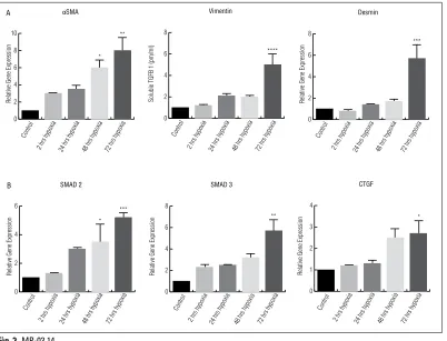
Isolated hypoxia-induced inflammation and fibrotic pathways in human bladder smooth muscle cells
Wiafe, Bridget1; Adesida, Adetola1; Churchill, Thomas1; Metcalfe, Peter1
1Surgery, University of Alberta, Edmonton, AB, Canada
Introduction and Objectives: Several studies have implicated hypoxia in
the etiology of partial bladder outlet obstruction (pBOO). However, the isolated effects of hypoxia on bladder cells as far as the initiation and progression of pBOO is concerned are not yet known. Therefore, we aimed to characterize the progressive effects of pure hypoxia on bladder smooth muscle cells.
Methods: Sub-confluent normal human bladder smooth muscle cells
(hbSMC) were cultured in 3% O2 tension for two, 24, 48, and 72 hours. RNA was extracted for gene expression analysis using reverse transcrip-tion polymerase chain reactranscrip-tion (RT PCR) and protein produced in culture medium was analysed with enzyme-linked immunsorbant assay (ELISA).
Results: Transcription of HIF 1 and 2 were transiently induced after two
hours of hypoxia (p<0.05) whereas HIF 3 was upregulated after 72 hours (p<0.005). VEGF mRNA increased significantly after 24 and 72 hours (p<0.005). The inflammatory cytokines; TGFB (protein and mRNA), IL 1β, 1L6, and TNFα (mRNA) demonstrated a time-dependent increased expres-sion. Furthermore, the anti-inflammatory cytokine, IL-10, was downregu-lated after 72 hours (p<0.05). Evidence of myofibroblast activation and EMT included increased αSMA, vimentin, and desmin. Evidence of pro-fibrotic changes included increases in CTGF, SMAD 2 and 3, as well as collagens 1, 2, 3, 4, fibronectin, aggrecan and TIMP 1 transcripts (p<0.05). Total collagen proteins also increased time-dependently (p>0.05).
Conclusions: Together, these data show that the exposure of hbSMC to
low oxygen tension results in intense hypoxic cascade, including inflam-mation, myofibroblast activation, pro-fibrotic changes, and increased extracellular matrix expression. This elucidates mechanisms of
hypoxia-driven bladder deterioration after pBOO and may result in tailored in vivo experiments and, ultimately, translate into improved clinical outcomes.
MP-03.15
Erythropoietin accelerates the recovery of normal ureteral func-tion following complete unilateral obstrucfunc-tion
Janssen, Claudia1,2; Jäger, Wolfgang1,2; Moskalev, Igor3; Fazli, Ladan3;
Thüroff, Joachim2; Lange, Dirk1
1Urologic Sciences, University of British Columbia, Vancouver, BC,
Canada; 2Urology, University of Mainz, Mainz, Germany; 3Vancouver
Prostate Centre, University of British Columbia, Vancouver, BC, Canada
Introduction and Objectives: Obstructive uropathy facilitates ureteral
aperistalsis and ascending pyelonephritis. While physiologic smooth muscle contractions are lost in the dilated hydroureter during unilateral ureteral obstruction (UUO), the functional recovery of the ureter follow-ing obstruction reversal remains unclear. Erythropoietin (EPO) is protective in non-hematopoietic organs and restores peristalsis in hypocontractile intestinal smooth muscle cells. Here, we investigated the role of EPO in the (patho-) physiology of ureteral peristalsis and determined its thera-peutic value in obstructive uropathy.
Methods: Abdominal laparotomy and UUO were performed in female
CD-1 mice for 24 hours (Group 1; n=22), 48 hours (Group 2; n=22), or 72 hours (Group 3; n=22) using a non-traumatic vascular micro clip. Expression of EPO, EPO receptor (EPOR), and β-common receptor (βCR) in obstructed and unobstructed ureters was determined via reverse tran-scription polymerase chain reaction (RT PCR) in group 2 animals. Ten animals per group received 200 IU EPO on four consecutive days; con-trols received saline. The duration from removal of the obstruction until regression of hydronephrosis was assessed by ultrasonography. Peristaltic activity was tracked microscopically pre- and post-obstruction via open laparatomy.
Soluble TGFB 1 (pm/ml)
HBSMC secreted levels of TGFB 1 protein
Results: Both EPOR and βCR are expressed in murine ureters. Expression of endogenous EPO was remarkably up regulated in obstructed ureters compared to unobstructed ureters in untreated animals. The time until complete resolution of hydronephrosis after removal of the obstruction was found to depend on the duration of obstruction. Despite complete resolution of hydronephrosis after two, six, and eight days (Groups 1, 2, 3), the return of peristalsis was significantly delayed. EPO sigificantly accelerated regression of hydronephrosis and restoration of coordinated ureteral peristalsis in all groups.
Conclusions: EPO signaling may present a novel target for future
therapeu-tic agents for obstructive uropathy and motility disturbances of the ureter.
MP-03.16
Urothelial cells express a functional succinate receptor GPR91
Velasquez-Flores, Monica1; Cammisotto, Philippe1; Campeau, Lysanne1
1Lady Davis Institute for Medical Research, Montreal, QC, Canada
Introduction and Objectives: Lower urinary tract symptoms (LUTS) are
associated with the metabolic syndrome (MetS). Increased succinate pro-duction is detected in the presence of hyperglycemia and hypoxemia, as with diabetes mellitus and metabolic syndrome, which is strongly associated with overactive bladder syndrome. The aim of our study is to determine how succinate modulated bladder contractility.
Methods: Urothelial cells were isolated from Sprague-Dawley rat bladder
using a collagenase IV method and grown in collagen IV-coated petri dishes. After confluency, cells were exposed to succinate then assessed by microscopy and immunoblotting analysis.
Results: Immunohistochemistry revealed that cells were confluent and
express cytokeratin 17 and the receptor of succinate SUCNR1 (GPR91). Immunohistochemistry confirmed expression of GPR91. Incubation of cells with succinate (10-2 M) results in phosphorylation of Erk and c-Jun amino-terminal kinases (JNKs) JNK, with no effect on Akt-308P, Akt 473P, enos-1177P, or enos-405P. Erk phosphorylation was not observed with exposure to alpha-keto glutarate and citrate, two other intermediates of the
citric acid cycle witout affinity to the GPR91 receptor, or malonate, which increases intracellular succinate. On the other hand, succinate dose-dependently decreased the concentrations of intracellular cyclic AMP stimulated by forskolin. Finally, succinate dose-dependently increased the secretion of prostaglandin E2 (PGE2).
Conclusion: A functional succinate receptor is expressed in urothelial
cells. Succinate triggers activation of Erk and JNK by binding to its recep-tor GPR91, and increasing release of PGE2. These results suggest that succinate might be a major regulator of bladder contractility through its actions on urothelial cell signaling and secretion.
MP-03.17
A high-throughput minimally invasive murine model for the study of indwelling urinary biomaterial-associated urinary tract infections
Janssen, Claudia1,2; Jäger, Wolfgang1,2; Moskalev, Igor1; Chew, Ben H.1;
Lange, Dirk1
1Urologic Sciences, University of British Columbia, Vancouver, BC,
Canada; 2Urology, University of Mainz, Mainz, Germany
Introduction and Objectives: Indwelling urinary device-associated
infections are one of the most common healthcare-associated infec-tions frequently complicated by encrustation, causing potential block-age of the device. Preclinical research is limited by the lack of relevant high-throughput and cost effective animal models. Current models are restricted to female mice, associated with major transurethral loss of catheter materials during micturition, highly invasive and complex. We present an ultrasound-guided, minimally invasive model enabling the testing of novel antimicrobial biomaterials for indwelling urinary devices in a realistic environment.
Methods: Implantation of 4 mm catheter segments into murine bladders
was performed percutaneously (n=15 males; n=5 females) or transure-thrally (n=15 females) using the Seldinger technique under ultrasound guidance. Proteus mirabilis was instilled intraluminally. Catheter
encrusta-0
Soluble TGFB 1 (pm/ml)
tion was monitored by ultrasound. Bacteria were quantified from urine and catheters and encrustation analyzed on Days 6 or 21.
Results: Percutaneous and transurethral catheter implantations were
per-formed efficiently (mean time: 3.6 ± 0.8 min vs. 2.5 ± 0.5 min) in all animals. Ultrasound confirmed that 100% vs. 66%, of implanted cath-eters remained indwelling over the study period. Catheter encrustation
developed in P. mirabilis-infected urine 48 hours post-instillation and its
increase over time was detectable with ultrasonography. Fourier Transform Infrared Spectroscopy (FTIR) analysis of the encrustation confirmed a typical struvite spectrum. Control catheters remained sterile over 21 days.
Conclusions: Our minimally invasive and reproducible percutaneous
technique is suitable to study device-associated infection in both genders.
Infecting urine with P. mirabilis generates a preclinical model of catheter
encrustation within three days. The progression of encrustation can be monitored in vivo using ultrasonography, making this image-based model suitable to assess novel antibacterial and anti-encrustation therapies.
MP-03.18
Novel antimicrobial peptide coating to prevent catheter-associ-ated urinary tract infections
Lo, Joey1; Yu, Kai2; Haney, Evan3; Janssen, Claudia1,4; Hancock, Robert
E.3; Kizhakkedathu, Jayachandran2; Lange, Dirk1
1Urologic Sciences, University of British Columbia, Vancouver, BC,
Canada; 2Pathology and Lab Medicine, University of British Columbia,
Vancouver, BC, Canada; 3Microbiology & Immunology, University of
British Columbia, Vancouver, BC, Canada; 4Urology, University of Mainz,
Mainz, Germany
Introduction and Objectives: Urinary catheters are ideal surfaces for
bacterial biofilm formation, making them the main source of
hospital-acquired infections. With the increase in antibiotic resistance, there is a push for non antibiotic-based measures to prevent catheter-associated urinary tract infections (CAUTI). We are pursuing the use of novel anti-microbial catheter coatings using polymer-linked broad-spectrum antimi-crobial peptides (AMPs). Here, we present the efficacy of tethered AMPs against common uropathogens in vitro and in a relevant in vivo model.
Methods: Peptides Tet20, Tet26, and E6 were linked to surfaces at varying
densities using polymer brushes PDMA and PSBMA. The antimicrobial activity was determined via colony counts following exposure of uro-pathogens for up to 48 hours. To evaluate the in vivo efficacy, CAUTI models were developed in mice and rats using 5 mm and 10 mm cath-eter segments respectively, introduced transurethrally or transcutaneously
via ultrasound guidance, followed by transurethral inoculation using 105
S. aureus or P. mirabilis.
Results: E6-PSBMA and E6-PDMA were shown to be the most effective
peptide-brush combinations decreasing the bacterial load up to 108-fold.
Tet20-PSBMA and Tet20-PDMA were less effective at up to100-fold less bacterial load than controls, while all combinations with Tet26 were ineffective. Transcutaneous ultrasound-guided introduction of catheter pieces was more efficient in mice than rats, in which the transurethral method was more successful. Using the transurethral model, coated cath-eter pieces were found to contain significantly less bacteria compared to uncoated controls.
Conclusions: Based on our in vitro data, the AMP E6 tethered to surfaces